infusion of brain-derived neurotrophic factor into … of brain-derived neurotrophic factor into the...
TRANSCRIPT

Infusion of Brain-Derived Neurotrophic Factor into the LateralVentricle of the Adult Rat Leads to New Neurons in theParenchyma of the Striatum, Septum, Thalamus, and Hypothalamus
Viorica Pencea,1 Kimberly D. Bingaman,1,2 Stanley J. Wiegand,3 and Marla B. Luskin1
Departments of 1Cell Biology and 2Neurosurgery, Emory University School of Medicine, Atlanta, Georgia 30322, and3Regeneron Pharmaceuticals Inc., Tarrytown, New York 10591
The findings that brain-derived neurotrophic factor (BDNF) pro-motes in vitro the survival and/or differentiation of postnatalsubventricular zone (SVZ) progenitor cells and increases in vivothe number of the newly generated neurons in the adult rostralmigratory stream and olfactory bulb prompted us to investigatewhether the infusion of BDNF influences the proliferation and/ordifferentiation of cells in other regions of the adult forebrain. Weexamined the distribution and phenotype of newly generatedcells in the adult rat forebrain 16 d after intraventricular admin-istration of BDNF in conjunction with the cell proliferationmarker bromodeoxyuridine (BrdU) for 12 d. BDNF infusionresulted in numerous BrdU� cells, not only in the SVZ lining theinfused lateral ventricle, but moreover, in specific parenchymalstructures lining the lateral and third ventricles, including thestriatum and septum, as well as the thalamus and hypothala-mus, in which neurogenesis had never been demonstrated
previously during adulthood. In each region, newly generatedcells expressed the neuronal marker microtubule-associatedprotein-2, or neuron-specific tubulin, identified by the antibodyTuJ1. The percentage of the newly generated cells expressingTuJ1 ranged from 27 to 42%, suggesting that the adult fore-brain has a more profound capacity to produce neurons thanrecognized previously. The extent of cell proliferation afterBDNF infusion was correlated with the level of expression offull-length TrkB, the high-affinity receptor for BDNF, despite thefact that the BrdU� cells were not themselves TrkB�. Collec-tively, our results demonstrate that the adult brain parenchymamay recruit and/or generate new neurons, which could replacethose lost as a result of injury or disease.
Key words: brain-derived neurotrophic factor; cell prolifera-tion; forebrain parenchyma; intraventricular infusion; postnatalneurogenesis; subventricular zone
The majority of the cells in the mammalian forebrain ariseprenatally (Sidman and Rakic, 1973; Raedler and Raedler, 1978).However, it is now established that, in addition to the productionof sensory neurons and supporting cells in the olfactory epithe-lium (Graziadei and Monti Graziadei, 1976; Huard et al., 1998),both neurons and glia continue to be generated in restricted adultmammalian forebrain structures, including the hippocampus (Alt-man and Das, 1965; Schlessinger et al., 1975; Kaplan and Bell,1984; Kuhn et al., 1996; Palmer et al., 1997) and the subventricu-lar zone (SVZ) lining the lateral ventricles (Privat, 1975; Vaysseand Goldman, 1990; Levison and Goldman, 1993; Luskin, 1993;Lois and Alvarez-Buylla, 1994). Recent studies have raised thepossibility that the parenchyma of the adult forebrain also harborsprogenitor cells for neurons (Reynolds et al., 1992; Palmer et al.,1995; Marmur et al., 1998; Magavi et al., 2000). The source andproliferative potential of these progenitors have not been fullydetermined.
In vitro studies have revealed that the adult striatal SVZ canbe induced to generate both neurons and glia under the influ-ence of growth factors (Reynolds et al., 1992; Reynolds and
Weiss, 1992, 1996; Gritti et al., 1999). Furthermore, the in vivoexposure of the adult forebrain SVZ to neurotrophins yields anincrease in the number of progenitor cells, as well as theproduction of newly generated neurons (Craig et al., 1996;Kuhn et al., 1997; Zigova et al., 1998). Administration of eitherepidermal growth factor (EGF) or tumor growth factor-�(TGF-�) into mouse(Craig et al., 1996) or of either EGF orfibroblast growth factor-2 (FGF-2) into rat (Kuhn et al., 1997)resulted in an expansion of the SVZ surrounding the infusedlateral ventricle, as well as newborn cells in the nearby striatumand septum. However, although both EGF and FGF-2 ampli-fied the number of striatal SVZ progenitors in the adult brain,neither treatment resulted in a significant increase in theproduction of neurons in the forebrain parenchyma.
In this study, we analyzed the rat forebrain SVZ and paren-chyma for the presence of newly generated cells after intracere-broventricular infusion of brain-dervied neurotrophic factor(BDNF), in combination with the cell proliferation marker bro-modeoxyuridine (BrdU), to investigate whether and how theadult forebrain responds to BDNF exposure. The BDNF admin-istration resulted in numerous BrdU� cells, not only in the SVZlining the lateral ventricle, but also in the striatal and septalparenchyma. Moreover, a high number of BrdU-labeled cellswere found in discrete regions of the thalamus and hypothalamusbordering the third ventricle. Approximately 27–42% of thenewly generated cells expressed a neuron-specific marker. Fur-thermore, we found that the BrdU incorporation was correlatedwith the level of the high-affinity receptor for BDNF, TrkB.However, the BrdU� cells and TrkB� cells were in non-
Received Feb. 20, 2001; revised June 5, 2001; accepted June 7, 2001.This work was supported by National Institute of Deafness and Other Commu-
nicative Disorders Grant RO1 DC03190 (M.B.L.) and by Regeneron Pharmaceuti-cals, Inc. We are grateful to Dr. Giri Venkatraman, Dr. Volkan Coskun, and JoannaBonsall for their critical and helpful comments on this manuscript and to Dr. StuartC. Feinstein for his generous gift of anti-TrkB.
Correspondence should be addressed to Dr. Marla B. Luskin, Department of CellBiology, Emory University School of Medicine, 1648 Pierce Drive, Atlanta, GA30322. E-mail: [email protected] © 2001 Society for Neuroscience 0270-6474/01/210001-12$15.00/0
The Journal of Neuroscience, September 1, 2001, 21(17):6706–6717

overlapping populations. Collectively, our results underscore thepossibility that new neurons can be recruited to replace those lostas a result of disease or injury.
Parts of this work have been published previously in abstractform (Pencea et al., 1999).
MATERIALS AND METHODSImplantation of minipumps and administration of BDNF and BrdU. Todetermine whether the intracerebroventricular infusion of a neurotro-phic factor increases the proliferation of new neurons in the adultforebrain, we analyzed the distribution and number of newly generatedcells in the forebrain of Sprague Dawley rats, after the administration ofBDNF (n � 3) compared with that of the control vehicle, 0.1 M PBS,given alone (n � 3). The adult rats, weighing 220–250 gm, were anes-thetized with ketamine and implanted with an osmotic minipump (Alzet2002; Alza Scientific Products, Palo Alto, CA). The cannula was placedin the right lateral ventricle 4.0 mm deep to the pial surface and �0.0 mmanteroposterior relative to bregma and 1.8 mm lateral to the midline.Each rat was infused for 12 d with 12 �l /d of either human recombinantBDNF dissolved in 0.1 M PBS (1 �g/ml) (Regeneron Pharmaceuticals,Tarrytown, NY) or PBS only. To label the newly generated cells in theBDNF- or vehicle-infused brains, the cell proliferation marker BrdU wasdelivered at the same rate (12 �g/d) and through the same minipump asthe BDNF or PBS. After the cessation of the infusion of BDNF andBrdU or PBS and BrdU, the cannula was left in the lateral ventricle, andthe animals were allowed to survive another 16 d before perfusion (Fig.1). We also compared the distribution and phenotype of the newlygenerated cells after the intracerebroventricular infusion of BDNF andBrdU or PBS and BrdU to that in animals that received only dailyintraperitoneal injections of BrdU (5 mg/ml in 0.0007N NaOH saline, 50mg/kg) for 12 d.
Tissue processing and immunohistochemistry. Sixteen days after thecessation of the intracerebroventricular BDNF and BrdU, PBS andBrdU, or intraperitoneal BrdU, the animals were anesthetized withpentobarbital (50 mg/kg) and perfused transcardially with heparanizedsaline (5 U of heparin per milliliter of 0.9% NaCl), followed by 4%paraformaldehyde in 0.1 M phosphate buffer, pH 7.4. Brains were cryo-protected with 30% sucrose in 0.1 M phosphate buffer, pH 7.2, embeddedin Tissue-Tek OCT compound (Sakura Finetek, Torrance, CA), andsectioned on a cryostat in the coronal plane at 20 �m.
To reveal newly generated BrdU-positive cells, the sections wereincubated for 30 min in 1N HCl at 60°C to denature the DNA. Subse-quently, the sections were incubated first in blocking serum (10% normalgoat serum in 0.1 M phosphate buffer containing 0.02% Triton X-100, pH7.4), abbreviated NGS for 1 hr, and then for 48 hr with a 1:200 dilutionof a mouse IgG anti-BrdU (Accurate Chemicals, Westbury, NY) in NGS.For fluorescent visualization of BrdU-labeled cells, the sections wereincubated for 1 hr at room temperature in a rhodamine-conjugated goatanti-rat secondary antibody (1:200 dilution).
Some sections were processed to visualize only BrdU-labeled cells,whereas others were double-labeled with anti-BrdU, as well as an anti-body to a cell type-specific marker to determine the phenotype of thenewly generated cells. To identify neurons, we used the antibody TuJ1(1:400) (Covance, Richmond, CA), a mouse polyclonal IgG that recog-nizes neuron-specific �III-tubulin (Lee et al., 1990) usually expressed byimmature neurons, or a monoclonal antibody to microtubule-associatedprotein-2 (MAP-2) (1:200; Roche Products, Indianapolis, IN), whichrecognizes more differentiated neurons (Bernhardt et al., 1985; Johnsonand Jope, 1992). We also used a polyclonal antibody to glial fibrillaryacidic protein (GFAP) (1:500 dilution; Dako, Glostrup, Denmark) (Big-nami et al., 1972) to identify astrocytes and either a mouse monoclonalantibody to galactocerebrosidase (GalC) (gift from Dr. Rao, Universityof Utah, Salt Lake City, UT) at a 1:200 dilution (Ranscht et al., 1982,1987) or mouse monoclonal anti-myelin proteolipid protein (PLP)(Chemicon, Temecula, CA) at a 1:200 dilution (Cheng et al., 1998) toidentify oligodendrocytes. As a marker for undifferentiated cells, amouse monoclonal antibody to the intermediate filament protein nestin(Hockfield and McKay, 1985; Frederiksen and McKay, 1988; Lendahland McKay, 1990) was used as an undiluted supernatant (DevelopmentalStudies Hybridoma Bank, Iowa City, IA). In addition, to analyze theexpression pattern of TrkB, the high-affinity tyrosine kinase receptor forBDNF, some sections were also incubated with anti-TrkB606 –619, a poly-clonal antibody that recognizes the intracellular domain of the full-length rat TrkB (amino acids 606–619) (gift from Dr. S. Feinstein,
University of California, Santa Barbara, CA) at a 1:25 dilution. Forfluorescent visualization of all cell type-specific antibodies and anti-TrkB, fluorescein-conjugated secondary antibodies were used at a 1:200dilution. All secondary antibodies were from Jackson ImmunoResearch
Figure 1. I llustration of the intraventricular infusion site, time course ofdelivery of BDNF or PBS in conjunction with BrdU, and structuresanalyzed for the distribution and phenotype of BrdU-labeled cells. A, B,In both experimental BDNF-infused (12 �g/d) and control PBS-infusedanimals, an osmotic minipump was used for continuous delivery of thegrowth factor or vehicle into the lateral ventricle, at a rate of 5 �l /hr for12 d. The animals were concurrently infused with BrdU through the sameminipump to label dividing cells. The animals were perfused 16 d aftercessation of the BDNF or PBS infusate. C, A diagram of a parasagittalsection of the adult rat brain demonstrating the placement in the rightlateral ventricle of the cannula used to infuse the BDNF, or PBS, inconjunction with BrdU. D–H, Drawings of representative coronal sectionsof the adult rat brain at different anteroposterior levels, designating thestructures quantitatively analyzed after the intraventricular administra-tion of BDNF or PBS. The diagrams demonstrate that each structureanalyzed ( gray) for the presence of newly generated BrdU � cells isadjacent to the lateral or third ventricle. The anterior part of the subven-tricular zone (D), striatum (E), and septum (F) surround the lateralventricle, whereas the thalamus (G) and hypothalamus (H ) are adjacentto the third ventricle. Note that, in G and H, the third ventricle istransected both ventrally and dorsally. 3V, Third ventricle; Acb, nucleusaccumbens; CC, corpus callosum; CTX, cerebral cortex; DG, dentategyrus; HYP, hypothalamus; IC, internal capsule; LV, lateral ventricle; OB,olfactory bulb; RMS, rostral migratory stream; SPT, septum; ST, striatum;SVZa, anterior part of the subventricular zone; TH, thalamus.
Pencea et al. • Infusion of BDNF Leads to New Neurons in the Forebrain J. Neurosci., September 1, 2001, 21(17):6706–6717 6707

(West Grove, PA). The slides were coverslipped with VectaShield (Vec-tor Laboratories, Burlingame, CA) and viewed using a Zeiss(Oberkochen, Germany) Axiophot fluorescent microscope equippedwith rhodamine and fluorescein filters, as well as a dual filter for visual-izing rhodamine and fluorescein fluorescence simultaneously. For con-firmation of the phenotype of individual BrdU � cells, sections were alsoviewed using a confocal scanning laser microscope (Zeiss Axioplanequipped with LSM 510). To reveal the cytoarchitecture of the structuresanalyzed, some sections were dehydrated in ethanol, counterstained withcresyl violet, rehydrated, and coverslipped with DPX (BDH LaboratorySupplies, Poole, UK). All microscopic images were processed usingAdobe Photoshop (Adobe Systems, Mountainview, CA).
Quantitative analyses. The density of BrdU-labeled cells, expressed ascells per cubic millimeter, was determined in the striatum, septum,thalamus, and hypothalamus, in both the BDNF-infused and PBS-infused brains. We selected for comparisons corresponding coronalsections exhibiting the same cytoarchitectonic features, determined usingthe rat atlas of Paxinos and Watson (1982), in all of the BDNF- andPBS-infused brains. This allowed cells to be counted in coronal sectionsat comparable rostrocaudal positions. The counts were made, using a40� objective, by placing an optical grid (field size, 250 � 250 �m)starting from the wall of the lateral or third ventricle and proceeding intothe parenchyma until BrdU � cells were no longer detectable. Thedensity of BrdU-labeled cells was calculated for each structure analyzedin every animal, and statistical analyses were performed using the Stu-dent’s t test (IBM Statistica; StatSoft Inc., Tulsa, OK).
The number of double-labeled BrdU �–TuJ1 � and BrdU �–GFAP �
cells in the striatum, septum, hypothalamus, and subventricular zonesurrounding the lateral ventricle was counted when viewed with a con-ventional or confocal microscope in at least three sections per brain. Ineach animal, the phenotype of 330–850 striatal cells, 200–900 septalcells, 200–1200 hypothalamic cells, and 600–2500 subventricular cellswas analyzed. The counts of double-labeled cells were analyzed statisti-cally using two-way and one-way ANOVA.
To compare the nuclear diameter of the newly generated cells in thestriatum, septum, hypothalamus, and thalamus, in BDNF- and PBS-infused animals, 25– 40 cells per structure were analyzed in eachanimal. The maximum nuclear diameter was measured using IP LabScientific Processing (Scanalytics Inc., Fairfax, VA), and an averagenuclear diameter was calculated for each structure in each animal.These means were then combined to determine the average diameterof newly generated cells in the parenchyma of the BDNF- and PBS-infused brains. In the same sections, �250 BrdU � cells per animalwere analyzed with IP Lab Scientific Processing to determine thepercentage of newly generated cells present in pairs. The cells wereconsidered to form a pair if their BrdU � nuclei were adjacent (�3 �mapart) to one another.
RESULTSWe examined the effect of intracerebroventricular administrationof BDNF on the distribution and phenotype of newly generatedcells in the adult forebrain. BDNF or PBS (control) was infusedcontinuously for 12 d into the right lateral ventricle of adult rats(Fig. 1A,B). A steady-state level of BDNF is achieved by intra-cerebroventricular infusion within 3 d (Anderson et al., 1995). Tolabel the newly generated cells, the cell proliferation markerBrdU was delivered by intracerebroventricular infusion concur-rently with the BDNF (BDNF–BrdU) or PBS (PBS–BrdU) (Fig.1A,B). The animals were perfused 16 d after BDNF or PBSwithdrawal (day 28) (Fig. 1A,B) to permit newly generated cellsto integrate in the host brain. The distribution and phenotype ofthe newly generated BrdU� cells were analyzed along the ros-trocaudal extent of the lateral and third ventricles (Fig. 1D–H). Athird group of animals received daily BrdU intraperitoneal injec-tions for 12 d, without any intracerebroventricular infusion.These animals were also perfused 16 d after the last BrdUinjection.
Newly generated cells appear in distinct regionssurrounding the lateral and third ventricles aftercombined intracerebroventricular administration ofBDNF and BrdUTo determine which regions surrounding the lateral and thirdventricles have the capacity to produce new cells in response toBDNF infusion into the adult lateral ventricle, we systematicallyanalyzed the distribution of BrdU� cells in the subventricularzone and parenchyma along the rostrocaudal extent of the fore-brain. Our analysis revealed that newly generated cells werepresent surrounding the lateral ventricle on the infused side butnot on the contralateral side, indicating that the BDNF–BrdU didnot diffuse contralaterally. An unexpected finding was that, inaddition to newly generated cells along the rostrocaudal extent ofthe rostral migratory stream (RMS) as reported previously(Zigova et al., 1998), we also detected newly generated cells: (1)in other regions of the SVZ immediately surrounding the lateralventricle, (2) in the parenchyma adjacent to the lateral ventricle(Fig. 2A), and (3) in the parenchyma of specific structures sur-rounding the third ventricle, in which cell proliferation has notbeen described previously (Fig. 2B). In particular, newly gener-ated cells were visualized in the striatum, septum, corpus callo-sum, and cerebral cortex, as well as in the thalamus and hypo-thalamus (Figs. 2, 3). The presence of newly generated cells inspecific parts of the thalamus and hypothalamus surrounding thethird ventricle suggests that BDNF–BrdU may have flowed cau-dally from the infused lateral ventricle into the third ventricle.
To determine whether there was a relationship between theparenchymal structures in which the BrdU� cells were found andthe morphology of the newly generated cells, in addition todouble-labeling with BrdU and cell type-specific markers (asdescribed below), we used the nuclear shape and diameter of thelabeled cells as a way to compare the diversity of labeled cells indifferent regions of the forebrain. In the BDNF-infused brains,the nuclear diameter and morphology of the newly generated cellsvaried according to the regions in which they were located, andthe largest mean nuclear diameter (range of 9.22–9.53 �m) wasfound in the striatum. The nuclei of the BrdU� cells in thestriatum and septum tended to be round, whereas the nuclei in thehypothalamus and thalamus were usually elongated. These resultsindicate that a diversity of cell types was generated as a functionof the location of the cells. However, despite this diversity, in allof the structures analyzed, the BrdU� cells were present fre-quently in close apposition to each other, such that they wereaggregated in pairs (Fig. 4). In the parenchymal structures con-taining BrdU� cells, the percentage of paired cells was relativelyuniform within a given structure, varying from an average of 22%in the hypothalamus to 37% in the striatum. The presence ofpairs of BrdU� cells may indicate that cell division occurs in situin the parenchyma.
Our analysis showed that the number of newly generated cellsin the brains of rats infused with PBS–BrdU was substantiallylower than in the brains infused with BDNF–BrdU, although thedistribution of the newly generated cells was similar in both setsof animals (Figs. 3, 5). However, the distribution of BrdU� cellsafter intracerebroventricular infusion of BrdU combined witheither BDNF or PBS differs from that observed after only intra-peritoneal administration of BrdU. In the animals that receivedintraperitoneal injections, the BrdU-labeling was present mainlyin the subventricular zone adjacent to the lateral ventricle, withjust a few BrdU� cells in the parenchyma surrounding the lateraland third ventricles. The pronounced discrepancy between the
6708 J. Neurosci., September 1, 2001, 21(17):6706–6717 Pencea et al. • Infusion of BDNF Leads to New Neurons in the Forebrain

distribution of BrdU labeling in the brains of the intracerebro-ventricular infused and intraperitoneal injected animals under-scores the limited ongoing proliferation that ordinarily occurs inthe parenchyma versus the subventricular zone of the adult brain.
The pattern of distribution of BrdU� cells in the SVZand parenchyma of the forebrain afterintracerebroventricular administration of BDNF andBrdUOur data indicated that there was a differential response toBDNF infusion in different regions along the ventricular lumen.In the SVZ lining the striatum, there was a prominent dorsal toventral gradient of BrdU� cells (Fig. 2A). In the dorsal aspect ofthe striatal SVZ, BrdU� cells formed a band multiple cell layersthick, whereas the ventral part of the striatal SVZ was consider-ably thinner and contained widely dispersed BrdU� cells. More-over, the BrdU� cells in the dorsal aspect of the striatal SVZoften occurred in clusters (Fig. 3D), suggesting that there were“hot spots” of proliferation around the lateral ventricle, similar tothose described in the telencephalon of the adult canary (Alvarez-Buylla et al., 1990). The gradient in the BrdU labeling of the SVZparallels the differences ordinarily observed in the relative thick-ness and density of the SVZ, as described by Chiasson et al.(1999).
In the parenchyma of each structure containing BrdU� cells,the density of the newly generated cells declines as a function ofdistance from the ventricular wall (Fig. 4). However, there was noapparent correlation between the extent and thickness of theSVZ and the number of newly generated cells present in theadjoining parenchyma. In some structures, such as the septum,there were numerous BrdU� cells in the parenchyma, up to 1 mmfrom the ventricular wall (Figs. 2A, 3F, 5B), although there wasonly marginal labeling of the SVZ. Conversely, in some regionscontaining a prominent SVZ, such as the striatum, there was a
high density of BrdU� cells in the SVZ adjacent to the lateralventricle (Figs. 2A, 3D). Moreover, there were numerous BrdU�
cells in the adjacent striatal parenchyma, up to 2 mm away fromthe lateral ventricle (Figs. 2A, 3D, 5A). In addition, a considerablenumber of BrdU� cells were observed in the parenchyma of thethalamus and hypothalamus surrounding the third ventricle, de-spite the absence of a distinguishable SVZ (Figs. 2B, 3H,J, 5C).Thus, the extent of the SVZ is not the only determining factor forthe production of new cells in the adult forebrain.
The BrdU� cells in the parenchyma were not evenly distrib-uted. Rather, the distribution of BrdU� cells was restricted byboundaries between and within structures of the forebrain, sim-ilar to the restriction of newly generated cells in the RMS(Luskin, 1993; Zigova et al., 1996, 1998). For example, althoughthe BrdU� cells in the septum on the infused side were wide-spread and numerous, they appeared to “avoid” the midlinefimbria fornix and to form a dorsal and a ventral stream aroundthe fimbria (Fig. 2A). The presence of BrdU� cells in the septumadjacent to the uninfused lateral ventricle, combined with theabsence of BrdU� cells in the SVZ and striatum also liningthe uninfused ventricle, suggests that some cells generated on theinfused side may have migrated across the midline to the con-tralateral septum. Alternatively, BrdU may have directly diffusedacross the midline in which it then became incorporated in theseptal cells on the contralateral side. The labeling pattern ob-served in the septum indicates that the distribution of BrdU�
cells is neither the result of the random migration of BrdU� cellsnor the effect of the passive diffusion of the BDNF–BrdU.
Prominent differences in the extent of BrdU incorporationwere also observed surrounding the third ventricle. In particular,although there were numerous BrdU� cells in the habenularnucleus of the thalamus, there were no BrdU� cells in the part ofthe dentate gyrus immediately adjacent to the third ventricle (Fig.
Figure 2. The distribution of newly generated cells in the parenchyma surrounding the lateral and third ventricles after the coinfusion of BDNF andBrdU into the lateral ventricle of an adult rat brain. The newly generated cells (bright orange) are identified in 20 �m coronal sections with an antibodyto BrdU and visualized with a rhodamine-conjugated secondary antibody. A, A representative fluorescent photomicrograph demonstrating BrdU � cellsin the parenchyma surrounding the infused lateral ventricle 16 d after a 12 d infusion of BDNF–BrdU. The striatal SVZ (arrows) has numerous BrdU �
cells, whereas the rest of the SVZ, including that lining the septum, is almost devoid of newly generated cells. Moreover, the dorsal half of the striatalSVZ appears thicker than the ventral part. The distribution of the BrdU � cells in the striatal parenchyma exhibits a medial to lateral gradient, with thenumber of BrdU � cells decreasing as a function of distance from the lateral ventricle. The distribution of BrdU � cells in the septal parenchyma is morehomogenous, although there is a relatively sharp decrease in the number of BrdU � cells at the border between septum and fornix (dashed line). Notethat, on both sides of the lateral ventricle, the BrdU � cells extend more than a few hundred micrometers into the parenchyma. A small number of thenewly generated cells can also be observed in the corpus callosum overlying the lateral ventricle. The midline of the section is approximately at the leftedge of the photomicrograph. B, A representative fluorescent photomicrograph showing BrdU � cells in the parenchyma surrounding the third ventricleof a BDNF-infused brain. In the hypothalamus, the newly generated cells extend bilaterally at least a few hundred micrometers into the parenchyma,and their distribution is relatively homogenous. Similar to the septal SVZ (shown in A), the hypothalamic ventricular lining is devoid of BrdU � cells.3V, Third ventricle; CC, corpus callosum; HYP, hypothalamus; LV, lateral ventricle; SPT, septum; ST, striatum. Scale bar, 100 �m.
Pencea et al. • Infusion of BDNF Leads to New Neurons in the Forebrain J. Neurosci., September 1, 2001, 21(17):6706–6717 6709

6B). However, after infusion of BDNF, there were newly gener-ated cells in the subgranular layer of the dentate gyrus, as hasbeen described previously in the normal brain (Altman and Das,1965; Schlessinger et al., 1975; Kaplan and Bell, 1984; Kuhn et al.,1996; Palmer et al., 1997). The restriction of BrdU labeling todiscrete regions surrounding the third ventricle is further illus-trated by the paucity of BrdU� cells in other thalamic nucleiadjacent to the habenula, although some of them are also adja-cent to the third ventricle (data not shown).
The hypothalamus, another structure bordering the third ven-tricle, also exhibits a well defined pattern of BrdU labeling after
4
cells in the striatal SVZ is higher, and more of these cells tend to occur inclusters. E, F, Representative fluorescent photomicrographs of infusedhemispheres demonstrating the newly generated cells in the septumadjacent to the infused lateral ventricle. The number of BrdU � cells (e.g.,arrowheads) in the septal parenchyma of the BDNF-infused brain (F) ismuch higher than the number of labeled cells in the PBS-infused brain(E). In both cases, however, the septal SVZ is almost devoid of BrdU �
cells. G, H, Representative fluorescent photomicrographs showing BrdU-positive cells in the thalamus, adjacent to the dorsal lumen of the thirdventricle, after the infusion of PBS–BrdU ( G) or BDNF–BrdU (H ). Thenumber of BrdU � cells (e.g., arrowheads) present in the thalamic paren-chyma of the BDNF-infused brain (H ) is higher than the numberobserved in the PBS-infused brain (G). In both sections, there are a fewBrdU � cells lining the wall of the third ventricle. I, J, Representativefluorescent photomicrographs demonstrating the relative number ofBrdU � cells in the hypothalamus of the PBS-infused ( I ) compared withBDNF-infused ( J) brain. After BDNF infusion ( J), numerous BrdU �
cells (e.g., arrowheads) are dispersed throughout the hypothalamic paren-chyma, whereas after the PBS infusion ( I ), fewer BrdU � cells (e.g.,arrowheads) are present. Note that very few BrdU � cells line the thirdventricle in the sections from both the BDNF-infused and PBS-infusedbrains. 3V, Third ventricle; CTX, frontal cortex; LV, lateral ventricle;HYP, hypothalamus; SPT, septum; ST, striatum; TH, thalamus. Scalebars, 100 �m.
Figure 3. A comparison of the number and distribution of the newlygenerated cells in forebrain structures surrounding the lateral and thirdventricles after the intraventricular coinfusion of BDNF and BrdU orPBS and BrdU. The newly generated BrdU-positive cells (bright orange)were identified in 20 �m coronal sections with an antibody to BrdU andvisualized with a secondary antibody conjugated to rhodamine. A, B,Representative fluorescent photomicrographs of infused hemispheresshowing BrdU � cells in the anterior part of the subventricular zone andthe adjacent frontal cortex after infusion of PBS–BrdU (A) or BDNF–BrdU (B) into the ipsilateral lateral ventricle. After BDNF infusion, theSVZa (B) is expanded in diameter relative to the SVZa of the PBS-infused brain (A). Moreover, after BDNF infusion (B), the number ofnewly generated cells (e.g., arrowheads) present in the frontal cortexsurrounding the SVZa is much higher than that observed in the PBS-infused brain (A). C, D, Representative fluorescent photomicrographs ofinfused hemispheres demonstrating the number of BrdU � cells in thestriatum in brains infused with PBS (C) or with BDNF (D). After BDNFor PBS infusion, BrdU � cells are dispersed throughout the striatalparenchyma, although there are substantially fewer new cells in thePBS-infused brain (C). Moreover, in the BDNF-infused brain (D) com-pared with the PBS-infused brain (C), the number of newly generated
Figure 4. Pairs of newly generated cells in the parenchyma of thestriatum after intraventricular infusion of BDNF. A, A representativefluorescent photomicrograph of a 20 �m coronal section demonstratingthat, 16 d after withdrawal of a 12 d infusion of BDNF–BrdU, a highproportion of the newly generated cells within the striatal parenchymaoccur in pairs. The cleavage plane between pairs of cells (e.g., arrows)appears random, with no preferential orientation relative to the ventric-ular surface. Midline is to the right, and dorsal is up. B, A representativephotomicrograph of the septal parenchyma, viewed with confocal micros-copy, showing a pair of newly generated BrdU � cells. The short distance(�2 �m) between the pairs of BrdU � nuclei combined with the highfrequency of pairs (shown in A) suggests that, after BDNF administra-tion, cell division may occur in situ. Scale bars: A, 50 �m; B, 10 �m.
6710 J. Neurosci., September 1, 2001, 21(17):6706–6717 Pencea et al. • Infusion of BDNF Leads to New Neurons in the Forebrain

BDNF infusion. Whereas in the rostral hypothalamus there wasa relatively uniform density of BrdU� cells surrounding the thirdventricle (Fig. 2B), more caudally, the newly generated cells wereconcentrated in particular hypothalamic nuclei. For example,there was a relatively high density of BrdU� cells in the parvo-cellular region of the paraventricular nucleus, although it is dis-placed from the wall of the third ventricle (Fig. 6F). Conversely,the periventricular nucleus immediately surrounding the thirdventricle has a comparatively low number of BrdU� cells. To-gether, the pattern of BrdU labeling in the hypothalamus suggeststhat regions presumably exposed to the highest concentration ofBDNF (e.g., those closest to the ventricular lumen) do not nec-essarily contain the highest density of newly generated BrdU�
cells.
TrkB expression in the forebrain parenchyma of BDNF-infused brains correlates with sites of cell proliferationTo determine the correlation between the increased cell prolif-eration after intracerebroventricular infusion of BDNF and theexpression of TrkB, the high-affinity receptor for BDNF, weanalyzed the relationship between the BrdU incorporation andthe pattern of the full-length TrkB expression in the structuressurrounding the lateral and third ventricles. In a previous study(Zigova et al., 1998), we found that the anterior part of the SVZ(SVZa) and the RMS of BDNF-infused adult brains contained ahigher number of BrdU� cells and expressed higher levels ofTrkB compared with the surrounding areas. In the present study,a similar correlation was observed in the areas surrounding thelateral (data not shown) and third (Fig. 6) ventricles. This corre-lation, such that the extent of BrdU incorporation and TrkBexpression parallel each other, can be observed in the regions ofthe hypothalamus examined. There were numerous BrdU� cellsand a high level of TrkB expression in the paraventricular nucleusof the hypothalamus, whereas in the periventricular nucleus, thenumbers of BrdU-labeled cells and TrkB expression were bothlow (Fig. 6E–G). Nevertheless, TrkB expression is not sufficientfor cell proliferation. For example, whereas TrkB is expressed ata uniformly high level throughout the habenular nucleus (Fig.6D), BrdU� cells are much more numerous along the medialedge (Fig. 6B). This disparity cannot be accounted for by thedifferences in BDNF exposure, because the dorsal edge of thehabenular nucleus, which has lower BrdU incorporation, alsofaces the third ventricle.
To investigate whether the TrkB expression was influenced bythe infusion of BDNF, we compared the pattern of TrkB expres-sion in the BDNF-infused hemispheres versus uninfused hemi-spheres or PBS-infused hemispheres. The levels of TrkB expres-sion in different structures of the BDNF-infused hemisphereswere similar to those in the contralateral uninfused hemispheresand in the PBS-infused brains. These findings indicate that theinfusion of BDNF or PBS did not result in an overt change in thelevel of TrkB expression in the regions containing BrdU� cells.
In the regions in which we observed both high levels of TrkBand numerous BrdU-labeled cells, we further investigatedwhether the cells expressing TrkB were able to incorporate BrdUduring the administration of BDNF–BrdU. Our confocal analysisrevealed that the full-length TrkB receptor was not expressed bythe BrdU� cells in the parenchyma (Fig. 7). Nevertheless, theTrkB� cells were frequently adjacent to the BrdU� cells (Fig.7C,D, insets), suggesting that BDNF may have an indirect effecton the proliferation and/or survival of the newly generated cells(see Discussion).
Figure 5. Density of newly generated BrdU � cells in the striatum,septum, and hypothalamus after BDNF infusion relative to PBS infusion.For each structure analyzed, the gradient of the cell density is plottedfrom the wall of the ventricle (corresponding to the origin of axes) to 2.25mm into the parenchyma. A, In the striatum, adjacent to the lateralventricle, the density of the BrdU � cells is two to three times higher afterBDNF infusion than after PBS infusion, and in both cases the cell densitydeclines gradually as a function of distance from the ventricular wall.Nevertheless, the newly generated cells extend farther into the paren-chyma after BDNF infusion compared with PBS infusion. B, At eachposition in the septum, the cell density is �1.5 times higher after BDNFinfusion than after PBS infusion. In both groups, the cell density remainsrelatively constant in the septal parenchyma (�0.75 mm from the ven-tricular wall) and then decreases gradually in the fimbria fornix. C, In theproximal 0.5 mm adjacent to the third ventricle, the cell density in thehypothalamus is more than two times higher after BDNF administrationthan after PBS infusion. The number of the newly generated cells declinessteeply beyond the hypothalamic border in both the BDNF- and PBS-infused brains. *p � 0.05; **p � 0.005.
Pencea et al. • Infusion of BDNF Leads to New Neurons in the Forebrain J. Neurosci., September 1, 2001, 21(17):6706–6717 6711

Numerous newly generated neurons appear in the SVZand parenchyma of the forebrain after BDNF infusionWe analyzed the phenotype of the BrdU� cells in the SVZ andparenchyma after intracerebroventricular infusion of BDNF todetermine what proportion of the newly generated cells wereneurons. The phenotype of the newly generated BrdU� cells wasidentified using cell type-specific markers. In all regions exam-ined, a significant proportion of BrdU� cells colocalized TuJ1(Fig. 8A–G), an antibody against neuron-specific �III-tubulin,expressed by immature neurons (Lee et al., 1990; Easter et al.,1993). The percentage of double-labeled TuJ1�–BrdU� cells inthe SVZ was �27% (Table 1). A similar percentage of the BrdU�
cells in the parenchyma of the striatum and septum were neurons;the percentage was slightly higher (�42%) in the hypothalamus(Table 1). In each of the regions containing TuJ1�–BrdU� cells,we also observed that a small percentage of the newly generatedcells expressed MAP-2�, a marker for mature neurons (Bern-hardt et al., 1985; Johnson and Jope, 1992) (Fig. 8H–K). Thisdiscrepancy between the percentage of TuJ1� and anti-MAP-2�
cells suggests that the majority of the newly generated neuronsmay not have sufficiently matured to express MAP-2.
The percentages of the newly generated cells that were TuJ1�
were similar in the BDNF- and PBS-infused brains in the SVZ,striatum, and septum but not in the hypothalamus, indicatingthat, in most regions, the in vivo administration of BDNF did notalter the phenotype of the newly generated cells. In all regions,however, the total number of new neurons was significantly higherafter BDNF infusion than PBS infusion. In the hypothalamus,there was a higher percentage of newly generated neurons in theBDNF-infused than in the PBS-infused brains (e.g., �41% afterBDNF vs �21% after PBS), suggesting that the cells of thehypothalamus may respond differently than other forebrain struc-
tures to the BDNF administration. In contrast to the BDNF- orPBS-infused brains, no new neurons were found outside thesubventricular zone in the brains of the animals that had receivedonly intraperitoneal injections of BrdU (data not shown).
Because the multipotential progenitor cells in the adult SVZordinarily give rise to glia (Reynolds et al., 1992; Chiasson et al.,1999), we investigated what percentage of the newly generatedBrdU� cells are astrocytes or oligodendrocytes after BDNFinfusion. We used an antibody to GFAP to identify astrocytes andantibodies against GalC and PLP to identify oligodendrocytes. Inboth the BDNF- and PBS-infused brains, the parenchyma of thestriatum (Fig. 9A), septum, and hypothalamus (Fig. 9B) containeda low percentage (2–7%) of newly generated GFAP� cells (Table1). The percentage of BrdU� astrocytes was similar (�3%) in thehypothalamus in the BDNF- and PBS-infused brains, whereas inthe striatum and septum, the percentage of BrdU� astrocytes washigher in the BDNF-infused brains (Table 1). In both the BDNF-and PBS-infused brains, however, the percentage of newly gen-erated astrocytes was much higher (17–19%) in the SVZ than inthe parenchyma. In each of the areas examined, the percentage ofBrdU� oligodendrocytes was negligible and not quantified.
The SVZ of the adult brain contains cells that express theintermediate neurofilament protein nestin, characteristic of un-differentiated neuroepithelial and radial glial cells (Hockfield andMcKay, 1985; Lendahl and McKay, 1990). Because nestin� SVZcells have the ability to incorporate BrdU and proliferate (Mor-shead et al., 1994), we sought to determine whether there was anincrease in the percentage of nestin�–BrdU� cells after theadministration of BDNF. Although we observed a few nestin�
cells in the SVZ surrounding the lateral ventricle as well asaround the cortical lesion made by the infusion catheter, theywere absent from the parenchyma. Moreover, only a low percent-
Figure 6. The correlation between the distribution of newlygenerated cells and the expression of TrkB in the parenchymasurrounding the third ventricle. A–G, Representative fluores-cent photomicrographs of 20 �m coronal sections demonstratingthe relationship between TrkB expression and BrdU incorpora-tion in structures surrounding the third ventricle, 16 d after a12 d interval of BDNF and BrdU coinfusion. BrdU� cells wereidentified with an antibody to BrdU and visualized by arhodamine-conjugated secondary antibody (B, F), whereasTrkB expression was detected in adjacent sections with an an-tibody to TrkB and visualized by a fluorescein-conjugated sec-ondary antibody (C, D, G). The cytoarchitecture of the struc-tures analyzed was visualized by either their pattern of TuJ1staining (A) or viewing sections stained with cresyl violet andviewed with a 4�,6�-diamidino-2-phenylindole filter (E). A–D,Photomicrographs of the thalamic habenular nucleus and den-tate gyrus adjacent to the dorsal lumen of the third ventriclestained with TuJ1 (A), anti-BrdU (B), and anti-TrkB (C, D).The habenular nucleus, with numerous BrdU� cells, has a highlevel of TrkB expression (D), whereas the cells in the dentategyrus, overlying the habenula, neither incorporate BrdU (B)nor express TrkB (D). E–G, Photomicrographs of the periven-tricular and paraventricular nuclei of the hypothalamus sur-rounding the ventral lumen of the third ventricle demonstratingtheir differential response to BDNF administration. The para-ventricular nucleus has a much higher number of newly gener-ated cells than the periventricular nucleus, despite its positionfarther away from the wall of the third ventricle. The TrkBexpression (G) correlates with the level of BrdU expression; it ishigher in the paraventricular nucleus than in the periventricularnucleus. The asterisks in E–G designate corresponding regionsof the paraventricular nucleus. 3V, Third ventricle; DG, dentategyrus; Hb, thalamic habenular nucleus; Pa, paraventricular hy-pothalamic nucleus; Pe, periventricular hypothalamic nucleus.Scale bars: A, B; 200 �m; C, D, 50 �m; E–G, 200 �m.
6712 J. Neurosci., September 1, 2001, 21(17):6706–6717 Pencea et al. • Infusion of BDNF Leads to New Neurons in the Forebrain

age of the nestin� cells in the SVZ incorporated BrdU. The lowpercentage of nestin�–BrdU� cells, in conjunction with the rel-atively high percentage of BrdU� cells expressing neuronal orastrocytic markers, suggests that the majority of the newly gen-erated cells undergo differentiation.
Infusion of BDNF and PBS results in the formation of“polyp-like” hyperplasias of the ventricular wallTo determine whether the infusion of BDNF or PBS induces theformation of “polyps” similar to those described after intracere-broventricular infusion of EGF (Kuhn et al., 1997), we analyzedthe BDNF- and PBS-infused brains for the incidence of polypformation and their phenotypic composition. Intraventricularpolyps were observed protruding into the infused lateral ventriclein both the BDNF- and PBS-infused brains. These polyps werenot present in the uninfused lateral or third ventricles. AfterBDNF infusion, several small (�200 �m in diameter) polypswere found emanating from the wall of the lateral ventricle liningthe striatum (Fig. 10B), septum, and corpus callosum. Thesehyperplasias consisted of cells that were predominantly TuJ1�;very few of the cells were GFAP� (data not shown). A lowproportion of the cells within the polyp were BrdU�, but theunderlying ventricular wall usually contained a high proportion ofBrdU� cells. In contrast, the PBS-infused brains contained con-siderably larger polyps (up to 600 �m in diameter) that weredevoid of astrocytic and neuronal markers, except for a centralcore containing predominantly neurons (Fig. 10A) and a lownumber of glia (data not shown). The polyps formed after PBS-infusion consist of a much higher density of BrdU� undifferen-tiated cells. The characteristics of the polyps observed afterBDNF-infusion suggest that the BDNF promotes the differenti-ation, but perhaps not the formation, of these polyps.
DISCUSSIONOur study disputes the belief that the SVZ and hippocampus arethe only areas of the forebrain that generate new neuronsthroughout life. In this study, we demonstrate that, 16 d after a12 d interval of BDNF–BrdU administration, new neurons occurnot only in the SVZ lining the lateral ventricle but also in thestriatum, septum, thalamus, and hypothalamus. After BDNFinfusion, numerous BrdU-labeled neurons were identified in re-stricted regions bordering the rostrocaudal extent of the lateraland third ventricles. The BrdU-immunoreactive cells were onlypresent in regions expressing TrkB, the high-affinity receptor forBDNF. However, the TrkB� cells were not themselves BrdU�.Our study demonstrates that the adult forebrain has a greatercapacity to produce new neurons than recognized previously andthat exogenous BDNF can trigger an immense proliferation andappearance of new neurons in the parenchyma of the forebrain.
BDNF leads to the production of newly generated cellsin the adult forebrainPrevious experiments have suggested that BDNF can promotethe survival and/or differentiation of cells in vitro and in vivo. Inthe adult forebrain, BDNF can rescue newly formed cells thatwould otherwise undergo cell death (Morshead and van derKooy, 1992). Moreover, in vitro studies have demonstrated thatBDNF can promote the survival of SVZ cells in both young andsenescent rats (Kirschenbaum and Goldman, 1995; Goldman etal., 1997). However, in our study, simply the prevention of celldeath seems insufficient to account for the immense number ofBrdU-immunoreactive cells in the SVZ, striatum, and septum, as
Figure 7. The newly generated cells in the parenchyma of the adult ratforebrain do not express TrkB receptor after coinfusion of BDNF andBrdU. A–D, Representative fluorescent photomicrographs of coronalsections, captured by confocal microscopy, showing the distribution of thenuclei of newly generated BrdU � cells and the cells expressing thefull-length TrkB receptor in the striatum (A), septum (B), thalamus (C),and hypothalamus (D). The BrdU � cells were identified with arhodamine-conjugated secondary antibody (bright orange), and the TrkBwas visualized with a fluorescein-conjugated secondary antibody ( green).The sections were visualized with either a dual fluorescein–rhodaminefilter or with only a fluorescein filter (insets). In none of the four regionsanalyzed did the BrdU � cells (e.g., asterisks) express the TrkB receptor.Frequently, however, the BrdU � cells were adjacent to TrkB � cells. Theabsence of double-labeled cells suggests that the BDNF may have anindirect effect on the proliferation and/or survival of newly generatedcells. Scale bar: A–D, 30 �m.
Pencea et al. • Infusion of BDNF Leads to New Neurons in the Forebrain J. Neurosci., September 1, 2001, 21(17):6706–6717 6713

well as in regions in which cell proliferation has never beendescribed previously, such as the thalamus and hypothalamus.
Our data suggests that the BDNF infusion triggers cell prolif-eration in the adult forebrain. The BDNF effects, however, mustbe distinguished from those of the implantation of a cannula andthe infusion of the vehicle (PBS) used for BDNF delivery. Wein-stein et al. (1996) showed that the insertion of a cannula into thelateral ventricle increases the proliferation of SVZ cells, suggest-ing that a low rate of proliferation might be induced by the
mechanical disruption of the SVZ. Whether SVZ “trauma” re-leases neurotrophins or cytokines that cause a proliferative re-sponse remains to be determined. However, the level of prolifer-ation after PBS–BrdU infusion is much lower than that afterBDNF–BrdU administration. This discrepancy indicates that theBDNF actions are above and beyond those attained by PBS alone.Therefore, we conclude that BDNF profoundly increases the cellproliferation and/or survival of progenitor cells and theirprogeny.
Figure 8. After the coinfusion of BDNF and BrdU into the lateral ventricle of the adult rat brain, newly generated cells in the SVZ and within theparenchyma express a neuronal phenotype. A–K, To analyze the phenotype of the newly generated cells, 20 �m coronal sections were immunostainedwith anti-BrdU (red) and the neuronal antibody TuJ1 ( green) (A–G) or anti-MAP-2 ( green) (H–K) and then visualized with either conventional (A, F,G, J, K ) or confocal (B–E, H, I ) microscopy. A, A representative photomicrograph viewed with a dual fluorescein–rhodamine filter, demonstrating thepresence of numerous BrdU � cells in the SVZ and the absence of BrdU � cells in the ependyma lining the lateral ventricle. The arrow designates adouble-labeled BrdU �/TuJ1 � cell with the typical bipolar morphology of a migrating neuron. B–E, Representative photomicrographs of the striatalparenchyma visualized by confocal microscopy and viewed with either a dual fluorescein–rhodamine filter (B, D) or with only a fluorescein filter (C, E).In B and C, the two large cells, with morphology typical of striatal neurons (e.g., arrowheads), display prominent cytoplasmic TuJ1 staining surroundingtheir nucleus. The lower cell (arrows) is double-labeled (BrdU �/TuJ1 �). Several cells in the striatal parenchyma adjacent to the subventricular zone,shown in D and E, are BrdU �. The neuronal phenotype of one of these cells (arrows) is established by the TuJ1 � cytoplasm (E) surrounding its nucleus.The upper BrdU � cells (arrowheads) also colocalize TuJ1, but because of the plane of focus, the TuJ1 staining is not limited to the periphery of thenuclei, and therefore their neuronal phenotype cannot be definitively established. F, G, Representative photomicrographs of the septal parenchymaviewed with a dual fluorescein–rhodamine filter ( F) or with a fluorescein filter (G). The BrdU �/TuJ1 � newly generated neuron (arrows) is flanked bynumerous TuJ1 � fibers (e.g., arrowheads). H–K, Representative photomicrographs of the hypothalamic parenchyma visualized by confocal (H, I ) orconventional (J, K ) microscopy and viewed with either a dual fluorescein–rhodamine filter (H, J ) or with only a fluorescein filter (I, K ). In H and I, twocells with a neuronal morphology display prominent MAP-2 staining of their somata and proximal processes (e.g., arrowheads). The lower cell (arrows)is a double-labeled (BrdU �/MAP-2 �) neuron. In J and K, one of the MAP-2 � hypothalamic neurons is also BrdU �. ep, Ependyma; LV, lateral ventricle;ST, striatum; SVZ, subventricular zone. Scale bars: A, 25 �m; B, C, 10 �m; D, E, 10 �m; F, G, 10 �m; H, I, 10 �m; J, K, 10 �m.
6714 J. Neurosci., September 1, 2001, 21(17):6706–6717 Pencea et al. • Infusion of BDNF Leads to New Neurons in the Forebrain
Presumptive subventricular and nonsubventricularorigins of the newly generated cells in theadult forebrainIn our study, numerous newly generated cells were found inseveral parenchymal structures after BDNF infusion. One hy-pothesis to account for this distribution is that the SVZ cellsdivide and their progeny migrate into the nearby parenchyma,much like after the intracerebroventricular infusion of EGF(Craig et al., 1996). This scenario is also similar to the generationof new neurons in the adult primate brain reported by Gould et al.(1999). However, in our study, in contrast to that of Craig et al.(1996), we did not observe an expansion of the SVZ, with theexception of the anterior part of the SVZ and the RMS leading tothe olfactory bulb. Moreover, in restricted regions such as thehypothalamus, we observed a significant number of new BrdU�
cells in the apparent absence of a SVZ. This suggests that, inagreement with Magavi et al. (2000), the SVZ may not be the onlysource of new neurons in the adult forebrain. Furthermore, theoccurrence of newly generated cells in the parenchyma mayindicate that progenitor cells are normally present in situ and areinduced to divide after BDNF exposure. In fact, multipotentprogenitor cells have been shown to reside in parenchymal struc-tures of the adult forebrain (Reynolds et al., 1992; Palmer et al.,1995; Marmur et al., 1998; Laywell et al., 2000). These progenitorcells exhibit a robust proliferation in response to the in vitroexposure to particular growth factors. Furthermore, Magavi et al.(2000) demonstrated that newly generated neurons can be ob-served in the adult mouse cerebral cortex after lesion of layer VI
neurons. The authors concluded that at least some neurons weregenerated in situ. In our study, a high percentage of the newlygenerated parenchymal cells occurred in pairs. This could ariseby a number of mechanisms, including the division of “activated”cells residing in the parenchyma or, alternatively, the divisionwithin the parenchyma of progenitor cells that originated in theSVZ. Additional experiments are needed to distinguish betweenthese possibilities. The sparse ongoing proliferation present in theadult mammalian brain may therefore be attributable to a limi-tation in the availability of certain growth factors and regulatorysignals rather than an absence of progenitor cells.
Although the division of parenchymal progenitor cells in situcould explain the occurrence of newly generated cells in theparenchymal structures lacking a distinguishable SVZ, it is notclear whether BDNF can diffuse more than a few hundred mi-crometers away from the ventricular wall to directly affect distantprogenitors. Previous studies have shown a negligible parenchy-mal diffusion of BDNF after its administration into the lumen ofthe lateral ventricle for 14 d (Yan et al., 1994; Anderson et al.,1995). Because TrkB is abundantly expressed in the SVZ sur-rounding the lateral ventricle (Yan et al., 1994; Zigova et al.,1998), the BDNF binding to TrkB receptors might limit thediffusion of BDNF. Therefore, the actions of BDNF could berestricted to progenitor cells situated in proximity to the ventricle,and the migration of progenitor cells into the depth of theparenchyma might be at least partially responsible for the exten-sive distribution of the newly generated cells.
Table 1. Percentage of neurons and astrocytes in the SVZ and parenchyma
Infusate
Subventricular zone Striatum Septum Hypothalamus
% Neurons % Astrocytes % Neurons % Astrocytes % Neurons % Astrocytes % Neurons % Astrocytes
BDNF 26.64 � 11.22 16.85 � 3.36 29.81 � 7.42 6.69 � 3.22* 33.62 � 13.77 3.11 � 0.31** 41.66 � 2.63** 3.31 � 1.26PBS 21.82 � 12.49 18.64 � 9.59 23.14 � 10.21 3.06 � 1.67 27.03 � 7.65 1.78 � 0.74 21.38 � 12.83 3.19 � 1.67
The percentages of neurons and astrocytes in the SVZ and parenchyma were counted using conventional and confocal microscopy in at least three sections per structure ineach animal (3 animals per group). In all structures analyzed, the conventional and confocal microscopy counts were combined because of their similarity. All measurementsare mean � SEM. Statistical analyses were performed using one-way and two-way ANOVA. *p � 0.05; **p � 0.005.
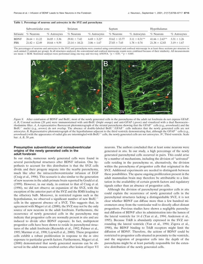
Figure 9. After coinfusion of BDNF and BrdU, most of the newly generated cells in the parenchyma of the adult rat forebrain do not express GFAP.A, B, Coronal sections (20 �m) were immunostained with anti-BrdU (bright orange) and anti-GFAP ( green) and visualized with a dual fluorescein–rhodamine filter. A, A representative fluorescent photomicrograph of the striatal parenchyma showing that the GFAP � cells (e.g., arrowheads) and theBrdU � nuclei (e.g., arrows) do not overlap. The absence of double-labeled BrdU �/GFAP � cells indicates that the newly generated cells are notastrocytes. B, Representative photomicrograph of the hypothalamus adjacent to the third ventricle demonstrating that, although the GFAP � cells (e.g.,arrowheads) with the appearance of radial glia are intermingled with BrdU � cells, the newly generated cells are not astrocytes. 3V, Third ventricle. Scalebar: A, B, 50 �m.
Pencea et al. • Infusion of BDNF Leads to New Neurons in the Forebrain J. Neurosci., September 1, 2001, 21(17):6706–6717 6715

The possible direct and indirect involvement of TrkBreceptors in the production of newly generatedneurons in the adult forebrainAn unexpected finding of this study was the variable level ofBrdU incorporation in structures presumably exposed to a uni-form concentration of BDNF–BrdU. For example, although therewere numerous BrdU� cells in the thalamic habenular nuclei,there were no BrdU� cells in the overlying part of the dentategyrus, also adjacent to the third ventricle. Moreover, the struc-tures closest to the ventricle did not always exhibit the highestlevel of BrdU incorporation. For example, in the hypothalamus,there is a higher density of BrdU� cells in the paraventricularnucleus, despite its location farther away from the ventricle. Anexplanation for the difference in the density of BrdU� cells is thatthe regions with high cell proliferation have high TrkB expression,whereas the regions with a very low level of cell proliferation havelow TrkB expression. Together, these findings suggest that thedistribution of newly generated cells after BDNF infusion may beat least partially influenced by the pattern of BDNF expression.
Although the regions with increased cell proliferation coincidewith areas of high TrkB expression, the newly generated BrdU�
cells were not TrkB�. The absence of the TrkB receptor from themembrane of the BrdU� cells may indicate that BDNF does notact directly on progenitor cells but rather initially influences thecells surrounding them. This notion is supported by our confocalanalysis in which TrkB� cells were frequently adjacent to BrdU�
cells. The possibility that TrkB� cells exert a paracrine influenceon progenitors cannot be ruled out. Another hypothesis to ac-count for the absence of TrkB receptor from the membrane of thenewly generated cells is the downregulation of the TrkB receptorafter BDNF exposure. Alternatively, after binding BDNF, theprogenitor cells may internalize their TrkB receptor, which re-sults in the masking of the antigenic sites recognized by ourantibody. Previous studies have shown that internalization andtransport of the ligand–receptor complex are required to initiate
some responses to neurotrophins (Bergeron et al., 1995; Zhang etal., 2000). Hence, it will be important to elucidate whether theinternalization of the BDNF–TrkB complex plays a role in thesurvival and/or differentiation of the newly generated cells afterBDNF infusion of the adult forebrain.
Concluding remarks on the potential relevance ofBDNF infusion to strategies for neuronal replacementRecent studies have demonstrated that, in brains affected byneurodegenerative diseases, discrete regions characterized byloss of neurons have abnormally low levels of BDNF expression.In particular, Ferrer et al. (2000) have shown that, in Hunting-ton’s disease, BDNF is reduced by 53–82% in the caudate andputamen, two regions exhibiting neuronal loss. Similar studiesalso suggest that, in Alzheimer’s disease, decreased levels ofBDNF leads to lack of trophic support for susceptible neurons,and thus, contributes to the degeneration of specific neuronalpopulations (Hock et al., 2000). Collectively, these findings indi-cate that BDNF is important for the survival of certain neuronalpopulations in the adult forebrain. Thus, if BDNF could beprovided exogenously, it could potentially serve to promote theformation of numerous new neurons in extensive regions of themammalian brain. Our future studies will seek to reveal whetherBDNF can rescue degenerated neurons or help achieve replace-ment of neurons already lost as a result of a disease process.
REFERENCESAltman J, Das GD (1965) Autoradiographic and histological evidence of
postnatal hippocampal neurogenesis in rats. J Comp Neurol124:319–335.
Alvarez-Buylla A, Theelen M, Nottebohm F (1990) Proliferation “hotspots” in adult avian ventricular zone reveals radial cell division.Neuron 5:101–109.
Anderson KD, Alderson RF, Altar CA, DiStefano PS, Corcoran TL,Lindsay RM, Wiegand SJ (1995) Differential distribution of exoge-nous BDNF, NGF, and NT-3 in the brain corresponds to the relativeabundance and distribution of high-affinity and low-affinity neurotro-phin receptors. J Comp Neurol 357:296–317.
Bergeron JJ, Di Guglielmo GM, Baass PC, Authier F, Posner BI (1995)Endosomes, receptor tyrosine kinase internalization and signal trans-duction. Biosci Rep 15:411–418.
Bernhardt R, Huber G, Matus A (1985) Differences in the developmen-tal patterns of three microtubule associated proteins in the rat cerebel-lum. J Neurosci 5:977–991.
Bignami A, Eng LF, Dahl D, Uyeda CT (1972) Localization of the glialfibrillary acidic protein in astrocytes by immunofluorescence. Brain Res43:429–435.
Cheng CM, Joncas G, Reinhardt RR, Farrer R, Quarles R, Janssen J,McDonald MP, Crawley JN, Powell-Braxton L, Bondy CA (1998)Biochemical and morphometric analyses show that myelination in theinsulin-like growth factor 1 null brain is proportionate to its neuronalcomposition. J Neurosci 18:5673–5681.
Chiasson BJ, Tropepe V, Morshead CM, van der Kooy D (1999) Adultmammalian forebrain ependymal and subependymal cells demonstrateproliferative potential, but only subependymal cells have neural stemcell characteristics. J Neurosci 19:4462–4471.
Craig CG, Tropepe V, Morshead CM, Reynolds BA, Weiss S, van derKooy D (1996) In vivo growth factor expansion of endogenous sub-ependymal neural precursor cell populations in the adult mouse brain.J Neurosci 16:2649–2658.
Easter Jr SS, Ross LS, Frankfurter A (1993) Initial tract formation in themouse brain. J Neurosci 13:285–299.
Ferrer I, Goutan E, Marin C, Rey MJ, Ribalta T (2000) Brain-derivedneurotrophic factor in Huntington disease. Brain Res 866:257–261.
Frederiksen K, McKay RD (1988) Proliferation and differentiation of ratneuroepithelial precursor cells in vivo. J Neurosci 8:1144–1151.
Goldman SA, Kirschenbaum B, Harrison-Restelli C, Thaler HT (1997)Neuronal precursors of the adult rat subependymal zone persist intosenescence, with no decline in spatial extent or response to BDNF.J Neurobiol 32:554–566.
Gould E, Reeves AJ, Graziano MS, Gross CG (1999) Neurogenesis inthe neocortex of adult primates. Science 286:548–552.
Graziadei PPC, Monti Graziadei GA (1976) Neurogenesis and neuronregeneration in the olfactory system of mammals. III. Deafferentation
Figure 10. The protrusion of hyperplastic polyps into the lateral ventri-cle of the brain of an adult rat after the intracerebroventricular adminis-tration of BDNF–BrdU or PBS–BrdU. A, B, Coronal sections (20 �m)stained with anti-BrdU (bright orange) and TuJ1 ( green) and visualized bya dual fluorescein–rhodamine filter. A, A representative hyperplasticpolyp situated along the striatal wall formed 16 d after a 12 d interval ofintracerebroventricular PBS–BrdU administration. The polyp consists ofmultiple layers of BrdU �/TuJ1 � cells surrounding a central TuJ1 �/BrdU � core. Notice the abundance of BrdU � cells in the polyp comparedwith the low number of BrdU � cells in the adjacent striatal SVZ (e.g.,arrows). B, A representative hyperplastic polyp from the striatal wall afterintracerebroventricular administration of BDNF and BrdU. The polypcontains cells with a high level of TuJ1 immunoreactivity and a low levelof BrdU incorporation and is considerably smaller than the polyp in Aresulting from the PBS infusion, indicating that the hyperplastic polypformed after BDNF administration is more differentiated than that afterPBS infusion. Also note that, in B, there are numerous BrdU � cells in thestriatal SVZ immediately underlying the polyp. LV, Lateral ventricle;SPT, septum; ST, striatum. Scale bars: A, 100 �m; B, 50 �m.
6716 J. Neurosci., September 1, 2001, 21(17):6706–6717 Pencea et al. • Infusion of BDNF Leads to New Neurons in the Forebrain

and reinnervation of the olfactory bulb following section of the filaolfactoria in rat. J Neurocytol 9:145–162.
Gritti A, Frolichsthal-Schoeller P, Galli R, Parati EA, Cova L, Pagano SF,Bjornson CR, Vescovi AL (1999) Epidermal and fibroblast growthfactors behave as mitogenic regulators for a single multipotent stemcell-like population from the subventricular region of the adult mouseforebrain. J Neurosci 19:3287–3297.
Hock C, Heese K, Hulette C, Rosenberg C, Otten U (2000) Region-specific neurotrophin imbalances in Alzheimer disease: decreased lev-els of brain-derived neurotrophic factor and increased levels of nervegrowth factor in hippocampus and cortical areas. Arch Neurol57:846–851.
Hockfield S, McKay RD (1985) Identification of major cell classes in thedeveloping mammalian nervous system. J Neurosci 5:3310–3328.
Huard JM, Youngentob SL, Goldstein BJ, Luskin MB, Schwob JE(1998) Adult olfactory epithelium contains multipotent progenitorsthat give rise to neurons and non-neural cells. J Comp Neurol400:469–486.
Johnson GV, Jope RS (1992) The role of microtubule-associated protein2 (MAP-2) in neuronal growth, plasticity, and degeneration. J NeurosciRes 33:505–512.
Kaplan MS, Bell DH (1984) Mitotic neuroblasts in the 9-day-old and11-month-old rodent hippocampus. J Neurosci 4:1429–1441.
Kirschenbaum B, Goldman SA (1995) Brain-derived neurotrophic fac-tor promotes the survival of neurons arising from the adult rat fore-brain subependymal zone. Proc Natl Acad Sci USA 92:210–214.
Kuhn HG, Dickinson-Anson H, Gage FH (1996) Neurogenesis in thedentate gyrus of the adult rat: age-related decrease of neuronal pro-genitor proliferation. J Neurosci 16:2027–2033.
Kuhn HG, Winkler J, Kempermann G, Thal LJ, Gage FH (1997) Epi-dermal growth factor and fibroblast growth factor-2 have differenteffects on neural progenitors in the adult rat brain. J Neurosci17:5820–5829.
Laywell ED, Rakic P, Kukekov VG, Holland EC, Steindler DA (2000)Identification of a multipotent astrocytic stem cell in the immature andadult mouse brain. Proc Natl Acad Sci USA 97:13883–13888.
Lee MK, Rebhun LI, Frankfurter A (1990) Posttranslational modifica-tion of class III beta-tubulin. Proc Natl Acad Sci USA 87:7195–7199.
Lendahl U, McKay RD (1990) The use of cell lines in neurobiology.Trends Neurosci 13:132–137.
Levison SW, Goldman JE (1993) Both oligodendrocytes and astrocytesdevelop from progenitors in the subventricular zone of postnatal ratforebrain. Neuron 10:201–212.
Lois C, Alvarez-Buylla A (1994) Long-distance neuronal migration inthe adult mammalian brain. Science 264:1145–1148.
Luskin MB (1993) Restricted proliferation and migration of postnatallygenerated neurons derived from the forebrain subventricular zone.Neuron 11:173–189.
Magavi SS, Leavitt BR, Macklis JD (2000) Induction of neurogenesis inthe neocortex of adult mice. Nature 405:951–955.
Marmur R, Mabie PC, Gokhan S, Song Q, Kessler JA, Mehler MF(1998) Isolation and developmental characterization of cerebral corti-cal multipotent progenitors. Dev Biol 204:577–591.
Morshead CM, van der Kooy D (1992) Postmitotic death is the fate ofconstitutively proliferating cells in the subependymal layer of the adultmouse brain. J Neurosci 12:249–256.
Morshead CM, Reynolds BA, Craig CG, McBurney MW, Staines WA,Morassutti D, Weiss S, van der Kooy D (1994) Neural stem cells in the
adult mammalian forebrain: a relatively quiescent subpopulation ofsubependymal cells. Neuron 13:1071–1082.
Palmer TD, Ray J, Gage FH (1995) FGF-2-responsive neuronal progen-itors reside in proliferative and quiescent regions of the adult rodentbrain. Mol Cell Neurosci 6:474–486.
Palmer TD, Takahashi J, Gage FH (1997) The adult rat hippocampuscontains primordial neural stem cells. Mol Cell Neurosci 8:389–404.
Paxinos G, Watson C (1982) The rat brain in stereotaxic coordinates.New York: Academic.
Pencea V, Bingaman KB, Wiegand SJ, Luskin MB (1999) Infusion ofBDNF into the lateral ventricle of the adult rat leads to an increase inthe number of newly generated cells in the fore, mid and hindbrainparenchyma. Soc Neurosci Abstr 25:2045.
Privat A (1975) Postnatal gliogenesis in the mammalian brain. Int RevCytol 40:281–323.
Raedler E, Raedler A (1978) Autoradiographic study of early neurogen-esis in rat neocortex. Anat Embryol (Berl) 154:267–284.
Ranscht B, Clapshaw PA, Price J, Noble M, Seifert W (1982) Develop-ment of oligodendrocytes and Schwann cells studied with a monoclonalantibody against galactocerebroside. Proc Natl Acad Sci USA79:2709–2713.
Ranscht B, Wood PM, Bunge RP (1987) Inhibition of in vitro peripheralmyelin formation by monoclonal anti-galactocerebroside. J Neurosci7:2936–2947.
Reynolds BA, Weiss S (1992) Generation of neurons and astrocytesfrom isolated cells of the adult mammalian central nervous system.Science 255:1707–1710.
Reynolds BA, Weiss S (1996) Clonal and population analyses demon-strate that an EGF-responsive mammalian embryonic CNS precursor isa stem cell. Dev Biol 175:1–13.
Reynolds BA, Tetzlaff W, Weiss S (1992) A multipotent EGF-responsive striatal embryonic progenitor cell produces neurons andastrocytes. J Neurosci 12:4565–4574.
Schlessinger AR, Cowan WM, Gottlieb DI (1975) An autoradiographicstudy of the time of origin and the pattern of granule cell migration inthe dentate gyrus of the rat. J Comp Neurol 159:149–175.
Sidman RL, Rakic P (1973) Neuronal migration, with special referenceto developing human brain: a review. Brain Res 62:1–35.
Vaysse PJ, Goldman JE (1990) A clonal analysis of glial lineages inneonatal forebrain development in vitro. Neuron 5:227–235.
Weinstein DE, Burrola P, Kilpatrick TJ (1996) Increased proliferationof precursor cells in the adult rat brain after targeted lesioning. BrainRes 743:11–16.
Yan Q, Matheson C, Sun J, Radeke MJ, Feinstein SC, Miller JA (1994)Distribution of intracerebral ventricularly administered neurotrophinsin rat brain and its correlation with trk receptor expression. Exp Neurol127:23–36.
Zhang Y, Moheban DB, Conway BR, Bhattacharyya A, Segal RA (2000)Cell surface Trk receptors mediate NGF-induced survival while inter-nalized receptors regulate NGF-induced differentiation. J Neurosci20:5671–5678.
Zigova T, Betarbet R, Soteres BJ, Brock S, Bakay RA, Luskin MB(1996) A comparison of the patterns of migration and the destinationsof homotopically transplanted neonatal subventricular zone cells andheterotopically transplanted telencephalic ventricular zone cells. DevBiol 173:459–474.
Zigova T, Pencea V, Wiegand SJ, Luskin MB (1998) Intraventricularadministration of BDNF increases the number of newly generatedneurons in the adult olfactory bulb. Mol Cell Neurosci 11:234–245.
Pencea et al. • Infusion of BDNF Leads to New Neurons in the Forebrain J. Neurosci., September 1, 2001, 21(17):6706–6717 6717