influence of excitation/inhibition imbalance on local processing
TRANSCRIPT
Influence of Excitation/Inhibition Imbalance on Local Processing Bias inAutism Spectrum Disorder
Yukie Nagai, Takakazu Moriwaki1, and Minoru Asada({yukie, takakazu.moriwaki, asada}@ams.eng.osaka-u.ac.jp)
Graduate School of Engineering, Osaka University2-1 Yamada-oka, Suita, Osaka 565-0871, Japan
AbstractPeople with autism spectrum disorder (ASD) tend to detect lo-cal patterns of visual stimuli more quickly than global patterns,which is opposite to the behavior of typically developing peo-ple. We hypothesized that the imbalance between excitationand inhibition neurons in the visual cortex causes the local pro-cessing bias observed in ASD. Stronger inhibitory connectionscould diminish the neural activities and thus prevent global fea-ture integration, whereas properly balanced connections wouldenable the cortex to detect features of any size. We verifiedour hypothesis by employing a computational neural networkcalled a neocognitron. Our experimental results demonstratedthat the network with stronger inhibitory connections exhib-ited a local processing bias, whereas the network with properlyadjusted connections showed a moderate global bias. More-over, the networks with extremely strong or weak inhibitionsrevealed no perception bias. These results suggest that an ex-citation/inhibition imbalance causes multiple types of atypicalperception in ASD.Keywords: autism spectrum disorder; local processing bias;excitation/inhibition balance; neocognitron
IntroductionAutism spectrum disorder (ASD) is a neurodevelopmentaldisorder that is characterized by impaired social interactionand communication (Baron-Cohen, 1995). For example, peo-ple with ASD show difficulties in making eye contact, es-tablishing joint attention, and reading others’ intentions—abilities that are otherwise usually acquired in the first fewyears of life. Unlike the traditional view of ASD, recent stud-ies in cognitive science, neuroscience, and Tojisha-Kenkyu2
suggest that a core problem of ASD lies in the lower per-ception and action rather than in higher cognition (Frith &Happe, 1994; Happe & Frith, 2006; Ayaya & Kumagaya,2008). A new hypothesis was proposed called “weak cen-tral coherence” (Frith & Happe, 1994; Happe & Frith, 2006),suggesting that a diminished ability to integrate informationor an enhanced ability in the lower perception causes socialdifficulties in ASD. Researchers in Tojisha-Kenkyu has pro-vided further insight into the mechanism. They argue thatASD may be associated with an abnormal sensitivity to pre-diction error, and thus individuals with ASD acquire a differ-ent internal model from typically developing people (Ayaya& Kumagaya, 2008). Difficulties in social communicationmay therefore be caused by the difference between the inter-nal model of ASD and that of non-ASD rather than by dis-abilities intrinsic to ASD (Nagai, 2015).
1T. Moriwaki is with Ishida Co., Ltd. since April 2014.2A developing research area, in which researchers suffering from
ASD investigate their atypical sensorimotor experiences from a first-person perspective.
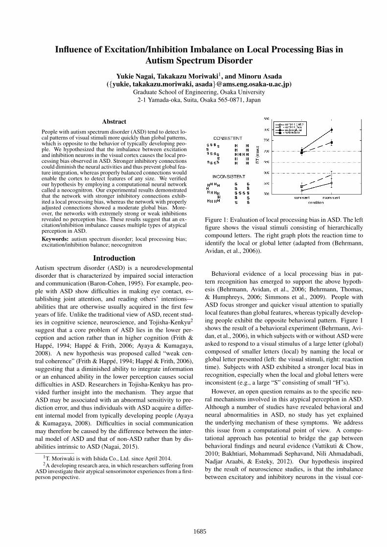
Figure 1: Evaluation of local processing bias in ASD. The leftfigure shows the visual stimuli consisting of hierarchicallycompound letters. The right graph plots the reaction time toidentify the local or global letter (adapted from (Behrmann,Avidan, et al., 2006)).
Behavioral evidence of a local processing bias in pat-tern recognition has emerged to support the above hypoth-esis (Behrmann, Avidan, et al., 2006; Behrmann, Thomas,& Humphreys, 2006; Simmons et al., 2009). People withASD focus stronger and quicker visual attention to spatiallylocal features than global features, whereas typically develop-ing people exhibit the opposite behavioral pattern. Figure 1shows the result of a behavioral experiment (Behrmann, Avi-dan, et al., 2006), in which subjects with or without ASD wereasked to respond to a visual stimulus of a large letter (global)composed of smaller letters (local) by naming the local orglobal letter presented (left: the visual stimuli, right: reactiontime). Subjects with ASD exhibited a stronger local bias inrecognition, especially when the local and global letters wereinconsistent (e.g., a large “S” consisting of small “H”s).
However, an open question remains as to the specific neu-ral mechanisms involved in this atypical perception in ASD.Although a number of studies have revealed behavioral andneural abnormalities in ASD, no study has yet explainedthe underlying mechanism of these symptoms. We addressthis issue from a computational point of view. A compu-tational approach has potential to bridge the gap betweenbehavioral findings and neural evidence (Vattikuti & Chow,2010; Bakhtiari, Mohammadi Sephavand, Nili Ahmadabadi,Nadjar Araabi, & Esteky, 2012). Our hypothesis inspiredby the result of neuroscience studies, is that the imbalancebetween excitatory and inhibitory neurons in the visual cor-
1685
tex causes a local processing bias in ASD. Previous studieshave reported an atypical excitation/inhibition balance (E/Ibalance) and relevant gamma-band activity in ASD (Sun etal., 2012; Snijders, Milivojevic, & Kemner, 2013). Further-more, the association between the E/I imbalance and socialdysfunction was demonstrated in a mouse experiment (Yizharet al., 2011). Based on this evidence, we consider that theE/I balance might influence the integration of spatial infor-mation. For example, stronger inhibitory connections coulddiminish the response of succeeding neurons and thus hinderglobal feature integration. We verify our hypothesis by em-ploying a computational neural network called a neocogni-tron (Fukushima & Miyake, 1982; Fukushima, 1988, 2003).
Neocognitron: A Hierarchical Neural Networkfor Pattern Recognition
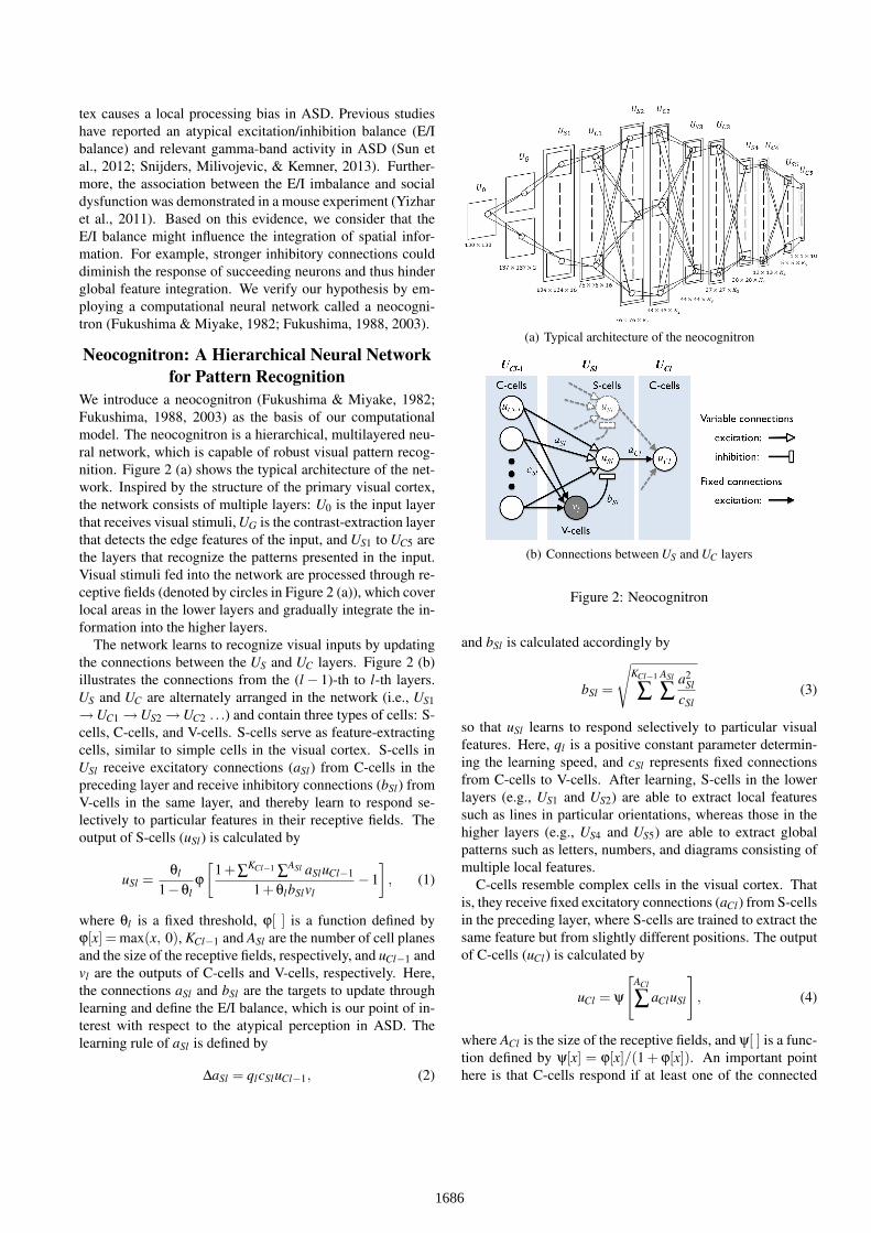
We introduce a neocognitron (Fukushima & Miyake, 1982;Fukushima, 1988, 2003) as the basis of our computationalmodel. The neocognitron is a hierarchical, multilayered neu-ral network, which is capable of robust visual pattern recog-nition. Figure 2 (a) shows the typical architecture of the net-work. Inspired by the structure of the primary visual cortex,the network consists of multiple layers: U0 is the input layerthat receives visual stimuli, UG is the contrast-extraction layerthat detects the edge features of the input, and US1 to UC5 arethe layers that recognize the patterns presented in the input.Visual stimuli fed into the network are processed through re-ceptive fields (denoted by circles in Figure 2 (a)), which coverlocal areas in the lower layers and gradually integrate the in-formation into the higher layers.
The network learns to recognize visual inputs by updatingthe connections between the US and UC layers. Figure 2 (b)illustrates the connections from the (l − 1)-th to l-th layers.US and UC are alternately arranged in the network (i.e., US1→UC1 →US2 →UC2 . . .) and contain three types of cells: S-cells, C-cells, and V-cells. S-cells serve as feature-extractingcells, similar to simple cells in the visual cortex. S-cells inUSl receive excitatory connections (aSl) from C-cells in thepreceding layer and receive inhibitory connections (bSl) fromV-cells in the same layer, and thereby learn to respond se-lectively to particular features in their receptive fields. Theoutput of S-cells (uSl) is calculated by
uSl =θl
1−θlϕ
[1+∑KCl−1 ∑ASl aSluCl−1
1+θlbSlvl−1
], (1)
where θl is a fixed threshold, ϕ[ ] is a function defined byϕ[x] = max(x, 0), KCl−1 and ASl are the number of cell planesand the size of the receptive fields, respectively, and uCl−1 andvl are the outputs of C-cells and V-cells, respectively. Here,the connections aSl and bSl are the targets to update throughlearning and define the E/I balance, which is our point of in-terest with respect to the atypical perception in ASD. Thelearning rule of aSl is defined by
∆aSl = qlcSluCl−1, (2)
(a) Typical architecture of the neocognitron
(b) Connections between US and UC layers
Figure 2: Neocognitron
and bSl is calculated accordingly by
bSl =
√KCl−1
∑ASl
∑ a2Sl
cSl(3)
so that uSl learns to respond selectively to particular visualfeatures. Here, ql is a positive constant parameter determin-ing the learning speed, and cSl represents fixed connectionsfrom C-cells to V-cells. After learning, S-cells in the lowerlayers (e.g., US1 and US2) are able to extract local featuressuch as lines in particular orientations, whereas those in thehigher layers (e.g., US4 and US5) are able to extract globalpatterns such as letters, numbers, and diagrams consisting ofmultiple local features.
C-cells resemble complex cells in the visual cortex. Thatis, they receive fixed excitatory connections (aCl) from S-cellsin the preceding layer, where S-cells are trained to extract thesame feature but from slightly different positions. The outputof C-cells (uCl) is calculated by
uCl = ψ
[ACl
∑aCluSl
], (4)
where ACl is the size of the receptive fields, and ψ[ ] is a func-tion defined by ψ[x] = ϕ[x]/(1 + ϕ[x]). An important pointhere is that C-cells respond if at least one of the connected
1686

S-cells yields an output. C-cells thus spatially blur the re-sponses of S-cells and make the network robust against posi-tion errors in visual stimuli. The mechanism of the neocogni-tron is described in greater detail in Fukushima and Miyake(1982) and Fukushima (1988, 2003).
Influence of E/I Balance onLocal/Global Information Processing
We hypothesize that the E/I balance influences the lo-cal/global processing bias in pattern recognition. Figure 3illustrates how the relative strength of the inhibitory connec-tions bSl from V-cells affects the output of S-cells uSl in theneocognitron: (a) proper inhibitory connections, (b) strongerinhibitory connections, and (c) weaker inhibitory connec-tions. We suppose that the neocognitron has been trained withvisual stimuli, each of which contains a single number butwith various types, sizes, and positions. The network is thenpresented with hierarchically compound numbers, as shownin the leftmost of Figure 3 (a), where multiple small number“3”s form a large number “2” according to their close posi-tioning.
If the network has properly adjusted connections, it shouldbe able to detect the numbers presented in the input imageregardless of their sizes and positions. As illustrated in therightmost image of Figure 3 (a), the output of S-cells, whichreceive proper inhibitory connections from V-cells, maintainsthe features of both “3” and “2” as the integration of multi-ple “3”s. The extracted features then enable the network torecognize both the local and global patterns in the succeed-ing layers. In contrast, stronger or weaker inhibitory connec-tions cause local or global processing bias in the recognition.If inhibitory neurons have stronger connections, as shown inFigure 3 (b), they suppress the global response of S-cells sothat they extract only the core features of the input. Underthis scenario, only the local number “3” becomes visible, butnot the global number “2”, because of the sparse activationof S-cells. On the other hand, weaker inhibitory connectionsproduce the opposite effect. Figure 3 (c) depicts how weakerinhibitions enhance the responses of S-cells and therefore di-minish the local features of the input. In this case, only theglobal number “2” becomes visible by connecting the localnumber “3”s, while “3” per se becomes increasingly difficultto recognize.
Taken together, our hypothesis suggests that the atypicalperception in ASD as well as the typical perception in non-ASD can be modeled by the E/I balance in the visual cortex.Of particular importance, the use of a unified architecture al-lows for not only the local processing bias but also anothertype of symptom in ASD to be modeled. Behavioral studiessuggest two types of ASD symptoms: hyperesthesia and hy-poesthesia. Hyperesthesia corresponds to the local process-ing bias because it exhibits increased sensitivity to perceptualstimuli. Hypoesthesia, on the other hand, corresponds to asuper-global or no bias in pattern recognition (i.e., difficultyin pattern recognition), because it shows reduced sensitivity
(a) Proper inhibitory connections maintain both local and globalfeatures.
(b) Stronger inhibitory connections ex-tract only local features.
(c) Weaker inhibitory connections empha-size global features.
Figure 3: Influence of E/I balance on local/global processingbias
to the stimuli. Our computational model thus reveals the un-derlying neural mechanism based on a common architecture:stronger inhibitory connections (i.e., a lower E/I balance) maycause hyperesthesia, whereas weaker inhibitory connections(i.e., a higher E/I balance) may cause hypoesthesia.
Pattern Recognition ExperimentTraining and Testing DataTo verify our hypothesis, we conducted a pattern recognitionexperiment using the neocognitron. The visual stimuli usedfor the training and testing of the network are presented inFigures 4 (a) and (b), respectively. Each stimulus was a blackand white image with a size of 130 × 130 pixels. The trainingdata contained a single number per image, where the numberwas “0” to “9” drawn in different sizes and positions (for a to-tal of 400 images). The neocognitron was trained in a super-
1687
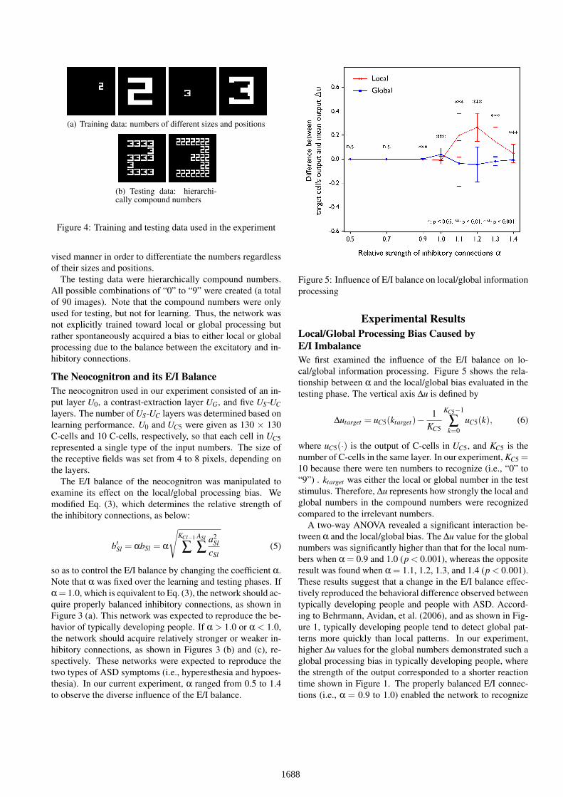
(a) Training data: numbers of different sizes and positions
(b) Testing data: hierarchi-cally compound numbers
Figure 4: Training and testing data used in the experiment
vised manner in order to differentiate the numbers regardlessof their sizes and positions.
The testing data were hierarchically compound numbers.All possible combinations of “0” to “9” were created (a totalof 90 images). Note that the compound numbers were onlyused for testing, but not for learning. Thus, the network wasnot explicitly trained toward local or global processing butrather spontaneously acquired a bias to either local or globalprocessing due to the balance between the excitatory and in-hibitory connections.
The Neocognitron and its E/I BalanceThe neocognitron used in our experiment consisted of an in-put layer U0, a contrast-extraction layer UG, and five US-UClayers. The number of US-UC layers was determined based onlearning performance. U0 and UC5 were given as 130 × 130C-cells and 10 C-cells, respectively, so that each cell in UC5represented a single type of the input numbers. The size ofthe receptive fields was set from 4 to 8 pixels, depending onthe layers.
The E/I balance of the neocognitron was manipulated toexamine its effect on the local/global processing bias. Wemodified Eq. (3), which determines the relative strength ofthe inhibitory connections, as below:
b′Sl = αbSl = α
√KCl−1
∑ASl
∑ a2Sl
cSl(5)
so as to control the E/I balance by changing the coefficient α.Note that α was fixed over the learning and testing phases. Ifα = 1.0, which is equivalent to Eq. (3), the network should ac-quire properly balanced inhibitory connections, as shown inFigure 3 (a). This network was expected to reproduce the be-havior of typically developing people. If α > 1.0 or α < 1.0,the network should acquire relatively stronger or weaker in-hibitory connections, as shown in Figures 3 (b) and (c), re-spectively. These networks were expected to reproduce thetwo types of ASD symptoms (i.e., hyperesthesia and hypoes-thesia). In our current experiment, α ranged from 0.5 to 1.4to observe the diverse influence of the E/I balance.
Figure 5: Influence of E/I balance on local/global informationprocessing
Experimental ResultsLocal/Global Processing Bias Caused byE/I ImbalanceWe first examined the influence of the E/I balance on lo-cal/global information processing. Figure 5 shows the rela-tionship between α and the local/global bias evaluated in thetesting phase. The vertical axis ∆u is defined by
∆utarget = uC5(ktarget)−1
KC5
KC5−1
∑k=0
uC5(k), (6)
where uC5(·) is the output of C-cells in UC5, and KC5 is thenumber of C-cells in the same layer. In our experiment, KC5 =10 because there were ten numbers to recognize (i.e., “0” to“9”) . ktarget was either the local or global number in the teststimulus. Therefore, ∆u represents how strongly the local andglobal numbers in the compound numbers were recognizedcompared to the irrelevant numbers.
A two-way ANOVA revealed a significant interaction be-tween α and the local/global bias. The ∆u value for the globalnumbers was significantly higher than that for the local num-bers when α = 0.9 and 1.0 (p < 0.001), whereas the oppositeresult was found when α = 1.1, 1.2, 1.3, and 1.4 (p < 0.001).These results suggest that a change in the E/I balance effec-tively reproduced the behavioral difference observed betweentypically developing people and people with ASD. Accord-ing to Behrmann, Avidan, et al. (2006), and as shown in Fig-ure 1, typically developing people tend to detect global pat-terns more quickly than local patterns. In our experiment,higher ∆u values for the global numbers demonstrated such aglobal processing bias in typically developing people, wherethe strength of the output corresponded to a shorter reactiontime shown in Figure 1. The properly balanced E/I connec-tions (i.e., α = 0.9 to 1.0) enabled the network to recognize
1688
the global numbers more strongly than the local numbers. Incontrast, higher ∆u values for the local numbers demonstratedthe local processing bias in ASD. Inhibitory connections en-hanced by α = 1.1 to 1.4 forced the network to extract thelocal features more strongly than the global ones, which gen-erated an ASD-like bias toward the local information.
Moreover, our results suggest that our computationalmodel with a variable E/I balance can reproduce multipletypes of ASD symptoms. ASD is characterized by both hy-poesthesia as well as hyperesthesia in sensory perception. Asmentioned above, hyperesthesia might be related to a localprocessing bias, whereas hypoesthesia might be linked to asuper-global or no processing bias. When α = 0.5 to 0.7,the neocognitron did not show any bias toward the local orglobal patterns. Indeed, the network could recognize neitherthe local nor global numbers. This inability to recognize thepatterns also appeared when α > 1.4. These results indicatethat hypoesthesia in ASD shares a common neural mecha-nism with hyperesthesia, and that a change in the E/I balanceproduces the difference between these symptoms.
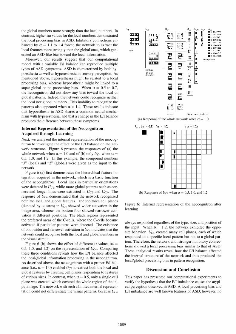
Internal Representation of the NeocognitronAcquired through LearningNext, we analyzed the internal representation of the neocog-nitron to investigate the effect of the E/I balance on the net-work structure. Figure 6 presents the responses of (a) thewhole network when α = 1.0 and of (b) only UC4 when α =0.5, 1.0, and 1.2. In this example, the compound numbers“3” (local) and “2” (global) were given as the input to thenetwork.
Figure 6 (a) first demonstrates the hierarchical feature in-tegration acquired in the network, which is a basic functionof the neocognitron. Local lines in particular orientationswere detected in UC1, while more global patterns such as cor-ners and longer lines were extracted in UC2 and UC3. Theresponse of UC4 demonstrated that the network recognizedboth the local and global features. The top three cell planes(denoted by squares) in UC4 showed wider activation in theimage area, whereas the bottom four showed narrower acti-vation at different positions. The black regions representedthe preferred areas of the C-cells, where the C-cells becameactivated if particular patterns were detected. The existenceof both wider and narrower activation in UC4 indicates that thenetwork could recognize both the local and global numbers inthe visual stimuli.
Figure 6 (b) shows the effect of different α values (α =0.5, 1.0, and 1.2) on the representation of UC4. Comparingthese three conditions reveals how the E/I balance affectedthe local/global information processing in the neocognitron.As described above, the neocognitron with a proper E/I bal-ance (i.e., α = 1.0) enabled UC4 to extract both the local andglobal features by creating cell planes responding to featuresof various sizes. In contrast, when α = 0.5, only a single cellplane was created, which covered the whole region of the in-put image. The network with such a limited internal represen-tation could not differentiate the visual patterns, because UC4
(a) Response of the whole network when α = 1.0
(b) Response of UC4 when α = 0.5, 1.0, and 1.2
Figure 6: Internal representation of the neocognitron afterlearning
always responded regardless of the type, size, and position ofthe input. When α = 1.2, the network exhibited the oppo-site behavior. UC4 created many cell planes, each of whichresponded to a specific local pattern but not to a global pat-tern. Therefore, the network with stronger inhibitory connec-tions showed a local processing bias similar to that of ASD.These analytical results reveal how the E/I balance affectedthe internal structure of the network and thus produced thelocal/global processing bias in pattern recognition.
Discussion and ConclusionThis paper has presented our computational experiments toverify the hypothesis that the E/I imbalance causes the atypi-cal perception observed in ASD. A local processing bias andE/I imbalance are well known features of ASD; however, no
1689

previous study has demonstrated their association. Our com-putational model revealed that the E/I balance affects the in-ternal representation of the visual cortex and thus produces alocal/global processing bias in pattern recognition.
One of our most notable experimental results is that theunified neural architecture could reproduce multiple symp-toms of ASD as well as typical non-ASD behavior by chang-ing only one parameter (i.e., the E/I balance). A proper E/Ibalance led to a behavioral pattern representative of typicallydeveloping people, whereas a higher or a lower E/I balanceresulted in hyperesthesia or hypoesthesia, which is represen-tative of ASD. Only a change in the E/I balance generatedthe behavioral differences between typical and atypical vi-sual perception. This result provides new insight into ASDand further supports a recent argument that the hyperesthe-sia and hypoesthesia of ASD are two sides of the same coin;although their behaviors appear different, they may share acommon underlying mechanism (Ayaya et al., 2013). Ourcomputational model is therefore highly plausible because ofits unified architecture.
There has been another hypothesis about the neural mech-anism underlying local processing bias. A magnocellulardeficit or its abnormal activity might be a cause of the ob-served local processing bias in ASD (Sutherland & Crewther,2010; McCleery, Allman, Carver, & Dobkins, 2007). The hu-man brain has two parallel pathways for visual processing:the magnocellular pathway conveying the global and coarseinformation of visual input, and the parvocellular pathwayconveying the local and fine information of the input. There-fore, a magnocellular deficit in ASD could result in difficultyof recognizing the global feature of a stimulus. Our results donot deny this possibility but instead provide another potentialexplanation for these observed ASD symptoms. Furthermore,our model has an advantage of reproducing multiple aspectsof the ASD symptoms, instead of only the local processingbias, by employing a shared neural architecture. We intend tofurther investigate the relationship between different neuralmodels for ASD.
AcknowledgementsThis work is partially supported by MEXT/JSPS KAK-ENHI (Research Project Numbers: 24119003, 24000012,25700027).
ReferencesAyaya, S., Kawano, T., Mukaiyachi, I., Tojisha-Kenkyukai,
N., Ishihara, K., Ikeda, T., & Kumagaya, S. (2013). Tojishakenkyu no kenkyu (in japanese) (K. Ishihara, Ed.). Igaku-shoin.
Ayaya, S., & Kumagaya, S. (2008). Hattatsu shougai tojishakenkyu (in japanese). Igaku-shoin.
Bakhtiari, R., Mohammadi Sephavand, N., Nili Ahmadabadi,M., Nadjar Araabi, B., & Esteky, H. (2012). Computa-tional model of excitatory/inhibitory ratio imbalance role inattention deficit disorders. Journal of Computational Neu-roscience, 33(2), 389–404.
Baron-Cohen, S. (1995). Mindblindness. MIT Press.Behrmann, M., Avidan, G., Leonard, G. L., Kimchi, R., Luna,
B., Humphreys, K., & Minshew, N. (2006). Configuralprocessing in autism and its relationship to face processing.Neuropsychologia, 44(1), 110–129.
Behrmann, M., Thomas, C., & Humphreys, K. (2006). See-ing it differently: visual processing in autism. Trends inCognitive Sciences, 10(6), 258–264.
Frith, U., & Happe, F. (1994). Autism: beyond ”theory ofmind”. Cognition, 50, 115–132.
Fukushima, K. (1988). Neocognitron: A Hierarchical NeuralNetwork Capable of Visual Pattern Recognition. NeuralNetworks, 1, 119–130.
Fukushima, K. (2003). Neocognitron for handwritten digitrecognition. Neurocomputing, 51, 161–180.
Fukushima, K., & Miyake, S. (1982). Neocognitron: A newalgorithm for pattern recognition tolerant of deformationsand shifts in position. Pattern Recognition, 15(6), 455–469.
Happe, F., & Frith, U. (2006). The Weak Coherence Ac-count: Detail-focused Cognitive Style in Autism SpectrumDisorders. Journal of Autism and Developmental Disor-ders, 36(1), 5–25.
McCleery, J. P., Allman, E., Carver, L. J., & Dobkins, K. R.(2007). Abnormal Magnocellular Pathway Visual Process-ing in Infants at Risk for Autism. Biological Psychiatry,62, 1007–1014.
Nagai, Y. (2015). Mechanism for cognitive development. InH. Ishiguro, M. Osaka, T. Fujikado, & M. Asada (Eds.),Cognitive neuroscience robotics: A: Synthetic approachesto human understanding. Springer (in press).
Simmons, D. R., Robertson, A. E., McKay, L. S., Toal, E.,McAleer, P., & Pollick, F. E. (2009). Vision in autismspectrum disorders. Vision Research, 49(22), 2705–2739.
Snijders, T. M., Milivojevic, B., & Kemner, C. (2013). Atyp-ical excitation-inhibition balance in autism captured by thegamma response to contextual modulation. NeuroImage:Clinical, 3, 65–72.
Sun, L., Grutzner, C., Bolte, S., Wibral, M., Tozman, T.,Schlitt, S., . . . Uhlhaas, P. J. (2012). Impaired Gamma-Band Activity during Perceptual Organization in Adultswith Autism Spectrum Disorders: Evidence for Dysfunc-tional Network Activity in Frontal-Posterior Cortices. Neu-robiology of Disease, 32(28), 9563–9573.
Sutherland, A., & Crewther, D. P. (2010). Magnocellularvisual evoked potential delay with high autism spectrumquotient yields a neural mechanism for altered perception.Brain: A Journal of Neurology, 133, 2089–2097.
Vattikuti, S., & Chow, C. C. (2010). A computational modelfor cerebral cortical dysfunction in autism spectrum disor-ders. Biological Psychiatry, 67(7), 672–678.
Yizhar, O., Fenno, L. E., Prigge, M., Schneider, F., Davidson,T. J., O’Shea, D. J., . . . Deisseroth, K. (2011). Neocorticalexcitation/inhibition balance in information processing andsocial dysfunction. Nature, 477(7363), 171–178.
1690