inflammation objectives/rationale inflammation is a normal response to infection or injury and is...
TRANSCRIPT
Inflammation
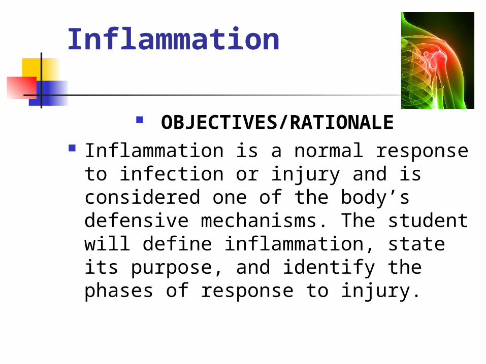
OBJECTIVES/RATIONALE Inflammation is a normal response
to infection or injury and is considered one of the body’s defensive mechanisms. The student will define inflammation, state its purpose, and identify the phases of response to injury.

I. The Cell A. Most signs of disease can be
traced back to damage of individual cells and their attempts at repair - in order to understand what is happening when a person is ill, one must first comprehend the events at the basic level of the cell or sometimes in the area of biochemistry.

I. The Cell (Continued)
B. The cell is the foundation of the structure of the human body. It is seen as a self-sustaining factory that carries on all processes of life
a. respiration b. use & production of energy c. Reproduction d. elimination

I. The Cell (Continued) C. large group of cells that join together
for a specific activity is called tissue D. different tissues that join together for
a specific function is called an organ E. organs that perform together for a
specific function is called an organ system
F. Cell Dysfunction = Organ Dysfunction = Clinical Disease

Defense Mechanisms Immunity- ability of the body to
defend itself against infectious agents, foreign cells, and even abnormal body cells, such as cancer cells
Immunity includes: Nonspecific Defense Mechanisms Specific Defense Mechanisms

Nonspecific Defense Mechanisms
Mechanisms that are effective against any foreign agent that enters the body and are referred to as innate immunity Physical and Chemical Barriers:
Skin Tears, saliva, sweat, oil Mucous membranes Cilia

Nonspecific Defense Mechanisms
Phagocytosis- leukocytes engulf and destroy bacteria and other material
Natural Killer Cells- type of leukocyte that recognizes body cells with abnormal membranes
Fever- sign the body is defending itself; phagocytes find and destroy foreign invaders; releasing substances that raise the body temp
Interferon- group of substances that stimulates the immune system (used with cancer)

II. Cellular Injury A. Inflammation the body’s
response to injury

II. Cellular Injury (Continued) B. Limiting factor of inflammation:
cannot occur in tissue that does not have a blood supply.
a. This factor is important in forensic medicine because evidence of inflammation in tissue confirms that an injury occurred while the individual was alive.

II. Cellular Injury (Continued)
b. If no evidence exists, the pathologist can be assured that the person was dead when the injury was inflicted.
c. When Tissue is Destroyed by an Injury. The inflammatory process will only occur along borders of injury where blood supply is maintained, i.e.gangrene

II. Cellular Injury (Continued) D. Inflammation is designed to be
a Beneficial Protective Mechanism - in some instances reaction may become so intense that it becomes harmful to tissues.

II. Cellular Injury (Continued) E. Inflammation vs. Infection a. Where as inflammation is a response
to injury, infection is the invasion of living tissue by pathogens.
b. An infection causes inflammation, but tissue that is inflamed may not be infected. Therefore, inflammation may exist without the presence of microbial pathogens, i.e. sunburn

II. Cellular Injury (Continued) F. Inflammatory Process a. The cells of the immune
system are widely distributed throughout the body, but if infection or tissue damage occurs it is necessary to concentrate them and their products at the site of damage.

Types of Leukocytes (cells) Neutrophils- type of phagoctye
Most common WBC (50-70%) and live for 4-10 hours if not activated; they die as soon as they ingest another cell
1st to arrive at infection site through chemotaxis
Main component of pus and is whitish color and liquefy in tissue
After phagocytosis they release superoxide which is hypoclorous acid (chlorine bleach) to kill microbes

Types of Leukocytes Monocytes- type of phagocyte
1-3% of WBC; low count indicates no infection
Made in bone marrow and spread in body over 1-3 days
Macrophages- eat other cells and attach pathogens and foreign materials; will increase during times of infection
Dendrites- antigen presenting cells because they acquire antigens and show them to T-cells so they can recognize dangerous antigens

Types of Leukocytes Eosinphil- granulocyte that is a
bilobate (2 nucleus) Stains red 1-3% of WBC count increase with
allergic reactions and parasites

Types of Leukocytes Basophils- granulocytes
Rare type of WBC- less than 1% of WBC Stains blue Can release histamine and heparin for
infection Histamine- aids in immune response and
neurotransmittor; makes capillaries more permeable to WBCs
Heparin- an anticoagulant produced in liver and lungs and prevents clotting
Types of Leukocytes Lymphocytes- responsible for immune
response B Cells- make antibodies responsible for
attacking bacteria and toxins-mature in the bone marrow-humoral immunity (antibody production)-enlarge and divide when antibodies are present-some remain dormant until reactivated by same type of antigen

Types of Leukocytes T Cells- type of lymphocyte that
seek out and destroy specific targeted invaders-Produced in the bone marrow but mature
in the thymus1. Helper T Cells- Activates and
deactivates other immune cells; suppressor cells will halt immune system response when needed
2. Killer T Cells- cytotoxic- kills antigens

F. Inflammatory Process (Continued) b. Three major events occur during this
response: 1) Congestion phase: Initially, the
capillaries become engorged and dilated with blood. This increases capillary permeability caused by the reaction of the endothelial cells. This increases amount of blood, hyperemia, causes the heat and redness associated with inflammation

F. Inflammatory Process 2) Leakage - Leukocytes migrate out of the
capillaries into the surrounding Tissues. In the earliest stages of inflammation, neutrophils
are particularly prevalent, but later monocytes and lymphocytes also migrate towards the site of infection. Neutrophils line up within the capillary wall. Monocytes will clear up the debris.
3) Phagocytosis- specialized cells that defend the body against invading microorganisms and speed healing by engulfing cell debris in injured tissues
Inflammatory response The damaged cells release histamine;
this causes the capillary walls to become more permeable.
Plasma and Neutrophils are then able to move out of the blood vessels to tissue; a process called chemotaxis
Plasma and WBCs that escape from capillaries makes up the inflammatory exudate (swelling)

Chemotaxis

Inflammatory response
Polymorphs die after digesting bacteria and toxins and release a substance that liquefy into tissue- causing pus
Monocytes- then clear up the debris The inflammatory exudate contains
plasma protein- fibrin. Fibrin is needed for clotting.

G. Two Major Categories of
Inflammation: Acute and Chronic a. Acute inflammation– a condition of sudden
onset; if resolved, lasts a relatively short time Characteristic Signs that Accompany Acute Inflammation (referred to as cardinal signs)
1. Redness 2. Swelling 3. Pain 4. Increased warmth or heat 5. Loss of movement or function

G. Two Major Categories of Inflammation: Acute and Chronic (Continued)
b. Chronic inflammation– refers to a long duration (weeks, months, years, or even a lifetime)
i. Example of chronic diseases: asthma, allergy or hay fever, diabetes, etc.
ii. Chronic conditions often worsen (chronic progressive diseases) as result of aging process, environment, and cumulative damage of inflammation process

H. Serous fluids associated with inflammation a. Serous Transudates - serum
fluid that passes through membrane or tissue (very watery with low protein content)
i. Due to increased hydrostatic or decreased osmotic pressure in vascular system

H. Serous fluids associated with inflammation (Continued) ii. Example: pulmonary edema -
fluid filling lungs during congestive heart failure (result of decreased osmotic pressure)
iii. Inflammatory reactions involving pleural, pericardial, and peritoneal cavities are associated with serous discharge

Pulmonary Edema

H. Serous fluids associated with inflammation (Continued) b. Serous Exudates - serum fluid that is
cloudy, thick, protein-rich fluid i. created by decreased hydrostatic
pressure and increased osmotic pressure ii. Example: most common in acute
inflammations such as minor burns (resulting in formation of blisters)
Iii. Fibrinous exudate produces layer of fibrinogen, which forms a mesh of fibrin and becomes a scab

Serous Exudates
Associated with CVD
Blisters associated with sun burn

H. Serous fluids associated with inflammation (Continued) c. Purulent exudate – pus producing
fluid i. referred to as pyogenic; inflammation
with pus is called suppurative Abscesses, boils, styes
Bladder infection at autopsy

Specific Defense Mechanism Acquired Immunity- defends against
specific types of microorganisms and pathogens Responds specifically to future
exposures to the same microorganism or pathogen
Immunity can be acquired naturally by infection (ex: Chicken pox)
Immunity can be acquired artificially; (vaccination)

Specific Defenses Antigens- foreign element that
triggers the immune response The antigen is often a protein from
the microorganism or pathogen; the specificity of acquired immunity it the body’s ability to recognize these antigens

Specific Defenses Specific defenses against antigens
include special cells and the body’s lymphatic system
Lymph system- removes lymph and any microorganism before it enters blood Spleen, tonsils, and adenoids
I. Inflammatory Lesions
Inflammatory Lesions – inflammatory reactions result in production of lesion - lesions vary according to level of severity.

I. Inflammatory Lesions
Types of lesions: a. abscess- localized spherical
lesion filled with pus and pyogenic bacteria (usually stphylococci) usually found in many areas of body
including: skin (boils: furuncles and carbuncles), teeth, appendix, bowel, breast, and lung empyema – pus that fills pleural cavity

I. Inflammatory Lesions (Continued)
b. cellulitis – spreading, diffuse infection most commonly involving streptococcal infection of subcutaneous tissues (body is unable to confine infection to localized area) - characterized by congestion and edema

I. Inflammatory Lesions (Continued)
c. ulcers – depressed or excavated lesions on skin or mucosa may appear almost anywhere in body
and may involve many types of organs
stomach and duodenum may be ulcerated by gastric acids
pressure sores (decubitus ulcers) result from wasting away of tissue in bedridden patients

J. Tissue Repair
a. body’s attempt to return to normal; can only occur when bacteria have been destroyed
b. Fibroblasts allow for cut tissue edges to grow back together forming scar tissue Adhesions- connective tissue fibers anchoring
together Keloid- raised and often hard scar (benign
tumor)