infectious bronchitis virus e protein is targeted to the golgi
TRANSCRIPT
JOURNAL OF VIROLOGY,0022-538X/00/$04.0010
May 2000, p. 4319–4326 Vol. 74, No. 9
Copyright © 2000, American Society for Microbiology. All Rights Reserved.
Infectious Bronchitis Virus E Protein Is Targeted to the GolgiComplex and Directs Release of Virus-Like Particles
EMILY CORSE AND CAROLYN E. MACHAMER*
Department of Cell Biology and Anatomy, The Johns HopkinsUniversity School of Medicine, Baltimore, Maryland 21205
Received 14 December 1999/Accepted 3 February 2000
The coronavirus E protein is a poorly characterized small envelope protein present in low levels in virions.We are interested in the role of E in the intracellular targeting of infectious bronchitis virus (IBV) membraneproteins. We generated a cDNA clone of IBV E and antibodies to the E protein to study its cell biologicalproperties in the absence of virus infection. We show that IBV E is an integral membrane protein whenexpressed in cells from cDNA. Epitope-specific antibodies revealed that the C terminus of IBV E is cytoplasmicand the N terminus is translocated. The short luminal N terminus of IBV E contains a consensus site forN-linked glycosylation, but the site is not used. When expressed using recombinant vaccinia virus, the IBV Eprotein is released from cells at low levels in sedimentable particles that have a density similar to that ofcoronavirus virions. The IBV M protein is incorporated into these particles when present. Indirect immuno-fluorescence microscopy showed that E is localized to the Golgi complex in cells transiently expressing IBV E.When coexpressed with IBV M, both from cDNA and in IBV infection, the two proteins are colocalized in Golgimembranes, near the coronavirus budding site. Thus, even though IBV E is present at low levels in virions, itis apparently expressed at high levels in infected cells near the site of virus assembly.
Coronaviruses are enveloped positive-strand RNA viruses.In contrast to many of the well-studied enveloped viruses thatbud from the plasma membrane of cells, coronaviruses acquiretheir membrane envelope by budding into the lumen of Golgiand pre-Golgi compartments. After budding, virions arethought to move in vesicles through the secretory pathway andto exit the cell when these vesicles fuse with the plasma mem-brane (11, 34). The specific compartment into which corona-viruses bud is the cis-Golgi network (CGN), also known as theendoplasmic reticulum-Golgi intermediate compartment (15).The mechanism of budding-site selection is unclear. Just asenveloped viruses that bud from the plasma membrane mustdirect the accumulation of their envelope proteins at the cellsurface, coronaviruses must localize their envelope proteins tothe membranes of the cis-Golgi network. The possible role ofthe targeting of the individual membrane proteins in corona-virus budding-site selection has been studied by expressingthem from cDNA in cultured cells.
The coronavirus avian infectious bronchitis virus (IBV) hasthree known membrane proteins. The spike (S) protein is alarge glycoprotein involved in target cell recognition and fusion(6). When IBV S is expressed alone, it is transported to theplasma membrane (36), and so it is unlikely that S alone isresponsible for determining the site of virus budding. Thematrix (M) protein is a glycoprotein with three transmembranedomains; its large C terminus is thought to bind to the nucleo-capsid during budding (17, 32). IBV M is found in the cis-Golginetwork and cis-Golgi complex when expressed alone (23), andthus it reaches a slightly later compartment than the IBVbudding site (13). The envelope (E) protein, due to its smallsize and low level in virions, has not been well characterized.However, it is associated with the virion envelope (20, 30).
Evidence from studies of other coronaviruses suggests the
coronavirus E protein is likely to play an important role in virusassembly. When the E and M proteins from either mousehepatitis virus (MHV) or transmissible gastroenteritis virus(TGEV) are expressed together in cells from cDNA, virus-likeparticles (VLPs), roughly the same size and shape as virions,are released from the cells (3, 25, 37). These results have beensuggested to indicate that coronavirus E and M proteins con-stitute the minimal assembly machinery. However, expressionof the MHV E protein alone was recently found to be sufficientfor VLP production (25). The envelope protein S is incorpo-rated into VLPs when present but is not necessary for particleformation (37). “Infectious” MHV VLPs containing S and theviral nucleocapsid protein (N) require E and M to transfer asynthetic viral RNA to new cells (4). Studies of MHV defectiveinterfering (DI) RNAs, which are incomplete genomic RNAs,have shown that a naturally occurring DI RNA, containingonly the coding sequence for the viral polymerase and nucleo-capsid proteins, can be complemented with a synthetic DIRNA encoding the E and M proteins to generate particles thatare released from cells (12). Finally, viruses with mutations inMHV E, generated by RNA recombination techniques, haveaberrantly shaped virions (7).
We have studied the cell biological properties of the IBV Eprotein as a prerequisite to investigating its role in virus as-sembly. We show that IBV E is integrally associated withcellular membranes, with its C terminus in the cytoplasm. Ex-pression of IBV E from cDNA resulted in its release from cellsin sedimentable particles, which incorporated IBV M protein ifpresent. Indirect immunofluorescence and confocal micros-copy of cells expressing IBV E showed that it is targeted to theGolgi complex. When IBV E and M proteins were coex-pressed, either by infection with recombinant vaccinia virusesor by infection with IBV, the two proteins colocalized in theGolgi complex near the virion budding site.
MATERIALS AND METHODS
Cells and viruses. BHK-21 and HeLa cells were maintained in Dulbecco’smodified Eagle’s medium (DMEM) containing 5% fetal calf serum (FCS) and
* Corresponding author. Mailing address: Department of Cell Biol-ogy and Anatomy, The Johns Hopkins University School of Medicine,725 N. Wolfe St., Baltimore, MD 21205. Phone: (410) 955-1809. Fax:(410) 955-4129. E-mail: [email protected].
4319
on March 23, 2018 by guest
http://jvi.asm.org/
Dow
nloaded from
antibiotics, and Vero cells were maintained in DMEM with 10% FCS andantibiotics. 143B cells were grown in DMEM with 10% FCS, antibiotics, and 25mg of 5-bromodeoxyuridine per ml. The adaptation of IBV (Beaudette strain) toVero cells has been described previously (22). The recombinant vaccinia virusesencoding phage T7 RNA polymerase (vTF7-3 [8]), and IBV M (vvIBVM [22])have been previously described. The recombinant vaccinia virus encoding IBV E(vvIBVE) was made by established methods, by subcloning E from pBS/IBVE(see below) into pSC11MCS1 (10), which contains the early vaccinia virus pro-moter p7.5, using the ApaII and SacI retriction sites. The resulting plasmid wastransfected into HeLa cells infected with wild-type vaccinia virus (WR strain)and allowed to recombine with the viral thymidine kinase gene. Recombinantviruses were selected in 143B cells, which are null for thymidine kinase, andplaque purified. Large-scale preparations of recombinant viruses were grown andsubjected to titer determination as described previously (39).
Expression vectors. The coding region of IBV E was subcloned by PCR fromp57-6 (22). The 59 primer was designed to contain an EcoRI site and the firstthree codons of IBV E, which are not present in p57-6, and the 39 primercontained a BamHI site. IBV E was cloned into pBluescript SK (Stratagene, LaJolla, Calif.) behind the T7 promoter, using EcoRI and BamHI to generatepBS/IBV E, and the sequence of the IBV E open reading frame was confirmedby dideoxy sequencing. The pBS/IBV E plasmid was used to express IBV E invTF7-3-infected cells. IBV M was expressed in vTF7-3-infected cells from pAR/IBVM, which contains a T7 promoter and was generated by subcloning thecoding sequence for M from pSV/IBVE1 (22) into pAR2529-X at the XhoI site.Gm1, a Golgi-retained chimeric protein consisting of the ectodomain and cyto-plasmic tail of vesicular stomatitis virus (VSV) G protein and the first trans-membrane domain of IBV M, was expressed in vTF7-3-infected cells behind theT7 promoter as described (33).
Antibodies. Synthetic peptides corresponding to the 14 amino-terminal and 14carboxy-terminal amino acids of IBV E (each with an added cysteine residue)were synthesized, purified, and coupled to keyhole limpet hemocyanin by BostonBiomolecules, Inc. (Boston, Mass.). Polyclonal antibodies recognizing the aminoterminus and carboxy terminus of IBV E were made in rabbits by immunizingwith these peptides. The rat polyclonal anti-E antibody was made against thecarboxy-terminal E peptide. The rabbit anti-E antibodies were affinity purifiedfor use in indirect immunofluorescence by using the Reduce-Imm reducing kitand the Sulfolink kit (Pierce, Rockford, Ill.) as specified by the manufacturer.The affinity-purified polyclonal anti-IBV M antibody used in immunofluores-cence has been described previously (22). The polyclonal anti-IBV M antibodyused in immunoprecipitations was generated in rabbits against a peptide corre-sponding to the carboxy-terminal 14 amino acids as described previously (22).The polyclonal antibody against whole IBV virions was made by immunizingrabbits with purified UV-inactivated IBV virions. Virions were prepared asdescribed previously (31), except that the virus was grown in Vero cells. Anti-bodies to Gm1 were the mouse monoclonal antibody I1, which recognizes theluminal domain of VSV G (18), and a polyclonal antibody, recognizing thecytoplasmic tail, raised in rabbits to the C-terminal 14 amino acids of VSV Gprotein. The mouse monoclonal antibodies to GM130 and syntaxin 6 werepurchased from Transduction Laboratories, (Lexington, Ky.), and the mousemonoclonal anti-mannosidase II antibody was purchased from Berkeley Anti-body Co. (Richmond, Calif.). Texas Red-conjugated goat anti-rabbit, anti-mouse, and anti-rat immunoglobulin G (IgG) and fluorescein-conjugated goatanti-rabbit and anti-mouse IgG were from Jackson ImmunoResearch Laborato-ries, Inc. (West Grove, Pa.).
Immunofluorescence and confocal microscopy. For localization studies of IBVE and IBV M in recombinant vaccinia virus-infected cells, BHK-21 cells wereplated on coverslips in 35-mm dishes 1 day before infection. vvIBVE or vvIBVEplus vvIBVM were adsorbed at a multiplicity of infection of 5 for each virus in0.5 ml of serum-free DMEM for 30 min at 37°C. At 6 h postinfection, the cellswere fixed in 3% paraformaldehyde in phosphate-buffered saline for 20 min atroom temperature, permeabilized with 0.5% Triton X-100, and stained as pre-viously described (33), using affinity-purified anti-IBV E raised to the C terminus.For selective permeabilization of the plasma membrane with digitonin, BHK-21cells were infected with vTF7-3 (adsorption as above except that Opti-MEM[Life Technologies, Rockville, Md.] was used) and then transfected with 5 mg ofa plasmid encoding Gm1 using 10 ml of Lipofectin (Life Technologies), asspecified by the manufacturer, or infected with vvIBVE as described above. At6 h postinfection, the cells were transferred to ice, rinsed in KHM (110 mMpotassium acetate, 20 mM HEPES [pH 7.2], 2 mM magnesium acetate), andpermeabilized for 5 min on ice with 25 mg of digitonin per ml in KHM asdescribed previously (29). Fixation and staining were as described above, with nosubsequent permeabilization step. Images were collected with a Pascal 510 con-focal laser-scanning microscope (Zeiss) or a Noran OZ confocal laser-scanningmicroscope using Intervision software on a Silicon Graphics Indy R5000 plat-form. All images shown are 0.5-mm optical slices in the z axis near the center ofthe cell.
Metabolic labeling, alkaline carbonate extraction, glycosidase digestion, andimmunoprecipitation. Cells infected with vTF7-3 and transfected with eitherpBS/IBVE, pAR/IBVM, pAR/VSVG, or pAR/VSVGsoluble as described abovewere radiolabeled from 3.5 to 4.5 h postinfection with 50 mCi of 35S-Promix(Amersham Pharmacia Biotech, Inc., Piscataway, N.J.) in methionine- and cys-teine-free medium. For alkaline carbonate extraction, cells were homogenized in
15 mM NaCl–10 mM Tris-HCl (pH 7.4)–1 mM MgCl2–8% sucrose by 60 strokesin a tight-fitting Dounce homogenizer. Nuclei were pelleted by centrifugation,and the supernatant was adjusted to 0.1 M Na2CO3 (pH 11.5) and incubated 10min on ice. A control sample was incubated in 0.1 M NaCl under the sameconditions. Membranes were pelleted in a Beckman TLA100 rotor at 132,000 3g for 1 h. The supernatant was adjusted to pH 7, 1% Triton X-100, and 0.2%sodium dodecyl sulfate (SDS), and the pellet was resuspended in detergentsolution (62.5 mM EDTA, 50 mM Tris [pH 8], 0.4% deoxycholate, 1.0% NonidetP-40). The supernatant and pellet were immunoprecipitated with the appropriateantibodies as previously described (22). The immunoprecipitates were analyzedby polyacrylamide gel electrophoresis (PAGE) in the presence of SDS andvisualized by fluorography. For N-glycanase digestion, cells were lysed in deter-gent solution with protease inhibitors and immunoprecipitated with the appro-priate antibodies as described previously (22). The immunoprecipitates weretreated with N-glycanase as described previously (23), separated by SDS-PAGEon a 15% polyacrylamide gel, and visualized by fluorography.
Western blotting. Vero cells infected with IBV were lysed in 23 SDS samplebuffer–5% b-mercaptoethanol, and the lysates were subjected to SDS-PAGE.Proteins were transferred to nitrocellulose, and the membrane was blocked with5% nonfat milk in 10 mM Tris (pH 7.4)–150 mM NaCl–0.05% Tween 20 for 1 hat room temperature. Incubation with primary antibodies was carried out over-night at 4°C in the same buffer. The membrane was washed extensively beforebeing incubated with peroxidase-conjugated sheep anti-rabbit IgG for 1 h atroom temperature in the same buffer, followed by extensive washing. Boundantibody was detected with SuperSignal West Pico chemiluminescent substrate(Pierce).
Detection and equilibrium centrifugation of VLPs. Dishes (diameter, 10 cm)of BHK-21 cells were infected with vvIBVE and or vvIBV E and vvIBVM at amultiplicity of infection of 10 for each virus. At 3.5 h postinfection, the cells wereradiolabeled with 500 mCi of 35S-Promix (Amersham) in methionine- and cys-teine-free medium for 1 h and chased for 3 h with medium containing excessunlabeled cysteine and methionine. Medium was collected and cleared by cen-trifugation at 1,000 3 g for 10 min. Concentrated detergent solution was added,and the samples were immunoprecipitated with anti-E and anti-M antibodies.The immunoprecipitates were separated by SDS-PAGE on 15% polyacrylamidegels. Each lane contained the immunoprecipitate from two 10-cm dishes. Forequilibrium density centrifugation, chase medium was collected, cleared by cen-trifugation at 1,000 3 g for 10 min, and concentrated by pelleting onto 55%sucrose in TNE (50 mM Tris [pH 7.4], 100 mM NaCl, 1 mM EDTA) by centrif-ugation at 130,000 3 g for 2 h in a Beckman SW41 rotor. The medium/sucroseinterface was collected, diluted in TNE, and loaded onto a continuous gradientof 20 to 55% sucrose in TNE. The gradients were centrifuged at 130,000 3 g for18 h, and 1-ml fractions were collected from the top. Each fraction was immu-noprecipitated with the appropriate antibodies, and the immunoprecipitateswere electrophoresed and visualized by fluorography. Each gradient was loadedwith the supernatant from four 10-cm dishes of vaccinia virus-infected cells.
RESULTS
Detection of IBV E in transfected and IBV-infected cells.For expression of IBV E in the absence of the other viralproteins, a recombinant vaccinia virus encoding the proteinwas generated. Antipeptide polyclonal antibodies were madeagainst the amino and carboxy termini of IBV E in rabbits andtoward the carboxy terminus in rats. BHK-21 cells expressingIBV E by infection with vvIBVE were radiolabeled and immu-noprecipitated with each of these antibodies and the corre-sponding preimmune sera. Upon electrophoresis of the immu-noprecipitates, a specific band of 12 kDa corresponding to theE protein was visualized in each of the samples immunopre-cipitated with immune sera but was not seen in samples im-munoprecipitated with preimmune sera (Fig. 1a). Figure 1bshows Western blots of IBV-infected Vero cell lysates col-lected at various times postinfection. The blot in the top panelof Fig. 1b was probed with antibodies to IBV E and IBV M andshows that these two proteins were detected by 36 and 24 hpostinfection, respectively. The bottom panel of Fig. 1b dem-onstrates that the IBV E protein was not detected when thesame lysates were probed with antibody generated againstwhole virions. This result was not surprising, given the low levelof IBV E present in virions (20).
IBV E is an integral membrane protein. IBV E was shown tobe associated with purified virions (20), and its sequence pre-dicts a single hydrophobic domain. Microsomal membranesfrom radiolabeled BHK-21 cells expressing IBV E were sub-
4320 CORSE AND MACHAMER J. VIROL.
on March 23, 2018 by guest
http://jvi.asm.org/
Dow
nloaded from

jected to alkaline carbonate extraction to assess whether IBVE was an integral membrane protein. After extraction for 10min at 0°C with 0.1 M NaCl (control) or 0.1 M Na2CO3 (pH11.5), samples were centrifuged and the membrane pellets andsupernatants were subjected to immunoprecipitation with an-ti-E antibodies. The association of IBV E with the pelletedmembranes in the presence of 0.1 M Na2CO3 indicates that itis an integral membrane protein (Fig. 2). Controls were per-formed using VSV G protein, a known integral membraneprotein, and VSV Gsoluble, a secreted form of G which lacks thetransmembrane domain (data not shown). VSV G was associ-ated with the pelleted membranes in the presence of both 0.1M NaCl and 0.1 M Na2CO3, which is consistent with its inte-gral membrane association. VSV Gsoluble pelleted with mem-branes after 0.1 M NaCl treatment but was extracted from
membranes with 0.1 M Na2CO3, as expected for a nonintegral,secreted protein.
Membrane topology of IBV E. To determine the orientationof IBV E in membranes, we used antibodies specific for the Nand C termini of the protein. Preliminary indirect-immunoflu-orescence microscopy suggested that IBV E was localized to anintracellular juxtanuclear compartment when expressed alonefrom cDNA (see Fig. 6). BHK-21 cells transiently expressingthe E protein were permeabilized with digitonin, which at lowconcentrations selectively permeabilizes the plasma membranebut leaves intracellular membranes intact (29). To confirm thatintracellular membranes were indeed not permeabilized whenthe cells were treated with digitonin, BHK-21 cells transientlyexpressing the Golgi-resident Gm1 protein (33) were stainedwith antibodies specific either to the luminal head domain (Fig.3b and f) or to the cytoplasmic tail domain (Fig. 3a and e). Thecytoplasmic tail epitope should always be accessible to anti-body when the plasma membrane is permeabilized, while theluminal epitope should be accessible only when Golgi mem-branes are also permeabilized. As expected, the cytoplasmictail domain of Gm1 was accessible to antibody when the cellswere permeabilized with either digitonin (Fig. 3e) or TritonX-100 (Fig. 3a), while the luminal head domain was not acces-sible to antibody when cells were permeabilized with digitonin(Fig. 3f), consistent with an unpermeabilized Golgi apparatus.The IBV E protein was accessible to C-terminus-specific anti-bodies in the presence of either digitonin (Fig. 3g) or TritonX-100 (Fig. 3c) but was accessible to N-terminus-specific anti-bodies only in the presence of Triton X-100 (Fig. 3d). Theseresults are consistent with IBV E possessing a luminal N ter-minus, a cytoplasmic C terminus, and a single transmembranedomain.
Posttranslational modifications of IBV E. IBV E contains aconsensus site for N-linked glycosylation very near its N ter-minus. Given that the N terminus is luminal (Fig. 3) and giventhe utility of glycosylation in studying trafficking of proteins, weperformed N-glycanase digestion to determine if the site isused. BHK-21 cells transiently expressing the IBV E proteinwere radiolabeled and immunoprecipitated with anti-E anti-
FIG. 1. Antibodies recognizing the IBV E protein are specific. (a) BHK-21cells infected with vvIBVE were radiolabeled from 3.5 to 4.5 h postinfection andlysed in detergent solution. The lysates were immunoprecipitated with polyclonalpeptide antibodies to IBV E and the corresponding preimmune sera. The im-munoprecipitates were analyzed by SDS-PAGE on a 15% polyacrylamide geland visualized by fluorography. 3012 is the rabbit anti-C-terminal peptide anti-body, 3230 is the rat anti-C-terminal peptide antibody, and 3028 is the rabbitanti-N-terminal peptide antibody. (b) Vero cells infected with IBV were har-vested after infection for the times shown in SDS sample buffer. The sampleswere subjected to SDS-PAGE and Western blotting with anti-E and anti-Mantibodies (top) or anti-IBV antibodies (bottom). The lanes labeled 2 containlysate from mock-infected cells. h.p.i., hours postinfection.
FIG. 2. IBV E is an integral membrane protein. BHK-21 cells infected withvvIBVE were radiolabeled from 3.5 to 4.5 h postinfection, and microsomes wereprepared from homogenized cells. The microsomes were extracted with either0.1 M NaCl (left) or 0.1 M Na2CO3, (pH 11.5) (right), and the membranes werepelleted. Pellets (P) and supernatants (S) were immunoprecipitated with anti-IBV E in the presence of detergent, and the immunoprecipitates were analyzedby SDS-PAGE on a 15% polyacrylamide gel and visualized by fluorography.
VOL. 74, 2000 GOLGI TARGETING OF IBV E 4321
on March 23, 2018 by guest
http://jvi.asm.org/
Dow
nloaded from

body. The immunoprecipitates were digested with N-gly-canase. The results, shown in Fig. 4, indicate that IBV E doesnot undergo N-linked glycosylation, since its electrophoreticmobility does not change after treatment with N-glycanase. Asa positive control for N-glycanase digestion, IBV M, which isknown to have N-linked sugars (31), was also treated withN-glycanase and shown to increase in mobility under theseconditions (Fig. 4).
IBV E, like other coronavirus E proteins, contains cysteineresidues adjacent to its transmembrane domain, and there isevidence that MHV E is posttranslationally acylated (40).However, we were unable to detect incorporation of [3H]palmi-tate into the IBV E protein in BHK-21 cells (data not shown).
Release of VLPs from cells expressing IBV E and M. Wedetermined if VLPs could be generated by expression of IBVE and M, as reported for other coronaviruses (3, 25, 37). IBVE and M were expressed using the recombinant vaccinia vi-ruses vvIBVE and vvIBVM (22). Cells were radiolabeled from3.5 to 4.5 h postinfection and chased for 3 h. Supernatants were
collected, cleared of debris, and immunoprecipitated with an-tibodies to E and M (Fig. 5a). IBV E was released into thesupernatant regardless of whether IBV M was present in trans-fected cells. However, IBV M was not released into the super-natant unless IBV E was present. These results suggest that Eis required for the release of these proteins from cells, becauseM was not released when expressed alone.
To determine whether the proteins are released in sediment-able particles, as shown for other coronaviruses, the superna-tants from cells expressing E or E and M together were loadedonto 20 to 55% sucrose gradients and centrifuged to equilib-rium. Fractions were collected and immunoprecipitated withappropriate antibodies. The immunoprecipitates were electro-phoresed and visualized by fluorography, and the results areshown in Fig. 5b (E and M) and c (E alone). The presence ofE and M proteins in fractions 4, 5, and 6 (Fig. 5b) shows thatthe proteins released into the supernatant are present in sedi-mentable particles. Fraction 5, which contained the majority ofthe M protein, had a density of 1.11 g/cm3. The E protein wasalso released from cells in sedimentable particles when ex-pressed alone, as shown in Fig. 5c. The peak density for the Eprotein, both when coexpressed with M and when expressedalone, occurred between fractions 5 and 6 and was measured tobe 1.14 g/cm3. These results differ from those reported forMHV E and M proteins, where particles containing both pro-teins were shown to be denser than those containing MHV Ealone (25). The difference might be explained by our observa-tion that the level of IBV E protein released from cells ex-pressing E alone was consistently greater than that of E re-leased from cells expressing both E and M proteins (Fig. 5a).We noted that the release of both types of particles occurred atextremely low efficiency, since the supernatants contained lessthan 0.01% of cellular E or M proteins.
We also attempted to assess IBV E and IBV M interactions
FIG. 3. The C terminus of IBV E is cytoplasmic. BHK-21 cells were infected with vTF7-3 and transfected with a plasmid encoding the Golgi resident protein Gm1(a, b, e, and f) or infected with vvIBV E (c, d, g, and h) as described in Materials and Methods. At 6 h postinfection, cells were either permeabilized with digitonin(e to h) and fixed for immunofluorescence or fixed and permeabilized with Triton X-100 prior to staining (a to d). The cells were stained with antibodies to thecytoplasmic tail of Gm1 (a and e), the luminal head of Gm1 (b and f), the C terminus of IBV E (c and g), or the N terminus of IBV E (d and h). Secondary antibodieswere fluorescein-conjugated goat anti-mouse IgG (b and f) and Texas red-conjugated goat anti-rabbit IgG (all other panels).
FIG. 4. The N-terminal N-linked glycosylation site of IBV E is not used.BHK-21 cells infected with vTF7-3 were transfected with plasmids encoding IBVE or IBV M, radiolabeled from 3.5 to 4.5 h postinfection and lysed in detergentsolution. The lysates were immunoprecipitated with appropriate antibodies to Eor M. The immunoprecipitates were mock treated (2) or treated with N-gly-canase (1), analyzed by SDS-PAGE on a 15% polyacrylamide gel, and visualizedby fluorography.
4322 CORSE AND MACHAMER J. VIROL.
on March 23, 2018 by guest
http://jvi.asm.org/
Dow
nloaded from

by coimmunoprecipitation from recombinant vaccinia virus-infected cells, but we found no evidence for stable associationbetween the two proteins (data not shown). Also, the rate ofIBV M trafficking, measured by monitoring the processing ofits N-linked oligosaccharides at different times after synthesis,was unchanged in cells coexpressing IBV E (data not shown).Thus, it appears that IBV E and IBV M do not interactstrongly in transfected cells or in detergent lysates.
IBV E is targeted to the Golgi complex. To establish theintracellular localization of IBV E, BHK-21 cells infected withvvIBVE were analyzed by indirect immunofluorescence usingconfocal microscopy. A juxtanuclear staining pattern was ob-served, which was compared with the localizations of threeendogenous Golgi marker proteins (Fig. 6). The endogenousGolgi proteins that were analyzed were GM130, a cis-Golgiresident (26), mannosidase II, a Golgi stack marker (35), andsyntaxin 6, a trans-Golgi network and endosomal resident (5).The red images (Fig. 6a, e, and i) show the intracellular dis-tribution of the IBV E protein, and the green images (Fig. 6b,f, and j) show that of each marker protein. The third image inevery row (Fig. 6c, g, and k) corresponds to the overlay of red
and green images; yellow represents regions of overlap be-tween the red- and green-staining patterns. The staining pat-tern of IBV E was most similar to that of mannosidase II,whereas there was somewhat less overlap with either GM130or syntaxin 6. These results demonstrate that IBV E is targetedto Golgi membranes in the absence of the other IBV proteinsand that it reaches the Golgi stacks, where it appears to accu-mulate. No plasma membrane staining was observed in IBVE-expressing cells.
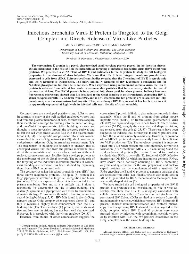
Colocalization of IBV E and IBV M in vaccinia virus- andIBV-infected cells. We also investigated the distribution of IBVE in cells coexpressing IBV M. Figure 7a to d shows the resultsof a double labeling experiment in which BHK-21 cells coin-fected with vvIBVE and vvIBVM were stained with antibodiesto E and M. Confocal microscopy indicated that the distribu-tion of the proteins completely overlapped (Fig. 7c). IBV Eand M also colocalized in cells infected with IBV at 6 h postin-fection (Fig. 7e to h). In IBV-infected cells, there was someadditional reticular staining for IBV M, which probably corre-sponds to the endoplasmic reticulum (Fig. 7f). Even so, therewas nearly complete overlap between the distribution of IBV Eand M in IBV-infected cells. These results demonstrate thatboth IBV E and M proteins accumulate in infected cells nearthe site of virus budding.
DISCUSSION
Coronaviruses are positive-strand RNA viruses that obtaintheir membrane envelope by budding into the CGN, alsoknown as the endoplasmic reticulum-Golgi intermediate com-partment (13, 15). The mechanism of budding-site selection isunclear, but the possible role of the targeting of individualcoronavirus envelope proteins in this process has been exam-ined by expressing them from cDNA. IBV M is localized to thecis-Golgi network and cis-Golgi complex when expressed alone(23) and thus reaches a slightly later compartment than that ofthe budding site. MHV M is found in the trans-Golgi networkwhen expressed from cDNA (14), a compartment even moredistant from the budding compartment. Thus, it is unlikely thatthe M protein is solely responsible for selection of the coro-navirus budding site. The coronavirus S protein is found at theplasma membrane when expressed alone (36) and thus prob-ably does not play a major role in selection of the CGN forbudding. It seems likely that there is a general mechanism forthe accumulation of coronavirus envelope proteins at the bud-ding site so that efficient virus assembly can occur. The onlyother known coronavirus envelope protein, E, is incompletelycharacterized, and its subcellular localization has not beencarefully studied. The work described here was carried out asa prerequisite to determining the role of IBV E in budding-siteselection and virus assembly.
Topology and intracellular localization of IBV E. Using in-direct-immunofluorescence microscopy in conjunction with thedetergent digitonin, which selectively permeabilizes the plasmamembrane of cells when used at low concentrations (29), anddomain-specific antibodies generated against the IBV E pro-tein, we showed that the carboxyl terminus of E is cytoplasmic.This topology is predicted from the positive-inside rule (38),and the E proteins of all coronaviruses contain a positivelycharged residue(s) on the C-terminal sides of their predictedtransmembrane domains. This topology places IBV E in thetype III class of integral membrane proteins (38), since it lacksa cleaved signal sequence. Given this topology of the IBV Eprotein, it was possible that the protein was N glycosylated ata consensus site near the N terminus. We showed that theglycosylation site is not used, probably because of its extreme
FIG. 5. IBV E is released from transfected cells in VLPs that incorporateIBV M if present. (a) BHK-21 cells were mock infected (2), infected withvvIBVE (E), vvIBVM (M), or vvIBVE plus vvIBVM (E1M), radiolabeled from3.5 to 4.5 h postinfection, and chased for 3 h. Medium was collected, cleared ofdebris, and immunoprecipitated with anti-E and anti-M antibodies. The immu-noprecipitates were analyzed by SDS-PAGE and visualized by fluorography. (b)BHK-21 cells coinfected with vvIBVE and vvIBVM were radiolabeled from 3.5to 4.5 h postinfection and chased for 3 h. Concentrated supernatants were loadedonto a continuous 20 to 55% sucrose gradient and centrifuged to equilibrium.Fractions were collected and immunoprecipitated with anti-E and anti-M anti-bodies. The immunoprecipitates were separated by SDS-PAGE and visualized byfluorography. Fraction 1 corresponds to the top of the gradient, and fraction 12corresponds to the bottom of the gradient. (c) BHK-21 cells were infected withvvIBVE only and treated exactly as described for panel b.
VOL. 74, 2000 GOLGI TARGETING OF IBV E 4323
on March 23, 2018 by guest
http://jvi.asm.org/
Dow
nloaded from

proximity to the membrane (27). The topology of IBV E de-termined here differs from that previously published for TGEVE (9). Godet et al. showed that the C terminus of cell surfaceTGEV E was extracellular because it was accessible to anti-body in nonpermeabilized cells (9).
We showed by indirect immunofluorescence and confocalmicroscopy that the IBV E protein is localized to the Golgicomplex in both transfected and IBV-infected cells. We did notdetect IBV E at the cell surface, as described for bovine coro-navirus E and TGEV E proteins (1, 9). When we compared thelocalization of IBV E to that of three marker proteins thatreside in different regions of the Golgi complex, we found thatthe distribution of IBV E most closely overlapped that ofmannosidase II, a Golgi stack marker. There was less, butsignificant, overlap with the trans-Golgi resident syntaxin 6 andthe cis-Golgi protein GM130. The proximity of the subcellulardistribution of the E protein to the coronavirus budding site isinteresting in light of the hypothesis that E plays a role indirecting the accumulation of coronavirus envelope proteins atthe budding site. The IBV M protein is targeted to the cis-Golgi complex when it is expressed from cDNA, at least in partby information contained in its first transmembrane domain(23, 24, 33). Here we have shown that IBV encodes anotherenvelope protein (E) that possesses Golgi targeting informa-tion. It will be interesting to dissect the targeting information
in IBV E to determine whether E and M have a commonmechanism for Golgi localization.
When the IBV E and M proteins were expressed together incells, either by coinfection with recombinant vaccinia viruses orby IBV infection, they colocalized when analyzed by indirectimmunofluorescence and confocal microscopy. This result isintriguing, since it may point to interactions between the E andM proteins that result in their localization in the same com-partment. However, we were unable to demonstrate interac-tions between IBV E and M directly. We are interested ininteractions that the E protein may have with the other IBVproteins and the possible role of these interactions in gatheringthe envelope proteins at the CGN for budding. We plan tofurther examine the targeting of the IBV E protein and theeffect of its expression on the localization of IBV M at theultrastructural level in order to address these questions.
IBV VLPs are produced inefficiently. When the IBV E andM proteins were transiently expressed in cells, they were re-leased into the culture supernatants in sedimentable particles.We also observed that the IBV E protein was released insimilar particles when expressed alone. These particles, whencentrifuged to equilibrium on sucrose density gradients, haddensities of 1.11 g/cm3 (particles containing both E and M) and1.14 g/cm3 (particles containing E alone). The release of coro-navirus E protein from cells in membranous particles has ob-
FIG. 6. The IBV E protein is localized to the Golgi complex. BHK-21 cells infected with vvIBVE were fixed for immunofluorescence at 6 h postinfection,permeabilized with Triton X-100, and double labeled with antibodies to IBV E and different endogenous Golgi resident proteins. (a to d) Cells were stained with rabbitanti-IBV E and mouse anti-GM130; (e to h) cells were stained with rabbit anti-IBV E and mouse anti-mannosidase II; (i to l) cells were stained with rabbit anti-IBVE and mouse anti-syntaxin 6. Secondary antibodies were Texas red-conjugated goat anti-rabbit IgG and fluorescein-conjugated goat anti-mouse IgG. In each row, thered image corresponds to IBV E staining and the green image corresponds to the appropriate Golgi marker. The third image in each row (c, g, and k) is a merged image,where yellow represents overlap between the red- and green-staining patterns. The fourth image in each row (d, h, and l) is a phase image of each field of labeled cells.Bar, 10 mm.
4324 CORSE AND MACHAMER J. VIROL.
on March 23, 2018 by guest
http://jvi.asm.org/
Dow
nloaded from
vious implications for the importance of the role of E in virusassembly. However, at least for IBV proteins in BHK-21 cells,particle release was extremely inefficient, since the amounts ofE and M proteins released into the supernatant were less than0.01% of the cellular E and M levels. Clearly, other IBVcomponents must be required for efficient budding and/or re-lease of virus from cells. The efficiency of VLP formation incells expressing MHV and TGEV E and M proteins was notreported (3, 25, 37), and so it is not yet clear if the assemblymechanism of IBV differs from that of these other coronavi-ruses.
Can the E protein drive IBV assembly? It was proposed thatcoronavirus M and E proteins are the minimal assembly unit,since expression of these proteins resulted in their release inVLPs (37). This is unusual, since the assembly of many envel-oped viruses is nucleocapsid dependent. Recently, it was shownthat expression of MHV E alone induces VLPs (25), as wehave shown here for IBV E. However, the efficiency of VLPrelease from cells expressing IBV E with or without M isextremely low. Therefore, it is not clear if IBV VLP productionis relevant to virus assembly. IBV E is expressed from a tricis-tronic RNA (21), and the two open reading frames upstream ofIBV E are expressed in IBV-infected cells (19). The proteinencoded by open reading frame 3a is extremely hydrophobicand is predicted to be a membrane protein. It remains possiblethat the proteins encoded by open reading frames 3a and/or 3bare involved in assembly and budding-site selection, and per-haps they function in concert with the IBV E protein in theseprocesses. We are currently investigating this possibility.
Possible additional functions of IBV E. The IBV E proteinis abundant in IBV-infected cells at late times postinfection(Fig. 1B), and most or all of the overexpressed protein is foundin the Golgi complex (Fig. 7). However, the IBV E protein isonly a minor component of virions (20). Thus, some of thecellular E protein might be excluded from assembling virions.It will be interesting to quantitate the IBV E protein in cellsand in virions. The high level of E expression in infected cells
suggests that it might have additional functions besides itspotential role in virus assembly. One possibility is that it in-duces apoptosis in infected cells, as reported for the MHV Eprotein (2).
An interesting example of a small membrane protein withpotentially more than one function is found in the influenzavirus M2 protein, which has some similarities to the coronavi-rus E protein. It is also a type III integral membrane proteinfound in much lower levels in virions than in infected cells (16,41, 43). The function of M2 as a tetrameric ion channel is wellcharacterized (28), but it has also been implicated in assembly.Viruses resistant to plaque growth inhibition induced by amonoclonal antibody to the M2 protein were shown to havecompensating mutations in the M1 matrix protein, suggestingthat critical interactions between these two proteins occur dur-ing budding at the plasma membrane (42). It will be importantto address the possible functions of the IBV E protein ininfected cells in addition to its potential role in virus assembly.
ACKNOWLEDGMENTS
We thank M. Delannoy for confocal microscopy expertise and C.Buck for the pSCIIMCS1 vector and helpful advice regarding coinfec-tion with recombinant vaccinia viruses.
This work was supported by National Institutes of Health grantGM42522.
REFERENCES
1. Abraham, S., T. E. Kienzle, W. E. Lapps, and D. A. Brian. 1990. Sequenceand expression analysis of potential nonstructural proteins of 4.9, 4.8, 12.7,and 9.5 kDa encoded between the spike and membrane protein genes of thebovine coronavirus. Virology 177:488–495.
2. An, S., C. J. Chen, J. L. Leibowitz, and S. Makino. 1999. Induction ofapoptosis in murine coronavirus-infected cultured cells and demonstrationof E protein as an apoptosis inducer. J. Virol. 73:7853–7859.
3. Baudoux, P., C. Carrat, L. Besnardeau, B. Charley, and H. Laude. 1998.Coronavirus pseudoparticles formed with recombinant M and E proteinsinduce alpha interferon synthesis by leukocytes. J. Virol. 72:8636–8643.
4. Bos, E. C. W., W. Luytjes, H. Van der Meulen, H. K. Koerten, and W. J. M.Spaan. 1996. The production of recombinant infectious DI-particles of a
FIG. 7. IBV E and IBV M colocalize in transfected and IBV-infected cells. BHK-21 cells infected with vvIBVE and vvIBVM (a to d) or IBV-infected Vero cells(e to h) were fixed for immunofluorescence at 6 h postinfection and double labeled with rat anti-E antibody and rabbit anti-M antibody. Secondary antibodies wereTexas red-conjugated goat anti-rat IgG and fluorescein-conjugated goat anti-rabbit IgG. The red images correspond to IBV E staining, and the green images correspondto IBV M staining. The third image in each row (c and g) is a merged image, where yellow represents overlap between the red- and green-staining patterns. The fourthimage in each row (d and h) is a phase image of the field of labeled cells. Bar, 10 mm.
VOL. 74, 2000 GOLGI TARGETING OF IBV E 4325
on March 23, 2018 by guest
http://jvi.asm.org/
Dow
nloaded from

murine coronavirus in the absence of helper virus. Virology 218:52–60.5. Bock, J. B., J. Klumperman, S. Davanger, and R. H. Scheller. 1997. Syntaxin
6 functions in trans-Golgi network vesicle trafficking. Mol. Biol. Cell 8:1261–1271.
6. de Groot, R. J., R. W. van Leen, M. J. M. Dalderup, H. Vennema, M. C.Horzinek, and W. J. M. Spaan. 1989. Stably expressed FIPV peplomerprotein induces cell fusion and elicits neutralizing antibodies in mice. Virol-ogy 171:493–502.
7. Fischer, F., C. F. Stegen, P. S. Masters, and W. A. Samsonoff. 1998. Analysisof constructed E gene mutants of mouse hepatitis virus confirms a pivotalrole for E protein in coronavirus assembly. J. Virol. 72:7885–7894.
8. Fuerst, T. R., E. G. Niles, F. W. Studier, and B. Moss. 1986. Eukaryotictransient expression system based on recombinant vaccinia virus that syn-thesizes bacteriophage T7 RNA polymerase. Proc. Natl. Acad. Sci. USA83:8122–8126.
9. Godet, M., R. l’Haridon, J.-F. Vautherot, and H. Laude. 1992. TGEV coro-navirus ORF4 encodes a membrane protein that is incorporated into virions.Virology 188:666–675.
10. Hammond, S. A., R. P. Johnson, S. A. Kalams, B. D. Walker, M. Takiguchi,J. T. Safrit, R. A. Koup, and R. F. Siliciano. 1995. An epitope-selective,transporter associated with antigen presentation (TAP)-1/2-independentpathway and a more general TAP-1/2-dependent antigen-processing path-way allow recognition of the HIV-1 envelope glycoprotein by CD81 CTL. J.Immunol. 154:6140–6156.
11. Holmes, K. V., E. W. Doller, and L. S. Sturman. 1981. Tunicamycin resistantglycosylation of a coronavirus glycoprotein: demonstration of a novel type ofviral glycoprotein. Virology 115:334–344.
12. Kim, K. H., K. Narayanan, and S. Makino. 1997. Assembled coronavirusfrom complementation of two defective interfering RNAs. J. Virol. 71:3922–3931.
13. Klumperman, J., J. Krijnse Locker, A. Meijer, M. C. Horzinek, H. J. Geuze,and P. J. M. Rottier. 1994. Coronavirus M proteins accumulate in the Golgicomplex beyond the site of virion budding. J. Virol. 68:6523–6534.
14. Krijnse Locker, J., G. Griffiths, M. C. Horzinek, and P. J. M. Rottier. 1992.O-glycosylation of the coronavirus M protein. J. Biol. Chem. 267:14094–14101.
15. Krijnse Locker, J., M. Ericsson, P. J. M. Rottier, and G. Griffiths. 1994.Characterization of the budding compartment of mouse hepatitis virus: ev-idence that transport from the RER to the Golgi complex requires only onevesicular transport step. J. Cell Biol. 124:55–70.
16. Lamb, R. A., S. L. Zebedee, and C. D. Richardson. 1985. Influenza virus M2protein is an integral membrane protein expressed on the infected-cell sur-face. Cell 40:627–633.
17. Lanser, J., and C. R. Howard. 1980. The polypeptides of infectious bronchitisvirus (IBV-41 Strain). J. Gen. Virol. 46:349–361.
18. Lefrancois, L., and D. S. Lyles. 1982. The interaction of antibody with themajor surface glycoprotein of vesicular stomatitis virus. Virology 121:168–174.
19. Liu, D. X., D. Cavanagh, P. Green, and S. C. Inglis. 1991. A polycistronicmRNA specified by the coronavirus infectious bronchitis virus. Virology184:531–544.
20. Liu, D. X., and S. C. Inglis. 1991. Association of the infectious bronchitisvirus 3c protein with the virion envelope. Virology 185:911–917.
21. Liu, D. X., and S. C. Inglis. 1992. Internal entry of ribosomes on a tricistronicmRNA encoded by infectious bronchitis virus. J. Virol. 66:6143–6154.
22. Machamer, C. E., and J. K. Rose. 1987. A specific transmembrane domain ofa coronavirus EI glycoprotein is required for its retention in the Golgiregion. J. Cell Biol. 105:1205–1214.
23. Machamer, C. E., S. A. Mentone, J. K. Rose, and M. G. Farquhar. 1990. TheE1 glycoprotein of an avian coronavirus is targeted to the cis Golgi complex.
Proc. Natl. Acad. Sci. USA 87:6944–6948.24. Machamer, C. E., M. G. Grim, A. Esquela, S. W. Chung, M. Rolls, K. Ryan,
and A. M. Swift. 1993. Retention of a cis Golgi protein requires polarresidues on one face on a predicted a-helix in the transmembrane domain.Mol. Biol. Cell 4:695–704.
25. Maeda, J., A. Maeda, and S. Makino. 1999. Release of coronavirus E proteinin membrane vesicles from virus-infected cells and E protein-expressingcells. Virology 263:265–272.
26. Nakamura, N., C. Rabouille, R. Watson, T. Nilsson, N. Hui, P. Slusarewicz,T. E. Kries, and G. Warren. 1995. Characterization of a cis-Golgi matrixprotein, GM130. J. Cell Biol. 131:1715–1726.
27. Nilsson, I. M., and G. von Heijne. 1993. Determination of the distancebetween the oligosaccharyltransferase active site and the endoplasmic retic-ulum membrane. J. Biol. Chem. 268:5768–5801.
28. Pinto, L. H., L. J. Holsinger, and R. A. Lamb. 1992. Influenza virus M2protein has ion channel activity. Cell 69:517–528.
29. Plutner, H., H. W. Davidson, J. Saraste, and W. E. Balch. 1992. Morpho-logical analysis of protein transport from the ER to the Golgi membranes indigitonin-permeabilized cells: role of the p58 containing compartment.J. Cell Biol. 119:1097–1116.
30. Smith, A. R., M. E. G. Boursnell, M. M. Binns, T. D. K. Brown, and S. C.Inglis. 1990. Identification of a new membrane-associated polypeptide spec-ified by the coronavirus infectious bronchitis virus. J. Gen. Virol. 71:3–11.
31. Stern, D. F., and B. M. Sefton. 1982. Coronavirus proteins: structure andfunction of the oligosaccharides of the avian infectious bronchitis virus.J. Virol. 44:804–812.
32. Sturman, L. S., K. V. Holmes, and J. Behnke. 1980. Isolation of coronavirusenvelope glycoproteins and interaction with the viral nucleocapsid. J. Virol.33:449–462.
33. Swift, A. M., and C. E. Machamer. 1991. A Golgi retention signal in amembrane-spanning domain of coronavirus E1 protein. J. Cell Biol. 115:19–30.
34. Tooze, J., S. A. Tooze, and S. D. Fuller. 1987. Sorting of progeny coronavirusfrom condensed secretory proteins at the exit from the trans-Golgi networkof AtT20 cells. J. Cell Biol. 105:1215–1226.
35. Velasco, A., L. Hendricks, K. W. Moremen, D. R. Tulsiani, O. Touster, andM. G. Farquhar. 1993. Cell type-dependent variations in the subcellulardistribution of alpha-mannosidase I and II. J. Cell Biol. 122:39–51.
36. Vennema, H., L. Heijnen, A. Zijderveld, M. C. Horzinek, and W. J. M.Spaan. 1990. Intracellular transport of recombinant coronavirus spike pro-teins: implications for viral assembly. J. Virol. 64:339–346.
37. Vennema, H., G.-J. Godeke, J. W. A. Rossen, W. F. Voorhout, M. C.Horzinek, D.-J. E. Opstelten, and P. J. M. Rottier. 1996. Nucleocapsid-independent assembly of coronavirus-like particles by co-expression of viralenvelope protein genes. EMBO J. 15:2020–2028.
38. von Heijne, G., and Y. Gavel. 1988. Topogenic signals in integral membraneproteins. Eur. J. Biochem. 174:671–678.
39. Weisz, O. A., and C. E. Machamer. 1994. Use of recombinant vaccinia virusvectors for cell biology. Methods Cell Biol. 43:137–159.
40. Yu, X., W. Bi, S. R. Weiss, and J. L. Leibowitz. 1994. Mouse hepatitis virusgene 5b protein is a new virion envelope protein. Virology 202:1018–1023.
41. Zebedee, S. L., and R. A. Lamb. 1988. Influenza A virus M2 protein: mono-clonal antibody restriction of virus growth and detection of M2 in virions.J. Virol. 62:2762–2772.
42. Zebedee, S. L., and R. A. Lamb. 1989. Growth restriction of influenza A virusby M2 protein antibody is genetically linked to the M1 protein. Proc. Natl.Acad. Sci. USA 86:1061–1065.
43. Zebedee, S. L., C. D. Richardson, and R. A. Lamb. 1985. Characterization ofthe influenza virus M2 integral membrane protein and expression at theinfected-cell surface from cloned cDNA. J. Virol. 56:502–511.
4326 CORSE AND MACHAMER J. VIROL.
on March 23, 2018 by guest
http://jvi.asm.org/
Dow
nloaded from