infect. immun. doi:10.1128/iai.01199-07 accepted · for communicable diseases control and...
TRANSCRIPT

Enteropathogenic Escherichia coli (EPEC) O125:H6 triggers attaching
and effacing lesions on human intestinal biopsies independently of Nck
and TccP
1
2
3
4
5
6
7
8
9
10
11
12
13
14
15
16
17
18
19
20
Li BaiP
1+P, Stephanie SchüllerP
2+P, Andrew WhaleP
3P, Aurelie MousnierP
3P, Olivier MarchesP
3P,
Lei WangP
1P, Tadasuke OokaP
4P, Robert HeuschkelP
2P, Franco TorrenteP
2P, James B. KaperP
5P,
Tânia A.T. GomesP
6P, Jiangou XuP
1P, Alan D. PhillipsP
2P and Gad FrankelP
3*P
P
1PState Key Laboratory for Infectious Disease Prevention and Control, National Institute
for Communicable Diseases Control and Prevention, China CDC, Beijing, China;
P
2PCentre for Paediatric Gastroenterology, Royal Free and University College Medical
School, London, UK; P
3PDivision of Cell and Molecular Biology, Imperial College London
London, UK; P
4PDivision of Bioenvironmental Science, Frontier Science Research Center,
University of Miyazaki, 5200 Kiyotake, Miyazaki 889-1692, Japan P
5PCenter for Vaccine
Development, University of Maryland School of Medicine, Baltimore, MD USAP
6PDepartamento de Microbiologia, Imunologia e Parasitologia, Universidade Federal de
São Paulo, São Paulo, Brazil
Running title: Actin polymerization by EPEC O125:H6
+These authors are equal contributors
*Corresponding author. Division of Molecular and Cellular Biology, Flowers Building
Imperial College London, London SW7 2AZ, Tel:+44 020 2594 5253; Fax: +44 020
5794 3069 E-mail: [email protected] 21
1
ACCEPTED
Copyright © 2007, American Society for Microbiology and/or the Listed Authors/Institutions. All Rights Reserved.Infect. Immun. doi:10.1128/IAI.01199-07 IAI Accepts, published online ahead of print on 5 November 2007
on Novem
ber 9, 2018 by guesthttp://iai.asm
.org/D
ownloaded from

Abstract 22
23
24
25
26
27
28
29
30
31
32
33
34
35
36
37
38
39
Typical enteropathogenic Escherichia coli (EPEC) and enterohemorrhagic E. coli employ
either Nck, TccP or Nck and TccP pathways to activate N-WASP and to trigger actin
polymerization in cultured cells. This phenotype is used as a marker for the pathogenic
potential of EPEC and EHEC strains. In this paper we report that EPEC O125:H6, which
represent a large category of strains, lack the ability to utilize either Nck or TccP and
hence trigger actin polymerization in vitro only inefficiently. However, we show that
infection of human intestinal biopsies with EPEC O125:H6 results in formation of typical
attaching and effacing lesions. Expressing TccP in EPEC O125:H6, which harbor an
EHEC O157-like Tir, resulted in efficient actin polymerization in vitro and enhanced
colonization of human intestinal in vitro organ cultures with detectable N-WASP and
electron dense material at the site of bacterial adhesion. These results show the existence
of a natural category of EPEC that colonizes the gut mucosa using Nck and TccP
independent mechanisms. Importantly, the results highlight yet again the fact that
conclusions made on the basis of in vitro cell culture models cannot be extrapolated
wholesale to infection of mucosal surfaces and that the ability to induce actin
polymerization on cultured cells should not be used as a definitive marker for EPEC and
EHEC virulence.
2
ACCEPTED
on Novem
ber 9, 2018 by guesthttp://iai.asm
.org/D
ownloaded from

Introduction 40
41
42
43
44
45
46
47
48
49
50
51
52
53
54
55
56
57
58
59
60
61
62
63
64
Enteropathogenic Escherichia coli (EPEC) comprise a category of diarrheagenic E. coli
that was the first to be implicated in human disease. In 1987 the World Health
Organization assigned EPEC to serogroups O26, O55, O86, O111, O114, O119, O125,
O126, O127, O128, O142, and O158 (reviewed in (7)). EPEC are divided into two
evolutionary related lineages termed EPEC 1 (typified by expression of flagellar antigen
H6 and intimin c) and EPEC 2 (typified by expression of flagellar antigen H2, intimin
d"and TccP2) (10, 34, 35). Enterohemorrhagic E. coli (EHEC) constitute a subgroupP
Pof
Shiga-toxin producing E. coli (STEC) that can cause bloody diarrhea,P
Phemorrhagic
colitis, and hemolytic-uremic syndrome. EHEC O157:H7 is the most common and
virulent serotype that is implicated worldwide in human disease (reviewed in (17)).
While colonizing the gut mucosa EPEC and EHEC trigger widespread ultrastructural
changes, which are characterized by localized disintegration of the brush border
microvilli and close association of the bacteria with the enterocyte plasma membrane,
termed attaching and effacing (A/E) lesions (27). Formation of A/E lesions can be
reproduced ex vivo by infection of cultured human intestinal explants (in vitro organ
culture, IVOC) with EPEC (22). The genes necessary for A/E lesion formation in vitro
are carried on the Locus of Enterocyte Effacement (LEE) (26) that encodes a type III
secretion system (T3SS) (15), the adhesin intimin (16), chaperones, translocator and six
effector proteins, including Tir (translocated intimin receptor) (20).
Once translocated, Tir is integrated into the host cell plasma membrane in a hairpin loop
topology (13). The extracellular loop, presented above the plasma membrane, serves as a
receptor for the bacterial adhesin intimin. Results from EPEC infection of cultured
epithelial cells in vitro have shown that clustering of Tir by intimin (4) leads to
phosphorylation of a Tir tyrosine residue (19) which is present in the context of a
3
ACCEPTED
on Novem
ber 9, 2018 by guesthttp://iai.asm
.org/D
ownloaded from

65
66
67
68
69
70
71
72
73
74
75
76
77
78
79
80
81
82
83
84
85
86
87
consensus binding site (YBPBDEP/D/V) for the mammalian adaptor proteins Nck1 and 2
(referred to collectively as Nck throughout). Binding of Nck to phosphorylated Tir leads
to recruitment and activation of the neuronal Wiskott-Aldrich syndrome protein (N-
WASP), initiating actin polymerization via the actin-related protein 2/3 (Arp2/3) complex
(reviewed in (6)).
Strains belonging to EPEC 1, commonly represented by O127:H6 strain E2348/69,
trigger actin polymerization predominantly via the Nck actin polymerization pathway,
while strains belonging to EPEC 2, commonly represented by O111:NM strain B171, can
trigger actin polymerization in vitro by redundant mechanisms involving either Nck or
TccP2, which is functionally interchangeable with TccP of EHEC O157:H7 (34).
TccP/EspFBUB is a bacterial effector protein that although has not been shown to bind Tir
directly binds directly to N-WASP, leading to recruitment of the Arp2/3 complex and
localized actin polymerization (5, 11). Recent studies have shown that a conserved NPY
carboxy terminal Tir motif in EPEC and EHEC is involved in Nck-independent actin
polymerization in the former and TccP-dependent actin polymerization pathway in the
latter (1). Importantly, the Nck, TccP and TccP2 actin polymerization pathways are all
dispensable for A/E lesion formation on human IVOC (11, 32, 34) and mouse gut
following infection with Citrobacter rodentium (9).
Recently while screening for the presence of tccP and tccP2 in clinical EPEC isolates we
discovered that strains belonging to EPEC O125:H6 naturally encode non-tyrosine
phosphorylated Tir and yet are tccP and tccP2 gene negative (30). The aim of this study
was to investigate whether EPEC O125:H6 triggered actin polymerization in vitro and ex
vivo.
4
ACCEPTED
on Novem
ber 9, 2018 by guesthttp://iai.asm
.org/D
ownloaded from

Material and Methods 88
89
90
91
92
93
94
95
96
97
98
99
100
101
102
103
104
105
106
107
108
109
110
111
Bacterial strains, plasmids and growth conditions
The bacterial strains and plasmids used in this study are listed in Table I. Bacteria were
grown for 8 h in Luria Bertani (LB) medium before being diluted into Dulbecco’s
Modified Eagle’s Medium (DMEM) and incubated at 37°C in 5% COB2 Bstatically
overnight. Growth media was supplemented with ampicillin (50 µg/ml) or kanamycin
(50og/ml), when necessary.
Infection of cultured cells
Bacterial cultures were used to infect HeLa cells grown on cover slips in 24 well-plates
for 3 h (E2348/69), 6 h (TUV 93-0) or 5 h (all other strains) as described (34). Cell
monolayers were then fixed in 3.7% paraformaldehyde for 15 min and permeabilized
with 0.1% Triton X-100 for 4 min. EHEC O157:H7 and EPEC E2348/69 were visualized
with goat anti-O157 (Fitzgerald industries) and anti O127 antiserum (a gift from Dr.
Roberto La Ragione, VLA, UK), respectively. O125 bacteria were visualized with
Hoechst-33342 DNA stain (Molecular probes) or anti O125 antiserum (a gift from Dr.
Roberto La Ragione, VLA, UK), actin was stained with Oregon green phalloidin
(Invitrogen), and HA-tagged TccP was detected by anti HA as described (21, 34). TirBEPECB
and TirBEHECB were detected as described in (34) and (11), respectively. Samples were
analyzed using a Zeiss axioimager fluorescence microscope and images processed using
axiovision and Adobe photoshop software.
Recombinant DNA
The tir gene of ICC223 was amplified by PCR using primers 125tir-F2 and 125tir-R2
(Table 2) and cloned into pSA10 (generating plasmid pICC368) and sequenced
(accession number AB355659). A tir deletion mutant in strain ICC223 was made using
5
ACCEPTED
on Novem
ber 9, 2018 by guesthttp://iai.asm
.org/D
ownloaded from

112
113
114
115
116
117
118
119
120
121
122
123
124
125
126
127
128
129
130
131
132
133
134
135
the lambda red system (8) with primers 125tir-F1 and 125tir-R1 and pKD4 as template,
generating strain ICC224. Primers flanking the deleted region and inside the kanamycin
cassette were used in PCR to verify the deletion (pairs of primers: kt and tir-flank-F, k2
and tir-flank-R/ Table 3). E2348/69Ftir (ICC225, Table 1) was generated using the
lambda red system (8) with primers EPEC FRT and Tir EPEC FRT rev; the deletion was
confirmed by PCR using the primer pairs Map303 for and kt, and 3CesT rev and k2
(Table 3).
To create TccP-HA, the coding sequence for tccP was amplified (primers pkk-tccP-F1
and pkk-tccP-R1; Table 3) and cloned into pSA10 as described (11)
Sequence comparison of Tir proteins
Clustal W was used for making a multiple alignment of the TirBEPEC_O125:H6 Bsequences with
14 known Tir sequences which were retrieved from the GenBank database. A phylogenic
tree was constructed with the neighbor-joining algorithm with the MEGA 3.1 software
(24). Poisson correction with the complete deletion of gaps was used to calculate protein
distances. Bootstrap analysis with 1000 replicates was performed to evaluate the
significance of the internal branches.
In vitro organ cultures
Paediatric tissue was obtained with fully informed parental consent and local ethical
committee approval using grasp forceps during routine endoscopic investigation of
intestinal disorders. Small intestinal mucosal biopsies which appeared macroscopically
normal were taken for organ culture experiments as described previously (14). Adherence
was examined using tissue from six patients (aged between 139 and 201 months) by
scanning electron microscopy (SEM) and five further cases (aged between 141-200
months) by cryosectioning, immunostaining and transmission electron microscopy
6
ACCEPTED
on Novem
ber 9, 2018 by guesthttp://iai.asm
.org/D
ownloaded from

136
137
138
139
140
141
142
143
144
145
146
147
148
149
150
151
152
153
(TEM) as described (32). IVOC infected with EHEC O157:H7 strain TUV 93-0 was used
as a positive control. In each experiment a non-infected sample was included to exclude
endogenous bacterial adhesion.
For immunofluorescence, samples were embedded in OCT compound (Sakura), snap-
frozen in liquid nitrogen and stored at &70°C until use. Serial sections of 8 µm were cut
with an MTE cryostat (SLEE Technik), picked up on poly- TLT-lysine-coated slides and air-
dried. Tissue sections were fixed in formalin for 10 min and blocked with 0.5% BSA, 2%
normal goat serum in PBS for 20 min at room temperature. Slides were incubated with
rabbit anti-TirBEHEC_O157:H7B, anti-TccP, or anti-N-WASP (kindly provided by Silvia
Lommel, Institute for Cell Biology, University of Bonn, Germany) for 60 min at room
temperature, washed and incubated in Alexa Fluor 488-conjugated goat anti-rabbit IgG
(Molecular Probes) for 30 min. Counterstaining of bacteria and cell nuclei was performed
using propidium iodide (Sigma). Epithelial cells were stained with mouse anti-
cytokeratin (Dako) and Alexa Fluor 647-conjugated goat anti-mouse IgG (Molecular
Probes). Sections were analyzed with a Radiance 2100 confocal laser scanning
microscope (Bio-Rad, UK).
7
ACCEPTED
on Novem
ber 9, 2018 by guesthttp://iai.asm
.org/D
ownloaded from

Results 154
155
156
157
158
159
160
161
162
163
164
165
166
167
168
169
170
171
172
173
174
175
176
177
EPEC O125:H6 cannot trigger efficient actin polymerization in vitro
In order to characterize the ability of EPEC O125:H6 to trigger actin polymerization in
vitro, HeLa cells were infected with four different strains isolated in Brazil and the UK
and with control EPEC 1 O127:H6 (E2348/69) and EHEC O157:H7 (EDL933 stx
negative strain TUV 93-0) strains (Table 1). All four O125:H6 strains failed to trigger
detectable actin polymerization under attached bacteria (data not shown). We selected
one of the O125:H6 EPEC strains, isolated in Brazil (ICC223), for further detailed
analysis.
The competence of ICC223-induced actin remodeling was further quantified by counting
the percentage of cell-associated bacteria which were also associated with intense F-actin
staining. Regions of 5-20 bacteria per cell were examined for each strain in three separate
experiments carried out in duplicate. 100 bacteria per coverslip were examined.
Quantifying the efficiency of actin polymerization revealed weak actin aggregation under
only 3% of adherent ICC223 despite efficient Tir translocation (Fig. 1; Table 2). This was
in sharp contrast to cells infected with control EPEC 1 O127:H6 (E2348/69) and EHEC
O157:H7 (TUV 93-0) strains which induce efficient actin polymerization under 75% and
70% of attached bacteria, respectively (Fig. 1; Table 2). The 3% of adherent ICC223
showing weak actin accretion is comparable to the frequency seen after infection with
E2348/69 expressing Tir Y474F (3).
Sequence and functional analysis of TirBEPEC_O125:H6B.
In order to characterize Tir of ICC223 the tir gene was amplified by PCR, cloned into
pSA10 (generating plasmid pICC368) and sequenced (accession number AB355659).
Multiple sequence alignment of the 569 amino acids of TirBICC223 Bwith representative Tir
8
ACCEPTED
on Novem
ber 9, 2018 by guesthttp://iai.asm
.org/D
ownloaded from

178
179
180
181
182
183
184
185
186
187
188
189
190
191
192
193
194
195
196
197
198
199
200
201
202
sequences in the database revealed that the two transmembrane domains (TMDs) and the
intimin-binding domain were highly conserved. The phylogenetic relationship of TirBEPEC-
_O125:H6 Bwith available Tir molecules revealed that it is on the same main branch of
TirBEHECB_BO157B, TirBEPECB_BO55:H7B and TirBEPECB_BO63B (Fig. 2A). Moreover, TirBICC223 Bshares 96.8%
and 56.9% sequence identity with TirBEHEC_O157:H7B and TirBEPEC_O127:H6B respectively and
100% sequence identity (with the exception of deletion of one of the GESKGA repeats)
with TirBEPECB_BO63B of an environmental EPEC isolate belonging to serogroup O63 (Fig.
2B). In particular, the sequence confirms that TirBEPEC_O125:H6B lacks the Y474 equivalent of
TirBEPEC_O127:H6B (Fig. 2B), which is consistent with our inability to detect phosphorylated
TirBEPECB_BO125:H6B during infection (30). The NPY motif that is implicated in the inefficient
Nck-independent actin polymerization pathway in EPEC O127:H6 and the TccP-
mediated actin polymerization pathway in EHEC O157:H7 (1) is conserved in TirBEPECB-
_BO125:H6B.
We next generated a tir deletion mutant in strain ICC223, generating strain ICC224.
Complementing the ICC224 mutant with pICC368, which over expresses TirBEPECB_BO125:H6B,
did not reveal Tir tyrosine phosphorylation, recruitment of Nck (data not shown) or
confer actin polymerization activity following infection of HeLa cells (Table 2). In
contrast, when the mutant was complemented with a plasmid encoding TirBEPEC_O127:H6
B(pACYC-P) (18) we detected Tir tyrosine phosphorylation, recruitment of Nck and
efficient actin polymerization under attached ICC224 bacteria (data not shown). In a
reciprocal experiment pICC368 encoding TirBEPECB_BO125:H6B was used to complement TUV
93-0Ftir (22) and E2348/69Ftir (ICC225, Table 1). While TUV 93-0Ftir was deficient in
actin polymerization (data not shown), expression of TirBEPECB_BO125:H6B restored actin
polymerization activity to the wild type level (73.3%) (Fig. 1; Table 2). In contrast, only
background actin staining was observed under E2348/69Ftir expressing TirBEPECB_BO125:H6B
9
ACCEPTED
on Novem
ber 9, 2018 by guesthttp://iai.asm
.org/D
ownloaded from

203
204
205
206
207
208
209
210
211
212
213
214
215
216
217
218
219
220
221
222
223
224
225
226
227
(Fig. 1; Table 2). Finally we transformed wild type EPEC O125:H6 strain ICC223 with a
plasmid encoding HA-tagged TccP_BO157:H7B (pICC369). Infection of HeLa cells showed
that ICC223 (stained with anti O125 antiserum) expressing TccP can effectively trigger
actin polymerization; anti-HA staining confirmed that TccP was concentrated at tip of the
pedestal (Fig. 3). Taken together these results show that EPEC O125:H6 strains have the
potential to trigger actin polymerization in HeLa cells, provided that they are equipped
with either TccP or with Tir that can undergo tyrosine phosphorylation. In this respect,
wild type EPEC O125:H6 exhibits a similar phenotype to that of EHEC O157:H7FtccP
(11).
EPEC O125:H6 induce A/E lesion on human intestinal in vitro organ cultures
We previously showed that EPEC O127:H6 (E2348/69) expressing Tir-Y454F/Y474F
(32) and EHEC O157:H7FtccP (11) can induce A/E lesions during infection of human
IVOC. Accordingly, we tested if EPEC O125:H6 strain ICC223, which naturally lacks
tccP/tccP2 and carries a non-tyrosine phosphorylated Tir can infect human intestinal
IVOC ex vivo. SEM analysis of 8 h IVOC samples showed that all EPEC O125:H6
strains adhered to human terminal ileum. Adherence patterns were similar to the EHEC
strain TUV 93-0 control, with intimate bacterial attachment and microvillous elongation
of the IVOC tissue in between adhering bacteria (Fig. 4).
Immunofluorescence staining of cryosections revealed localization of translocated Tir
underneath adherent ICC223 and TUV 93-0 (Fig. 5). In contrast, efficient N-WASP
recruitment could be observed beneath adherent TUV 93-0 whereas only a minority of
ICC223 bacteria showed a weak positive reaction. This phenotype is reminiscent of TUV
93-0FtccP which also showed intimate adherence to terminal ileum in the absence of N-
WASP recruitment (11). Importantly, expressing TccP in ICC223 resulted in efficient
recruitment of TccP and N-WASP at the site of bacterial attachment (Fig. 5). In addition,
10
ACCEPTED
on Novem
ber 9, 2018 by guesthttp://iai.asm
.org/D
ownloaded from

228
229
230
231
232
233
234
235
236
237
238
239
240
241
242
colonization of terminal ileum by ICC223 expressing TccP appeared to be enhanced
compared to ICC223 as intimately adhering bacteria were detected on 4/4 biopsies
infected with ICC223 expressing TccP compared to 2/4 biopsies infected with ICC223.
This result may suggest that expression of TccP, although not essential, may increase
colonization efficiency.
Finally, infected IVOC were analyzed by TEM. This revealed that while ICC223 can
efficiently trigger A/E lesions (Fig. 6), the intensity of electron dense staining under
attached bacteria, indicating actin polymerization, was variable and much less profound
than that seen after infection with wild type EPEC O127:H6 or EPEC O127:H6
expressing Tir Y474S (32). ICC223 expressing TccP was associated with an increased
presence of electron dense material compared to ICC223 (Fig. 6C). These results suggest
that EPEC O125:H6 like EHEC O157:H7FtccP can cause intimate attachment and
microvillous effacement without efficient recruitment of N-WASP or F-actin beneath
adhering bacteria. Expression of TccP restores efficient recruitment of N-WASP and
actin polymerization and increases colonization efficiency of human intestinal IVOC.
11
ACCEPTED
on Novem
ber 9, 2018 by guesthttp://iai.asm
.org/D
ownloaded from

Discussion 243
244
245
246
247
248
249
250
251
252
253
254
255
256
257
258
259
260
261
262
263
264
265
266
267
The ability of typical EHEC O157:H7 and EPEC strains to trigger efficient actin
polymerization on cultured cells is linked to activation of N-WASP. However, while
typical EHEC O157:H7 use the effector protein TccP (5, 11), EPEC strains use the host
adaptor protein Nck, which binds tyrosine phosphorylated Tir (2, 12). Interestingly, non-
O157 EHEC (29) and EPEC strains belonging to lineage 2 (34) can use both the Nck and
TccP2 actin polymerization pathways, while atypical sorbitol fermenting EHEC O157
express both TccP and TccP2 (29). The fact that EPEC and EHEC express what seems to
be redundant mechanisms to efficiently trigger actin polymerization is suggestive of an
essential role and of selective pressure to maintain this capability.
In this paper we have shown that EPEC O125:H6 express Tir which naturally lacks a
Y474 equivalent (19) and hence cannot trigger actin polymerization using the Nck
pathway (2, 12). All the tested EPEC O125:H6 strains also naturally lack TccP and
TccP2 (30) and hence cannot use these effector proteins to trigger efficient actin
polymerization. Accordingly, as we observed during infection of HeLa cells, EPEC
O125:H6 can only trigger inefficient actin polymerization presumably by using the NPY
motif, which is conserved in EPEC and EHEC strains (1). Expressing TccP in EPEC
O125:H6 enabled the strain to efficiently trigger actin polymerization in infected HeLa
cells. Consistent with these finding we found that TirBEPEC_125:H6B is phylogenetically
clustered with, and functionally interchangeable with, TirBEHEC_O157:H7B.
Importantly, we have demonstrated that in spite of their inability to trigger efficient actin
polymerization on cultured HeLa cells, EPEC O125:H6 strains can infect human
intestinal explants, intimately attach to the enterocytes and trigger effacement of the
brush border microvilli. These IVOC phenotypes parallel those reported for EHEC
O157:H7FtccP (11), EPEC 1 strain E2348/69 O127:H6 expressing TirY474S (32) and C.
12
ACCEPTED
on Novem
ber 9, 2018 by guesthttp://iai.asm
.org/D
ownloaded from

rodentium (9). It therefore appears that during infection of mucosal surfaces neither Nck
nor TccP is needed for A/E lesion formation. However, although the number of IVOC
used was relatively small we observed that EPEC O125:H6 expressing TccP colonize the
mucosa of the terminal ileum (4 IVOC out of 4) more efficiently than wild type O125:H6
(2 IVOC out of 4). Moreover, infection of IVOC with O125:H6 expressing TccP resulted
in detection of N-WASP at the site of bacterial attachment and accumulation of electron
dense material under attached bacteria, believed to be actin. A possible interpretation of
the data is that colonization and A/E lesion formation can be achieved by EPEC and
EHEC in the absence of efficient actin polymerization activity. However, the ability to
efficiently polymerize actin might stabilize initial adhesion and increase long term
colonization potential. Indeed, interfering with the actin cell signaling seems to modulate
the ability of EPEC to remain attached to IVOC as EPECFmap and EPECFespH mutants
detach from IVOC in high frequency leaving behind pedestal footprints (33). As Map and
EspH cooperate with Tir in coordinating actin dynamics our results suggest that timely
and efficient polymerization of actin, although not essential for colonization, might
provide a subtle advantage over EPEC and EHEC strain lacking this capability. In order
to address this hypothesis experimentally we are currently engineering site directed Tir
mutants in Citrobacter rodentium, the mouse pathogen equivalent of EPEC and EHEC
(28), that will be used in competitive index studies with the wild type strain.
268
269
270
271
272
273
274
275
276
277
278
279
280
281
282
283
284
285
286
287
288
289
290
291
292
The ability of EPEC and EHEC to trigger actin polymerization in cultured cells has been
used for many years as the main virulence marker for EPEC and EHEC, since Knutton et
al. (21) developed the fluorescent actin staining (FAS) test. The current study shows that
relying on the FAS test alone is not sufficient. While FAS positive strains are likely to be
pathogenic, LEE-positive strains that fail to trigger actin polymerization in vitro cannot
be classified as non-pathogenic and alternative assays should be employed. Indeed, the
13
ACCEPTED
on Novem
ber 9, 2018 by guesthttp://iai.asm
.org/D
ownloaded from

293
294
295
296
297
298
299
phenotype we described for EPEC O125:H6 is not uncommon. A previous study has
shown that 29% of eae-positive strains isolated from children in the United Kingdom
were FAS negative on HEp-2 cells but produced typical A/E lesion on human IVOC (23).
These findings reinforce the important differences in signal transduction between
cultured epithelial cells and mucosal surfaces (32) and suggest the existence of an
important subgroup of EPEC strains that utilize a TccP- and Nck-independent pathway to
adhere and trigger A/E lesion formation on mucosal surfaces.
14
ACCEPTED
on Novem
ber 9, 2018 by guesthttp://iai.asm
.org/D
ownloaded from

Acknowledgments 300
301
302
303
304
305
We thank Junkal Garmendia for the HA-TccP construct, Silvia Lommel, Institute for Cell
Biology, University of Bonn, Germany for the anti-N-WASP and Dr. Roberto La
Ragione, VLA, UK for anti O125. Work in the laboratory of ADP was supported by the
NIH (Grant R37AI21657 to JB Kaper). The work in the laboratory of GF was supported
by a BBSRC China partnering award and the Wellcome Trust.
15
ACCEPTED
on Novem
ber 9, 2018 by guesthttp://iai.asm
.org/D
ownloaded from

References 306
307
308
309
310
311
312
313
314
315
316
317
318
319
320
321
322
323
324
325
326
327
328
329
1. Brady, M. J., K. G. Campellone, M. Ghildiyal, and J. M. Leong. 2007.
Enterohaemorrhagic and enteropathogenic Escherichia coli Tir proteins trigger a common
Nck-independent actin assembly pathway. Cell. Microbiol. :doi: 10.1111/j.1462-
5822.2007.00954.x.
2. Campellone, K. G., A. Giese, D. J. Tipper, and J. M. Leong. 2002. A tyrosine-
phosphorylated 12-amino-acid sequence of enteropathogenic Escherichia coli Tir binds the
host adaptor protein Nck and is required for Nck localization to actin pedestals. Mol.
Microbiol. 43:1227-1241.
3. Campellone, K. G., and J. M. Leong. 2005. Nck-independent actin assembly is mediated
by two phosphorylated tyrosines within enteropathogenic Escherichia coli Tir. Mol.
Microbiol. 56:416-432.
4. Campellone, K. G., S. Rankin, T. Pawson, M. W. Kirschner, D. J. Tipper, and J. M.
Leong. 2004. Clustering of Nck by a 12-residue Tir phosphopeptide is sufficient to trigger
localized actin assembly. J. Cell Biol. 164:406-416.
5. Campellone, K. G., D. Robbins, and J. M. Leong. 2004. EspFBUB Is a translocated EHEC
effector that interacts with Tir and N-WASP and promotes Nck-independent actin
assembly. Dev. Cell 7:217-228.
6. Caron, E., V. F. Crepin, N. Simpson, J. Garmendia, and G. Frankel. 2006. Subversion
of actin dynamics by EPEC and EHEC. Curr. Opin. Microbiol. 9:40-45.
7. Chen, H. D., and G. Frankel. 2005. Enteropathogenic Escherichia coli: unravelling
pathogenesis. FEMS Microbiol. Rev. 29:83-98.
8. Datsenko, K. A., and B. L. Wanner. 2000. One-step inactivation of chromosomal genes
in Escherichia coli K12 using PCR products. Proc. Nat. Acad. Sci. USA 97:6640-6645.
16
ACCEPTED
on Novem
ber 9, 2018 by guesthttp://iai.asm
.org/D
ownloaded from

9. Deng, W., B. A. Vallance, Y. Li, J. L. Puente, and B. B. Finlay. 2003. Citrobacter
rodentium translocated intimin receptor (Tir) is an essential virulence factor needed for
actin condensation, intestinal colonization and colonic hyperplasia in mice. Mol. Microbiol.
48:95-115.
330
331
332
333
334
335
336
337
338
339
340
341
342
343
344
345
346
347
348
349
350
351
352
10. Donnenberg, M. S., and T. S. Whittam. 2001. Pathogenesis and evolution of virulence in
enteropathogenic and enterohemorrhagic Escherichia coli. J. Clin. Invest. 107:539-548.
11. Garmendia, J., A. Phillips, Y. Chong, S. Schuller, O. Marches, S. Dahan, E. Oswald,
R. K. Shaw, S. Knutton, and G. Frankel. 2004. TccP is an enterohaemorrhagic E. coli
O157:H7 type III effector protein that couples Tir to the actin-cytoskeleton. Cell.
Microbiol. 6:1167-1183.
12. Gruenheid, S., R. DeVinney, F. Bladt, D. Goosney, S. Gelkop, G. D. Gish, T. Pawson,
and B. B. Finlay. 2001. Enteropathogenic E. coli Tir binds Nck to initiate actin pedestal
formation in host cells. Nat. Cell Biol. 3`:856-859.
13. Hartland, E. L., M. Batchelor, R. M. Delahay, C. Hale, S. Matthews, G. Dougan, S.
Knutton, I. Connerton, and G. Frankel. 1999. Binding of intimin from enteropathogenic
Escherichia coli to Tir and to host cells. Mol. Microbiol. 32:151-158.
14. Hicks, S., G. Frankel, J. B. Kaper, G. Dougan, and A. D. Phillips. 1998. Role of intimin
and bundle foming pili in enteropathgenic Escherichia coli adhesion to paediatric intestine
in vitro. Infect. Immun. 66:1570-1578.
15. Jarvis, K. G., J. A. Giron, A. E. Jerse, T. K. McDaniel, M. S. Donnenberg, and J. B.
Kaper. 1995. Enteropathogenic Escherichia coli contains a putative type III secretion
system necessary for the export of proteins involved in attaching and effacing lesion
formation. Proc. Natl. Acad. Sci. USA 92:7996-8000.
17
ACCEPTED
on Novem
ber 9, 2018 by guesthttp://iai.asm
.org/D
ownloaded from

16. Jerse, A. E., J. Yu, B. D. Tall, and J. B. Kaper. 1990. A genetic locus of
enteropathogenic Escherichia coli necessary for the production of attaching and effacing
lesions on tissue culture cells. Proc. Natl. Acad. Sci. USA 87:7839-7843.
353
354
355
356
357
358
359
360
361
362
363
364
365
366
367
368
369
370
371
372
373
374
375
376
377
17. Karch, H., P. I. Tarr, and M. Bielaszewska. 2005. Enterohaemorrhagic Escherichia coli
in human medicine. Int. J. Med. Microbiol. 295:405-418.
18. Kenny, B. 2001. The enterohaemorrhagic Escherichia coli (serotype O157:H7) Tir
molecule is not functionally interchangeable for its enteropathogenic E. coli (serotype
O127:H6) homologue. Cell. Microbiol. 3:499-510.
19. Kenny, B. 1999. Phosphorylation of tyrosine 474 of the enteropathogenic Escherichia coli
(EPEC) Tir receptor molecule is essential for actin nucleating activity and is preceded by
additional host modifications. Mol. Microbiol. 31:1229-41.
20. Kenny, B., R. DeVinney, M. Stein, D. J. Reinscheid, E. A. Frey, and B. B. Finlay. 1997.
Enteropathogenic E. coli (EPEC) transfers its receptor for intimate adherence into
mammalian cells. Cell 91:511-520.
21. Knutton, S., T. Baldwin, P. H. Williams, and A. S. McNeish. 1989. Actin accumulation
at sites of bacterial adhesion to tissue culture cells: basis of a new diagnostic test for
enteropathogenic and enterohemorrhagic Escherichia coli. Infect. Immun. 57:1290-1298.
22. Knutton, S., D. R. Lloyd, and A. S. McNeish. 1987. Adhesion of enteropathogenic
Escherichia coli to human intestinal enterocytes and cultured human intestinal mucosa.
Infect. Immun. 55:69-77.
23. Knutton, S., R. Shaw, A. D. Phillips, H. R. Smith, G. A. Willshaw, P. Watson, and E.
Price. 2001. Phenotypic and genetic analysis of diarrhea-associated Escherichia coli
isolated from children in the United Kingdom. J. Pediatr. Gastroenterol. Nutr. 33:32-40.
24. Kumar, S., K. Tamura, and M. Nei. 2004. MEGA3: Integrated software for Molecular
Evolutionary Genetics Analysis and sequence alignment. Brief Bioinform. 5:150-163.
18
ACCEPTED
on Novem
ber 9, 2018 by guesthttp://iai.asm
.org/D
ownloaded from

25. Levine, M. M., E. J. Bergquist, D. R. Nalin, D. H. Waterman, R. B. Hornick, C. R.
Young, and S. Sotman. 1978. Escherichia coli that cause diarrhoea but do not produce
heat-labile or heat-stable enterotoxins and are non-invasive. Lancet i::119-122.
378
379
380
381
382
383
384
385
386
387
388
389
390
391
392
393
394
395
396
397
398
399
400
401
402
26. McDaniel, T. K., K. G. Jarvis, M. S. Donnenberg, and J. B. Kaper. 1995. A genetic
locus of enterocyte effacement conserved among diverse enterobacterial pathogens. Proc
Natl. Acad. Sci. USA 92:1664-1668.
27. Moon, H. W., S. C. Whipp, R. A. Argenzio, M. M. Levine, and R. A. Giannella. 1983.
Attaching and effacing activities of rabbit and human enteropathogenic Escherichia coli in
pig and rabbit intestines. Infect. Immun. 41:1340-1351.
28. Mundy, R., T. T. MacDonald, G. Dougan, G. Frankel, and S. Wiles. 2005. Citrobacter
rodentium of mice and man. Cell. Microbiol. 7:1697-1706.
29. Ogura, Y., T. Ooka, A. Whale, J. Garmendia, L. Beutin, S. Tennant, G. Krause, S.
Morabito, I. Chinen, T. Tobe, H. Abe, R. Tozzoli, A. Caprioli, M. Rivas, R. Robins
Browne, T. Hayashi, and G. Frankel. 2007. TccP2 of O157:H7 and non-O157
enterohemorrhagic Escherichia coli (EHEC): challenging the dogma of EHEC-induced
actin polymerization. Infect. Immun. 75:604-612.
30. Ooka, T., M. A. M. Vieira, Y. Ogura, L. Beutin, R. L. Ragione, P. M. van Diemen, M.
P. Stevens, I. Aktan, S. Cawthraw, A. Best, R. T. Hernandes, G. Krause, T. A. T.
Gomes, T. Hayashi, and G. Frankel. 2007. Characterisation of tccP2 carried by atypical
enteropathogenic Escherichia coli. FEMS Microbiol. Lett. 271:126-135.
31. Schlosser-Silverman, E., M. Elgrably-Weiss, I. Rosenshine, R. Kohen, and S. Altuvia.
2000. Characterization of Escherichia coli DNA lesions generated within J774
macrophages. J. Bacteriol. 182:5225-5230.
32. Schüller, S., Y. Chong, J. Lewin, B. Kenny, G. Frankel, and A. D. Phillips. 2007. Tir
phosphorylation and Nck/N-WASP recruitment by enteropathogenic and
19
ACCEPTED
on Novem
ber 9, 2018 by guesthttp://iai.asm
.org/D
ownloaded from

enterohaemorrhagic Escherichia coli during ex vivo colonization of human intestinal
mucosa is different to cell culture models. Cell. Microbiol. 9:1352-1364.
403
404
405
406
407
408
409
410
411
412
413
414
33. Shaw, R. K., J. Cleary, G. Frankel, and S. Knutton. 2005. Enteropathogenic Escherichia
coli interaction with human intestinal mucosa: role of effector proteins in brush border
remodelling and 'attaching & effacing' lesion formation. Infect. Immun. 73:1243-1251.
34. Whale, D., R. T. Hernandes, O. Tadasuke, B. L., S. Schüller, J. Garmendia, L.
Crowther, M. A. M. Vieira, Y. Ogura, G. Krause, A. D. Phillips, T. A. T. Gomes, T.
Hayashi, and G. Frankel. 2007. TccP2-mediated subversion of actin dynamics by EPEC 2
- a distinct evolutionary lineage of enteropathogenic Escherichia coli. Microbiology
153:1743-1755.
35. Whittam, T. S., and E. A. McGraw. 1996. Clonal analysis of EPEC serogroups. Rev.
Microbiol. Sao Paulo 27 (Suppl l):7-16.
20
ACCEPTED
on Novem
ber 9, 2018 by guesthttp://iai.asm
.org/D
ownloaded from

Table 1. Strains and Plasmids used in this study 415
416
Name Description Soure of reference Strains
TUV 93-0 EHEC O157:H7 strain EDL933 stxP
-
P(intimin i)
ATCC
E2348/69 Wild-type EPEC O127:H6 (intimin
c)
(25)
ICC223 E. coli O125:H6 isolated in Brazil
from a diarrheagenic case (intimin c)
This study
CPG35 E. coli O125:H6 isolated in the UK
from a diarrheagenic case (intimin
c)
This study
N67
E. coli O125:H6 isolated in the UK
from a diarrheagenic case (intimin c)
This study
E. coli O125:H6 isolated in Brazil
from a diarrheagenic case (intimin c)
2741-5
This study
ICC224 ICC223〉tir::Km This study
ICC225 〉tir in EPEC O127:H6 This study
TUV 93-0〉tir 〉tir in EHEC O157:H7 (22)
Plasmid
pACYC-P A pACYC derivative, encoding mtc
(map, tir, cesT, 5ガ eae) from
E2348/69
(18)
pKD46 Helper plasmid encoding そ red
recombinase
(8)
pKD4 Template (10)
pSA10 A pKK177-3 derivative containing
lacP
QPPP
(31)
pICC368 A pSA10 derivative encoding
TirBICC223 Bfusion protein
This study
pICC369 A pSA10 derivative encoding TUV
93-0 TccP–HA fusion protein
This study
21
ACCEPTED
on Novem
ber 9, 2018 by guesthttp://iai.asm
.org/D
ownloaded from

Table 2. Quantification of efficiency of actin polymerization triggered during bacterial infection 417
418 Strains Actin accretion beneath
attached bacteria (% of adherent bacteria)*
ICC223 (O125:H6) 3.8±1**
E2348/69 (O127:H6) 75±4
TUV 93-0 (O157:H7) 70.6±5
ICC224(pICC368) 3.7±1.5**
E2348/69Ätir(pICC368) 3.1±0.6**
TUV 93-0Ätir(pICC368) 73.3±4.7
ICC223(pICC369) 81±3.1
419
420
421
422
423
424
425
426
*Data are mean ±SD of three independent experiments carried out in duplicate. The efficiency of
actin accretion was quantified by counting the percentage of cell associated bacteria which were
also associated with aggregated F-actin staining. Regions of 5-20 attached bacteria per cell, 100
bacteria per coverslip were examined.
** Weak actin recruitment is detected beneath the adherent bacteria but is distinct from the intensly
stained F-actin pedestals triggered by strains E2348/69, TUV 93-0, TUV 93-0Ätir(pICC368) and
ICC223(pICC369).
22
ACCEPTED
on Novem
ber 9, 2018 by guesthttp://iai.asm
.org/D
ownloaded from
427
428
429
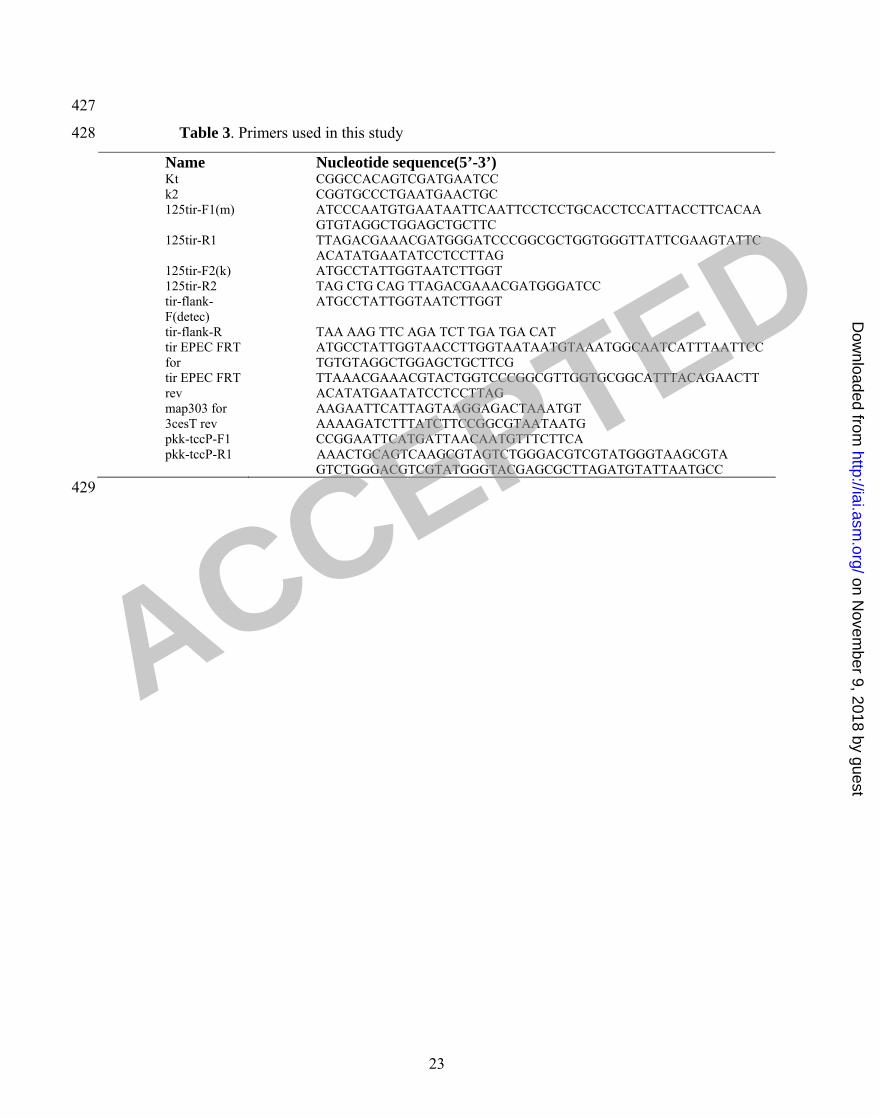
Table 3. Primers used in this study
Name Nucleotide sequence(5’-3’) Kt CGGCCACAGTCGATGAATCC
k2 CGGTGCCCTGAATGAACTGC
125tir-F1(m) ATCCCAATGTGAATAATTCAATTCCTCCTGCACCTCCATTACCTTCACAA
GTGTAGGCTGGAGCTGCTTC
125tir-R1 TTAGACGAAACGATGGGATCCCGGCGCTGGTGGGTTATTCGAAGTATTC
ACATATGAATATCCTCCTTAG
125tir-F2(k) ATGCCTATTGGTAATCTTGGT
125tir-R2 TAG CTG CAG TTAGACGAAACGATGGGATCC
tir-flank-
F(detec)
ATGCCTATTGGTAATCTTGGT
tir-flank-R TAA AAG TTC AGA TCT TGA TGA CAT
tir EPEC FRT
for
ATGCCTATTGGTAACCTTGGTAATAATGTAAATGGCAATCATTTAATTCC
TGTGTAGGCTGGAGCTGCTTCG
tir EPEC FRT
rev
TTAAACGAAACGTACTGGTCCCGGCGTTGGTGCGGCATTTACAGAACTT
ACATATGAATATCCTCCTTAG
map303 for AAGAATTCATTAGTAAGGAGACTAAATGT
3cesT rev AAAAGATCTTTATCTTCCGGCGTAATAATG
pkk-tccP-F1 CCGGAATTCATGATTAACAATGTTTCTTCA
pkk-tccP-R1 AAACTGCAGTCAAGCGTAGTCTGGGACGTCGTATGGGTAAGCGTA
GTCTGGGACGTCGTATGGGTACGAGCGCTTAGATGTATTAATGCC
23
ACCEPTED
on Novem
ber 9, 2018 by guesthttp://iai.asm
.org/D
ownloaded from

Figure legends 430
431
432
433
434
435
436
437
438
439
440
441
442
443
444
445
446
447
448
449
450
451
452
453
454
455
Fig. 1. EPEC O125:H6 strain ICC223 cannot efficiently induce actin polymerization in
infected HeLa cells, while the control EPEC O127:H6 strain E2348/69 and EHEC
O157:H7 strain TUV 93-0 trigger efficient actin polymerization. Expression of
TirBEPEC_O125:H6B in EPEC E2348/69Ftir did not restore actin polymerization while
expressing TirBEPEC_O125:H6B in EHEC TUV 93-0Ftir resulted in strong actin
polymerization. Bacterial DNA was visualized in blue using Hoechst-33342. Tir is
labeled red with anti-TirBEHECB or TirBEPECB (for strain E2348/69) antiserum. Actin was
labeled in green using Oregon green-conjugated phalloidin. Separate monochrome
images of the UV, red and green fluorescence channels, and a merged colour image are
shown. Bar = 5 om .
Fig. 2. A. The phylogenetic relationship of the tir genes from EPEC O125:H6 with the
previously selected published tir genes. B. Amino acid sequence of Tir proteins of EPEC
O125:H6 strain ICC223 was aligned with previously published Tir sequences as
described (30). The tir genes of amino acid residues identical in all the proteins are
indicated by black, the residues shared by no less than 50 % identity within all proteins
are gray. The intimin-binding domain and two predicted transmembrane domains are
indicated by dashed line and underline, respectively. Black triangles indicate the tyrosine
residues phosphorylated by host cell kinase(s). Underlining with *1 indicates the regions
containing Y454 that is involved in pedestal formation via the TirBEHEC_O157:H7B-
TccP/EspFu pathway and the alternative TirBEPEC_O127:H6B-Nck-independent pathway.
Underlining with *2 indicates the TirBEPEC_O127:H6B Y474 involved in the Nck pedestal
24
ACCEPTED
on Novem
ber 9, 2018 by guesthttp://iai.asm
.org/D
ownloaded from

456
457
458
459
460
461
462
463
464
465
466
467
468
469
470
471
472
473
474
475
476
477
478
479
480
formation pathway. The underlining with *3 indicates the region corresponding to the
O157 EHEC Tir 519-524 residues that may be related to the T3SS-dependent secretion
efficiency.
Fig. 3. A. ICC223 binds to HeLa cells but cannot trigger actin polymerization.
Expression of TccPBEHECO157:H7 Bconfers actin polymerization activity. B. HA staining
shows that TccP is concentrated under attached ICC223 bacteria.
Fig. 4. Scanning electron micrographs of EPEC O125:H6 strains (ICC223, 35, N67 and
2741-5) and EHEC O157:H7 (TUV 93-0) on human terminal ileum after 8 h of IVOC.
All strains show intimate adherence to the mucosa and microvillous elongation in
between attaching bacteria is evident. A non-infected sample (NI) was included as
negative control. Bar = 5 om.
Fig. 5. Immunofluorescence staining of cryosections of human terminal ileum infected
with EPEC O125:H6 (ICC223), its TccP-expressing derivative (ICC223 + TccP), and
EHEC O157:H7 (TUV 93-0). Whereas all strains show Tir translocation (green) into the
host cell membrane, N-WASP staining (green) can be observed underneath TccP-
expressing TUV 93-0 and ICC223 + TccP but is only very weakly recruited beneath a
minority of ICC223 bacteria. Sections were counterstained with propidium iodide (red)
and anti-cytokeratin (blue) to visualise bacteria/cell nuclei and epithelial cells
respectively. Shown are merged images of all fluorescence channels.
25
ACCEPTED
on Novem
ber 9, 2018 by guesthttp://iai.asm
.org/D
ownloaded from

Fig. 6. Transmission electron microscopy of human IVOC infected with ICC223. A.
ICC223 efficiently colonizes the gut mucosa. B. Typical A/E lesion with intimate
bacterial attachment and effacement of brush border microvilli; increased electron density
at the site of bacterial attachment (representing accumulated actin) is not apparent. C.
ICC223 expressing TccP attaching effacing lesion showing presence of increased
electron density in the epithelium at the site of attachment. Bars = 2 om (A), 0.5 om (B
and C).
481
482
483
484
485
486
487
26
ACCEPTED
on Novem
ber 9, 2018 by guesthttp://iai.asm
.org/D
ownloaded from

Fig. 1
ICC223
E2348/69
TUV 93-0
E2348/69 tir +TirO125
TUV 93-0 tir +TirO125
ACCEPTED on Novem
ber 9, 2018 by guesthttp://iai.asm
.org/D
ownloaded from

Fig. 2A
ACCEPTED on Novem
ber 9, 2018 by guesthttp://iai.asm
.org/D
ownloaded from

O63
O157:H-(95SF2)
O157:H7(Sakai)
O55:H7(CPG6)
O125:H6 (ICC223)
O127:H6(E2348/69)
O63
O157:H-(95SF2)
O157:H7(Sakai)
O55:H7(CPG6)
O125:H6 (ICC223)
O127:H6(E2348/69)
O63
O157:H-(95SF2)
O157:H7(Sakai)
O55:H7(CPG6)
O125:H6 (ICC223)
O127:H6(E2348/69)
O63
O157:H-(95SF2)
O157:H7(Sakai)
O55:H7(CPG6)
O125:H6 (ICC223)
O127:H6(E2348/69)
O63
O157:H-(95SF2)
O157:H7(Sakai)
O55:H7(CPG6)
O125:H6 (ICC223)
O127:H6(E2348/69)
O63
O157:H-(95SF2)
O157:H7(Sakai)
O55:H7(CPG6)
O125:H6 (ICC223)
O127:H6(E2348/69)
O63
O157:H-(95SF2)
O157:H7(Sakai)
O55:H7(CPG6)
O125:H6
O127:H6(E2348/69)
TMD
IBD
TMD
+1 +2
+3
Fig. 2B
ACCEPTED
on Novem
ber 9, 2018 by guesthttp://iai.asm
.org/D
ownloaded from

ICC223+TccPEDL933-HA)
F-a
ctin
+H
A
ICC223
ICC223+TccP
Bacteria Actin Merged
Fig. 3
ACCEPTED on Novem
ber 9, 2018 by guesthttp://iai.asm
.org/D
ownloaded from

Uninfected O125H6 ICC223
O125:H6 N67 O125:H6 2741-5O125:H6 CPG35
Fig. 4
O157:H7 TUV 93-0
ACCEPTED on Novem
ber 9, 2018 by guesthttp://iai.asm
.org/D
ownloaded from

Tir N-WASP
O125:H6ICC223
ICC223+ TccP
TccP
O157:H7TUV 93-0
Fig. 5
ACCEPTED on Novem
ber 9, 2018 by guesthttp://iai.asm
.org/D
ownloaded from

EPEC O125 EPEC O125 +TccPEPEC O125
Fig. 6
ACCEPTED on Novem
ber 9, 2018 by guesthttp://iai.asm
.org/D
ownloaded from