induction of nitric oxide synthase and activation of … · induction of nitric oxide synthase...
TRANSCRIPT

Induction of nitric oxide synthase and activation of NF-kB by interleukin-12 p40 in microglial cells
Kalipada Pahan, Faruk G. Sheikh* , Xiaojuan Liu, Shilo Hilger, Michael McKinney! and Thomas M. Petro
Department of Oral Biology, University of Nebraska Medical Center, Lincoln,
Nebraska 68583; and !Department of Pharmacology, Mayo Clinic at Jacksonville, Jacksonville, Florida 32224
Running title: IL-12 p40 induces iNOS in microglia
To whom correspondence should be addressed:Kalipada Pahan, Ph.D.Department of Oral BiologyUniversity of Nebraska Medical Center
40th and HoldregeLincoln, NE 68583-0740Tel#(402) 472-1324Fax#(402) 472-2551Email# [email protected]
*Present address: Department of Biology, Walter Reed Army Institute of Research, Washington, D.C.
1
Copyright 2000 by The American Society for Biochemistry and Molecular Biology, Inc.
JBC Papers in Press. Published on December 7, 2000 as Manuscript M008262200 by guest on Septem
ber 26, 2018http://w
ww
.jbc.org/D
ownloaded from

Abstract: Interleukin-12 (IL-12) is composed of two different subunits - p40 and p35.Expression of p40 mRNA but not that of p35 mRNA in excessive amount in thecentral nervous system of patients with Multiple Sclerosis (MS) suggests that IL-12 p40 may have a role in the pathogenesis of the disease. However, the mode ofaction of p40 is completely unknown. Since nitric oxide produced from theinduction of nitric oxide synthase (iNOS) also plays a vital role in thepathophysiology of MS, the present study was undertaken to explore the role ofp40 in the induction of NO production and the expression of iNOS in microglia.Both IL-12 and p402, the p40 homodimer, dose-dependently induced the
production of NO in BV-2 microglial cells. This induction of NO production wasaccompanied by an induction of iNOS protein and mRNA. Induction of NOproduction by the expression of mouse p40 cDNA but not that of the mouse p35cDNA suggests that the p40 but not the p35 subunit of IL-12 is involved in theexpression of iNOS. In addition to BV-2 glial cells, p402 also induced the
production of NO in mouse primary microglia and peritoneal macrophages.However, both IL-12 and p402 were unable to induce the production of NO in
mouse primary astrocytes. Since activation of NF-kB is important for theexpression of iNOS, we investigated the effect of p402 on the activation of NF-
kB. Induction of the DNA-binding as well as the transcriptional activity of NF-kBby p402 and inhibition of p402-induced expression of iNOS by SN50, a cell-
permeable peptide carrying the nuclear localization sequence of p50 NF-kB, butnot by SN50M, a nonfunctional peptide mutant, suggests that p402 induces the
expression of iNOS through the activation of NF-kB. This study delineates a novelrole of IL-12 p40 in inducing the expression of iNOS in microglial cells whichmay participate in the pathogenesis of neuroinflammatory diseases.
Key words: Nitric oxide, iNOS, NF-kB, IL-12 p40, Microglia
Introduction:Nitric oxide (NO), derived in excessive amount from the activation of induciblenitric oxide synthase (iNOS) in glial cells (microglia and astrocytes), is assumed tocontribute to oligodendrocyte degeneration in demyelinating diseases and neuronaldeath during neurodegenerative diseases (1-5). Evidence from several laboratories
2
by guest on September 26, 2018
http://ww
w.jbc.org/
Dow
nloaded from

emphasizes the involvement of NO in the pathophysiology of multiple sclerosis(MS) and experimental allergic encephalomyelitis (EAE), the animal model of MS(6-8). Analysis of CSF from MS patients has shown increased levels of nitrite and
nitrate compared with normal control (9). The reaction of NO with O2- forms
peroxynitrite, ONOO-, a strong nitrosating agent capable of nitrosating tyrosineresidues of a protein to nitrotyrosine. Increased levels of nitrotyrosine have beenfound in demyelinating lesions of MS brains as well as in spinal cords of mice withEAE (10,11). Subsequently, semiquantitative RT-PCR for iNOS mRNA in MSbrains also shows markedly higher expression of iNOS mRNA in MS brains thanin normal brains (12,13).On the other hand, interleukin-12 (IL-12) plays a critical role in the earlyinflammatory response to infection and in the generation of T helper type 1 Th-1cells, which favor cell-mediated immunity (14). Recently, it has been found thatoverproduction of IL-12 can be dangerous to the host as it is involved in thepathogenesis of a number of autoimmune inflammatory diseases (e.g. multiplesclerosis, arthritis, insulin-dependent diabetes) (15,16). IL-12 consists of a heavychain (p40) and a light chain (p35) linked covalently by disulfide bonds to give riseto a heterodimeric (p70) molecule (17,18). It is known that the heterodimeric p70molecule is the bioactive IL-12 cytokine and both subunits must be co-expressedin the same cell to generate the bioactive form (19). However, the level of p40 ismuch higher than that of p35 in IL-12 producing cells (19). Again, several reports(15,19-21) indicate that the level of p40 mRNA in the central nervous system(CNS) of patients with MS is much higher than the CNS of control subjectswhereas the level of p35 mRNA is about the same or decreases compared tocontrols. Similarly, in mice with experimental allergic encephalomyelitis (EAE),an animal model of MS, the expression of p40 mRNA but not that of p35 mRNAincreases in brain and spinal cord (22). However, the functional significance ofmarked overexpression of IL-12 p40 subunit in neural tissues of MS patients andEAE animals has not been delineated so far.
We herein report the first evidence that p402, the IL-12 p40 homodimer,
markedly induce the production of NO and the expression of iNOS through theactivation of NF-kB in mouse microglia.
Materials and Methods:Reagents: Fetal bovine serum, Hank’s balanced salt solution (HBSS) and
3
by guest on September 26, 2018
http://ww
w.jbc.org/
Dow
nloaded from
DMEM/F-12 were from GIBCO, USA. L-NG-Monomethylarginine (L-NMMA)
and D-NG-monomethylarginine (D-NMMA), NF-kB SN50 and NF-kB SN50Mwere purchased from Biomol, USA. LPS (Escherichia coli, serotype 0111:B4) andarginase were from Sigma, USA. Antibodies against mouse macrophage iNOSwere obtained from Calbiochem, USA. Recombinant mouse IL-12 and p40homodimer were obtained from Pharmingen, USA. Recombinant mouse IFN-γ, TNF-α and IL-1β were obtained from R&D, USA.Isolation of mouse microglia and astrocytes: Astrocytes were prepared from mousecerebral tissue as described by McCarthy and DeVellis (23). Cells were maintainedin DMEM/F-12 medium containing 10% fetal bovine serum (FBS). After 10 daysof culture astrocytes were separated from microglia and oligodendrocytes byshaking for 24 h in an orbital shaker at 240 rpm. To ensure the removal ofoligodendrocytes and microglia, the shaking was repeated twice after a gap of oneor two days. Astrocyte cultures were >95% positive for glial fibrillary acidicprotein, a specific marker for astrocytes. Cells were trypsinized, subcultured andstimulated with IL-12 p70, IL-12 p402 and other cytokines in serum-free
DMEM/F-12. Microglial cells were isolated from mixed glial cultures according to the procedureof Guilian and Baker (24). Briefly, on day 7 to 9 the mixed glial cultures werewashed 3 times with DMEM/F-12 and subjected to a shake at 240 rpm for 2 h at
370C on a rotary shaker. The floating cells were washed and seeded on to plastic
tissue culture flasks and incubated at 370C for 2 h. The attached cells wereremoved by trypsinization and seeded on to new plates for further studies. Ninetyto ninety-five percent of this preparation was found to be positive for Mac-1surface antigen. For the induction of NO production, cells were stimulated withIL-12 p70, IL-12 p402 and other cytokines in serum-free DMEM/F-12.
Mouse BV-2 microglial cells (kind gift from Virginia Bocchini of University ofPerugia) were also maintained and induced with different stimuli as indicatedabove.Isolation of mouse macrophages and induction of NO production: Residentmacrophages were obtained from mouse by peritoneal lavage with sterile RPMI1640 medium containing 1% fetal bovine serum and 100 µg/ml gentamicin (25).
Cells were washed three times with RPMI 1640 at 40C and were maintained at
370C in a humified incubator containing 5% CO2 in air. Macrophages at a concentration
of 2 X 10⁶/ml in RPMI 1640 medium containing L-glutamine and gentamicin
4
by guest on September 26, 2018
http://ww
w.jbc.org/
Dow
nloaded from

were added in volumes of 1 ml to a 35 mm plate. After 1 h, nonadherent cells wereremoved by washing and 1 ml of serum-free RPMI 1640 medium with variousstimuli were added to the adherent cells. After 24 h the culture supernatants weretransferred to measure NO production.Construction of mouse p40 and p35 cDNA expression constructs: Recombinantplasmids containing cDNA for mouse p40 (ATCC # 87595) and p35 (ATCC #87596) in pBluescript SK+ were obtained from the American Type CultureCollection ATCC. The p35 and p40 cDNA was cut out of the plasmids utilizingthe restriction enzymes Xho I and Not I. The enzyme reaction products were sizefractionated on 0.8 % agarose/0.5X TAE and visualized by ethidium bromidefluorescence. Bands of approximately 750 bp and 1050 bp corresponding to p35and p40 cDNA, respectively, were isolated from the gel using Qiagen gelminiprep kit according to manufacturer’s specifications. The isolated cDNA wasligated into Xho I/Not I cut pCIneo mammalian expression vector (Promega,Madison, WI) utilizing T4 DNA ligase according to manufacture’s specifications.The cloned cDNA was used to transform competent E. coli JM109. Severaltransformed clones were isolated and plasmids were prepared from each. Thesequence of the inserted DNA in several plasmid constructs was confirmed at thecore facilities of the Beadle Center for Biotechnology, University of Nebraska.Expression of mouse p40 and p35 cDNAs in BV-2 glial cells: Cells at 50 to 60%confluence were transfected with 1 µg of each of p40 and p35 cDNAs byLipofectamine Plus (GIBCO) following manufacturers protocol (26,27). Twenty-four h after transfection, cells were incubated with serum-free media. To excludethe influence of bioactive IL-12 p70 on this experiment, we also added anti-IL-12p70 (1 µg/ml) to the serum-free media. After 24 h of incubation, culturesupernatants were transferred to measure NO production. Assay for NO synthesis: Synthesis of NO was determined by assay of culturesupernatants for nitrite, a stable reaction product of NO with molecular oxygen.Briefly, 400 µl of culture supernatant was allowed to react with 200µl of Griessreagent (28-31) and incubated at room temperature for 15 min. The optical densityof the assay samples was measured spectrophotometrically at 570 nm. Freshculture media served as the blank in all experiments. Nitrite concentrations werecalculated from a standard curve derived from the reaction of NaNO2 in the assay.
Protein was measured by the procedure of Bradford (32).Immunoblot analysis for iNOS: Immunoblot analysis for iNOS was carried out asdescribed earlier (28-30). Briefly, cells were scraped off, washed with Hank’sbuffer, and homogenized in 50mM Tris-HCl (pH 7.4) containing protease
5
by guest on September 26, 2018
http://ww
w.jbc.org/
Dow
nloaded from

inhibitors (1 mM PMSF, 5 µg/ml aprotinin, 5 µg/ml pepstatin A, and 5 µg/mlleupeptin). After electrophoresis the proteins were transferred onto a nitrocellulosemembrane, and the iNOS band was visualized by immunoblotting with antibodiesagainst mouse macrophage iNOS and by chemiluminescence assay.RNA Isolation and Northern Blot Analysis: Cells were taken out of the culturedishes directly by adding Ultraspec-II RNA reagent (Biotecx Laboratories Inc.),and total RNA was isolated according to the manufacturer’s protocol. For Northernblot analyses, 20 µg of total RNA was electrophoresed on 1.2% denaturingformaldehyde-agarose gels, electrotransferred to Hybond nylon membrane
(Amersham Pharmacia Biotech), and hybridized at 68°C with 32P-labeled ,cDNAprobe using Express Hyb hybridization solution (CLONTECH) as described by themanufacturer. The cDNA probe was made by polymerase chain reactionamplification using two primers (forward primer: 5’-CTC CTT CAA AGA GGCAAA AAT A-3’; reverse primer: 5’-CAC TTC CTC CAG GAT GTT GT-3’)(28-30). After hybridization, the filters were washed two or three times in solutionI (2 × SSC, 0.05% SDS) for 1 h at room temperature followed by solution II (0.1 ×SSC, 0.1% SDS) at 50 °C for another hour. The membranes were then dried andexposed to x-ray films (Kodak). The same amount of RNA was hybridized withprobe for glyceraldehyde-3-phosphate dehydrogenase (GAPDH).Preparation of nuclear extracts and electrophoretic mobility shift assay: Nuclear
extracts from p402-stimulated or unstimulated cells (1 × 10⁷ cells) were prepared
usingthe method of Dignam et al. (33) with slight modifications. Cells were harvested,washed twice with ice-cold PBS, and lysed in 400 µl of buffer A (10 mM HEPES,pH 7.9, 10 mM KCl, 2 mM MgCl2, 0.5 mM DTT, 1 mM PMSF, 5 µg/ml aprotinin,
5 µg/ml pepstatin A, and 5 µg/ml leupeptin) containing 0.1% Nonidet P-40 for 15min on ice, vortexed vigorously for 15 s, and centrifuged at 14,000 rpm for 30 s.The pelleted nuclei were resuspended in 40 µl of buffer B (20 mM HEPES, pH 7.9,25% [vol/vol] glycerol, 0.42 M NaCl, 1.5 mM MgCl2, 0.2 mM EDTA, 0.5 mM
DTT, 1 mM PMSF, 5 µg/ml aprotinin, 5 µg/ml pepstatin A, and 5 µg/ml leupeptin).After 30 min on ice, lysates were centrifuged at 14,000 rpm for 10 min.Supernatants containing the nuclear proteins were diluted with 20 µl of modifiedbuffer C (20 mM HEPES, pH 7.9, 20% [vol/vol] glycerol, 0.05 M KCl, 0.2 mMEDTA, 0.5 mM DTT, and 0.5 mM PMSF) and stored at 70°C until use. Nuclearextracts were used for the electrophoretic mobility shift assay using the NF-kBDNA binding protein detection system kit (GIBCO/BRL) according to the
6
by guest on September 26, 2018
http://ww
w.jbc.org/
Dow
nloaded from

manufacturer’s protocol.Assay of transcriptional activity of NF-kB: To assay the transcriptional activity ofNF-kB, cells at 50 to 60% confluence were transfected with pNF-kB-Luc, anNF-kB-dependent reporter construct (obtained from Stratagene), using theLipofectamine Plus method (GIBCO-BRL) (26,27). All transfections included 50ng/µg total DNA of pRL-TK (a plasmid encoding Renilla luciferase, used astransfection efficiency control; Promega). After 24 h of transfection, cells weretreated with different stimuli for 6 h. Firefly and Renilla luciferase activities wereobtained by analyzing total cell extract according to standard instructions providedin the Dual Luciferase Kit (Promega) in a TD-20/20 Luminometer (TurnerDesigns, USA). Relative luciferase activity of cell extracts was typically
represented as (firefly luciferase value/Renilla luciferase value) × 10-3.
Results:Interleukin-12 (IL-12) induces the production of NO and the expression of iNOSin BV-2 microglial cells: IL-12 is a potent regulator of cell-mediated immuneresponses (14,19). To understand the role of IL-12 in the induction of iNOS, weexamined the effect of IL-12 p70 on the production of NO in mouse BV-2microglial cells. Results in table 1 show that mouse IL-12 markedly induced theproduction of NO. The inhibition of NO production by arginase, an enzyme thatdegrades the substrate (L-arginine) of NOS and L-NMA, a competitive inhibitorof NOS, but not by D-NMA, a negative control of L-NMA, suggest that IL-12-induced NO production in BV-2 glial cells is dependent on NOS-mediatedarginine metabolism (Table 1). To understand the mechanism of NO production inIL-12-stimulated BV-2 cells, we examined the effect of IL-12 on the proteinlevel of iNOS. Western blot analysis with antibodies against murine macrophageiNOS of IL-12-stimulated BV-2 cells clearly showed that IL-12 significantlyinduced the expression of iNOS protein (Fig. 1B). To understand the specificity ofinduction of iNOS, BV-2 glial cells were stimulated with different cytokines andLPS. Among all the inducers tested, LPS, IFN-γ and IL-12 efficiently induced theproduction of NO and the expression of iNOS protein whereas IL-1β was lessefficient in inducing the expression of iNOS (Fig. 1). However, there was noinduction of iNOS by TNF-α and IL-6 (Fig. 1). Similarly, TNF-α and IL-6 werealso ineffective in inducing the production of NO in mouse primary microglia (datanot shown). IL-12 p40 induces the production of NO and the expression of iNOS in BV-2
7
by guest on September 26, 2018
http://ww
w.jbc.org/
Dow
nloaded from

microglial cells: It is known that biologically active IL-12 is a 70 kDaheterodimeric glycoprotein comprised of disulfide-bonded 35 kDa (p35) and 40kDa (p40) subunits (17-19). However, the p40 but not the p35 mRNA is expressedin excessive amount in neural tissues of MS and EAE (15,19-21). Therefore, weexamined the effect of p402 on the expression of iNOS. Figure 2A shows that
p402 dose-dependently induced the production of NO. About 18 to 20 fold induction of
NO production was observed when p402 was used at a concentration of 5 or 10
ng/ml (Fig. 2A). This recombinant p402 (Pharmingen) was pure and it showed a
single 40 kDa protein band on SDS-PAGE (data not shown). The induction of NOproduction by p402 was also inhibited by anti-mouse p40 but not by anti-mouse
p70 (data not shown) suggesting that bioactive IL-12 p70 is not involved in p402-
mediated induction of NO production. Besides, recombinant mouse p402 obtained
from a different source (R&D, USA) also induced the production of NO in BV-2glial cells (data not shown). Taken together, these observations clearly show thatp402 is capable of inducing the production of NO in BV-2 glial cells. To
understand the mechanism of induction of NO production, we examined the effectof p402 on protein and mRNA levels of iNOS. Western blot analysis with
antibodies against murine macrophage iNOS and Northern blot analysis for iNOSmRNA of p402-stimulated BV-2 glial cells clearly showed that p402 induced the
expression of iNOS protein (Fig. 2B) and iNOS mRNA (Fig. 2C). Underphysiological conditions, IL-12 p40 exists as both monomer and dimer (19). Tounderstand the role of p40 monomer in the induction of iNOS, we examined theeffect of human p40 monomer (obtained from R&D, USA) on the induction of NOproduction in human THP1 monocytic cells. In contrast to the effect of mousep402 on NO production in mouse BV-2 glial cells, the human p40 monomer alone did
not induce the production of NO in THP1 cells. However, the human p40monomer markedly stimulated the production of NO in IFN-γ-treated THP1 cells(Pahan et al, unpublished observation).Expression of mouse p40 cDNA induces the production of NO and the expressionof iNOS in BV-2 glial cells: To further confirm the induction of iNOS by p40, weexamined the effect of transient expression of mouse p40 and p35 cDNAs on theproduction of NO and the expression of iNOS in BV-2 glial cells. Similar tomacrophages, microglial cells are also known to produce IL-12 (34,35). Mousep40 or p35 protein generated from transfected p40 or p35 cDNA may react withendogenous p35 or p40 protein to produce the bioactive IL-12 p70 heterodimer
8
by guest on September 26, 2018
http://ww
w.jbc.org/
Dow
nloaded from

which in turn may influence the effect of transfected p40 or p35 cDNA. Therefore,to exclude the possible influence by IL-12 p70, we also added anti-mouse p70 tothe serum-free media. Consistent to the induction of iNOS by p402, expression of
p40 cDNA but not that of p35 cDNA markedly induced the production of NO (Fig.3A) and the expression of iNOS protein (Fig. 3B) suggesting that p40 but not thep35 subunit of IL-12 is involved in the induction of iNOS in microglial cells andthat p40 induces iNOS independent of the so-called bioactive IL-12 p70.IL-12 p70 and IL-12 p40 induce the production of NO in mouse peritonealmacrophages and primary microglia but not in primary astrocytes: To understandwhether p402 induces the production of NO in primary cells, we examined the
effect of p402 on the production of NO in mouse primary glial cells (astrocytes and
microglia) and peritoneal macrophages (Table-2). Consistent to the induction ofiNOS in BV-2 glial cells, both IL-12 p70 and p402 markedly induced the
production of NO in mouse primary microglia and peritoneal macrophages.However, both IL-12 p70 and p402 were unable to induce the production of NO in
mouse primary astrocytes (Table-2) suggesting that p402 specifically induces
iNOS in mouse microglia and macrophages but not in astrocytes.IL-12 p40 induces the expression of iNOS through the activation of NF-kB inBV-2 glial cells: Since activation of NF-kB is necessary for induction of iNOS(26-30,36,37), to understand the basis of expression of iNOS, we examined theeffect of p402 on the activation of NF-kB in BV-2 glial cells. Activation of NF-
kB was monitored by both DNA binding and transcriptional activity of NF-kB(26,27,31). DNA binding activity of NF-kB was evaluated by the formation of adistinct and specific complex in a gel shift DNA binding assay. Treatment of BV-2 glial cells with different concentrations of p402 resulted in dose-dependent
induction of DNA binding activity of NF-kB with the maximum inductionobserved at 5 ng/ml (Fig. 4A). This gel shift assay detected a specific band inresponse to p402 that was competed off by an unlabeled probe suggesting that
p402 induces the DNA-binding activity of NF-kB. We then tested the effect of p402 on
NF-kB-dependent transcription of luciferase in BV-2 glial cells, using theexpression of luciferase from a reporter construct, pNF-kB Luc (Stratagene), as anassay. Consistent to the effect of p402 on the DNA binding activity of NF-kB,
p402 also induced NF-kB-dependent transcription of luciferase in a dose-dependent
fashion (Fig. 4B). To further confirm the activation of NF-kB by p402, we
9
by guest on September 26, 2018
http://ww
w.jbc.org/
Dow
nloaded from

expressed mouse p40 and p35 cDNAs in BV-2 glial cells and examined thetranscriptional activity of NF-kB. It is evident from figure 4C that expression ofmouse p40 but not that of p35 cDNA induced NF-kB-dependent expression ofluciferase suggesting that the p40 but not the p35 subunit of IL-12 is involved inthe activation of NF-kB. Since the activation of NF-kB is important for theinduction of iNOS, these results also suggest that unlike p40, p35 is unable toinduce iNOS due to its inability to induce the activation of NF-kB. To investigatefurther that p402-induced expression of iNOS in BV-2 microglial cells depends
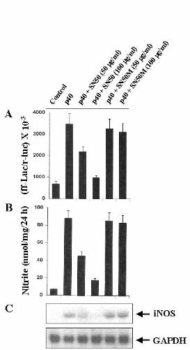
on the activation of NF-kB, we examined the effect of SN50 on p402-mediated
expression of iNOS. SN50 is a synthetic peptide containing signal sequences ofKaposi’s fibroblast growth factor and the nuclear localization sequence of NF-kBp50 (38). It has been reported to have the capacity to specifically block the nucleartranslocation of activated NF-kB (38). Inhibition of p402-mediated activation of
NF-kB (Fig. 5A), induction of NO production (Fig. 5B) and expression of iNOSmRNA (Fig. 5C) by SN50 but not by SN50M, a nonfunctional mutant of SN50,suggests that activation of NF-kB is necessary for the expression of iNOS inp402-stimulated BV-2 cells. However, p402 was unable to activate NF-kB in mouse
primary astrocytes (data not shown) suggesting that p402 is unable to induce iNOS
in mouse astrocytes (Table-2) due to its inability to induce the activation of NF-kB.The cytokines (TNF-α, IL-1β, IFN-γ, IL-12 p70, IL-12 p40, IL-6) or drugs(SN50 or SN50M) used under these experimental conditions had no effect on theviability of BV-2 glial cells, measured by trypan blue exclusion. Therefore, theconclusions drawn in this study are not due to any change in viability of the cells.
Discussion:IL-12, a heterodimeric cytokine, is most noted for its ability to regulate the
balance between type 1 and type 2 helper T cells (14,19). Neither IL-12 subunits(p35 or p40) alone was found to display significant biological activity over a largerange of concentrations (17-19). However, several evidences indicate that p40 isexpressed in excessive amount in the CNS of different demyelinating diseases suchas multiple sclerosis (MS), Guillain-Barre syndrome, and animal modelsexperimental autoimmune encephalomyelitis and neuritis (15,16,19-21). On theother hand, the expression of p35 remains almost constant or decreased to someextent in the CNS of these demyelinating diseases compared to the CNS of controlsubjects (19-21). However, the biological significance of this overexpression of
10
by guest on September 26, 2018
http://ww
w.jbc.org/
Dow
nloaded from

p40 in the CNS of patients with demyelinating diseases is not known.Several lines of evidence presented in this manuscript clearly support the
conclusion that IL-12 p40 homodimer, p402, induces the expression of iNOS in
mouse microglia and macrophages. This conclusion was based on the followingobservations. First, p402 induces the production of NO which is inhibited by
arginase, the enzyme which degrades the substrate of NOS, and by L-NMA, aninhibitor of NOS. Second, p402-mediated production of NO and expression of
iNOS is inhibited by anti-mouse p40 but not by anti-mouse p70. Third, theexpression of mouse p40 cDNA but not the mouse p35 cDNA induces theproduction of NO and the expression of iNOS suggesting that the p40 but not thep35 subunit of IL-12 is involved in the induction of iNOS. Since NO producedfrom the activation of iNOS in the CNS participates in the pathophysiology of MS(6-13), the overexpression of p40 mRNA in the CNS of MS patients (15,19-21)and the induction of iNOS by p40 suggest that p40 may participate in thepathophysiology of MS through the induction of iNOS.
The level of p40 is much higher (5- to 500-fold) than that of theheterodimeric p70 in IL-12-producing cells (19). This excess p40 produced eitherin vitro in activated cells or in vivo in serum of endotoxin-treated mice exists asboth dimer (20% to 40%) and monomer (the remainder) (19). Although thebiological role of the monomeric as well as the dimeric form of p40 is not known,it has been suggested that p402 may act as a physiologic regulator of bioactive IL-
12 since p402 possesses the IL-12 antagonist activity (19,39). Therefore, the
induction of iNOS by p402 suggests that p402 exhibits the IL-12 antagonist
activity possibly through the activation of iNOS. However, our observation thatboth p402 and the so-called bioactive IL-12 (heterodimeric p70) induce the
production of NO and the expression of iNOS prelude this possibility. If iNOS-derived NO mediates the IL-12 antagonist activity of p402 then IL-12 itself can
regulate its own function through the activation of iNOS. Apart from the IL-12antagonist activity of p40, experiments on Listeria monocytogenes infection inp40- and p35-deficient mice have shown that p40-deficient mice were susceptibleto infection but p35-deficient mice were able to eliminate bacteria despite themouse’s inability to produce biologically active heterodimeric IL-12 (19).Interestingly, it has also been found that p35-deficient mouse produce normallevels of p40 (19). Taken together, these observations suggest that p40 alone maycarry out some of the biological functions of heterodimeric IL-12. Here we present
11
by guest on September 26, 2018
http://ww
w.jbc.org/
Dow
nloaded from

the first evidence that similar to IL-12, p402 can also induce the expression of
iNOS and that iNOS-derived NO may account for the bacteria-eliminatingproperty of both IL-12 and p40.
The signaling events in cytokine-mediated induction of iNOS are notcompletely established so far. Proinflammatory cytokines (TNF-α, IL-1β, orIFN-γ) bind to their respective receptors and induce the expression of iNOS viaNF-kB activation (26-30,36,37). The presence of a consensus sequence in thepromoter region of iNOS for the binding of NF-kB (36) and the inhibition ofiNOS expression with the inhibition of NF-kB activation establishes an essentialrole of NF-kB activation in the induction of iNOS (26-30,36,37). Activation ofNF-kB by various cellular stimuli involves the proteolytic degradation of IkB, theinhibitory subunit of NF-kB complex, and the concomitant nuclear translocationof the liberated NF-kB heterodimer (40,41). Although the biochemical mechanismunderlying the degradation of IkB remains unclear, it appears that degradation ofIkB induced by various mitogens and cytokines occurs in association with thetransient phosphorylation of IkB on serines 32 and 36 (42). Consistently, twoclosely related kinases (IKKα and IKKβ) that directly phosphorylate IkBα havealso been described (43-45). Upon phosphorylation, IkB that is still bound to NF-kB apparently becomes a high affinity substrate for an ubiquitin-conjugatingenzyme (46). After phosphorylation-controlled ubiquitination, the IkB is rapidlyand completely degraded by the 20 S or 26 S proteosome, and the NF-kBheterodimer enter into the nucleus (47) and binds to the consensus DNA-bindingsite present in the promoter region of iNOS.
Our results have clearly shown that p402 induces the expression of iNOS
through the activation of NF-kB. First, p402 induces the DNA-binding as well as
the transcriptional activity of NF-kB. Second, expression of the mouse p40 cDNAbut not the mouse p35 cDNA induces the activation of NF-kB and the expressionof iNOS. Third, SN50, a cell-permeable peptide carrying the nuclear localizationsequence of p50 NF-kB, but not mutant SN50 (SN50M) inhibits p402-mediated
activation of NF-kB and expression of iNOS. It has been demonstrated that SN50specifically blocks the nuclear translocation of NF-kB, but does not affect theactivity of AP-1, SP-1 factor, and OCT-1 transcriptional factors (48) suggestingthat SN50 inhibits the expression of iNOS in p402-stimulated microglial cells by
inhibiting the activation of NF-kB. In addition, these results also suggest that IL-12 p402 is biologically active and that p402 alone can activate microglial cells.
12
by guest on September 26, 2018
http://ww
w.jbc.org/
Dow
nloaded from

At present, it is unclear how p402 activates NF-kB and induces iNOS in
microglial cells. IL-12 p402 has been shown to antagonize bioactive IL-12
heterodimer by binding to the IL-12 receptor complex (19). The high affinity IL-12 receptor is composed of a low affinity IL-12Rβ1 combined with a low affinityIL-12Rβ2 which are responsible for Tyk2/Jak2 activation, respectively, andSTAT4 activation (19,49). It appears that p402 binds to IL-12Rβ1 rather than IL-
12Rβ2 whereas bioactive IL-12 binds the receptor complex with high affinity (50).Therefore, it is possible that p402 activates NF-kB and induces the expression of
iNOS through the IL-12Rβ2.NO, a diffusible free radical, plays many roles as a signaling and as a
effector molecule in diverse biological systems including neuronal messenger,vasodilation and antimicrobial and antitumor activities (51,52). In the nervoussystem the NO appears to have both neurotoxic and neuroprotective effects andmay have a role in the pathogenesis of stroke and other neurodegenerative diseasesand in demyelinating conditions (e.g. multiple sclerosis, experimental allergicencephalopathy, X-adrenoleukodystrophy) associated with infiltratingmacrophages and the production of proinflamatory cytokines (53). NO and
peroxynitrite (reaction product of NO and O2-) are potentially toxic molecules to
neurons and oligodendrocytes that may mediate toxicity through the formation ofiron-NO complexes of iron containing enzyme systems (54), oxidation of proteinsulfhydryl groups (55), nitration of proteins and nitrosylation of nucleic acids andDNA strand breaks (56). Although monocytes/macrophages are the primary sourceof iNOS in inflammation, LPS and proinflammatory cytokines induce a similarresponse in microglia (35,57,58). NO derived from microglia has also beenimplicated in the damage of myelin producing oligodendrocytes in demyelinatingdisorders like multiple sclerosis and neuronal death during Alzheimer’s disease andbrain trauma (2-5).
Since IL-12 p40 is overexpressed in the CNS of the neuroinflammatorydiseases, the induction of iNOS expression by IL-12 p40 in microglia andmacropahges suggest that expression of p40 may induce/potentiate the neuralinjury in the inflamed CNS through the induction of NO production.
Acknowledgements: This study was supported by a grant from NIH (NS39940)and a seed grant from UNMC College of Dentistry (#00-09).
13
by guest on September 26, 2018
http://ww
w.jbc.org/
Dow
nloaded from

References:1. Galea, E., Feinstein, D. L., and Reis, D.J. (1992) Proc. Natl. Acad. Sci. USA. 89, 10945-109492. Koprowski, H., Zheng, Y.M., Heber-Katz, E., Fraser, N., Rorke, L., Fu, Z. F.,Hanlon, C., and Dietzshold, B. (1993) Proc. Natl. Acad. Sci. USA. 90, 3024-30273. Mitrovic, B., Ignarro, L. J., Montestruque, S., Smoll, A., and Merril, J. E. (1994) Neurosci. 61, 575-5854. Bo, L., Dawson, T. M., Wesselingh, S., Mork, S., Choi, S., Kong, P. A., Hanley,D., and Trapp. B. D. (1994) Ann. Neurol. 36, 778-7865. Merrill, J. E., Ignarro, L. J., Sherman, M. P., Melinek, J., and Lane, T. E. (1993) J. Immunol. 151, 2132-21416. Kolb, H., and Kolb-Bachofen, V. (1992) Immunol. Today 13, 157-1607. McCatney-Francis, N., Allen, J. B., Mizel, D. E., Albina, J. E., Xie, Q-W,Nathan, C. F. & Wahl, S. M. (1993) J. Ex. Med. 178, 749-7548. Koprowski, H., Zheng, Y. M., Heber-Katz, E., Fraser, N. et al. (1993) Proc.Natl. Acad. Sci. USA. 90, 3024-30279. Johnson, A. W., Land, J. M., Thompson, E. J., Bolanos, J. P., Clark, J. B. &Heales, S. J. R. (1995) J. Neurol. Neurosurg. Psychiatry 58, 107-11510. Brenner, T., Brocke, S., Szafer, F., Sobel, R. A., Parkinson, J. F., et al. (1997) J. Immunol. 158, 2940-294611. Hooper, D. C., Scott, G. S., Zborek, A., Mikheeva, T., Kean, R. B., Koprowski,H., and Spitsin, S.V. (2000) FASEB J. 14, 691-69812. Brosan, C. F., Battistini, L., Raine, C. S., Dickson, D. W. et al. 1994. Dev.Neurosci. 16, 152-16113. Bo, L., Dawson, T. M., Wesselingh, S., Mork, S., et al. 1994. Ann. Neurol. 36, 778-78614. Hsieh, C. S., Macatonia, S. E., Tripp, C. S., Wolf, S. F., O’Garra, A., andMurphy, K. M. (1993) Science 260, 547-54915. Constantinescu, C. S., Goodman, D. B., Hilliard, B., Wysocka, M., and Cohen,J.A. (2000) Neurosci. Lett. 287, 171-17416. Zipris, D., Greiner, D. L., Malkani, S., Whalen, B., Mordes, J. P., and Rossini,A. A. (1996) J. Immunol. 156, 1315-132117. Wolf, S.F., Temple, P. A., Kobayashi, M. et al. (1991) J. Immunol 146, 3074-308118. Schoenhaut, D.S., Chua, A. O. et al. (1992) J. Immunol. 148, 3433-344019. Gately, M.K., Renzetti, L.M., Magram, J., Stern, A.S., Adorini, L., Gubler, U.,and Presky, D.H. (1998) Annu. Rev. Immunol. 16, 495-521
14
by guest on September 26, 2018
http://ww
w.jbc.org/
Dow
nloaded from

20. van Boxel-Dezaire, A.H., Hoff, S.C. et al. (1999) Ann. Neurol. 45, 695-70321. Fassbender, K., Ragoschke, A., Rossol, S., Schwartz, A., Mielke, O., Paulig,A., and Hennerici, M. (1998) Neurology 51, 753-75822. Bright, J. J., Musuro, B. F., Du, C., and Sriram, S. (1998) J. Neuroimmunol. 82, 22-3023. McCarthy, K., and DeVellis, J. (1980) J. Cell Biol. 85, 890-90224. Giulian, D., and Baker, T. J. (1986) J. Neurosci. 6, 2163-217825. Pahan, K., Sheikh, F. G., Namboodiri, A. M. S., and Singh, I. (1998) Free Rad.Biol. Med. 24, 39-4826. Pahan, K., Liu, X., Wood, C., and Raymond, J.R. (2000) FEBS Lett. 472, 203-20727. Pahan, K., Liu, X., McKinney, M.J., Wood, C., Sheikh, F.G., and Raymond,J.R. (2000) J. Neurochem. 74, 2288-229528. Feinstein, D.L., Galea, E., Roberts, S., Berquist, H., Wang, H., and Reis, D. J.(1994) J. Neurochem. 62, 315-32129. Pahan, K., Namboodiri, A. M. S., Sheikh, F. G., Smith, B. T., and Singh, I.(1997) J. Biol. Chem. 272, 7786-779130. Pahan, K., Sheikh, F. G., Khan, M., Namboodiri, A. M. S., and Singh, I. 1998. J. Biol. Chem. 273, 2591-260031. Pahan, K., Raymond, J. R., and Singh, I. 1999. J. Biol. Chem. 274: 7528-753632. Bradford, M. (1976) Anal. Biochem. 72, 248-25433. Dignam, J.D., Lebovitz, R.M., and Roeder, R.G. (1983) Nucl. Acids Res. 11, 1475-148934. Aloisi, F., Penna, G., Cerase, J., Menendez Iglesias, B., and Adorini, L. (1997) J. Immunol. 159, 1604-161235. Suzumura, A., Sawada, M., and Takayanagi, T. (1998) Brain Res. 787, 139-14236. Xie, Q., Kashiwabara, Y., and Nathan, C. (1994) J. Biol. Chem. 269, 4705-470837. Pahan, K., Sheikh, F.G., Namboodiri, A.M.S., and Singh, I. (1997) J. Clin.Invest. 100, 2671-267938. Lin, Y-Z., Yao, S.Y., Veach, R.A., Torgerson, T.R., and Hawiger, J. (1995) J.Biol. Chem. 270, 14255-1425839. Germann, T., Rude, E., Mattner, F., and Gately, M. K. (1995) Immunol. Today 16, 500-50140. Stefanova, I., M. L. Corcoran, E. M. Horak, L. M. Wahl, J. B. Bolen, and I. D.Horak (1993) J. Biol. Chem. 268, 20725-20728
15
by guest on September 26, 2018
http://ww
w.jbc.org/
Dow
nloaded from

41. Salkowski, C. A., Detore, G., McNally, R., van Rooijen, N., and Vogel, S. N.(1997) J. Immunol. 158, 905-91242. Beg, A. A., Ruben, S. M., Scheinman, R. I., Haskil, S., Rosen, C. A., andBaldwin, A. S., Jr. (1992) Genes Dev. 6, 1899-191343. DiDonato, J. A., Hayakawa, M., Rothwarf, D. M., Zandi, E., and Karin, M.(1997) Nature 388, 548-55444. Maniatis, T. (1997) Science 278, 818-81945. Mercurio, F., Zhu, H., Murray, B. W. et al. (1997) Science 278, 860-86646. Sun, S.-C., Ganchi, P. A., Ballard, D. W., and Greene, W. C. (1993) Science 259, 1912-191547. Brown, K., Gerstberger, S., Carlson, L., Franzoso, G., and Siebenlist, U. (1995) Science 267, 1485-148848. Qin, Z. H., Wang, Y., Nakai, M., and Chase, T. N. (1998) Mol. Pharmacol. 53, 33-4249. Wang, X., Wilkinson, V. L., Podlaski, F. J., Wu, C., Stern, A. S., Presky, D. H.,Magram, J. (1999) Eur. J. Immunol. 29, 2007-201350. Presky, D. H., Yang, H., Minetti, L. J., Chua, A. O., Nabavi, N., Wu, C. Y.,Gately, M. K., Gubler, U. (1996) Proc. Natl. Acad. Sci. USA 93, 14002-1400751. Nathan, C. (1992) FASEB J. 6, 3051-306452. Jaffrey, S. R., and Snyder, S. H. (1995) Annu. Rev. Cell Dev. Biol. 11, 417-44053. Dawson, V.L., Dawson, T. M., London, E. D., Bredt, D. T., and Snyder, S. H.(1991) Proc. Natl. Acad. Sci. USA. 88, 6368-637154. Drapier, J-C., and Hibbs, J. B. (1988) J. Immunol. 140, 2829-283855. Radi, R., Beckman, J.S., Bush, K.M., and Freeman. B.A. (1991) J. Biol. Chem. 266, 4244-425056. Wink, D.A., Kasprazak, K.S. et al. (1991) Science 254, 1001-100357. Pahan, K., Sheikh, F.G., Namboodiri, A.M.S., and Singh, I. (1998) J. Biol.Chem. 273, 12219-1222658. Hu, S.X., Sheng, W.S., Peterson, P.K., and Chao. C.C. (1995) Glia 15, 491-494
Table-1. Induction of NO production by IL-12 in BV-2 glial cells
16
by guest on September 26, 2018
http://ww
w.jbc.org/
Dow
nloaded from

Treatments Nitrite (nmol/mg/24 h)Control 4.3 + 0.5
IL-12 (1 ng/ml) 35.6 + 4.1IL-12 (2 ng/ml) 66.3 + 7.1IL-12 (5 ng/ml) 85.5 + 9.8IL-12 (10 ng/ml) 86.3 + 8.7
IL-12 (5 ng/ml) + Arginase 8.4 + 1.0IL-12 (5 ng/ml) + L-NMA 12.9 + 1.5IL-12 (5 ng/ml) + D-NMA 83.7 + 7.5
BV-2 glial cells were cultured for 24 h in serum-free DMEM/F-12 with the listedreagents; and nitrite concentration in the supernatants was measured as describedunder “Materials and Methods”. Arginase (100 units/ml), L-NMA (0.1 mM) andD-NMA (0.1 mM) were added to the cells together with IL-12. Data are mean + S.D. of three different experiments.
Table-2. Induction of NO production by IL-12 p70 and IL-12 p402 in mouse primary astrocytes, microglia and macrophages
Treatments Nitrite (nmol/mg/24 h) Macrophages Astrocytes MicrogliaControl 9.2 + 1.2 3.4 + 0.3 6.7 + 0.8IL-12 (5 ng/ml) 112 + 13.5 3.5 + 0.4 92.5 + 11.2p402 (5 ng/ml) 120 + 15 3.4 + 0.4 95.2 + 10.8
Mouse peritoneal macrophages, and primary astrocytes and microglia werecultured for 24 h in serum-free DMEM/F-12 with the listed reagents; and nitriteconcentration in the supernatants was measured as described under “Materials andMethods”. Data are mean + S.D. of three different experiments.
Legends to figures:
17
by guest on September 26, 2018
http://ww
w.jbc.org/
Dow
nloaded from

Fig. 1. Induction of NO production and expression of iNOS by different cytokinesin BV-2 glial cells. Cells were cultured with different cytokines under serum-freecondition. A) After 24 h, supernatants were used for nitrite assay as mentionedunder “Materials and Methods”. Data are mean + S.D. of three differentexperiments. B) Cell homogenates were electrophoresed, transferred onnitrocellulose membrane and immunoblotted with antibodies against mousemacrophage iNOS as mentioned under “Materials and Methods”. Concentration ofdifferent stimuli were: LPS, 1.0 µg/ml; TNF-α, 50 ng/ml; IL-1β, 10 ng/ml; IFN-γ, 25 U/ml; IL-12, 5 ng/ml; IL-6, 20 ng/ml.Fig. 2. Dose-dependent induction of NO production and expression of iNOS byIL-12 p40 in BV-2 glial cells. Cells were cultured with different concentrations ofp402 under serum-free condition. A) After 24 h, supernatants were used for nitrite
assay. Data are mean + S.D. of three different experiments. B) Cell homogenateswere electrophoresed, transferred on nitrocellulose membrane and immunoblottedwith antibodies against mouse macrophage iNOS. C) After 6 h of incubation, cellswere taken out directly by adding Ultraspec-II RNA reagent (Biotecx LaboratoriesInc.) to the plates for isolation of total RNA, and Northern blot analysis for iNOSmRNA was carried out as described under "Materials and Methods."Fig. 3. Expression of p40 cDNA but not that of p35 cDNA induces the expressionof iNOS in BV-2 glial cells. Cells plated at 50 to 60% confluence in six-wellplates were transfected with 1 µg of each of p40 and p35 cDNAs by LipofectaminePlus (GIBCO) as described under Materials and Methods. Twenty-four h aftertransfection, cells were incubated with serum-free media in the presence of 1 µg/ml of anti-mouse IL-12 p70. A) After 24 h, supernatants were used for nitriteassay. Data are mean + S.D. of three different experiments. B) Cell homogenateswere electrophoresed, transferred on nitrocellulose membrane and immunoblottedwith antibodies against mouse macrophage iNOS.Fig. 4. IL-12 p40 induces the activation of NF-kB in BV-2 glial cells. A) Cellsincubated in serum-free DMEM/F-12 were treated with different concentrationsof p402. After 1 h of incubation, cells were taken out to prepare nuclear extracts,
and nuclear proteins were used for the electrophoretic mobility shift assay asdescribed under "Materials and Methods." B) Lanes 1-3 represent nuclear extractof control cells, nuclear extract of p402-treated cells, and nuclear extract of p402-
treated cells incubated with a 100-fold excess of unlabeled oligonucleotide. Theconcentration of p402 used in this experiment was 5 ng/ml. The upper arrow
indicates the induced NF-kB band, and the lower arrow indicates the unbound
18
by guest on September 26, 2018
http://ww
w.jbc.org/
Dow
nloaded from

probe. C) Cells plated at 50 to 60% confluence in six-well plates werecotransfected with 1 µg of pNF-kB-Luc (an NF-kB-dependent reporterconstruct) and 50 ng of pRL-TK (a plasmid encoding Renilla luciferase, used astransfection efficiency control) using Lipofectamine Plus as described underMaterials and Methods. After 24 h of transfection, cells were stimulated withdifferent concentrations of p402 for 6 h under serum-free condition. Firefly and
Renilla luciferase activities were obtained by analyzing total cell extract asdescribed under “Materials and Methods”.Fig. 5. Expression of p40 cDNA but not that p35 cDNA induces the activation ofNF-kB in BV-2 glial cells. Cells plated at 50 to 60% confluence in six-well plateswere cotransfected with 1 µg of pNF-kB-Luc (an NF-kB-dependent reporterconstruct) and 1 µg of p40, p35 or the empty vector using the Lipofectamine Plusmethod. All transfections included 50 ng/µg total DNA of pRL-TK. After 24 h oftransfection, cells were incubated with serum-free media for 24 h in the presenceof 1 µg/ml of anti-mouse IL-12 p70. Firefly and Renilla luciferase activities wereobtained by analyzing total cell extract as described under Materials and Methods.Fig. 6. SN50, a specific cell-permeable peptide inhibitor of NF-kB nucleartranslocation, inhibits p402-mediated activation of NF-kB and expression ofiNOS in BV-2 glial cells. A) Cells plated at 50 to 60% confluence in six-wellplates were cotransfected with 1 µg of pNF-kB-Luc and 50 ng of pRL-TK usingLipofectamine Plus. After 24 h of transfection, cells were incubated with differentconcentrations of SN50 or SN50M (a nonfunctional mutant of SN50) for 1 hfollowed by the stimulatation with 5 ng/ml of p402 for 6 h under serum-free
condition. Firefly and Renilla luciferase activities were obtained by analyzing totalcell extract as described above. B) Cells preincubated with different concentrationsof SN50 or SN50M for 1 h received 5 ng/ml of p402 under serum-free condition.
After 24 h, supernatants were used for nitrite assay. Data are mean + S.D. of threedifferent experiments. C) After 6 h of incubation, cells were taken out directly byadding Ultraspec-II RNA reagent (Biotecx Laboratories Inc.) to the plates forisolation of total RNA, and Northern blot analysis for iNOS mRNA was carried outas described above.
19
by guest on September 26, 2018
http://ww
w.jbc.org/
Dow
nloaded from


Thomas M. PetroKalipada Pahan, Faruk G. Sheikh, Xiaojuan Liu, Shilo Hilger, Michael McKinney and
in microglial cellsInduction of nitric oxide synthase and activation of NF-kappaB by interleukin-12 p40
published online December 7, 2000J. Biol. Chem.
10.1074/jbc.M008262200Access the most updated version of this article at doi:
Alerts:
When a correction for this article is posted•
When this article is cited•
to choose from all of JBC's e-mail alertsClick here
by guest on September 26, 2018
http://ww
w.jbc.org/
Dow
nloaded from