induced inflammation andvascular injury
TRANSCRIPT
INFECTION AND IMMUNITY, May 1982, p. 548-5570019-9567/82/050548-10$02.00/0
Vol. 36, No. 2
Effect of Immune Serum or Polymyxin B on Escherichia coli-Induced Inflammation and Vascular Injury
ANDREW C. ISSEKUTZ,t* SHABIR BHIMJI, AND ROBERT BORTOLUSSI
Departments of Microbiology and Pediatrics, Dalhousie University, Halifax, Nova Scotia, B3J 3G9 Canada
Received 12 August 1981/Accepted 23 December 1981
Bacterial invasion of the tissues often stimulates a vigorous inflammatoryreaction, which may limit the spread of microorganisms but may also beaccompanied by serious vascular injury and tissue damage. We previously studiedthe inflammatory reaction induced by the injection of killed Escherichia coli intorabbit skin, a model suitable for the quantitation of various parameters ofinflammation. Here we report the effect of immune serum treatment of the E. colion their capacity to induce inflammation and vascular injury. Injection of killed E.coli treated with immune serum elicited a reaction which had a smaller increase invascular permeability (protein exudation), measured with 1251I-labeled albumin,less increase in blood flow, measured with 86RbCl, less leukocyte infiltration,measured with 51Cr-labeled leukocytes, and a lesser degree of hemorrhage,measured with 59Fe-labeled erythrocytes, than E. coli treated with nonimmuneserum. Crossover experiments with four different E. coli serotypes and fourdifferent antisera indicated that antibody to specific 0 antigens or a relatedantigen, but not to K or H antigen, was important for modifying the inflammatoryresponse. Treatment of four different E. coli serotypes with antiserum to "core"glycolipid, produced by immunization with the E. coli J5 mutant, inhibited theinflammatory response to all four E. coli serotypes. Finally, treatment of killed E.coli with polymyxin B also inhibited their inflammation-inducing potential. Theseresults suggest that it may be possible to diminish the magnitude of local vascularand tissue injury associated with E. coli infections by the use of antisera orpolymyxin B, which bind to endotoxin on the E. coli.
Bacterial invasion of tissues often evokes asevere inflammatory response. Such a responseahd the subsequent phagocytosis and killing ofthe bacteria by the infiltrating polymorphonucle-ar leukocytes (PMNLs) are important factorslimiting the spread of the microorganisms (1,24). However, this type of a response may beaccompanied by local edema, vascular injury,and hemorrhage (4, 21). When these processesoccur in a vital organ such as the central nervoussystem, serious tissue damage and neurologicalsequellae may result (7, 9). We have recentlystudied the inflammatory reaction elicited byEscherichia coli, a bacterium that is a majorcause ofnewborn infection and meningitis. Men-ingitis caused by this organism is accompaniedby a high incidence of central nervous systemsequellae even with appropriate antibiotic thera-py (2, 19).We have found, in a rabbit model particularly
suitable for quantitating inflammatory reactions,that injection of killed E. coli into rabbit skininduces severe inflammation accompanied by
t Present address: Izaak Walton Killam Hospital, Halifax,Nova Scotia, B3J 3G9 Canada.
local protein exudation (permeability), increasein local blood flow, massive PMNL infiltration,and, shortly thereafter, extensive vascular inju-ry with hemorrhage (17). Because of the serious-ness of E. coli infections in humans and thesevere inflammatory reaction induced by thispathogen under clinical and experimental condi-tions, we have investigated approaches whichmay modify this reaction and perhaps shed lighton the bacterial and host factors that influencethe reaction. Here, using the rabbit model, wereport the effects of immune serum and poly-myxin B on E. coli-induced inflammation andvascular injury.
MATERIALS AND METHODSBacteria and antisera. All E. coli strains used were
clinical isolates. Three of the strains were serumresistant (30% serum) and possessed Ki capsularantigen (01:K1:H7, 0:18ac:K1:H7, and 07:K1), andone was a serum-sensitive stool isolate (055:K59). Aspontaneous mutant of 01:Kl:H7, which lacked Kl,that is, 01:Kneg:H7, was a kind gift from F. Orskov(Serumstaatsinstitut, Copenhagen, Denmark). Thesebacteria were grown in brain heart infusion broth(Difco Laboratories, Detroit, Mich.) for 18 h at 37°C
548

E. COLI-INDUCED INFLAMMATION AND IMMUNE SERUM
and then used live, Formalin killed (0.5% for 4 h), orboiled for 2.5 h in phosphate-buffered saline (PBS) asindicated. To raise antisera to "core" glycolipid, theUDP-galactose epimerase-deficient E. coli J5 mutant(kind gift from Peter Elsbach, New York University,New York, N.Y., and Loretta Leive, National Insti-tutes of Health, Bethesda, Md.), which does notsynthesize 0 antigen (10), was used. This strain wascultured in Davis minimal medium (6) to grow therough mutant or in Davis medium with 5 mM galactoseto grow the 0111 phenotype (34). The bacteria werewashed twice in PBS, and the number was adjustedspectrophotometrically at 540 nm, using a standardcurve that was verified by pour plate colony counts.
Antisera to the various serotypes were raised byimmunizing rabbits intravenously (i.v) at weekly inter-vals with 2.5 x 10⁷ Formalin-killed E. coli, boiled J5,or boiled O111:B4 for 3 weeks, then 5 x 10⁷ colony-forming units (CFU) at week 4, and 5 x 10⁷ CFU oflive E. coli, boiled J5, or boiled 0111:B4 for 2 moreweeks. One week later, serum was collected andtested for antibody as described below. All sera wereheat inactivated (56°C, 30 min) before use. Antiserumspecific for the Kl polysaccharide was produced byadsorption (90 min, room temperature) of anti-O1:K1:H7 serum with an excess (0.3 ml/1010 CFU) ofthe Formalin-killed Ki-deficient mutant (01:Kneg:H7).
Inflammation in the rabbit skin was induced byintradermal (i.d.) injection, on the clipped back of a
rabbit, of 108 Formalin-killed E. coli or 3 x 10⁷ live E.coli. These doses were previously determined to in-duce a reproducible reaction, with significant vascularinjury and hemorrhage (unpublished data). Bacteriafor skin injections were preincubated with saline orheat-inactivated nonimmune serum (prebleed) or im-mune serum (approximately 3 agglutinating units or0.5 to 2% serum) for 60 min at room temperature. Theywere then washed with 50 volumes of pyrogen-freesaline (3,000 x g, 20 min), resuspended in saline byusing a Vortex mixer, and injected i.d. in 0.2 ml with a30-gauge by 0.5-in. (1.27-cm) needle.Measurement of hemorrhage. Hemorrhage was
quantitated with 59Fe-labeled transfused erythrocytes(RBCs) as described previously (16). Briefly, RBCswere labeled with 59Fe by i.v. injection of 300 RCi of59Fe-ferrous citrate (New England Nuclear Corp.,Lachine, Quebec). Three days later the blood obtainedfrom such a "donor" rabbit contained 99% of theradioactivity in the RBC fraction. Blood was thencollected into acid-citrate-dextrose anticoagulant, andapproximately 12 x 106 cpm of RBCs, equivalent to 15to 20 ml of blood, was transfused i.v. into blood group-compatible 2- to 2.5-kg female New Zealand whiterabbits, which were bled for the same volume immedi-ately before transfusion. A few hours after transfu-sion, the skin sites were injected i.d. Before sacrifice,1 ml of venous blood was collected to determine thehematocrit and the amount of radioactivity circulatingper milliliter of blood. After the rabbit was killed by ani.v. overdose of sodium pentobarbital, the skin of theback was removed and the blood in the large vesselswas drained. Radioactivity in these sites was mea-
sured by a Packard gamma counter. The quantity ofblood in the sites was calculated (microliters of bloodequivalents per site) and adjusted to an average hemat-ocrit of 40%.Measurement of leukocyte infiltration. For the quan-
titation of leukocyte infiltration at the skin injectionsites, "Cr-labeled leukocytes were used as describedpreviously (12). Briefly, the rabbit receiving the skininjections was bled from the central ear artery for 30ml of blood into 0.2% EDTA and 3 ml into acid-citrate-dextrose. One volume of 1% hydroxyethylcellulose(Polysciences Inc., Warrington, Pa.) was mixed with 4volumes of EDTA-blood at 37°C to sediment theRBCs. The leukocyte-rich plasma was harvested andcentrifuged (200 x g, 10 min), and the leukocyte-RBCpellet was incubated for 30 min at 37°C in 4 ml of Ca2+-Mg2+-free Tyrode solution containing 10% autologousacid-citrate-dextrose plasma and 100 ,uCi of Na251CrO4 (New England Nuclear). After the incubation,the 52Cr-labeled leukocytes were washed in Ca2+-Mg2+-free Tyrode solution and injected i.d. back intothe donor rabbit. It has been shown that this type ofleukocyte preparation gives results comparable tothose of a highly purified PMNL preparation isolatedby hydroxyethylcellulose and Percoll (Pharmacia FineChemicals, Dorval, Que.) density gradient separation(12). This is true for inflammatory reactions in whichhistology confirms that more than 90% of the infiltrat-ing leukocytes are PMNLs, as was the case in thelesions studied here. For kinetic experiments, therabbit was injected i.d. with E. coli at various timesbefore labeled leukocyte injection. For cumulativemeasurements of leukocyte infiltration, the i.d. E. coliinjections were given at the time of leukocyte injection(see below).Measurement of vascular permeability. Exudation
due to enhanced vascular permeability was quantitat-ed with rabbit serum albumin (Sigma Chemical Co.,St. Louis, Mo.) labeled with 1251 (New England Nucle-ar) as described previously (32). Skin sites were inject-ed at various times (see below), and 20 min beforesacrifice, the animals were injected i.v. with 5 ,Ci of121-labeled albumin per kg.Measurement of blood flow. Blood flow was mea-
sured with 'Rb as previously described (11). Briefly,at the time of sacrifice, 75 ,uCi of 'RbCl (NewEngland Nuclear) was injected i.v. Forty-five secondslater an overdose of sodium pentobarbital and 10 ml ofsaturated KCI solution were injected by the sameroute.
E. coli i.d. injection protocol. Two injection proto-cols were used for studying the inflammatory reactionto E. coli. The kinetics of the development of thereaction was followed by giving i.d. injections inquadruplicate at various times before sacrifice in thesame rabbit. For example, sites were injected 6.5, 4.5,3, 1.5, and 0.75 h before sacrifice so that at deathlesions of different ages, and thus various stages ofevolution, were present. In the kinetic experiments,leukocyte infiltration was measured by administeringthe 51Cr-labeled leukocytes i.v. 60 min before sacri-fice, permeability was measured by injecting the 1251_labeled albumin 20 min before sacrifice, and bloodflow was measured by injecting 'RbCl in the last 45 s.In this way, in the lesions the content of 5"Cr was therate of accumulation of labeled leukocytes per hour,the content of 1251 was the rate of albumin extravasa-tion in 20 min, and the content of 'Rb was the bloodflow at the time of sacrifice. With the kinetic protocol,these three parameters of inflammation were mea-sured simultaneously in the same animal, and the ageof the lesions was expressed as the mean to the nearest
549VOL. 36, 1982

550 ISSEKUTZ, BHIMJI, AND BORTOLUSSI
PERMEABLITY
A._
E
C-L1-
UF
LEUKOCYTE NRLTRATION
- 2 5 HEMORRHAGE
- 20
> 15-
'= 10-
0
-" rf. ---Y--A-":o' 1 .5 3:0 4.5 6.0 co 0.0 1.5 3.0 4.5 6.0 24
AGE OF LESION (Hrs) AGE OF LESION (Hrs)
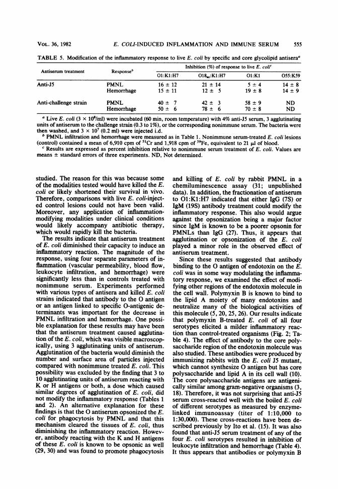
FIG. 1. Effect of immune serum on E. coli-induced inflammation. Killed E. coli 01:K1:H7 bacteria were
treated (60 min, room temperature) with nonimmune (prebleed) serum (x) or immune (anti-01:K1:H7) serum (0)and then washed. These E. coli were then injected i.d. in saline (108 CFU/0.2 ml) at various times so that lesionsof different ages were present at the time of sacrifice. The parameters were measured as described in the text.Control sites were injected with saline. These sites contained 80 cpm of 5"Cr, 280 cpm of 1251, 235 cpm of 86Rb,and 150 cpm of 59Fe, equivalent to 1.2 ,ul of blood, and these values were not influenced by the time of salineinjection. The blood content of control sites were subtracted from the hemorrhage values of the lesions. Pointsare means ± standard errors of quadruplicate sites in one representative experiment of three.
15 min at the time that the measurements were per-
formed.Hemorrhage was measured as a cumulative parame-
ter rather than as a rate because, unlike the case with5tCr-labeled leukocytes, '25N-labeled albumin, or'RbCl, the specific activity of the 59Fe-labeled RBCsin the circulation of these animals did not changesignificantly over the 24-h period of the experiment.Thus, the 59Fe content of the lesions was a measure ofthe labeled RBCs accumulating during the wholecourse of the reaction. In addition to the kineticexperiments, leukocyte infiltration could be measuredin a cumulative fashion simultaneously with hemor-rhage by injecting the sites with the E. coli at the timeof i.v. 5"Cr-labeled leukocyte injection. The reactionswere allowed to develop for 4.5 h, after which timerabbits were sacrificed.
In all of these experiments, 36 to 40 skin sites wereinjected in a random fashion so that control E. coli andtest E. coli lesions were present on the same animal forthe same time periods. Control experiments showedthat this many E. coli injections did not alter thereaction in any individual lesion regardless of the timeof injection. The content of 5"Cr, 1251, 86Rb, and 59Fein the lesions was analyzed with a Packard Auto-Gamma spectrometer. Corrections were made for thespill of radioactive emissions into adjacent channels.'Rb and 59Fe were not used in the same animalbecause of spectral overlap.
Antibody determinations. The antibody titer in theantisera was determined by bacterial agglutination,performed by serial 50-pl dilutions of antiserum inPBS in U-bottom microtiter plates (Falcon Plastics,Oxnard, Calif.) and addition of 50 ,ul of 1.5 x 10⁸ CFUof Formalin-killed (containing K, 0, and H antigens)or boiled (lacking K and H) E. coli per ml (22).Readings were performed after 18 h at 4°C. In addi-tion, a modification of an enzyme-linked immunoassaydescribed by Polin and Kennett (23) was used to detectantibodies to core glycolipid. Disposable flat-bottom1.5-ml polyvinylchloride vials (Chester Plastics, Ches-ter, N.S.) were treated sequentially with 0.75 ml ofpoly-L-lysine (0.001% x 2 h, room temperature) andwashed in PBS. Boiled or live E. coli (4 x 10⁷ CFU) in1 ml were then added, centrifuged at 2,000 rpm for 20min, fixed by addition of 1 ml of 1% glutaraldehyde(Polysciences Inc.), and incubated for 30 min at roomtemperature. The glutaraldehyde was decanted andfollowed by addition of 1% bovine albumin (SigmaChemical Co.) in 0.1 M glycine buffer, pH 7.6. Finally,the vials were washed and allowed to dry. Testing ofantisera was performed by making threefold dilutionsin 0.1% bovine serum albumin-PBS and incubating0.75 ml in the bacteria-coated vials for 60 min at roomtemperature. After extensive washing with PBS, 0.75ml of a 1:1,000 dilution of peroxidase-labeled goat anti-rabbit gamma globulin or goat anti-rabbit immunoglob-ulin G (IgG) (gamma chain specific) or IgM (mu chain
0
z0
>
0ca]
C
z
C-
0
INFECT. IMMUN.

E. COLI-INDUCED INFLAMMATION AND IMMUNE SERUM
specific) (Cappel Laboratories, Cochranville, Pa.) wasadded and incubated for 60 min at room temperature.After extensive washing, the peroxidase activity wasdetermined, using 0-dianisidine substrate (SigmaChemical Co.) (28). The reaction was linear up to an
absorbance of 0.350 at 460 nm. Therefore, titers areextrapolated and expressed as the greatest dilutiongiving 0.150 absorbance.
Immunization with a given E. coli serotype resultedin agglutinating antibody titers to K and H antigens of1:64 to 1:256. The titer to the 0 antigen of theimmunizing strain, as determined by agglutination ofboiled E. coli (lacking K and H), was in the range of1:2,500 to 1:10,000. The agglutination titer in nonim-mune (prebleed) sera to any of these antigens did notexceed 1:4 to 1:16. However, in several instances,immunization with one serotype caused a significant(fourfold or greater) rise in agglutinating titer againstone or more of the other three boiled E. coli serotypes.Therefore, these "cross-reacting" antibodies were re-moved by a single adsorption (90 min, room tempera-ture) of the antiserum with an excess (0.3 ml per 3 x10⁹ boiled E. coli) of the boiled cross-reacting strains.Such adsorption did not significantly decrease (lessthan fourfold) the titer against K, H, and 0 antigens ofthe serotype used for immunization. The sera ofrabbits used for the E. coli skin injections werescreened for agglutinating titers against the test E. coli.Animals with titers in excess of 1:16 were not used.
Fractionation of antiserum. Some antisera were frac-tionated by sucrose density gradient centrifugation(kindly performed by R. S. Faulkner), as previouslydescribed for the separation of 7S from 19S rubellaantibodies (8), except that the sera were not adsorbedwith chicken RBCs. Removal of IgG from the antise-rum was also performed by passing 2 ml through a 2-mIcolumn of protein A-Sepharose 4B (Pharmacia FineChemicals) as recommended by the manufacturer.
RESULTS
Effect of immune serum on the kinetics of E.coli inflammation. Figure 1 shows the kinetics offour parameters of inflammation during the reac-tion induced by the intradermal injection ofFormalin-killed E. coli 01:Kl:H7. Bacteria pre-treated with nonimmune serum evoked a rapidincrease in the vascular permeability, bloodflow, and leukocyte infiltration within the firsthour after injection. These parameters peaked in1.5-h-old (permeability) to 3-h-old (blood flowand for leukocyte infiltration) lesions. Changesin all three of these parameters then diminishedand were close to base-line values when thelesions were 6 h of age. Between 2 and 6 h afterinjection of E. coli, RBC extravasation andhemorrhage developed. The maximum degree ofhemorrhage was reached by 6 h. These resultsare similar to the reaction elicited by killed E.coli that were not serum treated, as described ina previous study (17). Figure 1 also shows theeffect of pretreating E. coli with approximately 3agglutinating units (1%) of heat-inactivated anti-serum to 01:Kl:H7. The antiserum-treated bac-teria induced milder reactions in permeability,
blood flow, and leukocyte infiltration, and, mostimportant, significantly less vascular injury andhemorrhage was observed.
Effect of antibody to K, H, or 0 antigens oninflammatory response induced by killed E. coli.The antiserum used in Fig. 1 contained antibod-ies reactive with at least three known antigenicstructures on the E. coli, namely, the capsularpolysaccharides (K), flagellum (H), and endo-toxin (0) (22). To determine whether antibody toone of these components of the bacterial surfacewas responsible for the modification of the in-flammatory reaction, various antisera were usedto treat a variety of E. coli serotypes beforeinjection (Table 1). Measurements were limitedto leukocyte infiltration and hemorrhage, be-cause previous work had shown a direct correla-tion between the degree of PMNL infiltrationand the severity of the vascular injury (13, 14;H. Z. Movat, M. M. Kopaniak, A. C. Issekutz,and B. J. Jeynes, Proc. 4th Int. Congr. Im-munol., abstr. 15.8.10, 1980). In contrast, theincreases in vascular permeability and bloodflow are transient and reversible phenomena,which are less clearly associated with irrevers-ible vascular damage.
It can be seen from Table 1 that inhibition ofPMNL infiltration (by 36 to 54%) and hemor-rhage (by 55 to 72%) occurred only when thechallenge E. coli was treated with antiserumcontaining antibody directed at the specific 0antigen. It should be noted that this was the caseeven though all of the Kl challenge strains wereagglutinated by each of the three anti-Kl-specif-ic antisera.The effect of anticapsular or flagellar antibody
was further investigated by adsorbing out the Kand H antibodies with Formalin-killed E. colicontaining Kl and H7 but differing at the 0antigen. Table 2 shows the effect of such adsorp-tion on the ability of the antisera to modify thePMNL infiltration and hemorrhage induced bytwo Kl- and H7-bearing E. coli strains. It can beseen that the adsorption of K and H antibodiesto the point where none were detectable byagglutination (<1:2) did not alter the capacity ofthe serum to inhibit PMNL infiltration (40 to45%) and hemorrhage (60 to 75%) when theantiserum contained antibodies to the 0 antigenof the challenge E. coli strain. The effect ofantibody to Kl capsular antigen was testeddirectly by treating the bacteria with an anti-Klantiserum. This antiserum was prepared by ad-sorbing antiserum raised against 01:Kl:H7 witha Formalin-fixed mutant of this strain, whichappears to differ only in that it does not synthe-size Kl antigen (01:Kneg:H7). After such ad-sorption, the antiserum retained an agglutinationtiter of 1:128 against Formalin-fixed 01:Kl:H7,but it did not agglutinate (<1:2) these E. coli
551VOL. 36, 1982

552 ISSEKUTZ, BHIMJI, AND BORTOLUSSI
TABLE 1. Modification of the inflammatory response to various strains of killed E. coli by various antisera"Inhibition (%) of response to killed E. colic
Responseb 01:K1:H7 018ac:K1:H7 07:K1 055:K59
Anti-01:K1:H7 PMNL 48 ± 5 10 ± 8 10 ± 5 NDHemorrhage 72 ± 6 5 ± 3 15 ± 5 8 ± 3
Anti-018ac:K1:H7 PMNL 8 ± 8 54 ± 5 9 ± 3 NDHemorrhage 10 ± 6 55 ± 5 2 ± 1 0 ± 2
Anti-07:K1 PMNL 0 ± 3 14 ± 7 40 ± 7 NDHemorrhage 8 ± 5 23 ± 7 62 ± 6 8 ± 4
Anti-055:K59 PMNL 8 ± 3 3 ± 5 ND 36 ± 3Hemorrhage 12 ± 5 15 ± 7 5 ± 5 56 ± 9
a Formalin-killed E. coli (109/ml) were incubated (60 min, room temperature) with 3 agglutinating units (0.5 to2%) of specific antiserum, of cross-reacting (with "K" or "H" antigens) antiserum (5 to 15%), or thecorresponding nonimmune (prebleed) serum at the same concentration. The E. coli isolates were washed, and108 (0.2 ml) were injected i.d. into rabbits.
b PMNL infiltration was measured cumulatively during the first 4.5 h, using 5 Cr-labeled leukocytes.Hemorrhage was measured simultaneously in 6.5-h-old lesions with 59Fe-labeled RBCs (see text). Control(nonimmune) serum E. coli lesions contained an average of 6,110 cpm of 51Cr and 1,805 cpm of 59Fe, equivalentto 18 ,ul of blood. Saline-injected sites contained 80 cpm of 51Cr and 110 cpm of 59Fe.
c Results are expressed as percent inhibition due to antiserum treatment relative to nonimmune serumtreatment of the E. coli. Values are means ± standard errors of at least three experiments performed inquadruplicate. ND, Not determined.
isolates after they were boiled to destroy Ki andH7 (01). Table 2 shows that this anti-Kl serumdid not modify the PMNL infiltration and hem-orrhage induced by 01:K1:H7 even though theequivalent of 10 agglutinating units of antiserumwas used to treat the bacteria.
Class of antibody influencing the inflammatoryresponse to E. coli. Antiserum to 01:K1:H7 wasfractionated into 7S and 19S antibodies by su-crose gradient centrifugation to determine thetype of antibody responsible for inhibiting theinflammatory reaction induced by the E. coli.Antibody to 0 antigen, as detected by agglutina-
tion of boiled 01 :K1 :H7, was present in both the7S and 19S fractions (Table 3). Not shown is thattwo fractions collected from the middle of thegradient, corresponding to the region with lessthan 19S but greater than 7S sedimentation, hadagglutinating titers of only 1:16, indicating rela-tively good separation of the 7S and 19S peaks.When 7S and 19S fractions were used to treat01:K1:H7 E. coli before injection, both frac-tions were found to inhibit PMNL infiltrationand the hemorrhagic reaction. Because it wasthought important to ensure that the observedeffect of the 19S IgM antibodies was not due to
TABLE 2. Effect of antiserum adsorption on modification of the inflammatory response to killed E. coliaInhibition (%) of response to
Antiserum Adsorbing strain Responseb killed E. col'01:K1:H7 018ac:K1:H7
Anti-O1:K1:H7 018ac:K1:H7 PMNL 40 ± 5Hemorrhage 60 ± 8
Anti-018ac:K1:H7 O1:K1:H7 PMNL 45 ± 4Hemorrhage 75 ± 5
Anti-O1:K1:H7 O1:K1 :H7 (K-negative mutant PMNL 5 ± 5of 01:K1:H7) Hemorrhage 7 ± 7
a Antisera were adsorbed (90 min at 4°C) until no agglutinating antibody against the adsorbing strain was
detectable. Killed E. coli O1:K1:H7 or 018,,,:K1:H7 were treated with similarly adsorbed nonimmune serum(control) or 3 agglutinating units of adsorbed antiserum before i.d. injection (108/0.2 ml), as in Table 1.
b Responses were measured simultaneously, as in Table 1.c Results are percent inhibition relative to control, nonimmune serum-treated E. coli. Values are means ±
standard errors of three rabbits performed in quadruplicate.
INFECT. IMMUN.

E. COLI-INDUCED INFLAMMATION AND IMMUNE SERUM
TABLE 3. Effect of antiserum fractions oninflammatory response to killed E. colia
Inhibition (%) of re-Anti-01:Kl:H7 0-antigen sponse to E. coli
fraction agglutination 01:K1:H7btiterPMNL Hemorrhage
7S 1:256 39 4 76 ± 319S 1:128 47 13 68 ± 819Safter 1:64 41 5 52±6
protein A-Sepharoseca Anti-01:K1:H7 serum and nonimmune serum
were fractionated on linear sucrose gradients. Thefractions were then used to treat (60 min, room tem-perature) killed E. coli 01:K1:H7 as in Table 1.
b Results are expressed as the percent inhibition ofthe responses relative to E. coli treated with fractionsof nonimmune serum. Values are means ± standarderrors offour experiments performed in quadruplicate.
c Antiserum was passed through a column of proteinA-Sepharose 4B and the 19S fraction was separated bysucrose gradient centrifugation.
contamination by small amounts of 7S IgG,these experiments were repeated by using pro-cedures to minimize this possibility. Two millili-ters of this antiserum was therefore first passedthrough a 2-ml column of protein A-Sepharose
J
z0
C-i
0
m
C.)0
J
co
PERMEABILITY
z
-4E^
4B to adsorb out nearly all of the IgG. Thematerial that passed through the column wasthen applied to the sucrose gradient. Agglutina-tion titers performed with these sucrose frac-tions indicated no agglutinating antibody in the7S fraction, whereas the 19S fraction containeda titer of 1:64. This doubly fractionated 19Santibody still inhibited the two inflammatoryparameters under study (PMNL infiltration,41%; and hemorrhage, 52%).
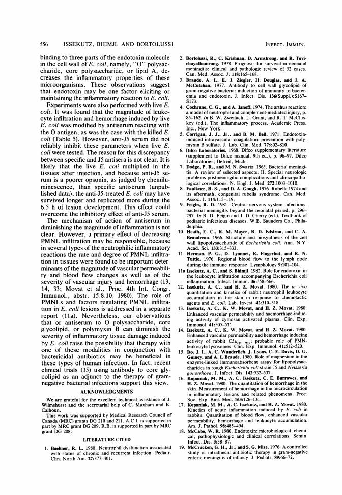
Effect of polymyxin B on E. coli-induced in-flammation. The results of the above experimentsuggested that antibodies reacting with the 0antigen of the endotoxin moiety on the E. colisurface could diminish the severity of inflamma-tion induced by Formalin-killed E. coli. Thisprompted us to test the effect of polymyxin B, adrug that is known to bind to the lipid A moietyof endotoxin and neutralize many of the biologi-cal actions of these molecules (5, 20, 25, 26).Figure 2 compares the development of the fourparameters of inflammation after the injection ofFormalin-killed 018ac:K1 :H7 E. coli treatedwith either saline or polymyxin B (50 ,ug/ml, 60min, room temperature) and washed. It can beseen that the polymyxin B treatment of the E.coli diminished the degree of vascular perme-ability, blood flow, leukocyte infiltration, and
LEUKOCYTE INRLTRATION
HEMORRHAGE
~, 15-z 12-
< 9-6-
El 00.0 1.5 3:0 4.5 6.0 ° 0.0 1.5 3.0 4.5 6.0 ' 24
AGE OF LESION (Hrs) AGE OF LESION (Hrs)
FIG. 2. Effect of polymyxin B on E. coli-induced inflammation. Killed E. coli 018ac:K1:H7 bacteria weretreated for 60 min at room temperature with saline (x) or 50 ,ug of polymyxin B per ml (0) and then washed. Thebacteria were then injected i.d. in saline (108 CFU/0.2 ml) at various times so that lesions of differing ages werepresent at the time of sacrifice. Inflammatory parameters and control itniections were described in the text.Control sites contained 260 cpm of 125I, 210 cpm of 'Rb, 75 cpm 51Cr, and 125 cpm 59Fe, equivalent to 1.1 ,ul ofblood. Points are means ± standard errors of quadruplicate sites in one representative experiment of three.
VOL. 36, 1982 553

554 ISSEKUTZ, BHIMJI, AND BORTOLUSSI
TABLE 4. Modification of the inflammatory response to various strains of killed E. coli by antiserum to coreglycolipid and by polymyxin B'
Inhibition (%) of response to killed E. colicTreatment Responseb 01:K1:H7 018ac:K1:H7 07:K1 055:K59
Polymyxin B (50 Lg/ml) PMNL 49 ± 6 30 ± 4 40 ± 5 37 ± 5Hemorrhage 68 ± 4 60 ± 5 72 ± 6 58 ± 7
Anti-J5 serum PMNL 29 ± 3 37 ± 5 39 ± 6 18 ± 5Hemorrhage 59 ± 5 39 ± 8 57 ± 8 30 ± 8
Anti-O11 serum PMNL 5 ± 3 6 ± 5Hemorrhage 8 ± 5 5 ± 2
Killed E. coli (109/ml) were incubated (60 min, room temperature) with 4% anti-J5 serum, anti-O111 serum,or the corresponding nonimmune serum as a control, or polymyxin B (50 ,ug/ml) or saline as a control. Thebacteria were washed, and 108 (0.2 ml) were injected i.d.
b PMNL infiltration and hemorrhage were measured as in Table 1. Control E. coli lesions contained a mean of5,210 cpm of 51Cr and 1,650 cpm of 59Fe, equivalent to 15 Ill of blood.
c Results are expressed as percent inhibition relative to nonimmune serum or saline treatment. Values aremeans ± standard errors of five expenments.
vascular injury with hemorrhage much as didantiserum treatment of the bacteria (Fig. 1).
In control experiments, zymosan particleswere treated with polymyxin B or injected to-gether with 50 ,ug of polymyxin B per ml. Theseconcentrations of polymyxin B did not alter thecourse of the reactions of zymosan (peak leuko-cyte infiltration in control zymosan lesions,4,100 cpm per site per h; polymyxin B treated,3,910 cpm; hemorrhage in control zymosan, 10.5,ul; polymyxin B, 9.7 [lI). Since polymyxin B canbind to the lipid A of most endotoxins (20), wetested the effect of this drug on all four E. coliserotypes. Table 4 shows that polymyxin Btreatment of any of the E. coli serotypes inhibit-ed the degree of PMNL infiltration (by 30 to49%) and hemorrhage (by 58 to 72%).
Effect of antiserum to core glycolipid on E. coli-induced inflammation. The core glycolipid orpolysaccharide region of endotoxin from a widevariety of gram-negative bacteria is antigenicallysimilar (reviewed in 3, 18). Therefore, antiserumto this region was produced by immunizingrabbits with the E. coli J5 mutant, which can-not synthesize 0 antigen (3, 10). The anti-J5serum was tested for cross-reacting antibody toboiled O1:Kl:H7, 018ac:K1:H7, and 07:K1,using the enzyme-linked immunoassay. Thisantiserum exhibited cross-reacting antibody ti-ters against these E. coli strains of 1:10,000 to1:30,000, whereas nonimmune titers were 1:40or less. The titer of cross-reacting antibody inthe anti-J5 antiserum was approximately one-third to one-fourth as high as in the anti-serum produced by specific immunization, e.g.,anti-01:K1:H7 against boiled 01:K1:H7 was1:40,000 by enzyme immunoassay. In contrastto the anti-J5 serum, such specific antisera
cross-reacted weakly (1:100 to 1:300) when mea-sured by this method.Table 4 shows the effect on PMNL infiltration
and hemorrhage of anti-J5 serum treatment ofthe four E. coli strains before i.d. injection. Thisantiserum inhibited the leukocyte response (15to 39%) and the degree of hemorrhage (30 to59%) induced by any of the four E. coli sero-types. Antiserum was also raised against E. coliJ5 grown in the presence of galactose to obtainthe phenotypic 0111:B4 parent (34). In contrastto the anti-J5 serum, treatment of E. coli withthis anti-O111 serum did not inhibit PMNLinfiltration or hemorrhage (Table 4). Anti-J5serum was also tested and compared with specif-ic 0 antiserum for modification of the inflamma-tory response induced by live E. coli. Table 5shows that, although anti-J5 treatment of For-malin-killed E. coli diminished somewhat theirability to induce leukocyte infiltration and hem-orrhage, this antiserum did not modify the re-sponse elicited by live E. coli. By contrast,specific 0 antiserum treatment diminished thereaction induced by live E. coli, as it did withkilled E. coli; this effect appeared to be specificfor the immunizing strain and is probably relatedto antibody to 0 antigen (not shown).
DISCUSSIONInvasion of host tissues by many types of
bacteria elicits a vigorous inflammatory reac-tion, often accompanied by tissue injury. Wepreviously found, in a rabbit model, that thesereactions are also elicited even by killed bacteriasuch as E. coli (17). Here we investigated somefactors which may modify the severity of theinflammatory reaction. In most of these experi-ments the reaction elicited by killed E. coli was
INFECT. IMMUN.
E. COLI-INDUCED INFLAMMATION AND IMMUNE SERUM
TABLE 5. Modification of the inflammatory response to live E. coli by specific and core glycolipid antisera'Inhibition (%) of response to live E. coWr
Antiserum treatment Responseb 1:K1:H7 018ac:Kl:H7 01:K1 055:K59
Anti-J5 PMNL 16 ± 12 21 ± 14 5 ± 4 14 ± 8Hemorrhage 15 ± 11 12 ± 5 19 ± 8 14 ± 9
Anti-challenge strain PMNL 40 ± 7 42 ± 3 58 ± 9 NDHemorrhage 50 ± 6 78 ± 6 70 ± 8 ND
a Live E. coti (3 x 10⁸/ml) were incubated (60 min, room temperature) with 4% anti-J5 serum, 3 agglutinatingunits of antiserum to the challenge strain (0.3 to 1%), or the corresponding nonimmune serum. The bacteria werethen washed, and 3 x 10⁷ (0.2 ml) were injected i.d.
b PMNL infiltration and hemorrhage were measured as in Table 1. Nonimmune serum-treated E. coli lesions(control) contained a mean of 6,910 cpm of 51Cr and 1,918 cpm of 59Fe, equivalent to 21 ,ul of blood.
c Results are expressed as percent inhibition relative to nonimmune serum treatment of E. coli. Values aremeans ± standard errors of three experiments. ND, Not determined.
studied. The reason for this was because someof the modalities tested would have killed the E.coli or likely shortened their survival in vivo.Therefore, comparisons with live E. coli-inject-ed control lesions could not have been valid.Moreover, any application of inflammation-modifying modalities under clinical conditionswould likely accompany antibiotic therapy,which would rapidly kill the bacteria.The results indicate that antiserum treatment
of E. coli diminished their capacity to induce aninflammatory reaction. The magnitude of theresponse, using four separate parameters of in-flammation (vascular permeability, blood flow,leukocyte infiltration, and hemorrhage) weresignificantly less than in controls treated withnonimmune serum. Experiments performedwith various types of antisera and killed E. colistrains indicated that antibody to the 0 antigenor an antigen linked to specific 0-antigenic de-terminants was important for the decrease inPMNL infiltration and hemorrhage. One possi-ble explanation for these results may have beenthat the antiserum treatment caused agglutina-tion of the E. coli, which was visible macroscop-ically, using 3 agglutinating units of antiserum.Agglutination of the bacteria would diminish thenumber and surface area of particles injectedcompared with nonimmune treated E. coli. Thispossibility was excluded by the finding that 3 to10 agglutinating units of antiserum reacting withK or H antigens or both, a dose which causedsimilar degrees of agglutination of E. coli, didnot modify the inflammatory response (Tables 1and 2). An alternative explanation for thesefindings is that the 0 antiserum opsonized the E.coli for phagocytosis by PMNL and that thismechanism cleared the tissues of E. coli, thusdiminishing the inflammatory reaction. Howev-er, antibody reacting with the K and H antigensof these E. coli is known to be opsonic as well(29, 30) and was found to promote phagocytosis
and killing of E. coli by rabbit PMNL in achemiluminescence assay (31; unpublisheddata). In addition, the fractionation of antiserumto O1:K1:H7 indicated that either IgG (7S) orIgM (19S) antibody treatment could modify theinflammatory response. This also would argueagainst the opsonization being a major factorsince IgM is known to be a poorer opsonin forPMNLs than IgG (27). Thus, it appears thatagglutination or opsonization of the E. coliplayed a minor role in the observed effect ofantiserum treatment.
Since these results suggested that antibodybinding to the 0 antigen of endotoxin on the E.coli was in some way modulating the inflamma-tory response, we examined the effect of modi-fying other regions of the endotoxin molecule inthe cell wall. Polymyxin B is known to bind tothe lipid A moiety of many endotoxins andneutralize many of the biological activities ofthis molecule (5, 20, 25, 26). Our results indicatethat polymyxin B-treated E. coli of all fourserotypes elicited a milder inflammatory reac-tion than control-treated organisms (Fig. 2; Ta-ble 4). The effect of antibody to the core poly-saccharide region of the endotoxin molecule wasalso studied. These antibodies were produced byimmunizing rabbits with the E. coli J5 mutant,which cannot synthesize 0 antigen but has corepolysaccharide and lipid A in its cell wall (10).The core polysaccharide antigens are antigeni-cally similar among gram-negative organisms (3,18). Therefore, it was not surprising that anti-J5serum cross-reacted well with the boiled E. coliof different serotypes as measured by enzyme-linked immunoassay (titer of 1:10,000 to1:30,000). These cross-reactions have been de-scribed previously by Ito et al. (15). It was alsofound that anti-J5 serum treatment of any of thefour E. coli serotypes resulted in inhibition ofleukocyte infiltration and hemorrhage (Table 4).It thus appears that antibodies or polymyxin B
VOL. 36, 1982 555
556 ISSEKUTZ, BHIMJI, AND BORTOLUSSI
binding to three parts of the endotoxin moleculein the cell wall of E. coli, namely, "O" polysac-charide, core polysaccharide, or lipid A, de-creases the inflammatory properties of thesemicroorganisms. These observations suggestthat endotoxin may be one factor eliciting ormaintaining the inflammatory reaction to E. coli.Experiments were also performed with live E.
coli. It was found that the magnitude of leuko-cyte infiltration and hemorrhage induced by liveE. coli was modified by antiserum reacting withthe 0 antigen, as was the case with the killed E.coli (Table 5). However, anti-J5 serum did notreliably inhibit these parameters when live E.coli were tested. The reason for this discrepancybetween specific and J5 antisera is not clear. It islikely that the live E. coli multiplied in thetissues after injection, and because anti-J5 se-rum is a poorer opsonin, as judged by chemilu-minescence, than specific antiserum (unpub-lished data), the anti-J5-treated E. coli may havesurvived longer and replicated more during the6.5 h of lesion development. This effect couldovercome the inhibitory effect of anti-J5 serum.The mechanism of action of antiserum in
diminishing the magnitude of inflammation is notclear. However, a primary effect of decreasingPMNL infiltration may be responsible, becausein several types of the neutrophilic inflammatoryreactions the rate and degree of PMNL infiltra-tion in tissues were found to be important deter-minants of the magnitude of vascular permeabili-ty and blood flow changes as well as of theseverity of vascular injury and hemorrhage (13,14, 33; Movat et al., Proc. 4th Int. Congr.Immunol., abstr. 15.8.10, 1980). The role ofPMNLs and factors regulating PMNL infiltra-tion in E. coli lesions is addressed in a separatereport (lla). Nevertheless, our observationsthat or antiserum to 0 polysaccharide, coreglycolipid, or polymyxin B can diminish theseverity of inflammatory tissue damage inducedby E. coli raise the possibility that therapy withone of these modalities in conjunction withbactericidal antibiotics may be beneficial inthese types of human infection. In fact, recentclinical trials (35) using antibody to core gly-colipid as an adjunct to the therapy of gram-negative bacterial infections support this view.
ACKNOWLEDGMENTS
We are grateful for the excellent technical assistance of J.Wilmshurst and the secretarial help of C. Maxham and K.Calhoun.
This work was supported by Medical Research Council ofCanada (MRC) grants DG 210 and 211. A.C.I. is supported inpart by MRC grant DG 209. R.B. is supported in part by MRCgrant DG 208.
LITERATURE CITED
1. Baehner, R. L. 1980. Neutrophil dysfunction associatedwith states of chronic and recurrent infection. Pediatr.Clin. North Am. 27:377-401.
2. Bortolussi, R., C. Krishnan, D. Armstrong, and R. Tovi-chayathamrong. 1978. Prognosis for survival in neonatalmeningitis: clinical and pathologic review of 52 cases.Can. Med. Assoc. J. 118:165-168.
3. Braude, A. I., E. J. Ziegler, H. Douglas, and J. A.McCutchan. 1977. Antibody to cell wall glycolipid ofgram-negative bacteria: induction of immunity to bacter-emia and endotoxin. J. Infect. Dis. 136(Suppl.):S167-S173.
4. Cochrane, C. G., and A. Janoff. 1974. The arthus reaction:a model of neutrophil and complement-mediated injury, p.85-162. In B. W. Zweifach, L. Grant, and R. T. McClus-key (ed.), The inflammatory process. Academic Press,Inc., New York.
5. Corrigan, J. J., Jr., and B. M. Bell. 1971. Endotoxin-induced intravascular coagulation: prevention with poly-myxin B sulfate. J. Lab. Clin. Med. 77:802-810.
6. Difco Laboratories. 1968. Difco supplementary literature(supplement to Difco manual, 9th ed.), p. 96-97. DifcoLaboratories, Detroit, Mich.
7. Dodge, P. R., and M. N. Swartz. 1965. Bacterial meningi-tis. A review of selected aspects. II. Special neurologicproblems postmeningitic complications and clinicopatho-logical correlations. N. Engl. J. Med. 272:1003-1010.
8. Faulkner, R. S., and D. A. Gough. 1976. Rubella 1974 andits aftermath, congenital rubella syndrome. Can. Med.Assoc. J. 114:115-119.
9. Feigin, R. D. 1981. Central nervous system infections:bacterial meningitis beyond the neonatal period, p. 296-297. In R. D. Feigin and J. D. Cherry (ed.), Textbook ofpediatric infectious diseases. W.B. Saunders Co., Phila-delphia.
10. Heath, E. C., R. M. Mayer, R. D. Edstron, and C. A.Beaudreau. 1966. Structure and biosynthesis of the cellwall lipopolysaccharide of Escherichia coli. Ann. N.Y.Acad. Sci. 133:315-333.
11. Herman, P. G., D. Lyonnet, R. Fingerhut, and R. N.Tuttle. 1976. Regional blood flow to the lymph nodeduring the immune response. Lymphology 9:101-104.
11a.Issekutz, A. C., and S. BhimJi. 1982. Role for endotoxin inthe leukocyte infiltration accompanying Escherichia coliinflammation. Infect. Immun. 36:558-566.
12. Issekutz, A. C., and H. Z. Movat. 1980. The in vivoquantitation and kinetics of rabbit neutrophil leukocyteaccumulation in the skin in response to chemotacticagents and E. coli. Lab. Invest. 42:310-318.
13. Issekutz, A. C., K. W. Movat, and H. Z. Movat. 1980.Enhanced vascular permeability and haemorrhage-induc-ing activity of zymosan activated plasma. Clin. Exp.Immunol. 41:505-511.
14. Issekutz, A. C., K. W. Movat, and H. Z. Movat. 1980.Enhanced vascular permeability and hemorrhage inducingactivity of rabbit C5ades Arg; probable role of PMN-leukocyte lysosomes. Clin. Exp. Immunol. 41:512-520.
15. Ito, J. I., A. C. Wunderlich, J. Lyons, C. E. Davis, D. G.Guiney, and A. I. Braude. 1980. Role of magnesium in theenzyme-linked immunoabsorbent assay for lipopolysac-charides in rough Escherichia coli strain J5 and Neisseriagonorrhoea. J. Infect. Dis. 142:532-537.
16. Kopaniak, M. M., A. C. Issekutz, C. E. Burrowes, andH. Z. Movat. 1980. The quantitation of hemorrhage in theskin. Measurement of hemorrhage in the microcirculationin inflammatory lesions and related phenomena. Proc.Soc. Exp. Biol. Med. 163:126-131.
17. Kopaniak, M. M., A. C. Issekutz, and H. Z. Movat. 1980.Kinetics of acute inflammation induced by E. coli inrabbits. Quantitation of blood flow, enhanced vascularpermeability, hemorrhage and leukocyte accumulation.Am. J. Pathol. 98:485-494.
18. McCabe, W. R. 1980. Endotoxin: microbiological, chemi-cal, pathophysiologic and clinical correlations. Semin.Infect. Dis. 3:38-87.
19. McCracken, G. H., Jr., and S. G. Mize. 1976. A controlledstudy of intrathecal antibiotic therapy in gram-negativeenteric meningitis of infancy. J. Pediatr. 89:66-72.
INFECT. IMMUN.

E. COLI-INDUCED INFLAMMATION AND IMMUNE SERUM
20. MorrIson, D. C., and D. M. Jacobs. 1976. Binding ofpolymyxin B to the lipid A portion of bacterial lipopoly-saccharides. Immunochemistry 13:813-818.
21. Movat, H. Z. 1979. The acute inflammatory reaction, p. 1-162. In H. Z. Movat (ed.), Inflammation, immunity andhypersensitivity. Harper and Row, Hagerstown, Md.
22. Orskov, I., F. 0rskov, B. Jann, and K. Jann. 1977.Serology, chemistry and genetics of 0 and K antigens ofEscherichia coli. Bacteriol. Rev. 41:667-710.
23. Polin, R. A., and R. Kennett. 1980. Use of monoclonalantibodies in an enzyme-linked inhibition assay for rapiddetection of streptococcal antigen. J. Pediatr. 97:540-544.
24. Quie, P. B., and K. L. Cates. 1978. Clinical manifestationsof disorders of neutrophil chemotaxis, p. 307-328. In J. I.
Gallin and P. G. Quie (ed.), Leukocyte chemotaxis:methods, physiology and clinical implications. RavenPress, New York.
25. RPfklnd, D. 1967. Prevention by polymixin B of endotoxinlethality in mice. J. Bacteriol. 93:1463-1464.
26. Rifkind, D., and R. B. Hill, Jr. 1967. Neutralization of theSchwartzmann reaction by polymyxin B. J. Immunol.99:564-569.
27. Scrlbner, D. J., and D. Fahrney. 1976. Neutrophil recep-tors for IgG and complement: their roles in the attachmentand ingestion phases of phagocytosis. J. Immunol.116:892-897.
28. Stelnman, R. M. 1976. Horseradish peroxidase as a mark-er for studies of pinocytosis, p. 379-386. In B. R. Bloom
and J. R. David (ed.), In vitro methods in cell-mediatedand tumor immunity. Academic Press, Inc., New York.
29. Stevens, P., C. L. Chu, and L. S. Young. 1980. K-1 antigencontent and the presence of an additional sialic acid-containing antigen among bacteremic K-1 Escherichiacoli: correlation with susceptibility to opsonophagocyto-sis. Infect. Immun. 29:1055-1061.
30. Stevens, P., S. N. Huang, W. D. Wdech, and L. W. Young.1978. Restricted complement activation by Escherichiacoli with the K-1 capsular serotype: a possible role inpathogenicity. J. Immunol. 121:2174-2180.
31. Stevens, P., and L. S. Young. 1977. Quantitative granulo-cyte chemiluminescence in the rapid detection of impairedopsonization of Escherichia coli. Infect. Immun. 16:796-804.
32. Udaka, K., Y. Takeuchi, and H. Z. Movat. 1970. Simplemethod for quantitation of enhanced vascular permeabili-ty. Proc. Soc. Exp. Biol. Med. 133:1384-1387.
33. Wedmore, C. V., and T. J. WilHams. 1981. Control ofvascular permeability by polymorphonuclear leukocytesin inflammation. Nature (London) 289:646-650.
34. Weiss, J., S. BecketQ iata, and P. Elsbach. 1980.Resistance of gram-negative bacteria to purified bacteri-cidal leukocyte proteins: relation to binding and bacteriallipopolysaccharide structure. J. Clin. Invest. 65:619-628.
35. Ziegler, E. J., J. A. McCutchan, and A. I. Braude. 1978.Clinical trial of core glycolipid antibody in gram-negativebacteremia. Trans. Assoc. Am. Physicians 91:253-258.
VOL. 36, 1982 557