increased chemical acetylation of peptides and proteins in ... · acetylphosphate (acp)....
TRANSCRIPT

Submitted 2 March 2018Accepted 10 April 2018Published 25 April 2018
Corresponding authorNilson A. Assuncao,[email protected],[email protected]
Academic editorVladimir Uversky
Additional Information andDeclarations can be found onpage 18
DOI 10.7717/peerj.4688
Copyright2018 Jedlicka et al.
Distributed underCreative Commons CC-BY 4.0
OPEN ACCESS
Increased chemical acetylation ofpeptides and proteins in rats afterdaily ingestion of diacetyl analyzed byNano-LC-MS/MSLeticia Dias Lima Jedlicka1,2, Sheila Barreto Guterres1,3, Aleksandro MartinsBalbino1, Giuseppe Bruno Neto1, Richardt Gama Landgraf1, Liliam Fernandes1,Emanuel Carrilho4, Etelvino José Henriques Bechara1,5 and Nilson A. Assuncao1
1 Institute of Environmental, Chemical and Pharmaceutical Sciences, Universidade Federal de São Paulo,Diadema, SP, Brazil
2 Institute of Studies in Health and Biological, Collective Health, Universidade Federal do Sul e Sudeste do Pará,Maraba, PA, Brazil
3Department of Chemistry, Fundacão Universidade Federal de Rondônia, Porto Velho, RO, Brazil4 São Carlos Institute of Chemistry, Universidade de São Paulo, São Carlos, SP, Brazil5Department of Fundamental Chemistry, Institute of Chemistry, Universidade de São Paulo,São Paulo, SP, Brazil
ABSTRACTBackground. Acetylation alters several protein properties including molecular weight,stability, enzymatic activity, protein–protein interactions, and other biological func-tions. Our previous findings demonstrating that diacetyl/peroxynitrite can acetylateL-lysine, L-histidine, and albumin in vitro led us to investigate whether diacetyl-treatedrats suffer protein acetylation as well.Methods. Wistar rats were administered diacetyl daily for four weeks, after which theywere sacrificed, and their lung proteins were extracted to be analysed by Nano-LC-MS/MS (Q-TOF). A C18 reversed-phase column and gradient elution with formicacid/acetonitrile solutions from 2 to 50% over 150 min were used to separate theproteins. Protein detection was performed using a microTOF-Q II (QTOF) equippedwith captive source and an electrospray-ionization source. The data from massspectrometry were processed using a Compass 1.7 and analyzed using Protein Scape,software that uses Mascot algorithms to perform protein searches.Results. A set of 3,162 acetylated peptides derived from 351 acetylated proteins in thediacetyl-treated group was identified. Among them, 23 targeted proteins were signifi-cantly more acetylated in the diacetyl-treated group than in the PBS control. Proteinacetylation of the group treated with 540 mg/kg/day of diacetyl was corroborated byWestern blotting analysis.Conclusions. These data support our hypothesis that diacetyl exposure in animals maylead to the generation of acetyl radicals, compounds that attach to proteins, affectingtheir functions and triggering adverse health problems.
Subjects Biochemistry, ToxicologyKeywords Radical acetylation, Diacetyl, Food additive, Lung diseases, Proteomics.
How to cite this article Jedlicka et al. (2018), Increased chemical acetylation of peptides and proteins in rats after daily ingestion of di-acetyl analyzed by Nano-LC-MS/MS. PeerJ 6:e4688; DOI 10.7717/peerj.4688

INTRODUCTIONDiacetyl is a flavoring commonly used in foodstuffs, as it lends a buttery flavor to productssuch as popcorn, coffee blends, cakes, cookies, wines and other goods (McCoy et al., 2017;Shibamoto, 2014; Ryan et al., 2014; Papetti, Mascherpa & Gazzani, 2014; Park, Gilbert &Whittaker, 2018; More, Raza & Vince, 2012). It is a volatile α-dicarbonyl and a highlyelectrophilic compound (Ryan et al., 2014) approved worldwide for use by food industries,despite ongoing health concerns dating back to 1986 (NIOSH, 1986), when the first casesof bronquiolitis obliterans involving diacetyl emerged (Park, Gilbert & Whittaker, 2018;Kreiss, 2014; Kanwal et al., 2011).
Recently, we reported that the reaction of peroxynitrite with α-dicarbonyls, namelydiacetyl and methylglyoxal, in aerated phosphate buffer pH 7.4 results in the acetylation offree amino acids, peptides and proteins added to the reaction mixture (Alves et al., 2013;Massari et al., 2010;Massari et al., 2011; Tokikawa et al., 2014). This reaction is initiated bynucleophilic addition of peroxynitrite to the carbonyl group of the α-dicarbonyl compoundyielding a peroxynitroso adduct, whose homolysis yields acetyl radicals.Dissolvedmolecularoxygen adds to the radical to ultimately produce acetate fromdiacetyl or acetate and formatefrom methylglyoxal (Massari et al., 2010;Massari et al., 2011). Formyl radical intermediategenerated by methylglyoxal/peroxynitrite was shown to add to the α-amino group ofL-lysine-containing synthesized tetrapeptides (Tokikawa et al., 2014). On the other hand,diacetyl/peroxynitrite-generated acetyl radicals have proven been proven to attack boththe α- and ε-amino groups of free and blocked L-Lys, L-Lys-containing peptides andserum albumin (Alves et al., 2013). These findings have raised the hypothesis that radicalacetylation of proteins contributes to transacetylase–promoted, post-translational proteinmodifications at sites where both methylglyoxal or diacetyl and peroxynitrite are present(Alves et al., 2013; Massari et al., 2010; Massari et al., 2011; Tokikawa et al., 2014). Fromthese facts, the competition of chemical (induced by diacetyl) and enzymatic (occurringnaturally in organisms) acetylation can be inferred, with the former process contributingto the increase of total protein acetylation.
Another source of acetylation in vivo is found through the action of acetyltransferases.These enzymes reversibly catalyze the transfer of the acetyl group from acetyl-CoA tothe ε-amino group of protein lysine residues (Drazic et al., 2016), a process promoted bylysine acetyltransferase and lysine deacetylase (Meng et al., 2018; Arif, Selvi & Kundu, 2010;Iyer, Fairlie & Brown, 2012) at the N-terminus during the synthesis of proteins. Proteinacetylation is highly conserved in eukaryotes and prokaryotes than phosphorylation, butit is less common than phosphorylation and ubiquitination (McEwan & Dikic, 2011).Acetylation can reportedly alter the protein function, size, enzymatic activity, stability,protein-protein interactions and other protein properties. When acetyltransferase isderegulated, and lysine acetylation is increased, modifications may occur in genes and inthe regulatory machinery, resulting in the manifestation of tumours in cells (Drazic et al.,2016). On the other hand, ATP-dependent acetylation has recently been reported to play arole in many cellular processes such as catalytic activity, immune responses and metabolicprocesses, including the generation of precursors of ‘‘energy-rich’’ metabolites such as
Jedlicka et al. (2018), PeerJ, DOI 10.7717/peerj.4688 2/22

acetylphosphate (acP). AcP-dependent acetylation tends to govern the translation of nu-cleotides, purine and pyrimidine metabolism and degradation of RNA (Kuhn et al., 2014).
In this work, we use proteomic and western blotting techniques to investigate if diacetylis also capable of leading to increases in vivo protein acetylation. Based on our findings, wesupport the hypothesis that diacetyl exposure in animals may lead to increases in proteinacetylation, which may affect protein functions and trigger adverse health problems.
METHODSAnimal treatmentAll animals were fed ad libitum and kept in a cabinet at 50–70% humidity, at a temperatureof 19–26 ◦C in a cycle of 12 h light/12 h dark. This study adheres to the guidelines establishedby the Brazilian College of Animal Experimentation (COBEA) and was approved by theEthical Committee of the SchoolMedicine of the FederalUniversity of SãoPaulo (UNIFESP,protocol no. 1949-11).
Eight-to-twelve-week-old male Wistar rats (250–300 g) were divided into two groups(6 animals each). The control group received phosphate-buffered saline (PBS), while thetreated group received 540 mg/kg/day of diacetyl (Cat B8530-7; Sigma Aldrich, St. Louis,MO, USA) dissolved in PBS Both groups were dosed using gavage. The concentration of540 mg/Kg/day of 2,3-butanedione and the treatment period of four weeks was based onthe experiment conducted by Colley and Cols (Colley et al., 1969).
After four weeks of treatment, the animals were anesthetized with ketamine and xylazine(Sigma Aldrich, St. Louis, MO, USA) and sacrificed. The lung tissue was collected andimmediately frozen in liquid nitrogen and stored at –80 ◦C.
Preparation of lung extractsTissue preparationFrozen lungs were ground into a fine powder in liquid nitrogen using a mortar andpestle. The homogenization process was used to avoid the activation of proteases andprevents protein degradation. The sample was lyophilized prior to analysis in order toremove residual water and stabilize the sample for handling at room temperature, therebyfacilitating the weighing process and preparation of the sample.
Protein extraction optimizationDue to the wide range of proteins and interfering substances in the final extracts, thesamples were obtained in the following three steps prior to proteomics analysis: tissuedisaggregation and cell homogenization; protein extraction from the biological matrix; andprotein precipitation and solubilisation in a urea buffer.
Three methods of protein extraction were tested to quantify the amount of protein inthe lysates before and after precipitation (Table 1). Thirteen milligrams of lyophilized lungsuspended in one mL of extraction buffer were used.
The lung powder was suspended and shaken for 1 h at 4 ◦C. After centrifugation (10min,5,000× RPM, 4 ◦C), 200 µL of the supernatant was mixed with 800 µL of DTT solution incold acetone (2 mg/mL) and incubated overnight at−20 ◦C. Afterwards, the samples were
Jedlicka et al. (2018), PeerJ, DOI 10.7717/peerj.4688 3/22

Table 1 Composition of the tested buffers.
Composition Buffer 1a Buffer 2b Buffer 3c
Bufferingagent
Tris 25 mM Tris 25 mM Tris 25 mM
Surfactant CHAPS 2% CHAPS4%TRITON 1%
CHAPS 4%
Proteaseinhibitors
Aprotinine,leupeptine,pepstatine,benzamidineand PMSF
Aprotinine,leupeptine,pepstatine,benzamidineand PMSF
Aprotinine,leupeptine,pepstatine,benzamidineand PMSF
Chaotropes 7 M Urea2 M Thiourea
Reducer 65 mM DTT
Notes.aBuffer 1 with power low solubility (few surfactants without chaotropes).bBuffer 2 with median solubilizing power (more surfactants).cBuffer 3 with maximum solubilization power (presence of chaotropes, surfactants and reducers).
centrifuged (10 min, 16,000 × RPM, 4 ◦C), and the sediments were washed four timeswith the DTT solution, dried in vacuum and solubilized in urea buffer (7 mol L−1 urea,2 mol L−1 thiourea, 4% CHAPS). The total protein concentration was determined by theBradford method (Bradford, 1976).
Tryptic digestionPrior to tryptic digestion, polypropylene microtubes were individually filled with an extractaliquot containing 250µg of the lung protein. The samples were reducedwithDithiothreitol(DTT) solution until achieving a final concentration of 5 mmol L−1, and they were thenincubated for 25 min at 56 ◦C. To achieve alkylation in the samples, iodoacetoamide (IAA)was added until reaching a final concentration of 14 mmol L−1. The samples were thenincubated for 30 min at room temperature and protected from light. Afterward, theywere diluted until the concentration of urea was reduced to 1,600 mmol L−1, and a CaCl2solution was added until reaching a final concentration of 1 mmol L−1 of CaCl2.
The enzymes trypsin and LysC endoproinase were added in the ratio of 1:50 of (enzyme:substrate). The samples were incubated for 18 h at 37 ◦C. The enzyme reaction was stoppedby adding TFA (trifluoroacetic acid) at the final concentration of 0.4%. The samples werecentrifuged at 2,500 rpm for 10 min at room temperature, and the pellet was discarded.The sample was evaporated until the volume was reduced to approximately 50 µl usinga vacuum concentrator (Speed Vacuum; Thermo Fisher Scientific, Waltham, MA, USA).Finally, 50 µl of 0.5% trifluoroacetic acid (TFA) were added thereto. Detergents wereremoved from the sample using a Pierce detergent removal spin column (# 87776; PierceBiotechnology/Thermo Fisher Scientific, Waltham, MA, USA), which was used accordingto the manufacturer’s specifications; the samples were filtered through a 22 µm PVDFsyringe filter stocked at 4 ◦C for mass spectrometry analysis.
Jedlicka et al. (2018), PeerJ, DOI 10.7717/peerj.4688 4/22

NanoLC–ESI/MS/MS analysesIn this study, on average six biological replicates and two replicate techniques were used.However, due to technical problems, we used 11 replicates of the control group and 10replicates of the group treated with 540 mg/kg/day of diacetyl. Each trypsinized samplewas dissolved with 100 µL of a mixture of water/acetonitrile/TFA (949:50:1 v/v). Allanalyses were performed using a Nano-UHPLC Advance (Bruker Daltonics, Bremen,Germany) equipped with a pump, an auto sampler, and a thermostatically controlledcolumn compartment. A C18 reversed-phase column (Magic C18 AQ, P/N: CP3/61271/00;Michrom, Boise, ID, USA), particle size 3 µm, internal diameter 0.1 mm, length 100 mmwas used. The column temperature was kept at 40 ◦C. Samples were separated using agradient mobile phase consisting of (A) formic acid/ACN/H2O (1:20:979) and (B) formicacid/ACN/H2O (1:950:50) in a gradient elution from 2 to 50 % over 150 min, as a graph inSM1. The flow rate was set at 0.500 µL/min, and the injection volume was 5 µL. Detectionswere performed using a micrOTOF-Q II (Bruker Daltonics, Billerica, MA, USA), anaccurate mass instrument equipped with captive source (Bruker Daltonics, Billerica, MA,USA) and an electrospray-ionization source (ESI). The mass spectrometer was runningin positive mode, with the desolvation temperature at 180 ◦C and the nebulizer set at 500V and 0.4 bars. All the operations, acquisition, and analysis of data were controlled byHystar software version 1.7 (Bruker Daltonics, Billerica, MA, USA). For MS/MS analyses,five precursor ions were automatically selected to undergo collision and fragmentationwith argon gas (≥ 2 L/min). Mass spectra were collected between 50 to 3,000 m/z, andcalibration was performed at the beginning of every day using the Tune-Mix ESI-G (AgilentTechnologies, Santa Clara, CA, USA). The collision energy was 12 eV, collision RF 600 Vpp,transfer time 140 µs, and pre-pulse storage 14 µs. MS/MS parameters were three precursorions, absolute threshold 2,000 cts, smart exclusion 5×, excluded after three spectra, andreleased after 1 min. The tune parameters were Funnel 1RF 300 Vpp, Funnel 2 RF 400Vpp, hexapole RF 400 Vpp, quadrupole ion energy 6.0 eV, and low mass 300 m/z. TheTOF (time of flight) conditions included the following: repetition rate 5 kHz, sample rate2 Ghz, flight tube 8,600 V, reflector 1,700 V, detector source 1,700 V, and detector TOF2140 V. Argon was used as a collision gas at a pressure of 2 ×10⁻⁶ mbar, and the collisionenergy values were 10–200 eV.
Bioinformatic analysisData deconvolution and database searchData from mass spectrometry were processed using a Compass 1.7 for OTOF (BrukerDaltonics, Billerica, MA, USA) and deconvoluted to generate a file compatible withMascot. This file was analyzed using Protein Scape (Bruker Daltonics, Billerica, MA,USA), a program that uses Mascot algorithms to perform the search. The databaseused was Swissprot, an annotated protein sequence database. The taxonomy was rattus,and the enzyme was trypsin with two missed cleavages. The fixed modification wascarbamidomethylation, and the variable modifications were oxidation of methionine andacetylation of lysine and arginine. Mass tolerance modification was 150 ppm to 1 Da.Mascot analysis of all proteins (p< 0.05) used a minimum score of 35.
Jedlicka et al. (2018), PeerJ, DOI 10.7717/peerj.4688 5/22
Protein network analysisThe protein-protein interaction analysis was performed using Cytoscape 3.3.0 software(http://www.cytoscape.org/) (Shannon et al., 2003), and the protein interaction networkwas obtained from the STRING8.2 database (http://string-db.org/) (Szklarczyk et al., 2011).STRING 8.2 uses the metric of ‘‘confidence score’’ to define the confidence of theinteractions. We selected only the interactions with proteins identified in our analyses.
Orthologs analysisOrthologs were subjected to Gene Ontology (GO) term analysis based on PANTHERclassification online tools (http://pantherdb.org/). To determine the biochemical functionsof acetylated proteins detected in the lungs of the group treated with diacetyl, GO wasperformed using IDs with the Rattus norvegicus genome found in the Uniprot database.This particular database was chosen as the reference database for the output report ofbiologicals process, proteins class, cellular components, pathways and molecular functions(Mi et al., 2013). These analyses were performed to acquire insights of the acetylationinvolved in the functions and pathways of proteins.
Analysis of sequence model around acetylated lysineThe software motif-x was employed to determine specific sequences of amino acid (15amino acids upstream and downstream of the acetylation site) in all protein sequencesacquired from NanoLC-MS/MS analysis. The entire database (IPI Rat Proteome) was usedas a background database parameter, and the significance was 0.000001 (Chou & Schwartz,2011; Schwartz & Gygi, 2005).
Western blottingSamples containing 25 µg of proteins from lung homogenate were subjected to 12%SDS–PAGE electrophoresis and electroblotted onto a nitrocellulose membrane (Millipore,USA). Following the blocking and washing steps, the membranes were incubated with theprimary Acetylated-Lysine antibody (Cell Signalling, Danvers, MA, USA) and anti-rabbitIgG HRP-linked (Cell Signalling, Danvers, MA, USA) as a secondary antibody. Themembranes were then detected using a chemiluminescence kit ‘‘Pierce ECL Plus WesternBlotting Substrate’’ (Thermo Scientific, Waltham, MA, USA) and chemiluminescencesoftware (GeneGnome System/Gene Tools Software; Syngene, Cambridge, UK).
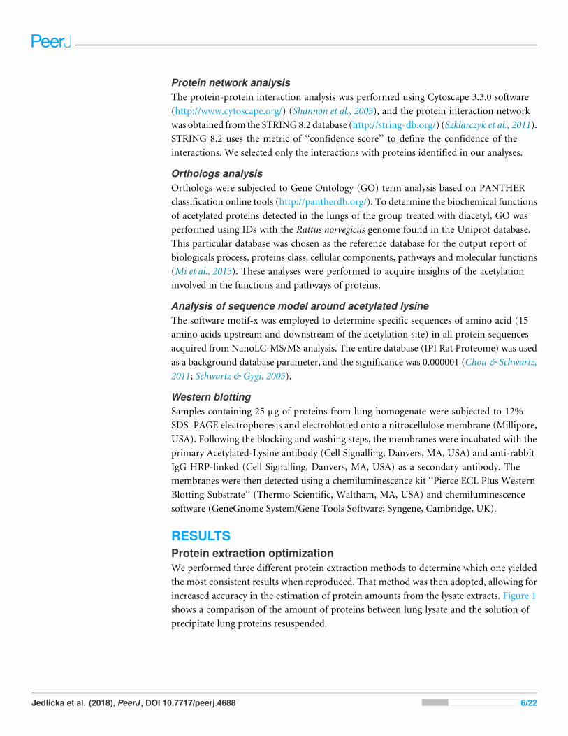
RESULTSProtein extraction optimizationWe performed three different protein extraction methods to determine which one yieldedthe most consistent results when reproduced. That method was then adopted, allowing forincreased accuracy in the estimation of protein amounts from the lysate extracts. Figure 1shows a comparison of the amount of proteins between lung lysate and the solution ofprecipitate lung proteins resuspended.
Jedlicka et al. (2018), PeerJ, DOI 10.7717/peerj.4688 6/22
Figure 1 Comparison of the amount of total protein present in the lysate and the resolubilizated pro-teins (precipitate proteins resuspended) using lung tissue.
Full-size DOI: 10.7717/peerj.4688/fig-1
MS/MS analyses of rat lung proteinsIn this work, qualitative proteomic analysis was used, specifically the bottom up technique.The lung extracts were analyzed by NanoLC-MS/MS, and significant differences wereshown among the protein profiles in the control and diacetyl-treated groups. Acetylationwas set in the search engine as a variable modification, and overall, the analyses showed10,302 peptides identified as belonging to 603 proteins in lung tissue. A set of 327 acetylatedproteins in the control group and 351 proteins in the group treated with 540 mg/kg/dayof diacetyl were detected. This increase in acetylation can occur either enzymatically orchemically, a phenomenon presently demonstrated in this work.
In this experiment, we identified 93 proteins which were common between the controlgroup and the 2,3-butanedione treated group. After this identification, we verified thepeptides present in these 93 proteins, the peptides common among the groups and theincidence of acetylation in these peptides. This peptide analysis was performed to ensurethat the acetylations found were due to the ingestion of 2,3-butanedione. We only validatedthe acetylations that were exclusively present in the treated group or that were expressed ina larger number in the treated group compared to the control group. After this analysis ofthe acetylated peptides, we selected 23 proteins, to which these acetylated peptides belong,and named them as ‘target proteins’.
Analysis of proteins and peptides revealed that acetylation is more abundant in the grouptreated with 2,3-butanedione than in the control group. The proteins that exhibited this
Jedlicka et al. (2018), PeerJ, DOI 10.7717/peerj.4688 7/22

Table 2 Target proteins, acetylated proteins in group treated with diacetyl but non-acetylated incontrol group.
Protein I.D. GeneI.D.
Meta scorecontrol
Meta scoretreated
Peptidescontrol
Peptidestreated
*SC [%]control
*SC [%]treated
AL1A1_RAT AL1A1 269.1 253.5 11 9 37.1 31.7ANXA2_RAT ANXA2 539.6 284.6 13 13 44.5 43.7ANXA5_RAT ANXA5 411 265 10 10 37.3 43.6ASSY_RAT ASS1 71 1,030.1 3 21 20.6 64.3BHMT1_RAT BHMT1 172.3 834.8 7 24 30 63.1CALR_RAT CARL 277.1 607.2 8 13 31.7 49.5CES1D_RAT CES1D 598.6 571.1 15 16 38.8 50.8DYH1_RAT DNAHC1 1,391.9 1,603.7 77 89 23.5 27.7EF2K_RAT EEF2K 354 304.5 18 17 38.5 34.1ENPL_RAT HSP90B1 742.6 492.2 25 16 29.6 23.5EPHA6_RAT EPHA6 486.4 361.4 27 20 32.4 24.5FABPL_RAT FABPL 120.5 779.4 3 16 36.2 81.9G3P_RAT GAPDH 486.7 763 14 18 53.8 62.2OGA_RAT MEGEA5 308.5 353.6 16 19 26.7 31.6PARK7_RAT PARK7 173.3 187.5 4 10 28.6 51.3PRC2A_RAT PRRC2A 776.2 670.4 43 36 25.3 23.6SI1L1_RAT SI1L1 575.9 672.1 32 37 27.9 30.7STIP1_RAT STIP1 189.5 258.9 10 14 22.1 30SYPM_RAT PARS2 171.2 183.4 9 10 32.6 46.3TBB4B_RAT TUBB4B 801.7 463.1 20 15 52.1 48.8TPP1_RAT TPP1 99.3 109.3 5 6 9.9 11.7UBR4_RAT UBR4 1,600.3 1,813.1 90 103 24.2 30.2UD2B2_RAT UGT2B 390.2 705.3 21 25 44.7 59.1
pattern of acetylation, described earlier as ‘target proteins’, and their respective peptidesare described in Table 2, which provides target protein identification and their respectivepeptide scores in both groups as well as descriptions of the peptide acetylation positions.
Some peptides showed post-translational modifications, and these peptides are listed inTable 3. As expected, L-lysine appears to be the predominant acetylated amino acid in thepeptide sequence, although arginine and histidine residues were found to be acetylated aswell.
The acetylation ratio from target proteins ratio was calculated in order tomore effectivelyvisualize the increase in acetylation. The increase in acetylation can be clearly seen inTable 4, which shows the increase in the acetylation ratio in the peptides identified in bothgroups. The student’s t -test was applied, and the difference was significant with p< 0.0001,demonstrating that there was a significant increase of the acetylation in these peptides.
Analysis of the distribution of acetylated proteins within the subcellular localizationrevealed that they were predominantly located in the cellular membrane and cytoplasm(53%). Nineteen percent are known to be present in the nucleus and 12% in the
Jedlicka et al. (2018), PeerJ, DOI 10.7717/peerj.4688 8/22

Table 3 Peptides from acetylated proteins in group treated with diacetyl but non-acetylated in control group.
Protein I.D. Gene I.D. Peptide sequence Peptidemetascorecontrol
Peptidemetascoretreated
Acetylationtreatedgroup
AL1A1_RAT ALDH1A1 -.MSSPAQPAVPAPLANLKIQHTK.I 15.4 16.1 7; 20; 22ANXA2_RAT ANXA2 K.ELPSAMKSALSGHLETVMLGLLK.T 15 21.7 3; 23ANXA2_RAT ANXA2 K.ELPSAMKSALSGHLETVMLGLLK.T 15 16.6 3; 23ANXA2_RAT ANXA2 K.GVDEVTIVNILTNR.S 71.9 18.2 14ANXA2_RAT ANXA2 K.SALSGHLETVMLGLLK.T 94 18.6 6ANXA5_RAT ANXA5 K.YMTISGFQIEETIDRETSGNLENLLLAVVK.S 16.4 16.7 15ANXA5_RAT ANXA5 K.YMTISGFQIEETIDRETSGNLENLLLAVVK.S 16.4 17.6 5; 30ASSY_RAT ASS1 R.GIYETPAGTILYHAHLDIEAFTMDR.E 39.8 16.1 13; 5BHMT1_RAT BHMT R.IASGRPYNPSMSKPDAWGVTK.G 16.3 17.5 5BHMT1_RAT BHMT R.IASGRPYNPSMSKPDAWGVTK.G 15.4 15.4 21; 30CALR_RAT CALR K.HEQNIDCGGGYVK.L 33 85.7 13CES1D_RAT CES1D K.GKVLGK.Y 24.4 15.1 2CES1D_RAT CES1D R.SHRDAGAPTFMYEFEYRPSFVSAMRPK.T 18.5 22.7 2; 25CES1D_RAT CES1D R.SHRDAGAPTFMYEFEYRPSFVSAMRPK.T 18.5 15.7 2; 7; 25DYH1_RAT DNAH1 R.SSLTRLASHMAEYECFQVELSK.N 19 16.7 5EF2K_RAT EEF2K R.SGDLYTQAAEAAMEAMK.G 30.7 21.1 7ENPL_RAT HSP90B1 R.MMKLIINSLYK.N 18.8 16.1 1; 3EPHA6_RAT EPHA6 R.EASIMGQFDHPNIIRLEGVVTK.R 18.3 16.8 0; 5EPHA6_RAT EPHA6 K.SVTEFNGDTITNTMTLGDIVYK.R 28.2 50.8 22FABPL_RAT FABPL K.SVTEFNGDTITNTMTLGDIVYK.R 16 36.2 22FABPL_RAT FABPL K.YQVQSQENFEPFMK.A 28.2 33.9 4G3P_RAT GAPDH K.RVIISAPSADAPMFVMGVNHEK.Y 18.6 23.1 1; 20; 22G3P_RAT GAPDH K.RVIISAPSADAPMFVMGVNHEK.Y 18.6 15.8 20; 22G3P_RAT GAPDH K.RVIISAPSADAPMFVMGVNHEK.Y 18.6 15.8 20; 22G3P_RAT GAPDH K.RVIISAPSADAPMFVMGVNHEK.Y 18.6 23.1 1; 20; 22OGA_RAT MGEA5 K.LDQVSQFGCRSFALLFDDIDHNMCAADK.E 20 15.4 21; 28PARK7_RAT PARK7 K.GAEEMETVIPVDIMR 28.2 16.1 5; 6PRC2A_RAT PRRC2A K.ALYPGALGRPPPMPPMNFDPRWMMIPPYVDPR.L 17.9 30.3 9PRC2A_RAT PRRC2A K.ALYPGALGRPPPMPPMNFDPRWMMIPPYVDPR.L 17.9 18.9 9PRC2A_RAT PRRC2A K.ALYPGALGRPPPMPPMNFDPRWMMIPPYVDPR.L 17.9 16.6 21PRC2A_RAT PRRC2A K.ALYPGALGRPPPMPPMNFDPRWMMIPPYVDPR.L 17.9 26.6 21; 32PRC2A_RAT PRRC2A K.ALYPGALGRPPPMPPMNFDPRWMMIPPYVDPR.L 17.9 19.4 32PRC2A_RAT PRRC2A K.ALYPGALGRPPPMPPMNFDPRWMMIPPYVDPR.L 17.9 15.1 9PRC2A_RAT PRRC2A K.AVGTPGGNSGGAGPGISTMSRGDLSQR.A 18.4 22 21; 27PRC2A_RAT PRRC2A R.ERSDSGGSSSEPFER.H 17.1 15.4 15SI1L1_RAT SIPA1L1 K.EKSKPYPGAELSSMGAIVWAVR.A 15.6 19.4 2SI1L1_RAT SIPA1L1 K.SLPLRRPSYTLGMK.S 19.5 16.8 5STIP1_RAT STIP1 R.RAMADPEVQQIMSDPAMR.L 20.7 18.9 1; 8
(continued on next page)
Jedlicka et al. (2018), PeerJ, DOI 10.7717/peerj.4688 9/22

Table 3 (continued)
Protein I.D. Gene I.D. Peptide sequence Peptidemetascorecontrol
Peptidemetascoretreated
Acetylationtreatedgroup
STIP1_RAT STIP1 R.RAMADPEVQQIMSDPAMR.L 20.7 20.4 8SYPM_RAT PARS2 K.GIEVGHTFYLGTKYSSIFNAHFTNA
HGESLLAEMGCYGLGVTR.I17.8 15 21; 26
TBB4B_RAT TUBB4B R.INVYYNEATGGKYVPR.A 21.9 15.4 6; 12TPP1_RAT TPP1 R.EREPELAQLLVDQIYENAMIAAGLVDDPR.A 15.2 19.3 29TPP1_RAT TPP1 R.INTLQAIWMMDPK.D 15.9 15.1 3UBR4_RAT UBR4 K.ALGTLGMTTNEKGQVVTK.T 15.7 21.7 2UBR4_RAT UBR4 K.EKAAPPPPPPPPPLESSPR.V 18.3 18.1 2; 9UBR4_RAT UBR4 K.EKEGESSGSQEDQLCTALVNQLNR.F 17.1 16.7 2; 24UBR4_RAT UBR4 K.FLSRPALPFILRLLR.G 15.1 30 5; 12UBR4_RAT UBR4 R.DNPEATQQMNDLIIGKVSTALK.G 28.8 17.2 6; 22UBR4_RAT UBR4 R.DNPEATQQMNDLIIGKVSTALK.G 17.3 21 22UBR4_RAT UBR4 R.MAGVMAQCGGLQCMLNRLAGVK.D 19.3 23.9 7UBR4_RAT UBR4 R.TGSTSSKEEDYESDAATIVQK.C 19.4 17.3 7; 21UD2B2_RAT UGT2B K.EWDTFYSEILGRPTTVDETMSKVEIWLIR.S 15.2 16.8 12; 22
cytoskeleton, while 14% are in different organelles, including mitochondria andendoplasmatic reticulum.
Protein interaction analysisFigure 2 consists of the acetylated protein network from treated group. This networkrepresents this protein interaction. Nodes represent the proteins in the network, and eachcolor represents a different situation in relation to protein acetylation, while the edgesrepresent the interactions between the proteins.
Orthology analysesIn order to reveal the involved cellular and metabolic processes as well as the subcellularlocation of the differentially expressed proteins in acetylation level with 2,3-butanedionetreatment, the GO-based analysis was conducted.
Analysis of the Molecular Function (Fig. 3A) revealed catalytic activity (57%), followedby specific binding function (19%). The analyses of biological functions (Fig. 3B) indicatedsome processes in which acetylated proteins are involved, including cellular processes(28.6%) and responses to stimulus (14.3%). The top three protein classes (Fig. 3C)display hydrolase (19%), chaperone (14.3 %) and oxidoreductase (14.3 %) activities. Thecellular component analyses (Fig. 3D) demonstrated that acetylated proteins belong tomacromolecular complexes (9.5%), cell organelles (9.5%), extracellular region (4.8%) andother cell parts (19%).
Motif analysis of proteins containing arginine-, lysine- andhistidine-acetylated peptidesIn order to characterize the possible specific sequence motifs surrounding acetylatedarginine, lysine and histidine residues in peptides of lung samples, a logo sequence to
Jedlicka et al. (2018), PeerJ, DOI 10.7717/peerj.4688 10/22

Table 4 Ratio of acetylation in both groups: control and treated with 2,3-butanedione.
Protein I.D. Gene I.D. Peptide sequence Acetylationratio controlgroup
Acetylationratio treatedgroup
AL1A1_RAT ALDH1A1 -.MSSPAQPAVPAPLANLKIQHTK.I 1 3ANXA2_RAT ANXA2 K.ELPSAMKSALSGHLETVMLGLLK.T 0.5 0.666666667ANXA2_RAT ANXA2 K.GVDEVTIVNILTNR.S 0 1ANXA2_RAT ANXA2 K.SALSGHLETVMLGLLK.T 0 1ANXA5_RAT ANXA5 K.YMTISGFQIEETIDRETSGNLENLLLAVVK.S 0 1ASSY_RAT ASS1 R.GIYETPAGTILYHAHLDIEAFTMDR.E 0 2BHMT1_RAT BHMT R.IASGRPYNPSMSKPDAWGVTK.G 0 2CALR_RAT CALR K.HEQNIDCGGGYVK.L 0 1CES1D_RAT CES1D K.GKVLGK.Y 0 1CES1D_RAT CES1D R.SHRDAGAPTFMYEFEYRPSFVSAMRPK.T 2 1.5DYH1_RAT DNAH1 R.SSLTRLASHMAEYECFQVELSK.N 0 1EF2K_RAT EEF2K R.SGDLYTQAAEAAMEAMK.G 0 1ENPL_RAT HSP90B1 R.MMKLIINSLYK.N 0 2EPHA6_RAT EPHA6 R.EASIMGQFDHPNIIRLEGVVTK.R 0 2FABPL_RAT FABPL K.SVTEFNGDTITNTMTLGDIVYK.R 0 1FABPL_RAT FABPL K.YQVQSQENFEPFMK.A 0 1G3P_RAT GAPDH K.RVIISAPSADAPMFVMGVNHEK.Y 1 1.5OGA_RAT MGEA5 K.LDQVSQFGCRSFALLFDDIDHNMCAADK.E 0 2PARK7_RAT PARK7 K.GAEEMETVIPVDIMR 1 0.5PRC2A_RAT PRRC2A K.ALYPGALGRPPPMPPMNFDPRWMMIPPYVDPR.L 1 0.5PRC2A_RAT PRRC2A K.AVGTPGGNSGGAGPGISTMSRGDLSQR.A 0 2PRC2A_RAT PRRC2A R.ERSDSGGSSSEPFER.H 0 1SI1L1_RAT SIPA1L1 K.EKSKPYPGAELSSMGAIVWAVR.A 0 1SI1L1_RAT SIPA1L1 K.SLPLRRPSYTLGMK.S 0 1STIP1_RAT STIP1 R.RAMADPEVQQIMSDPAMR.L 0 1SYPM_RAT PARS2 K.GIEVGHTFYLGTKYSSIFNAHFTNAH
GESLLAEMGCYGLGVTR.I1 2
TBB4B_RAT TUBB4B R.INVYYNEATGGKYVPR.A 0 2TPP1_RAT TPP1 R.EREPELAQLLVDQIYENAMIAAGLVDDPR.A 0 1TPP1_RAT TPP1 R.INTLQAIWMMDPK.D 0 1UBR4_RAT UBR4 K.ALGTLGMTTNEKGQVVTK.T 0 1UBR4_RAT UBR4 K.EKAAPPPPPPPPPLESSPR.V 0 2UBR4_RAT UBR4 K.EKEGESSGSQEDQLCTALVNQLNR.F 1 1UBR4_RAT UBR4 K.FLSRPALPFILRLLR.G 0 2UBR4_RAT UBR4 R.DNPEATQQMNDLIIGKVSTALK.G 2 3UBR4_RAT UBR4 R.MAGVMAQCGGLQCMLNRLAGVK.D 0 1UBR4_RAT UBR4 R.TGSTSSKEEDYESDAATIVQK.C 0 2UD2B2_RAT UGT2B K.EWDTFYSEILGRPTTVDETMSKVEIWLIR.S 0 2
Mean 0.283783784± 0.092(27)a
1.423423423± 0.104 (27)a
Notes.aMean± Std. Error(N).
Jedlicka et al. (2018), PeerJ, DOI 10.7717/peerj.4688 11/22

Figure 2 Network of acetylated proteins in group treated with Diacetyl. The green nodes are the pro-teins acetylated in group treated with Diacetyl but lack acetylation on treated groups. The red nodes arethe proteins presents only in group treated with Diacetyl; the blue nodes are the proteins presents only incontrol group and the purple nodes are proteins present in both groups.
Full-size DOI: 10.7717/peerj.4688/fig-2
compute the likelihood of amino acids at the positions surrounding the acetylation sitewas generated. Ten significantly enriched motifs were obtained from all the identifiedacetylated sites including *K, *R, *H (*K represents the acetylated lysine, *R represents theacetylated arginine and *H represents the acetylated histidine). As shown in Fig. 4, logoswith the highest scores were used and all motif analyses are available in SM 2–7. Figures 4Aand 4D show the motif surrounding acetylated arginine in samples from the control andtreated groups, respectively, and Figs. 4B and 4E show the motif surrounding acetylatedlysine. A number of reports have already demonstrated the occurrence of acetylation inarginine residue (Mathews et al., 2010). Figures 4C and 4F portray the motif surroundingacetylated histidine from control and groups treated with diacetyl, respectively.
Western blottingWestern blotting experiments indicated that the acetylation level was significantly higher inthe treated group as compared to the control group (Fig. 5). Figure 5A shows an increase inacetylation in bands that correspond between 35–70 KDa in lanes 5,7 and 8 that were filledwith samples from the group treated with 2,3-butanedione. The wells filled with samplesfrom the control group (lanes: 2–4) did not display the acetylation band.
Statistical analysis by the Student t -test revealed that mean values of protein intensitiesand variances are significantly different, with p= 0.0091 for means and p= 0.0015for variance. This Western blotting experiment data confirms the result of LC-MS/MS
Jedlicka et al. (2018), PeerJ, DOI 10.7717/peerj.4688 12/22

Figure 3 Orthology analyses frommore acetylated proteins in group treated with Diacetil than incontrol group. (A) Molecular function; (B) biological process; (C) protein class; (D) cellular component.
Full-size DOI: 10.7717/peerj.4688/fig-3
analysis, which revealed increases in protein acetylation from the group treated with2,3-butanedione in comparison with the control group.
DISCUSSIONProtein extraction optimizationThe amount of proteins obtained from precipitated and resolubilizated proteins usingthree buffers. Buffers 1 and 2 were slightly more efficient than Buffer 3, which led us tochoose Buffer 2 in all experiments.
Jedlicka et al. (2018), PeerJ, DOI 10.7717/peerj.4688 13/22

A
B
C
D
E
F
Figure 4 Motif analysis surrounding arginine, lysine and histidine acetylated peptides. (A) Motif anal-ysis of control group, surrounding acetylated arginine. (B) Motif analysis of control group, surroundingacetylated lysine. (C) Motif analysis of control group, surrounding acetylated histidine. (D) Motif analysisof group treated with diacetyl, surrounding arginine. (E) Motif analysis of group treated with diacetyl, sur-rounding lysine. (F) Motif analysis of group treated with diacetyl, surrounding histidine.
Full-size DOI: 10.7717/peerj.4688/fig-4
MS/MS analyses of rat lung proteinsThe results from provide evidence of an increase in protein acetylation in the group treatedwith diacetyl. Acetylation reportedly alters protein function, size, enzymatic activity,stability, protein-protein interactions and other protein properties. Some proteins regulateacetyltransferases and histone deacetylases and may induce acetylation of other proteins(Drazic et al., 2016). When acetyltransferases are deregulated, and lysine acetylation isincreased, modifications may occur in genes and the regulatory machinery (Drazic et al.,2016). These data show that diacetyl- triggered protein acetylation takes place in differentcell compartments and that it may be implicated in many cell functions.
Jedlicka et al. (2018), PeerJ, DOI 10.7717/peerj.4688 14/22

Figure 5 Western blotting for acetylated proteins from lung samples. (A) Western blotting 4 image:Lane 1: molecular weight; lanes 2–4: samples of the control group; lanes 5, 7, and 8: samples 5 of thediacetyl-treated group; and lanes 6, 9, and 10: sample buffer. (B) Western blotting quantification.
Full-size DOI: 10.7717/peerj.4688/fig-5
Protein interaction analysisThe protein interaction analysis showed that some acetylated proteins are interconnectedand/or connected with other proteins. To exemplify this interaction, we can cite the proteinASS1, found to be acetylated in the treated group, which interacts with both ALS and OTC.Present in our control group is the ALS enzyme, whose activity is regulated by acetylation,according to the UniProt database (http://www.uniprot.org). ASS1 interacts with OTC,which is present only in the diacetyl-treated group. OTC, one of the enzymes of the ureacycle, acts by detoxifying the excess of ammonium produced from amino acid catabolismand is negatively regulated by lysine acetylation (Yu et al., 2009).
Some acetylated proteins present in the network are involved in the cell redox balance(Drazic et al., 2016), in protein biosynthesis and have ATP and nucleotide binding activity,maturation, structural maintenance and regulation of specific proteins (Haase & Fitze,2016), along with cellular processes such as the basal metabolism, immunogenicity, cellcycle progression, DNA repair and apoptosis (Michalak et al., 2009). Some proteins alsoinduce anti-tumor immunity by inhibiting angiogenesis and have antioxidant activity inneurons and the heart, protecting against cell death (Kolisek et al., 2015; Singh et al., 2015).Additionally, the proteins play a cytoprotective role being a redox-responsive protein(Eltoweissy et al., 2011).
The increase in chemical acetylation of lung proteins of diacetyl-treated rats describedhere may be connected with the fact that diacetyl has been shown in vitro to generateacetyl radicals upon reaction with peroxynitrite, and more slowly with hydrogen peroxide(Tokikawa et al., 2014). The diacetyl/peroxynitrite system was then reported to promote
Jedlicka et al. (2018), PeerJ, DOI 10.7717/peerj.4688 15/22

acetylation of isolated amino acids, peptides and albumin. These data led us to postulatethat post-translational chemical acetylation of proteins may contribute to enzymaticacetylation at sites where both diacetyl and peroxynitrite at inflammation are formed.
Orthology analysesThe Gene Ontology (GO) function analysis of the target proteins revealed the distributionand function of these proteins. Protein acetylation regulates enzyme activities that mediate,for instance, the degradation of proteasomes and lysosomes by neutralizing the lysineresidues in the active sites, thereby causing conformational changes. In addition toregulating the catalytic activity of metabolic enzymes, acetylation controls substrateaccessibility, blocks substrate binding to the enzyme and modulates enzyme subcellularlocalization (Xiong & Guan, 2012).
The most crucial pathways are those related to the oxidative stress response (P00046),which causes cellular damage. In a normal functioning cell, several transcription factorsrespond to oxidative stress by modulating the expression of genes whose products relievethe altered redox status.
Motif analysis of proteins containing arginine-, lysine- andhistidine-acetylated peptidesThe possible motifs surround acetylated arginine, lysine and histidine. Despite lysinebeing the more common site of protein acetylation, some studies have demonstrated thatarginine can be acetylated as well, triggering biological responses (Rabbani et al., 2011;Slade, Subramanian & Thompson, 2014). Acetylation in both lysine and histidine residuewas previously demonstrated in vitro (Alves et al., 2013; Massari et al., 2010; Massari etal., 2011; Tokikawa et al., 2014), which reinforces our results about acetylation in theseresidues.
Western blottingWestern blotting was used to confirm the increase of acetylation previously found byNanoLc-ms/ms experiments. We used a specific acetylation antibody to detect bandswith a substantial increase in intensity in samples from the group treated with diacetylthat reveals the protein acetylation increase. These results confirm the protein acetylationidentified by NanoLc-ms/ms analyses.
CONCLUSIONSAltogether, our data strongly suggest that diacetyl gavage administered to rats mayconstitute a source of acetyl radical that can attack and acetylate lung proteins. It istempting to hypothesize that this is a contribution mechanism for the reported toxicity ofdiacetyl in workers dealing with ‘buttered’ food who subsequently acquire bronquiolitisobliterans.
Herein, we first optimized extraction conditions for the lung proteins of Wistar rats, forboth rats in the control group and those treated with diacetyl. Mass spectrometry results,confirmed by Western blotting analyses, revealed increased acetylation in the lung tissuesof groups treated with 2,3-butanedione.
Jedlicka et al. (2018), PeerJ, DOI 10.7717/peerj.4688 16/22

The proteins acetylated to different extents in the diacetyl-treated groupwere then relatedto reported interactions with other key proteins and enzymes of cell homeostasis. Diacetyltreatment, apparently, modifies the lung protein profile. Twenty-three diverse classes ofproteins were found to undergo preferential acetylation. They are present in differentregions of the cell and are involved in different molecular and biological processes. Ourdata indicate that the observed increased radical acetylation by diacetyl occurs randomly.
In a comprehensive view, we found more peptides acetylated in the group treated withdiacetyl than in the control group. The expected acetylation of lysine residues also occurredin arginine and histidine, suggesting that unlike acetylase-driven acetylation of proteins,radical acetylation occurs randomly, modifying residues of both the N-terminal, theC-terminal and the side chain of basic amino acid residues. The Western blotting analysisclearly demonstrated increased protein acetylation due to the daily intake of diacetyl.
Our study is consistent with early in vitro studies that showed increases in proteinacetylation in the presence of 2,3-butanedione. The data reported here reinforce ourhypothesis that diacetyl exposure is capable of increasing protein acetylation in vivo, thusraising a potential for diacetyl, a highly electrophilic α-dicarbonyl industrial xenobiotic, toplay a role in inflammatory bronquiolitis obliterans (Cavalcanti et al., 2012; Cummings etal., 2014; Egilman & Schilling, 2012).
List of abbreviations
ANXA2 Annexin-A2ANXA5 Annexin-A5ASS1 Argininosuccinate synthaseBHMT1 Betaine-homocysteine S-methyltransferase-1CALR CalreticulinCHAPS 3-[(3-cholamidopropyl)dimethylammonio]-1-propanesulfonateES1D Carboxylesterase-1DDTT DithiothreitolDNAH1 Dynein heavy chain-1EF2K Eukaryotic elongation factor-2 kinaseHSP90B1 EndoplasminEPHA6 Ephrin type-A receptorFABPL Fatty acid-binding proteinGAPDH Glyceraldehyde-3-phosphate dehydrogenaseMGEA5 O-GlcNAcasePARK7 Deglycase DJ-1PARS2 Protein Probable proline-tRNA ligase PBS] Phosphate-buffered salinePMSF Phenylmethylsulfonyl fluorideSIPA1L1 Signal-induced proliferation-associated 1-like protein-1STIP1 Stress-induced-phosphoprotein-1TUBB4B Tubulin beta-4B chainTFA Trifluoroacetic acidUBR4 Ubiquitin-protein ligaseUGT2B UDP-glucuronosyl- transferase-2B2.
Jedlicka et al. (2018), PeerJ, DOI 10.7717/peerj.4688 17/22

ADDITIONAL INFORMATION AND DECLARATIONS
FundingThis material is based upon work supported by the São Paulo Research Foundation(FAPESP, grants no. 2012/02514-9, 2013/07763-0, and 2010/01404-0) and the BrazilianInnovation Agency (FINEP). The funders had no role in study design, data collection andanalysis, decision to publish, or preparation of the manuscript.
Grant DisclosuresThe following grant information was disclosed by the authors:São Paulo Research Foundation (FAPESP): 2012/02514-9, 2013/07763-0, 2010/01404-0.Brazilian Innovation Agency (FINEP).
Competing InterestsThe authors declare there are no competing interests
Author Contributions• Leticia Dias Lima Jedlicka conceived and designed the experiments, performed theexperiments, analyzed the data, prepared figures and/or tables, authored or revieweddrafts of the paper, approved the final draft.• Sheila Barreto Guterres conceived and designed the experiments, performed theexperiments, authored or reviewed drafts of the paper, approved the final draft.• Aleksandro Martins Balbino and Giuseppe Bruno Neto performed the experiments,authored or reviewed drafts of the paper, approved the final draft.• Richardt Gama Landgraf, Liliam Fernandes, Emanuel Carrilho and Etelvino JoséHenriques Bechara conceived and designed the experiments, analyzed the data,contributed reagents/materials/analysis tools, authored or reviewed drafts of the paper,approved the final draft.• Nilson A. Assuncao conceived and designed the experiments, analyzed the data,contributed reagents/materials/analysis tools, prepared figures and/or tables, authoredor reviewed drafts of the paper.
Animal EthicsThe following information was supplied relating to ethical approvals (i.e., approving bodyand any reference numbers):
This study was approved by the Ethical Committee of the School Medicine of the FederalUniversity of São Paulo.
Data AvailabilityThe following information was supplied regarding data availability:
Project Name: Wistar rat, lung, NanoLC-MS/MSProject accession: PXD004504https://www.ebi.ac.uk/pride/archive/projects/PXD004504.
Jedlicka et al. (2018), PeerJ, DOI 10.7717/peerj.4688 18/22
Supplemental InformationSupplemental information for this article can be found online at http://dx.doi.org/10.7717/peerj.4688#supplemental-information.
REFERENCESAlves ANL, Jedlicka LDL, Massari J, JulianoMA, Bechara EJH, Assuncao NA. 2013.
Electrospray ionization mass spectrometry applied to study the radical acetylationof amino acids, peptides and proteins. Journal of the Brazilian Chemical Society24:1983–1990 DOI 10.5935/0103-5053.20130248.
Arif M, Selvi BR, Kundu TK. 2010. Lysine acetylation: the tale of a modificationfrom transcription regulation to metabolism. ChemBioChem 11:1501–1504DOI 10.1002/cbic.201000292.
BradfordMM. 1976. Rapid and sensitive method for quantitation of microgram quan-tities of protein utilizing principle of protein-dye binding. Analytical Biochemistry72:248–254 DOI 10.1016/0003-2697(76)90527-3.
Cavalcanti ZDR, De Albuquerque Filho APL, Pereira CCA, Coletta AMEN. 2012.Bronchiolitis associated with exposure to artificial butter flavoring in work-ers at a cookie factory in Brazil. Jornal Brasileiro De Pneumologia 38:395–399DOI 10.1590/S1806-37132012000300016.
ChouMF, Schwartz D. 2011. Biological sequence motif discovery using motif-x. CurrentProtocols in Bioinformatics 13:15–24 DOI 10.1002/0471250953.bi1315s35.
Colley J, Gaunt IF, Lansdown AB, Grasso P, Gangolli SD. 1969. Acute and short-term toxicity of diacetyl in rats. Food and Cosmetics Toxicology 7(6):571–580DOI 10.1016/S0015-6264(69)80460-8.
Cummings KJ, Boylstein RJ, StantonML, Piacitelli CA, Edwards NT, LeBouf RF, KreissK. 2014. Respiratory symptoms and lung function abnormalities related to workat a flavouring manufacturing facility. Occupational and Environmental Medicine71:549–554 DOI 10.1136/oemed-2013-101927.
Drazic A, Myklebust LM, Ree R, Arnesen T. 2016. The world of protein acety-lation. Biochimica et Biophysica Acta/General Subjects 1864(10):1372–1401DOI 10.1016/j.bbapap.2016.06.007.
Egilman DS, Schilling JH. 2012. Bronchiolitis obliterans and consumer exposure tobutter-flavored microwave popcorn: a case series. International Journal of Occupa-tional and Environmental Health 18:29–42 DOI 10.1179/1077352512Z.0000000005.
Eltoweissy M, Muller GA, Bibi A, Phuc VN, Dihazi GH, Muller CA, Dihazi H. 2011.Proteomics analysis identifies PARK7 as an important player for renal cell re-sistance and survival under oxidative stress.Molecular BioSystems 7:1277–1288DOI 10.1039/c0mb00116c.
Haase M, Fitze G. 2016.HSP90AB1: Helping the good and the bad. Gene 575:171–186DOI 10.1016/j.gene.2015.08.063.
Iyer A, Fairlie DP, Brown L. 2012. Lysine acetylation in obesity, diabetes and metabolicdisease. Immunology and Cell Biology 90:39–46 DOI 10.1038/icb.2011.99.
Jedlicka et al. (2018), PeerJ, DOI 10.7717/peerj.4688 19/22

Kanwal R, Kullman G, Fedan KB, Kreiss K. 2011. Occupational lung diseaserisk and exposure to butter-flavoring chemicals after implementation ofcontrols at a microwave popcorn plant. Public Health Reports 126:480–494DOI 10.1177/003335491112600405.
KolisekM,Montezano AC, Sponder G, Anagnostopoulou A, Vormann J, TouyzRM, Aschenbach JR. 2015. PARK7/DJ-1 dysregulation by oxidative stress leads tomagnesium deficiency: implications in degenerative and chronic diseases. ClinicalScience 129:1143–1150 DOI 10.1042/CS20150355.
Kreiss K. 2014.Work-related spirometric restriction in flavoring manufacturing workers.American Journal of Industrial Medicine 57:129–137 DOI 10.1002/ajim.22282.
KuhnML, Zemaitaitis B, Hu LI, Sahu A, Sorensen D, Minasov G, Lima BP, Scholle M,MrksichM, AndersonWF, Gibson BW, Schilling B,Wolfe AJ. 2014. Structuralkinetic and proteomic characterization of acetyl phosphate-dependent bacterialprotein acetylation. PLOS ONE 9(4):e94816 DOI 10.1371/journal.pone.0094816.
Massari J, Tokikawa R, Medinas DB, Angeli JPF, Di Mascio P, Assuncao NA, BecharaEJH. 2011. Generation of singlet oxygen by the glyoxal-peroxynitrite system. Journalof the American Chemical Society 133:20761–20768 DOI 10.1021/ja2051414.
Massari J, Tokikawa R, Zanolli L, Tavares MFM, Assuncao NA, Bechara EJH. 2010.Acetyl radical production by the methylglyoxal-peroxynitrite system: a possibleroute for l-lysine acetylation. Chemical Research in Toxicology 23:1762–1770DOI 10.1021/tx1002244.
Mathews JM,Watson SL, Snyder RW, Burgess JP, Morgan DL. 2010. Reaction ofthe butter flavorant diacetyl (2,3-Butanedione) with N-alpha-Acetylarginine: amodel for epitope formation with pulmonary proteins in the etiology of obliter-ative bronchiolitis. Journal of Agricultural and Food Chemistry 58:12761–12768DOI 10.1021/jf103251w.
McCoyMJ, Hoppe Parr KA, Anderson KE, Cornish J, Haapala M, Greivell J. 2017.Diacetyl and 2,3-pentanedione in breathing zone and area air during large-scalecommercial coffee roasting, blending and grinding processes. Toxicology Reports21(4):113–122 DOI 10.1016/j.toxrep.2017.01.004.
McEwan DG, Dikic I. 2011. The three musketeers of autophagy: phosphory-lation, ubiquitylation and acetylation. Trends in Cell Biology 21:195–201DOI 10.1016/j.tcb.2010.12.006.
Meng X, Lv Y, Mujahid H, EdelmannMJ, Zhao H, Peng X, Peng Z. 2018. Proteome-wide lysine acetylation identification in developing rice (Oryza sativa) seedsand protein co-modification by acetylation, succinylation, ubiquitination, andphosphorylation. Biochimica et Biophysica Acta/General Subjects 1866(3):451–463DOI 10.1016/j.bbapap.2017.12.001.
MiH,Muruganujan A, Casagrande JT, Thomas PD. 2013. Large-scale gene functionanalysis with the PANTHER classification system. Nature Protocols 8:1551–1566DOI 10.1038/nprot.2013.092.
Jedlicka et al. (2018), PeerJ, DOI 10.7717/peerj.4688 20/22

MichalakM, Groenendyk J, Szabo E, Gold LI, Opas M. 2009. Calreticulin, a multi-process calcium-buffering chaperone of the endoplasmic reticulum. BiochemicalJournal 417:651–666 DOI 10.1042/BJ20081847.
More SS, Raza A, Vince R. 2012. The butter flavorant, diacetyl, forms a covalent adductwith 2-deoxyguanosine, uncoils DNA, and leads to cell death. Journal of Agriculturaland Food Chemistry 60:3311–3317 DOI 10.1021/jf300180e.
NIOSH. 1986. Hazard evaluation and technical assistance report. International BakersServices, South Bend, 42. Available at https://www.cdc.gov/niosh/hhe/ reports/pdfs/1985-0171-1710.pdf .
Papetti A, Mascherpa D, Gazzani G. 2014. Free alpha-dicarbonyl compounds incoffee, barley coffee and soy sauce and effects of in vitro digestion. Food Chemistry164:259–265 DOI 10.1016/j.foodchem.2014.05.022.
Park RM, Gilbert SJ, Whittaker C. 2018. Pulmonary impairment and risk assess-ment in a diacetyl-exposed population: microwave popcorn workers. Journalof Occupational and Environmental Medicine Epub ahead of print Feb 13 2018DOI 10.1097/JOM.0000000000001303.
Rabbani N, Godfrey L, XueMZ, Shaheen F, GeoffrionM,Milne R, Thornalley PJ.2011. Glycation of LDL by methylglyoxal increases arterial atherogenicity a pos-sible contributor to increased risk of cardiovascular disease in diabetes. Diabetes60:1973–1980 DOI 10.2337/db11-0085.
Ryan DA, Miller RM, Lee K, Neal SJ, Fagan KA, Sengupta P, Portman DS. 2014.Sex, age, and hunger regulate behavioral prioritization through dynamicmodulation of chemoreceptor expression. Current Biology 24:2509–2517DOI 10.1016/j.cub.2014.09.032.
Schwartz D, Gygi SP. 2005. An iterative statistical approach to the identification ofprotein phosphorylation motifs from large-scale data sets. Nature Biotechnology23:1391–1398 DOI 10.1038/nbt1146.
Shannon P, Markiel A, Ozier O, Baliga NS,Wang JT, Ramage D, Amin N, SchwikowskiB, Ideker T. 2003. Cytoscape: a software environment for integrated mod-els of biomolecular interaction networks. Genome Research 13:2498–2504DOI 10.1101/gr.1239303.
Shibamoto T. 2014. Diacetyl: occurrence, analysis, and toxicity. Journal of Agriculturaland Food Chemistry 62:4048–4053 DOI 10.1021/jf500615u.
Singh Y, Chen H, Zhou Y, Föller M, Mak TW, Salker MS, Lang F. 2015. Differentialeffect of DJ-1/PARK7 on development of natural and induced regulatory T cells.Scientific Reports 5:17723 DOI 10.1038/srep17723.
Slade DJ, Subramanian V, Thompson PR. 2014. Citrulination unravels stem cells.Nature Chemical Biology 10:327–328 DOI 10.1038/nchembio.1504.
Szklarczyk D, Franceschini A, KuhnM, Simonovic M, Roth A, Minguez P, Doerks T,StarkM,Muller J, Bork P, Jensen LJ, VonMering C. 2011. The STRING databasein 2011: functional interaction networks of proteins, globally integrated and scored.Nucleic Acids Research 39:D561–568 DOI 10.1093/nar/gkq973.
Jedlicka et al. (2018), PeerJ, DOI 10.7717/peerj.4688 21/22

Tokikawa R, Loffredo C, UemiM,Machini MT, Bechara EJH. 2014. Radical acylationof L-lysine derivatives and L-lysine-containing peptides by peroxynitrite-treateddiacetyl and methylglyoxal. Free Radical Research 48:357–370DOI 10.3109/10715762.2013.871386.
Xiong Y, Guan KL. 2012.Mechanistic insights into the regulation of metabolic enzymesby acetylation. Journal of Cell Biology 198:155–164 DOI 10.1083/jcb.201202056.
YuW, Lin Y, Yao J, HuangW, Lei Q, Xiong Y, Zhao S, Guan KL. 2009. Lysine 88acetylation negatively regulates ornithine carbamoyltransferase activity in re-sponse to nutrient signals. Journal of Biological Chemistry 284:13669–13675DOI 10.1074/jbc.M901921200.
Jedlicka et al. (2018), PeerJ, DOI 10.7717/peerj.4688 22/22