increase in the protein-bound form of glutathione in human blood
TRANSCRIPT

Increase in the Protein-Bound Form of Glutathione in Human Bloodafter the Oral Administration of GlutathioneEun Young Park,† Nami Shimura,† Toru Konishi,‡ Yusuke Sauchi,‡ Sayori Wada,† Wataru Aoi,†
Yasushi Nakamura,† and Kenji Sato*,†
†Division of Applied Life Sciences, Graduate School of Life and Environmental Sciences, Kyoto Prefectural University, 1-5 Hangi-cho,Shimogamo, Sakyo-ku, Kyoto 606-8522, Japan‡KOHJIN Life Sciences Company, Ltd., 1-3 Yurakucho 1-chome, Chiyoda-ku, Tokyo 100-0006, Japan
ABSTRACT: The present study examined the impact of the supplementation of glutathione (GSH), γ-L-glutamyl-L-cysteinyl-glycine, on human blood GSH levels. Healthy human volunteers were orally supplemented with GSH (50 mg/kg body weight).Venous blood was collected from the cubital vein before and after ingestion. Plasma was mixed with 3 volumes of ethanol. Thesupernatant and precipitate were used for the deproteinized and protein fractions of plasma, respectively. Blood cell and plasmafractions were pretreated with 5% trichloroacetic acid−2% 2-mercaptoethanol to reduce the oxidized form of GSH and liberateprotein-bound GSH. The 2-mercaptoethanol-pretreated GSH was determined by precolumn derivatization with 6-aminoquinolyl-N-hydroxy succinimidyl carbamate and liquid chromatography−tandem mass spectrometry. There was nosignificant difference in GSH contents in the deproteinized fraction of plasma and blood cell fraction after GSH ingestion.However, the GSH contents in the protein-bound fraction of plasma significantly (P < 0.01) increased from 60 to 120 min afterGSH supplementation.
KEYWORDS: glutathione, GSH, blood, supplementation, oral administration, human trial
■ INTRODUCTION
Glutathione (GSH), γ-L-glutamyl-L-cysteinyl-glycine, is aubiquitous compound found in humans, animals, plants, andmicroorganisms.1 Within cells, GSH exists in both reduced andoxidized states. In healthy cells, the majority of GSH is presentin the reduced form.2 Most cells in the human body are capableof synthesizing GSH from glutamate, cysteine, and glycine.First, γ-glutamyl-cysteine is synthesized from L-glutamate and L-cysteine by γ-glutamyl-cysteine synthetase. Second, glycine isconjugated to the C-terminaus of γ-glutamyl-cysteine by GSHsynthetase.1,3
GSH is a cofactor or substrate for GSH peroxidase and GSHS-transferase (GST), which are involved in very importantantioxidant and detoxification systems, respectively.4 The mainfunction of GSH peroxidase is to reduce hydrogen peroxide towater and to reduce lipid hydroperoxides to their correspond-ing alcohols.3,5 The reduced form of GSH, which is involved inthese reactions as a cofactor, is converted to the oxidized form.Thus, the ratio of the oxidized form to the reduced form ofGSH has been thought to reflect oxidative status in cells,organs, and individuals.6 However, oxidized GSH is reduced byGSH reductase. The reduced form of nicotinamide adeninedinucleotide phosphate, which is a cofactor for GSH reductase,is supplied by the pentose phosphate pathway.7 GST catalyzesthe conjugation of reduced GSH to a wide variety of exogenouschemicals through a sulfhydryl group to make the compoundsmore soluble and to facilitate urinary excretion.8,9
Decreased GSH levels in cells have been demonstrated toincrease the risks of diseases and poisoning.1,10 GSH hastherefore been used to treat chronic liver diseases andpoisoning through intravenous injections.11,12 In addition tointravenous injections of GSH, oral administration has been
used for medical applications. Aside from medical applications,GSH has also been implicated in skin whitening. In vitrostudies have revealed that GSH inhibits melanin synthesis inthe reaction of tyrosinase and L-3,4-dihydroxyphenylalanine in adose-dependent manner.13,14 On the basis of these findings,GSH has been used as a supplement for skin whitening andimprovement of liver function.GSH supplementation has been prevalent worldwide.
However, bioavailability and the metabolic fate of orallyadministered GSH have not been fully understood. It hasbeen proposed that GSH is first degraded by γ-glutamyltransferase into γ-glutamyl-X (X = other amino acids) andcysteinyl-glycine. The cysteinyl-glycine is degraded intocysteine and glycine by peptidases.6,15 Orally administeredGSH could be degraded in this system. In addition, somehuman studies have demonstrated no significant increases inblood GSH levels in human volunteers after the oraladministration of a relatively large dose of GSH.16,17 Thesefindings suggest that GSH supplementation has no impact onblood and organ GSH levels. However, GSH can potentiallybind to proteins by disulfide bonds. Therefore, it is possiblethat orally administered GSH may be present and transportedas a protein-bound form in the blood, and this has not beenreported in previous studies.The objective of the present study was to elucidate the
impact of GSH supplementation on human blood GSH levelsin the free and protein-bound forms.
Received: March 18, 2014Revised: May 17, 2014Accepted: May 30, 2014Published: May 30, 2014
Article
pubs.acs.org/JAFC
© 2014 American Chemical Society 6183 dx.doi.org/10.1021/jf501338z | J. Agric. Food Chem. 2014, 62, 6183−6189

■ MATERIALS AND METHODSChemicals. The reduced form of L-glutathione was produced by
fermentation with Torula yeast by KOHJIN Life Sciences, a subsidiaryof Mitsubishi Corp. (Tokyo, Japan). This product has been qualifiedfor Generally Recognized As Safe status by the U.S. Food and DrugAdministration (U.S. FDA GRAS notified GRN000293). γ-L-Glutamyl-L-cysteine (γGlu-Cys) and L-cysteinyl-glycine (Cys-Gly)were obtained from Sigma-Aldrich (St. Louis, MO, USA). An aminoacid labeling kit (AccQ Tag) that consisted of 6-aminoquinolyl-N-hydroxysuccinimidyl carbamate reagent (AccQ), acetonitrile, and 0.2mM sodium borate buffer, pH 8.8, was purchased from Waters(Milford, MA, USA). Acetonitrile [high-performance liquid chroma-tography (HPLC) grade], triethylamine (TEA), formic acid, trichloro-acetic acid (TCA), and 2-mercaptoethanol were obtained from WakoPure Chemicals (Osaka, Japan). All other reagents were of analyticalgrade or higher.Clinical Study Design. The experimental protocol was submitted
to and approved by the experimental ethical committees of KyotoPrefectural University (KPU no. 63) and KOHJIN Life Sciences. Thehuman studies were performed according to the Helsinki Declarationand were under the control of medical doctors. The potential risks ofthe ingestion of GSH and the objective of the present study wererevealed to the volunteers. After careful reading of a consent form, allsubjects signed it. Seven healthy male subjects (mean age ± standarddeviation 39.8 ± 15.1 years) were fasted for 12 h before theexperiment and given the reduced form of L-glutathione at 50 mg/kgbody weight and 100 mL of water, which was the same dose as used inthe previous study.17 Approximately 10 mL of venous blood wascollected in the presence of heparin from the cubital vein before and15, 30, 60, 90, and 120 min after ingestion.Preparation of Blood Samples. One milliliter of the blood was
centrifuged at 3700g for 5 min to prepare the plasma and blood cellfractions. Four hundred microliters of the plasma was mixed with 1.2mL of ethanol and then centrifuged at 3700g for 5 min. Then, thesupernatant was collected. Aliquots of the supernatant (300 μL) were
dried under vacuum and added to 100 μL of 5% TCA containing 2%2-mercaptoethanol, and the mixture was allowed to stand for 30 min atroom temperature and used as the deproteinized plasma fraction in thefollowing experiments. The ethanol precipitate of the plasma waswashed with 200 μL of 75% (v/v) ethanol and then mixed with 160μL of 10% (w/v) TCA and stirred vigorously. To this suspension wereadded another 320 μL of 5% (w/v) TCA and 10 μL of 10% (v/v) 2-mercaptoethanol. The mixture was allowed to stand for 30 min atroom temperature and then centrifuged at 14800g for 5 min. Thesupernatant was collected and used as the protein-bound plasmafraction. The blood cell from 1 mL of blood was mixed with 500 μL of10% TCA and 1 mL of 5% TCA and stirred for 3 min. Thesupernatant was collected after centrifugation at 14800g for 5 min.Fifty microliters of the 5% TCA extract of the blood cell was mixedwith 450 μL of 5% TCA containing 2% 2-mercaptoethanol andallowed to stand for 30 min at room temperature and used as theblood cell fraction.
The weights of the blood cell and plasma ethanol precipitate weredetermined before the addition of TCA solutions. The samples werestored at −80 °C until use.
Derivatization of GSH with the AccQ Reagent. The reducedform of GSH was dissolved in 10 mM HCl to result in 2.5 μmol/mLand used as a stock solution. Suitable amounts of the standard stocksolution were dried under vacuum in a glass tube (50 mm × 6 mmi.d.) with a KLV-CC-105 centrifugal concentrator (Tomy, Tokyo,Japan). Fifty microliters of 5% TCA containing 2% 2-mercaptoethanolwas added to the tube and allowed to stand for 30 min, and thesolution was then dried under vacuum in a hydrolysis/derivatizationvial with a resealable valve (Waters). Twenty microliters of an alkalinesolution, which consisted of methanol, water, and TEA at a ratio of7:1:2 (v/v), was added to the tube, and the mixture was then driedunder vacuum to increase the pH for derivatization with AccQ. Insome cases, this treatment was deleted from the protocol. The driedstandards were resolved in 20 μL of 20 mM HCl, and 20 μL of AccQreagent and 60 μL of borate buffer were added in the AccQ Tag Kitand reacted at 50 °C for 10 min. The resultant AccQ derivatives were
Figure 1. Elution profiles of the 6-aminoquinolyl-N-hydroxysuccinimidyl carbamate (AccQ) derivatives of 2-mercaptoethanol-pretreated or intactglutathione (GSH). Pretreated and intact GSH was dried in the presence of triethylamine (TEA) before derivatization with AccQ. Peaks marked Aand B show GSH derivatives.
Journal of Agricultural and Food Chemistry Article
dx.doi.org/10.1021/jf501338z | J. Agric. Food Chem. 2014, 62, 6183−61896184
diluted with 5 mM sodium phosphate buffer, pH 7.4, containing 10%(v/v) acetonitrile. The solutions were filtered through a 0.45 μm filter(column guard LCR4; Millipore, Billerica, MA, USA).Fifty microliters of the plasma deproteinized and protein-bound
fractions and blood cell fraction were also dried under vacuum in thepresence of TEA and then derivatized with AccQ, as described above.Isolation of the AccQ Derivatives. The AccQ derivatives of
GSH with and without 5% TCA−2% 2-mercaptoethanol and TEApretreatments were isolated by reversed phase HPLC (RP-HPLC)with an Inertsil ODS-3 (4.6 mm i.d. × 250 mm, GL Sciences, Tokyo,Japan). Elution was performed with a binary gradient system with 0.1%formic acid (solvent A) and 60% (v/v) acetonitrile (solvent B). Thegradient profile was as follows: 0−1 min, 5−10% B; 1−10 min, 10−20% B; 10−25 min, 20−40% B; 25−30 min, 40−45% B; 30−35 min,45−50% B; 35−40 min, 50−100% B; 40−45 min, 100% B; 45−45.1min, 100−5% B; and 45.1−55 min, 5% B. The column wasequilibrated with 5% B at 1 mL/min. The column was maintainedat 45 °C, and elution was monitored by fluorescence at 395 nm thatwas excited at 250 nm.Mass Spectrometry Analysis. The isolated AccQ derivatives of
GSH were analyzed by electrospray ionization ion trap massspectrometry (ESI-MS) with an LCQ (Thermo Fisher Scientific,Waltham, MA, USA). The sample was delivered to the ion source by asyringe pump at 3 μL/min. Detection was conducted in positive modeand optimized and processed by Xcalibur version 2.07 (Thermo FisherScientific).Determination of GSH by Liquid Chromatography−Tandem
Mass Spectrometry (LC-MS/MS). The concentrations of GSH,γGlu-Cys, and Cys-Gly in the blood samples were determined byprecolumn derivatization with AccQ that was followed by an LC-MS/MS analysis with a Q-TRAP 3200 (AB SCIEX, Framingham, MA,USA). The AccQ derivative was prepared as described above andclarified by passing through a filter (0.45 μm column guard; Millipore).The samples (10 μL) were injected onto an Inertsil ODS-3 column(2.1 mm i.d. × 250 mm; GL Sciences), and a binary gradient elutionwas performed with 0.1% formic acid (solvent A) and 60% acetonitrile(solvent B) at a flow rate of 0.2 mL/min. The column was equilibratedwith 100% solvent A, and the gradient profile was as follows: 0−15min, 0−50% B; 15−20 min, 50−100% B; 20−24 min, 100% B; 24−24.1 min, 100−0% B; and 24.1−30 min, 0% B. The column wasmaintained at 40 °C throughout. The multireaction monitoring(MRM) condition was optimized in positive mode with Analystversion 4.2 (AB SCIEX) in autoselect mode.Statistics. Differences between the means were evaluated by
analyses of variance (ANOVA) that were followed by one-wayANOVAs with StatView 4.11 (Abacus Concepts, Berkeley, CA, USA).Significant differences between the groups were evaluated by Scheffe’spost hoc test.
■ RESULTSStructures of the AccQ Derivatives. The AccQ
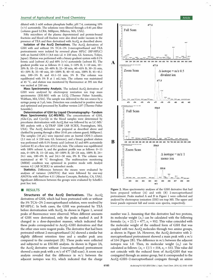
derivatives of GSH, which had been pretreated with or withoutthe 5% TCA−2% 2-mercaptoethanol solution, were resolved byRP-HPLC. In both cases, the GSH was pretreated by TEAbefore derivatization with AccQ. As shown in Figure 1, severalpeaks of fluorescence were observed. When different amountsof GSH were derivatized, only the peaks marked A and Bchanged in a dose-dependent manner, which indicated thatthese peaks corresponded to AccQ−GSH derivatives and thatthe other ones were reagent peaks. The derivative that had beenpretreated without 2-mercaptoethanol (A) showed a similar butslightly different retention time and a smaller peak areacompared to the counterpart (B). These peaks were collectedand subjected to an ESI-MS analysis. As shown in Figure 2A,the AccQ derivative without 2-mercaptoethanol pretreatmentshowed a main peak with a m/z of 477 (full scan). A zoom scananalysis revealed that the difference in m/z between theadjacent isotopes was 0.5, which indicated that the charge
number was 2. Assuming that this derivative had two protons,its molecular weight (x1) can be calculated with the followingformula: (x1 + 2)/2 = 477, x1 = 952. This value coincided withthe molecular weight of the oxidized form of GSH that wascoupled with two AccQ molecules through two amino groups,as shown in Figure 3A. However, the AccQ derivative with 2-mercaptoethanol pretreatment showed a main peak with a m/zof 554 (Figure 2B). The difference in m/z between the adjacentisotopes was 1.0. Then, its molecular weight (x2) can becalculated as follows: (x2 + 1)/1 = 554, x2 = 553. This value didnot coincide with the reduced form of AccQ-GSH that wasconjugated through an amino group, but it corresponded to theAccQ−GSH−2-mercaptoethanol conjugate through an amino
Figure 2. Mass spectrometry analyses of the GSH derivative that hadbeen prepared without (A) and with (B) 2-mercaptoethanolpretreatment. Peaks marked A and B in Figure 1 were collected andanalyzed by electrospray ionization (ESI) ion trap MS. The upper andlower panels represent full and zoom scan spectra, respectively.
Journal of Agricultural and Food Chemistry Article
dx.doi.org/10.1021/jf501338z | J. Agric. Food Chem. 2014, 62, 6183−61896185
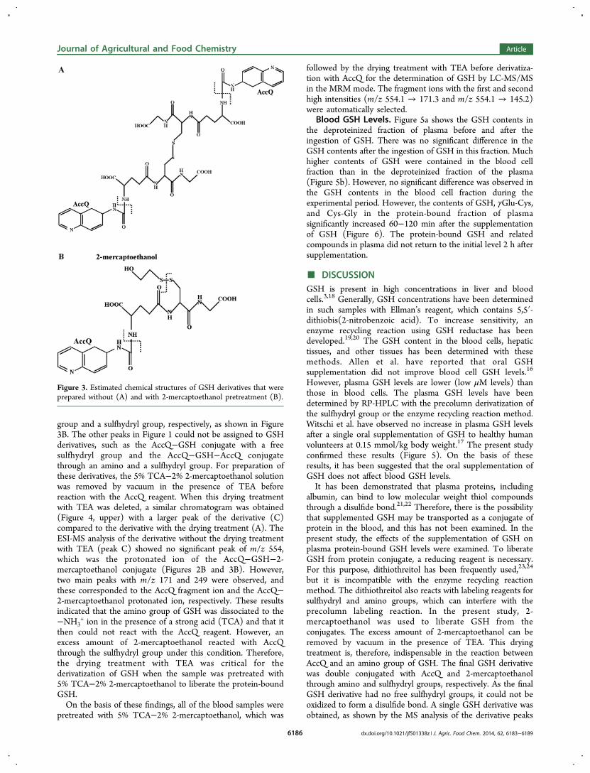
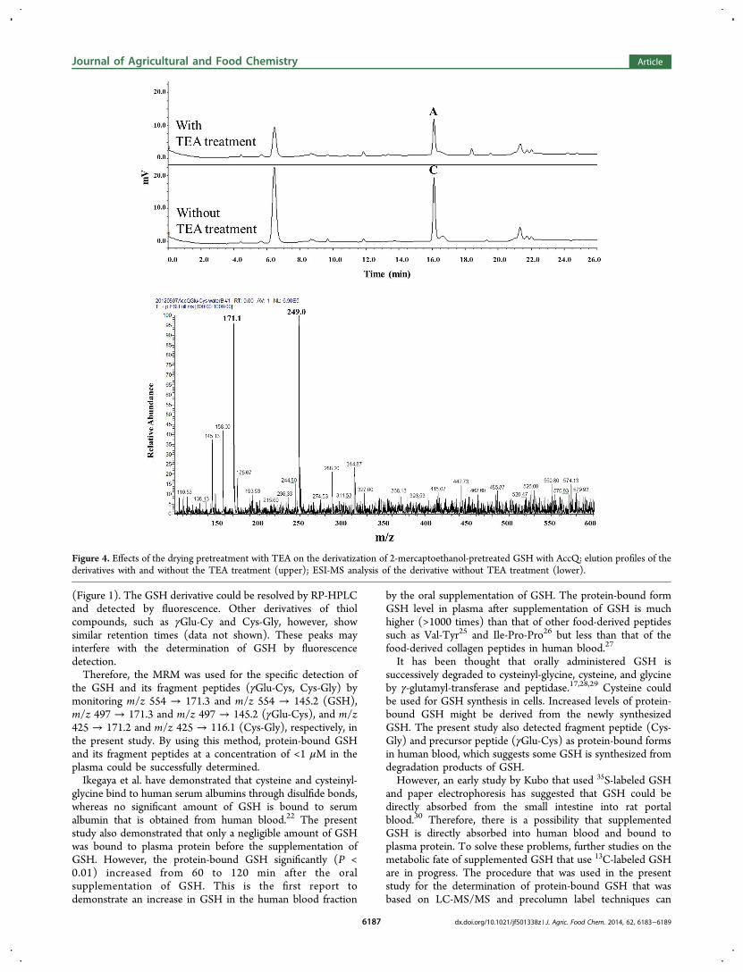
group and a sulfhydryl group, respectively, as shown in Figure3B. The other peaks in Figure 1 could not be assigned to GSHderivatives, such as the AccQ−GSH conjugate with a freesulfhydryl group and the AccQ−GSH−AccQ conjugatethrough an amino and a sulfhydryl group. For preparation ofthese derivatives, the 5% TCA−2% 2-mercaptoethanol solutionwas removed by vacuum in the presence of TEA beforereaction with the AccQ reagent. When this drying treatmentwith TEA was deleted, a similar chromatogram was obtained(Figure 4, upper) with a larger peak of the derivative (C)compared to the derivative with the drying treatment (A). TheESI-MS analysis of the derivative without the drying treatmentwith TEA (peak C) showed no significant peak of m/z 554,which was the protonated ion of the AccQ−GSH−2-mercaptoethanol conjugate (Figures 2B and 3B). However,two main peaks with m/z 171 and 249 were observed, andthese corresponded to the AccQ fragment ion and the AccQ−2-mercaptoethanol protonated ion, respectively. These resultsindicated that the amino group of GSH was dissociated to the−NH3
+ ion in the presence of a strong acid (TCA) and that itthen could not react with the AccQ reagent. However, anexcess amount of 2-mercaptoethanol reacted with AccQthrough the sulfhydryl group under this condition. Therefore,the drying treatment with TEA was critical for thederivatization of GSH when the sample was pretreated with5% TCA−2% 2-mercaptoethanol to liberate the protein-boundGSH.On the basis of these findings, all of the blood samples were
pretreated with 5% TCA−2% 2-mercaptoethanol, which was
followed by the drying treatment with TEA before derivatiza-tion with AccQ for the determination of GSH by LC-MS/MSin the MRM mode. The fragment ions with the first and secondhigh intensities (m/z 554.1 → 171.3 and m/z 554.1 → 145.2)were automatically selected.
Blood GSH Levels. Figure 5a shows the GSH contents inthe deproteinized fraction of plasma before and after theingestion of GSH. There was no significant difference in theGSH contents after the ingestion of GSH in this fraction. Muchhigher contents of GSH were contained in the blood cellfraction than in the deproteinized fraction of the plasma(Figure 5b). However, no significant difference was observed inthe GSH contents in the blood cell fraction during theexperimental period. However, the contents of GSH, γGlu-Cys,and Cys-Gly in the protein-bound fraction of plasmasignificantly increased 60−120 min after the supplementationof GSH (Figure 6). The protein-bound GSH and relatedcompounds in plasma did not return to the initial level 2 h aftersupplementation.
■ DISCUSSIONGSH is present in high concentrations in liver and bloodcells.3,18 Generally, GSH concentrations have been determinedin such samples with Ellman’s reagent, which contains 5,5′-dithiobis(2-nitrobenzoic acid). To increase sensitivity, anenzyme recycling reaction using GSH reductase has beendeveloped.19,20 The GSH content in the blood cells, hepatictissues, and other tissues has been determined with thesemethods. Allen et al. have reported that oral GSHsupplementation did not improve blood cell GSH levels.16
However, plasma GSH levels are lower (low μM levels) thanthose in blood cells. The plasma GSH levels have beendetermined by RP-HPLC with the precolumn derivatization ofthe sulfhydryl group or the enzyme recycling reaction method.Witschi et al. have observed no increase in plasma GSH levelsafter a single oral supplementation of GSH to healthy humanvolunteers at 0.15 mmol/kg body weight.17 The present studyconfirmed these results (Figure 5). On the basis of theseresults, it has been suggested that the oral supplementation ofGSH does not affect blood GSH levels.It has been demonstrated that plasma proteins, including
albumin, can bind to low molecular weight thiol compoundsthrough a disulfide bond.21,22 Therefore, there is the possibilitythat supplemented GSH may be transported as a conjugate ofprotein in the blood, and this has not been examined. In thepresent study, the effects of the supplementation of GSH onplasma protein-bound GSH levels were examined. To liberateGSH from protein conjugate, a reducing reagent is necessary.For this purpose, dithiothreitol has been frequently used,23,24
but it is incompatible with the enzyme recycling reactionmethod. The dithiothreitol also reacts with labeling reagents forsulfhydryl and amino groups, which can interfere with theprecolumn labeling reaction. In the present study, 2-mercaptoethanol was used to liberate GSH from theconjugates. The excess amount of 2-mercaptoethanol can beremoved by vacuum in the presence of TEA. This dryingtreatment is, therefore, indispensable in the reaction betweenAccQ and an amino group of GSH. The final GSH derivativewas double conjugated with AccQ and 2-mercaptoethanolthrough amino and sulfhydryl groups, respectively. As the finalGSH derivative had no free sulfhydryl groups, it could not beoxidized to form a disulfide bond. A single GSH derivative wasobtained, as shown by the MS analysis of the derivative peaks
Figure 3. Estimated chemical structures of GSH derivatives that wereprepared without (A) and with 2-mercaptoethanol pretreatment (B).
Journal of Agricultural and Food Chemistry Article
dx.doi.org/10.1021/jf501338z | J. Agric. Food Chem. 2014, 62, 6183−61896186
(Figure 1). The GSH derivative could be resolved by RP-HPLCand detected by fluorescence. Other derivatives of thiolcompounds, such as γGlu-Cy and Cys-Gly, however, showsimilar retention times (data not shown). These peaks mayinterfere with the determination of GSH by fluorescencedetection.Therefore, the MRM was used for the specific detection of
the GSH and its fragment peptides (γGlu-Cys, Cys-Gly) bymonitoring m/z 554 → 171.3 and m/z 554 → 145.2 (GSH),m/z 497 → 171.3 and m/z 497 → 145.2 (γGlu-Cys), and m/z425 → 171.2 and m/z 425 → 116.1 (Cys-Gly), respectively, inthe present study. By using this method, protein-bound GSHand its fragment peptides at a concentration of <1 μM in theplasma could be successfully determined.Ikegaya et al. have demonstrated that cysteine and cysteinyl-
glycine bind to human serum albumins through disulfide bonds,whereas no significant amount of GSH is bound to serumalbumin that is obtained from human blood.22 The presentstudy also demonstrated that only a negligible amount of GSHwas bound to plasma protein before the supplementation ofGSH. However, the protein-bound GSH significantly (P <0.01) increased from 60 to 120 min after the oralsupplementation of GSH. This is the first report todemonstrate an increase in GSH in the human blood fraction
by the oral supplementation of GSH. The protein-bound formGSH level in plasma after supplementation of GSH is muchhigher (>1000 times) than that of other food-derived peptidessuch as Val-Tyr25 and Ile-Pro-Pro26 but less than that of thefood-derived collagen peptides in human blood.27
It has been thought that orally administered GSH issuccessively degraded to cysteinyl-glycine, cysteine, and glycineby γ-glutamyl-transferase and peptidase.17,28,29 Cysteine couldbe used for GSH synthesis in cells. Increased levels of protein-bound GSH might be derived from the newly synthesizedGSH. The present study also detected fragment peptide (Cys-Gly) and precursor peptide (γGlu-Cys) as protein-bound formsin human blood, which suggests some GSH is synthesized fromdegradation products of GSH.However, an early study by Kubo that used 35S-labeled GSH
and paper electrophoresis has suggested that GSH could bedirectly absorbed from the small intestine into rat portalblood.30 Therefore, there is a possibility that supplementedGSH is directly absorbed into human blood and bound toplasma protein. To solve these problems, further studies on themetabolic fate of supplemented GSH that use 13C-labeled GSHare in progress. The procedure that was used in the presentstudy for the determination of protein-bound GSH that wasbased on LC-MS/MS and precolumn label techniques can
Figure 4. Effects of the drying pretreatment with TEA on the derivatization of 2-mercaptoethanol-pretreated GSH with AccQ: elution profiles of thederivatives with and without the TEA treatment (upper); ESI-MS analysis of the derivative without TEA treatment (lower).
Journal of Agricultural and Food Chemistry Article
dx.doi.org/10.1021/jf501338z | J. Agric. Food Chem. 2014, 62, 6183−61896187

distinguish food-derived (13C-labeled) and endogenous GSHand their metabolites, such as γGlu-Cys, Cys-Gly, and cysteine,and this is a powerful tool for the elucidation of the absorption,metabolism, and transportation of food-derived GSH.
■ AUTHOR INFORMATIONCorresponding Author*(K.S.) Mail (present address): Division of Applied Bio-sciences, Graduate School of Agriculture, Kyoto University,Kitashirakawa Oiwake-cho, Kyoto 606-8502, Japan. Phone:+81-75-753-6444. Fax: +81-75-723-3503. E-mail: [email protected].
NotesThe authors declare no competing financial interest.
■ ACKNOWLEDGMENTS
We express our appreciation to the Kyoto Integrated Scienceand Technology Bio-Analysis Center for the use of their LC-MS/MS.
■ ABBREVIATIONS USED
GSH, glutathione; γGlu-Cys-Gly, γ-L-glutamyl-L-cysteinyl-gly-cine; Cys-Gly, L-cysteinyl-glycine; γGlu-Cys, γ-L-glutamyl-L-cysteine; AccQ, 6-aminoquinolyl-N-hydroxysuccinimidyl carba-mate; TEA, triethylamine; TCA, trichloroacetic acid; RP-HPLC, reversed phase high-performance liquid chromatog-raphy; ESI-MS, electrospray ionization mass spectrometry; LC-MS/MS, liquid chromatography−tandem mass spectrometry;ESI-MS/MS, electrospray ionization tandem mass spectrome-try
■ REFERENCES(1) Anderson, M. E. Glutathione: an overview of biosynthesis andmodulation. Chem.−Biol. Interact. 1998, 112, 1−14.(2) Fahey, R. C. Novel thiols of prokaryotes. Annu. Rev. Microbiol.2001, 55, 533−556.(3) Meister, A. Glutathione metabolism and its selectivemodification. J. Biol. Chem. 1988, 263, 17205−17208.(4) Mannervik, B.; Danielson, U. H. Glutathione transferases-structure and catalytic activity. CRC Crit. Rev. Biochem. 1988, 23,283−337.(5) Mannervik, B. The enzymes of glutathione metabolism: anoverview. Biochem. Soc. Trans. 1987, 15, 717−718.(6) Meister, A.; Anderson, M. E. Glutathione. Annu. Rev. Biochem.1983, 52, 711−760.(7) Nuray, N.; Ulusu, M. S.; Aslihan, A.; Orhan, C.; Gulgun, O.;Nuray, A.; Musa, B.; Milan, S.; Svorad, S.; Andrej, G.; Cimen, K.Pentose phosphate pathway, glutathione-dependent enzymes andantioxidant defense during oxidative stress in diabetic rodent brain andperipheral organs: effects of stobadine and vitamin E. Neurochem. Res.2003, 28, 815−823.(8) Douglas, K. T. Mechanism of action of glutathione-dependentenzymes. Adv. Enzymol. Relat. Areas Mol. Biol. 1987, 59, 103−167.(9) Oakley, A. Glutathione transferases: a structural perspective. DrugMetab. Rev. 2011, 43, 138−151.(10) White, A. C.; Thannickal, V. J.; Fanburg, B. L. Glutathionedeficiency in human disease. J. Nutr. Biochem. 1992, 5, 218−226.(11) Dentico, P.; Volpe, A.; Buongiorno, R.; Grattagliano, I.;Altomare, E.; Tantimonaco, G.; Scotto, G.; Sacco, R.; Schiraldi, O.Glutathione in the treatment of chronic fatty liver diseases. Recent Prog.Med. 1995, 86, 290−293.(12) Altomare, E.; Colonna, P.; Dagostino, C.; Castellaneta, G.;Vendemiale, G.; Grattagliano, I.; Cirelli, F.; Bovenzi, F.; Colonna, L.High-dose antioxidant therapy during thrombolysis in patients withacute myocardial infarction. Curr. Ther. Res. 1996, 57, 131−141.(13) Matsuki, M.; Watanabe, T.; Ogasawara, A.; Mikami, T.;Matsumoto, T. Inhibitory mechanism of melanin synthesis byglutathione. Yakugaku Zasshi 2008, 128, 1203−1207.(14) Arjinpathana, N.; Asawanonda, P. Glutathione as an oralwhitening agent: a randomized, double-blind, placebo-controlledstudy. J. Dermatol. Treat. 2012, 23, 97−102.(15) Meister, A. Glutathione deficiency produced by inhibition of itssynthesis, and its reversal; applications in research and therapy.Pharmacol. Ther. 1991, 51, 155−194.(16) Allen, J.; Bradley, R. D. Effects of oral glutathionesupplementation on systemic oxidative stress biomarkers in humanvolunteers. J. Altern. Complement. Med. 2011, 17, 827−833.
Figure 5. Total GSH contents in the deproteinized (ethanol-soluble)plasma fraction (a) and blood cell fraction (b) after the ingestion ofGSH. No significant difference was observed between groups (P <0.01).
Figure 6. Contents of glutathione (GSH), γ-glutamyl-cysteine (γGlu-Cys), and cysteinyl-glycine (Cys-Gly) in the ethanol precipitatefraction of plasma after ingestion of GSH. (∗, ∗∗) Significantlydifferent from baseline P < 0.05 and 0.01, respectively, according toScheffe’s post hoc test.
Journal of Agricultural and Food Chemistry Article
dx.doi.org/10.1021/jf501338z | J. Agric. Food Chem. 2014, 62, 6183−61896188
(17) Witschi, A.; Reddy, S.; Stofer, B.; Lauterburg, B. H. Thesystemic availability of oral glutathione. Eur. J. Clin. Pharmacol. 1992,43, 667−669.(18) Monks, T. J.; Anders, M. W.; Dekant, W.; Stevens, J. L.; Lau, S.S.; van Bladeren, P. J. Glutathione conjugate mediated toxicities.Toxicol. Appl. Pharmacol. 1990, 106, 1−19.(19) Teitze, F. Enzymic method for quantitative determination ofnanogram amounts of total and oxidized glutathione. Anal. Biochem.1969, 27, 502−522.(20) Rahman, I.; Kode, A.; Biswas, S. K. Assay for quantitativedetermination of glutathione and glutathione disulfide levels usingenzymatic recycling method. Nat. Protoc. 2006, 1, 3159−3165.(21) Sengupta, S.; Chen, H.; Togawa, T.; DiBello, P. M.; Majors, A.K.; Budy, B.; Kettere, M. E.; Jacobsen, D. W. Albumin thiolate anion isan intermediate in the formation of albumin-S−S-homocysteine. J.Biol. Chem. 2001, 276, 30111−30117.(22) Ikegaya, K.; Nokihara, K.; Yasuhara, T. Characterization ofsulfhydryl heterogeneity in human serum albumin and recombinanthuman serum albumin for clinical use. Biosci., Biotechnol., Biochem.2010, 74, 2232−2236.(23) Griffith, O. W. Determination of glutathione and glutathionedisulfide using glutathione reductase and 2-vinylpyridine. Anal.Biochem. 1980, 106, 207−212.(24) Ates, B.; Ercal, B. C.; Manda, K.; Abraham, L.; Ercal, N.Determindation of glutathione disulfide levels in biological samplesusing thiol-disulfide exchanging agent, dithiothreitol. Biomed. Chroma-togr. 2009, 23, 119−123.(25) Matsui, T.; Tamaya, K.; Seki, E.; Osajima, K.; Matsumo, K.;Kawasaki, T. Absorption of Val-Tyr with in vitro angiotensin I-converting enzyme inhibitory activity into the circulating blood systemof mild hypertensive subjects. Biol. Pharm. Bull. 2002, 25, 1228−1230.(26) Foltz, M.; Meynen, E. E.; Bianco, V.; van Platerink, C.; Koning,T. M.; Kloek, J. Angiotensin converting enzyme inhibitory peptidesfrom a lactotripeptide-enriched milk beverage are absorbed intact intothe circulation. J. Nutr. 2007, 137, 953−958.(27) Iwai, K.; Hasegawa, T.; Taguchi, Y.; Morimatsu, F.; Sato, K.;Nakamura, Y.; Higashi, A.; Kido, Y.; Nakabo, Y.; Ohtsuki, K.Identification of food-derived collagen peptides in human bloodafter oral ingestion of gelatin hydrolysates. J. Agric. Food Chem. 2005,53, 6531−6536.(28) Flagg, E. W.; Coates, R. J.; Eley, J. W.; Jones, D. P.; Gunter, E.W.; Byers, T. E.; Block, G. S.; Greenberg, R. S. Dietary glutathioneintake in humans and the relationship between intake and plasma totalglutathione level. Nutr. Cancer 1994, 21, 33−46.(29) MacNee, W.; Rahman, I. Oxidants/antioxidants in idiopathicpulmonary fibrosis. Thorax 1995, 50, S53−S58.(30) Kubo, Y. Absorption of 35S-GSH and its incorporation intoprotein in rats. Kurume Med. J. 1968, 15, 113−125.
Journal of Agricultural and Food Chemistry Article
dx.doi.org/10.1021/jf501338z | J. Agric. Food Chem. 2014, 62, 6183−61896189