increase in caudate nuclei volumes of first-episode schizophrenia patients taking antipsychotic
TRANSCRIPT
i~ !,-"
.~
link these neuropathologic findings to pathogenicprocesses and the clinical dimensions of the disease.Two questions that have not been resolved, however,are 1) whether some of the neuropathologic findingsassociated with schizophrenia are a consequence ofthe disease or of the substantial treatment that mostpatients receive in the course of their illness and 2)whether the neuropathologic process is, in some regions, progressive.
Since the major pharmacologic action of antipsychotic drugs is in the dopamine neurons that project tothe basal ganglia nuclei-including the caudate nucleus,globus pallidus, and putamen-and produce extrapyramidal side effects as a consequence of this action, ifdrugs can affect brain morphology, these structuresmight be particularly susceptible. In this context, recentpost-mortem and magnetic resonance imaging (MRI)studies that have reported increased striatal and lenticular nuclei volumes in schizophrenic patients (4-8) are of'interest. These findings are in contrast to the usual pattern of neuropathology in schizophrenia, in which reduction in the size of soft-tissue brain structures andenlargement of fluid-containing structures are characteristically seen (1, 2). In a post-mortem study, Heckerset al. (5) reported significant increases in left striatal
Obiective: This study examined the pathomorphology of the caudate nuclei in first-episodeschizophrenic patients with minimal previous neuroleptic exposure. Method: Magnetic resonance imaging (MRI) of the brain was used to examine longitudinally the caudate pathomorphology in 29 first-episode schizophrenic patients and 10 healthy comparison subjects. MRIscans were obtained after the subjects entered the study and at 18-month follow-up. Thepatients were treated with standardized neuroleptic regimens during the 18-month period.Volumetric assessments of the cerebral cortex, lateral ventricles, and caudate nuclei were performed on T1-weighted coronal brain sections. In addition, the patients were systematicallyevaluated for psychopathology at baseline and during treatment. Results: Caudate volumesincreased 5. 7% in the patients during the 18-month treatment interval, whereas they decreased1.6% in the comparison subjects over the same time period. Greater amounts ofantipsychoticmedication received by patients before the first scan and younger age at the time of the firstscan were associated with larger increases in caudate volume. Conclusions: Caudate enlargement occurs early in the course oftreatment in young first-episode schizophrenic patients. Thismay be a result of an interaction between neuroleptic treatment and the plasticity of dopa-minergic neuronal systems in young patients. '
(Am J Psychiatry 1994; 151:1430-1436)
Increase in Caudate Nuclei Volumes of First~EpisodeSchizophrenic Patients Taking Antipsychotic Drugs
Miranda H. Chakos, M.D., Jeffrey A. Lieberman, M.D., Robert M. Bilder, Ph.D.,Michael Borenstein, Ph.D., Gail Lerner, M.S., Bernhard Bogerts, M.D.,
Houwei Wu, M.D., Bruce Kinon, M.D., and Manzar Ashtari, Ph.D.
Presented in part at the International Congress on Schizophrenia,Colorado Springs, Colo., April 17-21, 1993. Received May 13, 1993;revisions received Feb. 8 and April 12, 1994; accepted April 19, 1994.From the Department of Psychiatry, Hillside Hospital-Long IslandJewish Medical Center; the Department of Psychiatry, University ofMagdeburg, Magdeburg, Germany; and the Department of Radiology, Long Island Jewish Medical Center, Albert Einstein College ofMedicine, New Hyde Park, N.Y. Address reprint requests to Dr.Chakos, Hillside Hospital-Long Island Jewish Medical Center, 75-59263rd St., Glen Oaks, NY 11004.
Supported by NIMH grant MH-41646, Research Scientist AwardMH-00537 to Dr. Lieberman, and grant MH-41960 to the MentalHealth Clinical Research Center for the Study of Schizophrenia, Hillside Hospital. Magnetic resonance imaging studies were perfcrmed inassociation with the Brain Morphometry and Image Analysis Center,Hillside Hospital, supported by a grant from the Helen and IrvingSchneider family.
P ost-mortem and in vivo imaging studies conducted over the last three decades have made sub
stantial progress in identifying the neuropathology ofschizophrenia. Abnormalities reflected by volume,shape, cytoarchitecture, and histology have been described in specific brain structures, including the ventricular system, cerebral cortex, hippocampus, cingulate gyrus, entorhinal cortex, and parahippocampalgyrus (1-3). Current research efforts are attempting to
'I
jl:j
I'i'
l ii '
~i"I
"II.il1:!
LrI.:
11
i!
II,~j :'! .
1430 Am] Psychiatry 151:10~ October 1994
volumes and a trend toward increases in right striatalvolumes of brains of schizophrenic patients in comparison with brains of age- and gender-matched controlsubjects. MRI studies of subcortical structures havefound increased lenticular nuclei volumes (6) and putamen and caudate enlargement (7, 8) in young butchronic, medicated schizophrenic patients comparedwith age- and gender-matched normal control subjects.These findings of striatal enlargement have been interpreted as a feature of schizophrenia that is caused by a .disturbance in neurodevelopmental pruning of subcortical structures in patients with the disease, particularlythose with early onset of illness (6, 9). Alternatively,treatment with neuroleptic drugs and resultant dopamine blockade could result in disinhibition of thestriatum, with consequent activation and (analogous tomuscle cells after activation) hypertrophy of striatal synaptic and/or cellular elements. Various forms of activity-dependent synaptic plasticity have been described inmany neural systems (10). Since cross-sectional studiesreporting caudate enlargement have examined heterogeneous groups of chronically medicated patients, it isnot clear whether the observed enlargement reflects apreexisting abnormality or is an effect of drug treatment. To address this question we examined the caudate nuclei in MRI scans acquired in a longitudinal prospective study of first-episode schizophrenic patientswho were in the early phases of treatment.
METHOD
This study used MRI and clinical data from a longitudinal prospective study of first-episode schizophrenic patients and normal comparison subjects to examine changes in caudate volumes over time.The subjects included in the study were a subgroup of the subjects forwhom brain morphology studies of medial temporal lobe structuresand the ventricular system have previously been reported (11-13).We selected subjects who had baseline and 18-month follow-up MRlssuitable for examination of the caudate. Detailed descriptions of themethods of the parent study have been previously reported (12, 14,15). Patients admitted to the hospital for a first episode of psychosiswho had had fewer than 12 weeks of lifetime exposure to neurolepticsor none at all underwent a structured interview with the Schedule forAffective Disorders and Schizophrenia (SADS) (16). Eligible patientswere between the ages of 16 and 40 years and had diagnoses ofschizophrenia, schizophreniform disorder, or schizoaffective disorder, mainly schizophrenic, according to the Research Diagnostic Criteria (17). They had no prior psychotic episodes, no history of currentsubstance abuse, and no neuromedical illness that could influence thediagnosis or the major variables assessed in the study. Patients withprior neuroleptic exposure had at least a 2-week medication washoutperiod before entering the study. All patients were acutely psychoticwith severe psychopathology at the time of entry into the study.
Comparison subjects were recruired through advertisements in thelocal media and, if eligible, were offered $50 to undergo MRI. Thevolunteers were screened for medical and psychiatric illness and history of drug abuse. They were then given the Structured Clinical Interview for DSM-III-R (18) and a physical examination with laboratory testing. Volunteers with significant findings were excluded fromparticipation.
Measurements of the caudate nuclei were performed on two MRIscans of 29 first-episode patients (21 with schizophrenia and eightwith schizoaffective disorder) and 10 healthy comparison subjects.Twenty-one of the 29 primarily schizophrenic patients had had noprior exposure to neuroleptic treatment at the· time they entered the
Am] Psychiatry 151:10, October 1994
CHAKOS, LIEBERMAN, BILDER, ET AL.
study. The remaining eight patients had had fewer than 12 weeks oflifetime treatment exposure. Seventeen patients were male, and 12were female; 13 were white, eight black, four Hispanic, and fourAsian. The mean age of the patients was 25.2 years (SD=6.3), and themean age at onset of illness' was 24.4 years (SD=6.6).
Eight of the comparison subjects were male, and two were female;all of them were white. Their mean age was 30.5 years (SD=4.9).
Patients underwent the first MRI of the brain (scan 1) when theyentered the study or following initiation of neuroleptic treatment;they underwent scan 2 at 18-month follow-up. Scan 1 was done assoon as a patient could tolerate the procedure and allow images ofadequate quality to be obtained. When necessary, chloral hydrate(500-1000 mg p.o.) or amobarbital sodium (250-500 mg p.o. ori.m.) was used to sedate patients for the procedure. Comparison subjects had MRI scans at approximately the same intervals within thesame time period.
MRIs were acquired on a Siemens Magnetom operating at 1.0 Twith a 3-dimensional gradient-echo sequence FLASH (fast low-angleshots) (flip angle=50°, TR=40 msec, TE=15 msec) acquiring 63 contiguous 3.1-mm TJ-weighted slices in the coronal plane (19). Patients'and comparison subjects' MRls were transferred from a VAX 11/750onto a Sun SPARC 10 workstation operating from a Sun 670 serverfor morphometric analysis. Quantitative assessments of volumes ofthe caudate nuclei, cortex, and lateral ventricles were performed withthe use of a semiautomated computer mensuration system, the validity and interrater reliability of which had been established previouslythrough phantom and in vivo studies (19).
The manual function of the computerized mensuration system wasused to measure the caudate in order to demarcate precisely theboundaries of adjacent gray matter structures. The caudate nucleuswas selected for measurement because its anatomic boundaries couldbe more precisely delineated than those of the globus pal!idum andputamen. The caudate nucleus was measured in coronal sections,with the cisterna pontis as the posterior landmark; measurement wascontinued anteriorly until the caudate head was no longer visible. Atthe slice anterior to the section where the anterior commissure crossesthe midline, the nucleus accumbens was separated from the caudateby a line combining the most basal extents of the lateral ventricle andthe internal capsule. All caudate measurements were performed by asingle operator (M.H.C.) under blind conditions and with the scansrandomly ordered. The reliability coefficient on test-retest measurements of the caudate nuclei of all patients and comparison subjectswas 0.89.
Cortical volumes were determined by the sums of the volumes ofcortical regions (the occipitoparietal, sensorimotor, premotor, prefrontal, and temporal regions). The regional volumes included cortical gray and hemispheric white matter, but not subcortical structures;basal ganglia, thalamus, and ventricle volumes were excluded. Eachregion was measured separately in each hemisphere, with the interhemispheric fissure as a medial boundary. In the sensorimotor region,the temporal lobe, basal ganglia, and thalamus were excluded by aline from the most superior extent of the insular cisterns to the sulcusof the corpus callosum. In the premotor region, the basal ganglia wereexcluded by a line from the most superior extent of the insular cisternsto the sulcus between the corpus callosum and the cingulate gyrus. Inthe temporal region, the temporal stem was separated from the basalganglia by a line from the most inferior part of the insular cisterns tothe most lateral extent of the basal cisterns above the hippocampus.(Full delineation criteria for cortical regions have been described byBilder et al. [20].)
Lateral ventricular volumes were computed by adding the volumes of the frontal horn, body, and occipital horn of the lateralventricles. (The methods of ventricular measurement have been reported by Degreef et al. [13].) All cortical and ventricular volumemeasurements were made by a single operator (H.W.), using automated functions on the computer-based mensuration system underblind conditions and with the scans randomly ordered. The test-retest reliability was 0.90 for cortical measurements and 0.99 for ventricular system measurements.
The patients had baseline evaluations of psychopathology, extrapyramidal side effect status, and involuntary movements that included use of the SADS-Change Version (SADS-C) with psychoticand disorganization items, a modified Scale for the Assessment of
1431
INCREASE IN CAUDATE NUCLEI VOLUMES
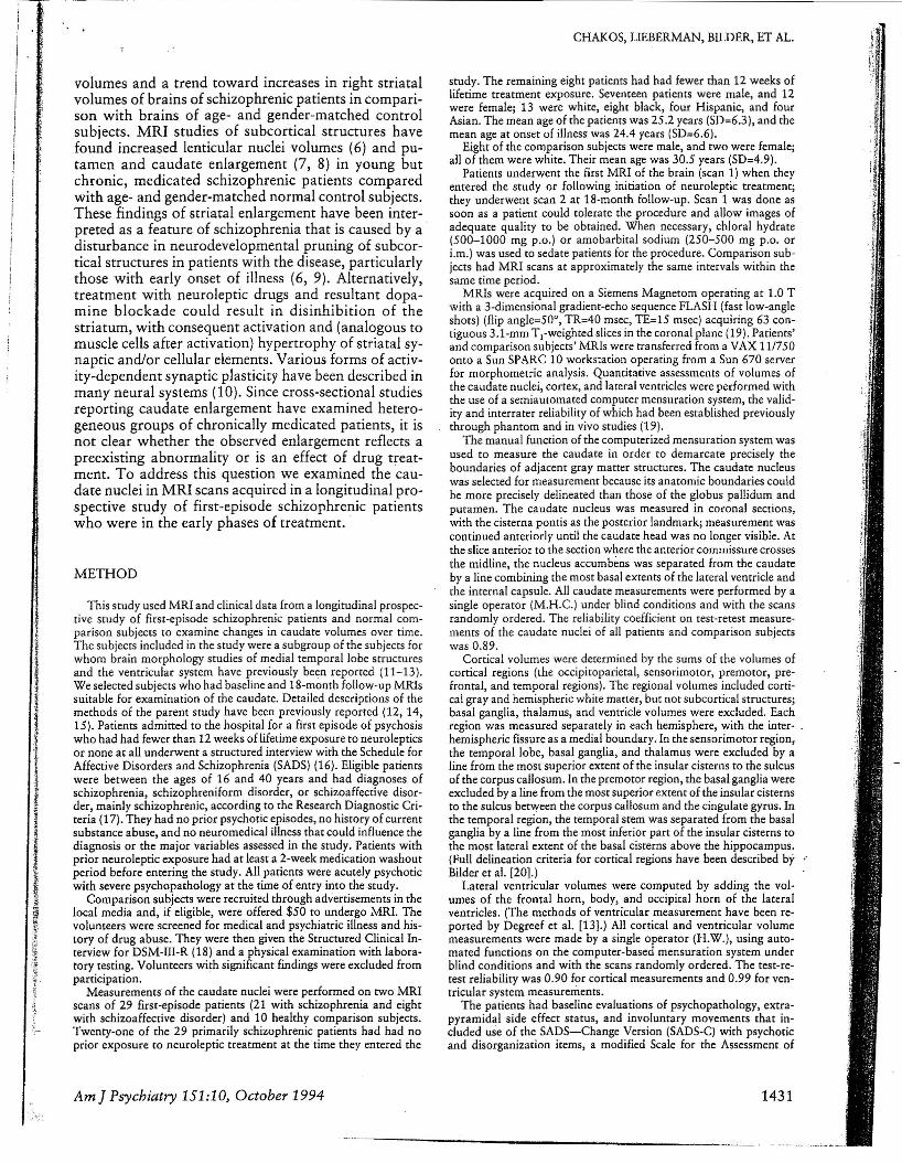
TABLE 1. Morphologic Measures of Brain Structures of First-Episode Schizophrenic Patients and Normal Comparison Subjects at BaselineMRI (Scan 1) and at IS-Month Follow-Up (Scan 2)
"Total group of patients versus comparison subjects: nonsignificant effect of group (F=0.40, df=l, 37, p=0.53); significant Group by Timeinteraction (F=6.01, df=l, 37, p<0.02). Treatment-naive subgroup versus comparison subjects: nonsignificant effect of group (F=0.17, df=l,29, p=0.69); significant Group by Time interaction (F=5.19, df=l, 29, p=0.03). Previously treated subgroup versus comparison subjects: nonsignificant effect of group (F=0.75, df=l, 16, p=0.40); nearly significant Group by Time interaction (F=4.13, df=l, 16, p<0.06).
bTotal group of patients versus comparison subjects: nonsignificant effect of group (F=0.43, df=l, 37, p=0.52)j nonsignificant Group by Timeinteraction (F=1.47, df=1, 37, p=O.23).
<Total group of patients versus comparison subjects: nonsignificant effect of group (F=1.30, df=l, 37, p=0.26)j nonsignificant Group by Timeinteraction (F=0.05, df=1, 37, p=O.83).
~ ,
r
II
IiII
Measure
Caudate volume (cc)"Scan 1Scan 2
Cortical volume (cc)bScan 1Scan 2
Lateral ventricular volume (cc)CScan 1Scan 2
Schizophrenic Patients
Treatment- PreviouslyTotal Naive Subgroup Treated Comparison
Group (N=29) (N=21) Subgroup (N=8) Subjects (N=10)
Mean SD Mean SD Mean SD Mean SD
5.58 0.86 5.50 8.45 5.77 9.17 5.59 0.945.90 0.88 5.84 8.13 6.07 1.08 5.50 0.89
780.63 162.40 849.22 321.15805.81 126.89 831.38 315.70
16.24 6.79 13.38 5.4915.59 7.65 13.21 6.68
f;
rI
1'1o-
r
i1
i
Negative Symptoms (21), a modified Simpson-Angus Rating Scale(22), and a modified Simpson Dyskinesia Scale (23). After baselineassessment, patients were treated openly in accordance with a standardized treatment algorithm (13, 14). They initially received fluphenazine, which was increased to 20 mg/day for 6 weeks. If they achievedremission or showed continuing improvement, they remained on thisregimen. Remission was operationally defined as a rating of no morethan 3 on any of the positive psychotic items on the SADS-C withpsychotic and disorganization items, a Clinical Global Impression(CGI) severity rating of 3 (mild) or less, and a CGI improvement rating of 2 (much improved) or 1 (very much improved). If not improvedin 6 weeks, patients progressed through the treatment algorithm, receiving full trials of up to three different neuroleptics. All study patients were followed as outpatients by research psychiatrists. Theywere evaluated biweekly with the SADS-C with psychotic and disorganization items, the Scale for the Assessment of Negative Symptoms,the'Simpson-Angus Rating Scale, and the CGI for 12 weeks and every4 weeks thereafter. Evaluations with the Simpson Dyskinesia Scalewere completed every 8 weeks. The general strategy of maintenancetreatment was to reduce the daily dose of antipsychotic medicationgradually to the lowest effective level.
RESULTS
Our analysis focused on changes in caudate volumein the first-episode patients and the effect of patientgroup versus comparison subject group on caudatechange. The size of our comparison group remainedsmall and limited our capacity to match for age andgender. Taking into account the small size of this group,we focused our primary analyses on the within-subjecteffect of time in the patients. We subsequently examined the between-group effect and the interaction ofgroup and time with respect to caudate volume.
All 29 patients had a remission of their illness withtreatment. The median time to remission was 11 weeks.
The patients had a significant mean increase of 0.32cc (SD=0.47) in total caudate volume from scan 1 toscan 2 (paired t=-3.68, df=28, p=O.OOl) but no signifi-
cant change in their ventricular and cortical volumes(table 1). To examine whether the apparent change incaudate volume noted in the patients was a discreteprocess or secondary to a more generalized process inthe brain, analyses of covariance were performed. The
-change in caudate volume in the patients during the 18month interval of treatment remained significant whenwe covaried height (F=11.68, df=1, 27, p=0.002) andtotal cortical volume (F=11.08, df=1, 27, p=0.003).
Between-Group Effects
The morphologic variables for the patients and comparison subjects are presented in table 1. There was nomain effect of group on caudate volume for the wholegroup of patients and the comparison subjects. Howevee-, the Group by Time interaction was significant,and it remained significant when we covaried height (F=5.47, df=l, 36, p<0.03) and cortical volume (F=4.60,df=1, 36, p<0.04). The mean total caudate volume forthe 29 patients increased 0.32 cc (SD=0.47) (paired t=3.68, df=28, p=O.OOl), while the mean caudate volumefor the normal comparison subjects was reduced by0.09 cc (SD=0.04) (paired t=0.69, df=9, p=0.51).
Right caudate volumes were larger than left caudatevolumes (main effect of hemisphere: F=9.56, df=l, 37,p=0.004) (table 2). This effect was not limited to eitherpatients or comparison subjects (Group by Hemisphereinteraction: F=0.14, df=l, 37, p=O.71). Nor did theasymmetry change over time (Hemisphere by Time interaction: F=0.09, df=1, 37, p=0.79; Group by Hemisphere by Time interaction: F=0.08, df=1, 37, p=O.77).When age was used as a covariate, the Group by Timeinteraction did not reach conventional levels of statistical significance (F=3.33, df=1, 36, p<0.08). There wasa negative association between caudate volume change
'II-,I~1i
.1)J
J-~-~
II
Am] Psychiatry 151:10, October 19941432
1I.
II-L..----------------~ .. ,-_..",!!!!!!!!~_!!!_._l_
Schizophrenic ComparisonPatients Subjects • •(N=29) (N=10) 1.0 • •u • '.Measure Mean SD Mean SD ~ -•
QI • •01 0.5 "I<Scan 1 c ••III ,
"1< •Right hemisphere 2.81 0.45 2.82 0.45 .c
0 • • ~Left hemisphere 2.77 0.42 2.77 0.48 • • "I<QI 0.0Total 5.58 0.86 5.59 0.94 E •:::I • "I<
Scan 2 ;g •Right hemisphere 2.98 0.44 2.77 0.47 -0.5 "I<
Left hemisphere 2.92 0.45 2.73 0.42 • "I< •Total 5.90 0.88 5.50 0.89 A
-1.010.0 20.0 30.0 40.0 50.0
Age (years)
CHAKOS) LIEBERMAN) BILDER, ET AL.
"I< ComparIson Subjects • Patients1.5
FIGURE 1. Change at IS-Month Follow-Up in Caudate Volume of,First-Episode Schizophrenic Patients and Normal Comparison Subjects by Age at the First MRI Scan"
Caudate Volume (cc)
TABLE 2. Caudate Volumes of First-Episode Schizophrenic Patientsand Normal Comparison Subjects at Baseline MRI (Scan I) and atIS-Month Follow-Up (Scan 2)
ri/.I· ,~
I!I
score and age among both the patients and the comparison subjects (figure 1). In patients this negative association appeared to be caused by greater increases in volumes in young patients, while in comparison subjects itwas largely due to reduction in the caudate volumes ofthe older subjects. Since the group of comparison subjects was relatively small, nonparametric statistics werealso used to confirm that the between-group differencein caudate change was not a spurious effect of a fewoutlying values. A Mann-Whitney U test performed onthe rank-ordered change scores again revealed a significant difference between the two groups (U=76.00, z=-2.22, p<0.03).
Clinical Correlates
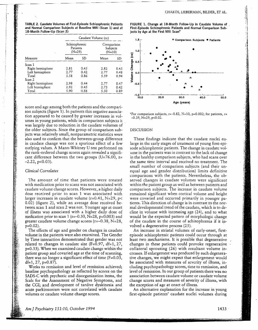
The amount of time that patients were treatedwith medication prior to scans was not associated withcaudate volume change scores. However, a higher dailydose received prior to scan 1 was associated withlarger increases in caudate volume (r=0.41, N=29, p<0.02) (figure 2), while an average dose received between scan 1 and scan 2 was not. Younger age at onsetof illness was associated with a higher daily dose ofmedication prior to scan 1 (r=-o.50, N=28) p=0.003) andgreater caudate volume change scores (r=-0.38, N=28,p=0.02).
The effects of age and gender on changes in caudatevolume in the patients were also examined. The Genderby Time interaction demonstrated that gender was notrelated to changes in caudate size (F=O.97, df=1, 27,p=0.33). When we examined caudate change within thepatient group and covaried age at the time of scanning,there was no longer a significant effect of time (F=0.03,dfd, 27, p=0.87).
Weeks to remission and level of remission achieved;baseline psychopathology as reflected by scores on theSADS-C with psychotic and disorganization items, theScale for the Assessment of Negative Symptoms, andthe CGI; and development of tardive dyskinesia andacute parkinsonism were not correlated with caudatevolumes or caudate volume change scores.
Am] Psychiatry 151:10, October 1994
"For comparison subjects, r=-0.82, N=10, p=0.002j for patients, r=-0.39, N=29) p<0.02.
DISCUSSION
These findings indicate that the caudate nuclei enlarge in the early stages of treatment of young first-episode schizophrenic patients. The change in caudate volume in the patients was in contrast to the lack of changein the healthy comparison subjects) who had scans overthe same time interval and received no treatment. Thesmall number of comparison subjects (and their unequal age and gender distribution) limits definitivecomparisons with the patients. Nevertheless, the observed changes in caudate volumes were significantwithin the patient group as well as between patients andcomparison subjects. The increase in caudate volumeremained significant when cortical volume and heightwere covaried and occurred primarily in younger patients. This direction of change is in contrast to the normal developmental trend of the caudate, which is to decline in volume with increasing age (24), and to whatwould be the expected pattern of morphologic changeof the caudate in the course of schizophrenia if it in-volved a degenerative process (25). '
An increase in striatal volumes of early-onset, firstepisode schizophrenic patients could occur through atleast two mechanisms. It is possible that degenerativechanges in these patients could provoke regenerativecollateral sprouting (26) with resultant volume increases. If enlargement was produced by such degenerative changes, we might expect that enlargement wouldbe associated with measures of severity of illness, including psychopathology scores, time to remission, andlevel of remission. In our group of patients there was noassociation between caudate volume or caudate volumechange scores and measures of severity of illness, withthe exception of age at onset of illness.
An alternative explanation for the increase in youngfirst-episode patients' caudate nuclei volumes during
1433
_...... - - - -~--~~-
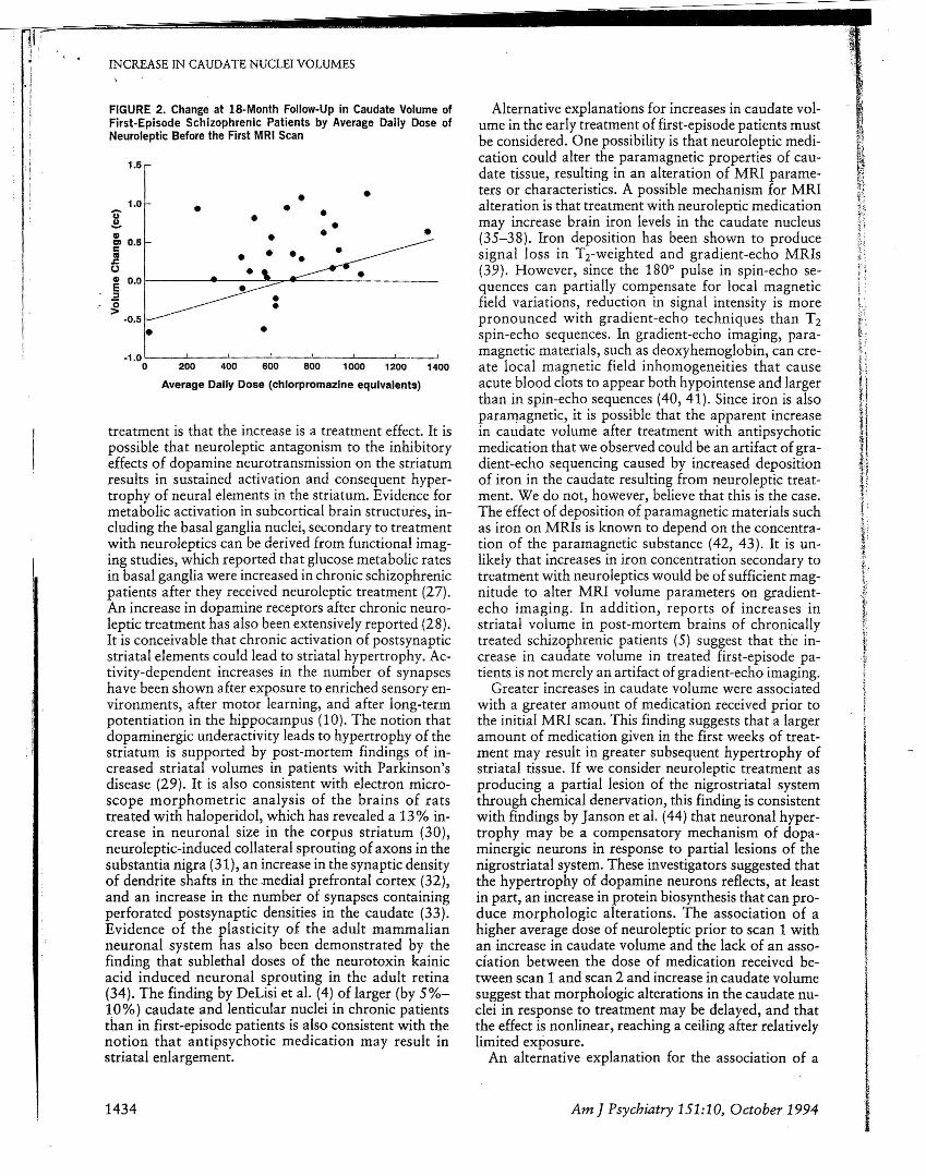
Alternative explanations for increases in caudate volume in the early treatment of first-episode patients mustbe considered. One possibility is that neuroleptic medication could alter the paramagnetic properties of caudate tissue, resulting in an alteration of MRI parameters or characteristics. A possible mechanism for MRIalteration is that treatment with neuroleptic medicationmay increase brain iron levels in the caudate nucleus(35-38). Iron deposition has been shown to producesignal loss in Trweighted and gradient-echo MRIs(39). However, since the 1800 pulse in spin-echo sequences can partially compensate for local magneticfield variations, reduction in signal intensity is morepronounced with gradient-echo techniques than T2spin-echo sequences. In gradient-echo imaging, paramagnetic materials, such as deoxyhemoglobin, can create local magnetic field inhomogeneities that causeacute blood clots to appear both hypointense and largerthan in spin-echo sequences (40,41). Since iron is alsoparamagnetic, it is possible that the apparent increasein caudate volume after treatment with antipsychoticmedication that we observed could be an artifact of gradient-echo sequencing caused by increased depositionof iron in the caudate resulting from neuroleptic treatment. We do not, however, believe that this is the case.The effect of deposition of paramagnetic materials suchas iron on MRIs is known to depend on the concentration of the paramagnetic substance (42, 43). It is unlikely that increases in iron concentration secondary totreatment with neuroleptics would be of sufficient magnitude to alter MRI volume parameters on gradientecho imaging. In addition, reports of increases instriatal volume in post-mortem brains of chronicallytreated schizophrenic patients (5) suggest that the increase in caudate volume in treated first-episode patients is not merely an artifact of gradient-echo imaging.
Greater increases in caudate volume were associatedwith a greater amount of medication received prior tothe initial MRI scan. This finding suggests that a largeramount of medication given in the first weeks of treatment may result in greater subsequent hypertrophy ofstriatal tissue. If we consider neuroleptic treatment asproducing a partial lesion of the nigrostriatal systemthrough chemical denervation, this finding is consistentwith findings by Janson et al. (44) that neuronal hypertrophy may be a compensatory mechanism of dopaminergic neurons in response to partial lesions of thenigrostriatal system. These investigators suggested thatthe hypertrophy of dopamine neurons reflects, at leastin part, an increase in protein biosynthesis that can produce morphologic alterations. The association of ahigher average dose of neuroleptic prior to scan 1 withan increase in caudate volume and the lack of an association between the dose of medication received between scan 1 and scan 2 and increase in caudate volumesuggest that morphologic alterations in the caudate nuclei in response to treatment may be delayed, and thatthe effect is nonlinear, reaching a ceiling after relativelylimited exposure.
An alternative explanation for the association of a
• •1.0 • •'U' • •.2- •II • o•
m 0.5cca •s::U •~ 0.0:I"0>
·0.5
• •-1.0 -------'
0 200 400 600 800 1000 1200 1400
Average Dally Dose (chlorpromazIne equivalents)
FIGURE 2. Change at IS-Month Follow-Up in Caudate Volume ofFirst-Episode Schizophrenic Patients by Average Daily Dose ofNeuroleptic Before the First MRI Scan
1.5
INCREASE IN CAUDATE NUCLEI VOLUMES
treatment is that the increase is a treatment effect. It ispossible that neuroleptic antagonism to the inhibitoryeffects of dopamine neurotransmission on the striatumresults in sustained activation and consequent hypertrophy of neural elements in the striatum. Evidence formetabolic activation in subcortical brain structures, including the basal ganglia nuclei, secondary to treatmentwith neuroleptics can be derived from functional imaging studies, which reported that glucose metabolic ratesin basal ganglia were increased in chronic schizophrenicpatients after they received neuroleptic treatment (27).An increase in dopamine receptors after chronic neuroleptic treatment has also been extensively reported (28).It is conceivable that chronic activation of postsynapticstriatal elements could lead to striatal hypertrophy. Activity-dependent increases in the number of synapseshave been shown after exposure to enriched sensory environments, after motor learning, and after long-termpotentiation in the hippocampus (10). The notion thatdopaminergic underactivity leads to hypertrophy of thestriatum is supported by post-mortem findings of increased striatal volumes in patients with Parkinson'sdisease (29). It is also consistent with electron microscope morphometric analysis of the brains of ratstreated with haloperidol, which has revealed a 13 % increase in neuronal size in the corpus striatum (30),neuroleptic-induced collateral sprouting ofaxons in thesubstantia nigra (31), an increase in the synaptic densityof dendrite shafts in the medial prefrontal cortex (32),and an increase in the number of synapses containingperforated postsynaptic densities in the caudate (33).Evidence of the plasticity of the adult mammalianneuronal system has also been demonstrated by thefinding that sublethal doses of the neurotoxin kainicacid induced neuronal sprouting in the adult retina(34). The finding by DeLisi et al. (4) of larger (by 5%10%) caudate and lenticular nuclei in chronic patientsthan in first-episode patients is also consistent with thenotion that antipsychotic medication may result instriatal enlargement.
1434 Am] Psychiatry 151:10, October 1994 I
" :~
larger daily dose of medication before scan 1 with agreater caudate volume change score is that patientswho required more medication in order to be stabilizedmight have had a more severe form of illness. However,if severity of illness caused the increase in caudate volume over time, we would have expected to see an association between caudate volume change scores andother measures of severity of illness, including psychopathology, level of remission, and time to remission.
The association of increased caudate nuclei volumeswith younger age at onset of illness is similar to theassociation between younger age at onset of illness andlarger lenticular nuclei in young, chronically treated patients that has been reported by other investigators (6).These findings suggest that an effect of neurolepticmedication on caudate nuclei volumes may be mitigated in older patients. One possible explanation forthis age effect is the greater neuronal plasticity in the
'; striatal systems of young patients, as a consequence of, which they are more likely to respond with activation: and hypertrophy of striatal tissue to the disinhibition
induced by dopamine-blocking neuroleptic medications.There are several limitations of the findings of this
study. Since the comparison group was small and notperfectly matched for age and gender, we cannot ruleout the possibility that the increases in caudate volumeamong the patients were secondary to differences in ageor gender between the patients and comparison subjects, rather than a treatment effect. A further limitationof this study is that, in a study of human subjects, it isnot possible to disentangle the confounding effect of severity of illness with dose of medication, since comparison subjects cannot be exposed to chronic neuroleptictreatment. An animal study with neuroleptic- and placebo-treated groups might help to disentangle thesevariables.
CONCLUSIONS
In summary, there was no change in ventricular andcortical volumes of first-episode schizophrenic patients18 months after entry into the study. Striatal enlargement did occur early in the course of illness of thesepatients, especially those with early onset of illness.This enlargement may be the result of an interactionbetween exposure to neuroleptic trearment and theneuronal plasticity of the dopaminergic neuronal systems of young patients. Alternatively, the enlargementmay be secondary to illness-related factors that provokeregenerative changes in striatal structures.
REFERENCES
1. Bogerts B: The neuropathology of schizophrenia: pathophysiological and neurodevelopmental implications, in Fetal NeuralDevelopment and Adult Schizophrenia. Edited by Mednick SA,Cannon TD, Barr CE, Lyon M. New York, Cambridge University Press, 1991
2. Hyde TM, Casanova MF, Kleinman JE, Weinberger DR: Neuroanatomical and neurochemical pathology in schizophrenia, in
Am] Psychiatry 151:10, October 1994
CHAKOS, LIEBERMAN, EllnER, ET AL.
American Psychiatric Press Review of Psychiatry, vol 10. Editedby Tasman A, Goldfinger SM. Washington, DC, American Psychiatric Press, 1991
3. Andreasen NC, Nasrallah HA, Dunn V, Olsen SC, Grove WM,Ehrhardt ]C, Coffman ]A, Crossett JH: Structural abnormalitiesin the frontal system in schizophrenia: a magnetic resonance imaging study. Arch Gen Psychiatry 1986; 43:136-144
4. Delisi LE, Hoff AL, Schwartz ]E, Shields GW, Halthore SN,Simhadri MG, Henn FA, Anand AK: Brain morphology in firstepisode schizophrenic-like psychotic patients: a quantitativemagnetic resonance imaging study. Bioi Psychiatry 1991; 29:159-175
5. Heckers S, Heinsen H, Heinsen Y, Beckmann H: Cortex, whitematter, and basal ganglia in a volumetric postmortem study. BioiPsychiatry 1991; 29:556-566
6, Jernigan TL, Zisook S, Heaton RK, Moranville ]T, Hesselink ]R,Braff DL: Magnetic resonance imaging abnormalities in lenticular nuclei and cerebral cortex in schizophrenia. Arch Gen Psychiatry 1991; 48:881-890
7. Swayze VW II, Andreasen NC, Alliger R], Yuh WT, Ehrhardt]C:Subcortical and temporal structures in affective disorder andschizophrenia: a magnetic resonance imaging study. BioI Psychiatry 1992; 31:221-240
8. Nasrallah HA, Chu 0, Olsen SC, Martin R: Increased caudatevolume in schizophrenia: a controlled MRI study (abstract).Schizophr Res 1993; 9:204
9. Feinberg I: Schizophrenia: caused by a fault in programmed synaptic elimination during adolescence? ] Psychiatr Res 1983; 17:319-334
10. Fields RD, Nelson PG: Activity-dependent development of thevertebrate nervous system, in International Review of Neurobiology, vol 34. Edited by Bradley B. San Diego, Academic Press,1992
11. Bogerts B, Ashtari M, Degreef G, Alvir ]M], Bilder RM, Lieberman ]A: Reduced temporal limbic structure volumes on magnetic resonance images in first episode schizophrenia. PsychiatryRes: Neuroimaging 1990; 35:1-13
12. Lieberman ]A, ]ody D, AJvir ]M], Ashtari M, Levy DL, BogertsB, Degreef G, Mayerhoff DI, Cooper T: Brain morphology, dopamine and eye tracking abnormalities in first-episode schizophrenia: prevalence and clinical correlates. Arch Gen Psychiatry1993; 50:357-368
13. Degreef G, Ashtari M, Bogerts B, Bilder RM, Jody DN, Alvir ],Lieberman ]A: Volumes of ventricular system subdivisions measured from magnetic resonance images in first-episode schizophrenic patients. Arch Gen Psychiatry 1992; 49:531-537
14. Lieberman ]A, Alvir ]M], Woerner M, Degreef G, Bilder R, Ashtari M, Bogerts B, Mayerhoff DI, Geisler SH, Loebel A, Levy DL,Hinrichsen GA, Szymanski S, Chakos MH, Koreen A, Borenstein MT, Kane ]M: Prospective study of psychobiology in firstepisode schizophrenia at Hillside Hospital: design, methodologyand summary of findings. Schizophr Bull 1992; 18:351-371
15. Lieberman], 10dy D, Geisler S, Alvir], Loebel A, Szymanski S,Woerner M, Borenstein M: Time course and biologic correlatesof treatment response in first-episode schizophrenia. Arch GenPsychiatry 1993; 50:369-376
16. Endicott], Spitzer RL: A diagnostic interview: the Schedule forAffective Disorders and Schizophrenia. Arch Gen Psychiatry.1978; 35:837-844
17. Spitzer RL, Endicott ], Robins E: Research Diagnostic Criteria:rationale and reliability. Arch Gen Psychiatry 1978; 35:773-782
18. Spitzer RL, Williams ]BW, Gibbon M: Structured Clinical Interview for DSM-III-R (SCID). New York, New York State Psychiatric Institute, Biometrics Research, 1987
19. Ashtari M, Zito ]L, Bennett IG, Lieberman ]A, Borenstein MT,Herman PG: Computerized volume mensuration of brain structure.] Invest Radio11990; 25:798-805
20. Bilder RM, Wu H, Bogerts B, Degreef G, Ashtari M, Alvir 1M],Snyder PI, Lieberman ]A: Absence of regional hemispheric volume asymmetries in first-episode schizophrenia. Am] Psychiatry1994; 151:1437-1447
21. Andreasen NC, Olsen S: Negative v positive schizophrenia: definition and validation. Arch Gen Psychiatry 1982; 39:789-794
1435