incidence and outcome of out-of-hospital cardiac …
TRANSCRIPT

Department of Perioperative, Intensive Care and Pain Medicine Helsinki University Hospital
University of Helsinki Helsinki, Finland
INCIDENCE AND OUTCOME OF OUT-OF-HOSPITAL
CARDIAC ARREST PATIENTS IN FINNISH INTENSIVE CARE
UNITS
Jukka Vaahersalo
ACADEMIC DISSERTATION
To be presented with the permission of the Medical Faculty of the University of Helsinki, for public examination in Lecture Hall 1 at Biomedicum, Haartmaninkatu 8,
on May 27th 2016, at 12 noon.
HELSINKI 2016

SUPERVISORS
Professor Ville Pettilä Department of Perioperative, Intensive Care and Pain Medicine,
University of Helsinki and Helsinki University Hospital Helsinki, Finland
Docent Tero Varpula
Department of Perioperative, Intensive Care and Pain Medicine Helsinki University Hospital
Helsinki, Finland
REVIEWERS
Docent Mika Laine Heart and Lung Center Helsinki University Hospital
Helsinki, Finland
Docent Mika Valtonen Department of Anaesthesiology and Intensive Care Medicine,
Turku University Hospital Turku, Finland
OFFICIAL OPPONENT
Professor Kjetil Sunde
Oslo University Hospital
ISBN 978-951-51-2177-6 (paperback) ISBN 978-951-51-2178-3 (PDF)
http://ethesis.helsinki.fi
Unigrafia Oy Helsinki 2016

May the Force be with you. -Obi-Wan Kenobi
To Hanna, Siiri, Lauri and Eero


Table of Contest
LIST OF ORIGINAL PUBLICATIONS 7 LIST OF ABBREVIATIONS 8 ABSTRACT 10 1 INTRODUCTION 13 2 REVIEW OF LITERATURE 15
2.1 Out of hospital cardiac arrest 15 2.1.1 Epidemiology 15 2.1.2 Pre hospital factors associated to survival 16
2.2 Post cardiac arrest syndrome 17 2.2.1 Post cardiac arrest brain injury 19 2.2.2 Post cardiac arrest myocardial dysfunction 19 2.2.3 Systemic ischemia/reperfusion response 20 2.2.4 Persistent precipitating pathology 20
2.3 Intensive care 21 2.3.1 Temperature control after cardiac arrest 22
2.3.1.1 Hypothermia 22 2.3.1.2 Clinical evidence of hypothermia and temperature control 23
2.3.2 Ventilation and blood gases 25 2.3.2.1 Oxygen 26 2.3.2.2 Carbon dioxide 28
2.4 Biomarkers after cardiac arrest 28 2.4.1 Inflammatory biomarkers- IL6 and CRP 29 2.4.2 Neurological biomarkers- S100B 30 2.4.3 Cardiac troponins 30
2.5 Outcome 31 2.5.1 Mortality 31 2.5.2 Neurological outcome 31 2.5.3 Factors associated to outcome variation 33 2.5.4 Outcome prediction 33
3 AIMS OF THE STUDY 37 4 PATIENTS AND METHODS 38
4.1 Patients 38 4.2 Study design 42
4.2.1 Study I 42 4.2.2 Study II 42 4.2.3 Study III 42 4.2.4 Study IV 43
4.3 Data collection 43 4.4 Blood samples 44 4.5 Measurements of biomarkers 44
4.5.1 Interleukin 6 and hs-CRP 45

4.5.2 Protein S-100B 45 4.5.3 High-sensitivity troponin-T 45
4.6 Disease severity scorings and definitions 45 4.7 Outcome measures 46
4.7.1 Neurological outcome 46 4.7.2 Mortality 46
4.8 Statistical methods 46 5 RESULTS 48
5.1 Incidence (I) 48 5.2 Temperature management (I) 48 5.3 Blood gases (II) 52
5.3.1 Oxygen 52 5.3.2 Carbon dioxide 54
5.4 Biomarkers after cardiac arrest 55 5.4.1 IL-6 and hs-CRP (III) 55 5.4.2 S100B (III) 58 5.4.3 Hs-TnT (IV) 59
5.5 Outcome (I) 60 5.5.1 OHCA patients with shockable and non-shockable rhythms 61
6 DISCUSSION 64 6.1 Incidence of OHCA (I) 64 6.2 Temperature management (I) 65 6.3 Blood gases (II) 66
6.3.1 Oxygen 66 6.3.2 Carbon dioxide 67
6.4 Biomarkers (III-IV) 69 6.4.1 IL-6 and hs-CRP 69 6.4.2 S100B 70 6.4.3 Hs-TnT 71
6.5 Outcome (I-IV) 72 6.5.1 Outcome of OHCA-shockable patients (I) 73 6.5.2 Outcome of OHCA-non-VF-patients (I) 74
6.6 Strengths and limitations 74 6.7 Clinical implications 76 6.8 Future perspectives 77
7 CONCLUSIONS 79 8 ACKNOWLEDGEMENTS 80 9 REFERENCES 83

7
LIST OF ORIGINAL PUBLICATIONS
This thesis is based on the following original publications, referred to in the text by their Roman numerals (I-IV).
I Vaahersalo J, Hiltunen P, Tiainen M, Oksanen T, Kaukonen KM, Kurola J, Ruokonen E, Tenhunen J, Ala-Kokko T, Lund V, Reinikainen M, Kiviniemi O, Silfvast T, Kuisma M, Varpula T, Pettilä V; FINNRESUSCI Study Group. Therapeutic hypothermia after out-of-hospital cardiac arrest in Finnish intensive care units: the FINNRESUSCI study. Intensive Care Med 2013; 39:826-837.
II Vaahersalo J, Bendel S, Reinikainen M, Kurola J, Tiainen M, Raj R, Pettilä V, Varpula T, Skrifvars M.B; FINNRESUSCI Study Group. Arterial blood gas tensions after resuscitation from out-of-hospital cardiac arrest: associations with long-term neurologic outcome. Crit Care Med. 2014; 42:1463-1470
III Vaahersalo J, Skrifvars M.B, Pulkki K, Stridsberg M, Røsjø H, Hovilehto S, Tiainen M, Varpula T, Pettilä V, Ruokonen E; FINNRESUSCI Laboratory Study Group. Admission interleukin-6 is associated with post resuscitation organ dysfunction and predicts long-term neurological outcome after out-of-hospital ventricular fibrillation. Resuscitation 2014; 85:1573-1579
IV Helge Røsjø, Jukka Vaahersalo, Tor-Arne Hagve, Ville Pettilä, Jouni Kurola, Torbjørn Omland, FINNRESUSCI Laboratory Study Group. Prognostic value of high-sensitivity troponin T levels in patients with ventricular arrhythmias and out-of-hospital cardiac arrest: data from the prospective FINNRESUSCI Study. Crit Care 2014; 18(6):605.
These articles have been printed with the kind permission of their copyright holders.

8
LIST OF ABBREVIATIONS
ABG Arterial Blood Gases AHA American Heart Association APACHE Acute Physiology and Chronic Health Evaluation ASY Asystole AUC Area Under (the ROC) Curve CA Cardiac Arrest CAHP Cardiac Arrest Hospital Prognosis CI Confidence Interval CPC Cerebral Performance Categories CPR Cardio Pulmonary Resuscitation CRF Case Report Form ECG Electrocardiogram EGDT Early Goal Directed Therapy ELISA Enzyme-Linked Immunosorbent Assay EMS Emergency Medical Service ERC European Resuscitation Council FICC Finnish Intensive Care Consortium Hs-CRP High sensitivity C-Reactive Protein Hs-TnT High sensitivity Troponin T ICU Intensive Care Unit IHCA In-hospital Cardiac Arrest IHD Ischemic Heart Disease IL-6 Interleukin 6 ILCOR International Liaison Committee of Resuscitation IQR Interquartile Range LOS Length Of Stay MODS Multi Organ Dysfunction MRI Magnetic Resonance Imagination NRI Net Reclassification Improvement NSE Neuron-specific Enolase OHCA Out of Hospital Cardiac Arrest

9
OR Odds Ratio PCAS Post Cardiac Arrest Syndrome PCI Percutaneous Coronary Intervention PEA Pulseless Electrical Activity RCT Randomized-Controlled Trial ROC Receiver Operating Characteristic ROSC Return of Spontaneous Circulation S100B Protein S100B SAPS Simplified Acute Physiology Score SOFA Sequential Organ Dysfunction Assessment TBI Traumatic Brain Injury TH Therapeutic Hypothermia TISS Therapeutic Intervention Scoring System TTM Targeted Temperature Management VF Ventricular Fibrillation VT Ventricular Tachycardia

10
ABSTRACT
Aims The objectives of this study were to evaluate the incidence and neurological outcomes of out-of-hospital cardiac arrest (OHCA) patients in Finnish intensive care units (ICU). This study also investigated the use of therapeutic hypothermia, arterial blood gas pressures and different biomarkers association with one-year neurological outcome, mortality or organ dysfunction in ICU-treated OHCA patients.
Materials and methods
A nationwide, prospective, observational FINNRESUSCI study was conducted in 21 out of 22 ICUs treating adult cardiac arrest patients in Finland during a one-year study period, 1st March 2010 to 28th February 2011. All successfully resuscitated adult patients after OHCA who were treated in ICU were included to this study. Blood samples for biomarker evaluation were collected from patients at four time points after informed consent, and neurological outcomes were determined 12 months after cardiac arrest. Study data were collected with Case Report Forms (CRF) and from the Finnish Intensive Care Consortium (FICC) database.
Study I included all FINNRESUSCI study patients and evaluated the incidence and the implementation of therapeutic hypothermia (TH) after OHCA in ICU and reported the mortality and 12-month neurological outcomes of OHCA patients resuscitated from shockable and non-shockable rhythms treated with or without TH. In Study II, all arterial blood gas samples obtained from all mechanically ventilated and comatose patients during the first 24h from ICU admission were analysed. The mean and time-weighted partial pressures of oxygen and carbon dioxide and their associations to mortality and neurological outcome were studied. In Study III, interleukin-6 (IL-6), high-sensitivity-CRP (hs-CRP) and protein S-100B were measured from patients resuscitated from shockable rhythm and their association to the duration of ischemia, organ dysfunction and neurological outcome were evaluated. In Study IV, high-sensitivity troponin T (hs-TnT) was analysed from a set of patients resuscitated from shockable rhythm and with a sample available from ICU admission (<6h).

11
This study evaluated the ability of hs-TnT to predict short- and long-term outcome.
Main results
Study I included 548 patients, of whom 311 (56.8%) had shockable (VF/VT) and 237 (43.2%) non-shockable (PEA or asystole) as an initial rhythm. The population-based incidence of OHCA patients in ICU was 13/100,000/year. Out of 504 (92%) unconscious patients at ICU admission, TH was induced in 311 patients, 241/281 (85.8%) patients resuscitated from VF/VT, and 70/223 (31.4%) patients resuscitated from PEA or asystole. Of the 504 unconscious patients 184 (37.2%) had a good neurological outcome (CPC 1-2) after 12 months, 147/281 (52.9%) in VF/VT group and 37/223 (17.1%) in PEA or asystole group. Good neurologic outcome was achieved 138/241 (58.0%) with shockable rhythms and 13/70 (19.4%) with non-shockable rhythms after TH treatment versus 9/40 (22.5%) and 24/153 (16.0%) without TH treatment.
Study II included 409 unconscious and mechanically ventilated patients. The mean carbon dioxide (PaCO2) tension during the first 24-hour in ICU was an independent predictor of a good outcome with an odds ratio (OR) of 1.054 (95% Confidence interval (CI) 1.006-1.104), for an increase of 1 mm Hg, but the mean oxygen (PaO2) tension was not OR 1.006 (95% CI 0.998-1.014). In multivariable regression analysis, the time spent above PaCO2 45 mmHg was associated with good neurologic outcome OR 1.015 (95% CI 1.002-1.029) for each percentage point increase in time, but time spent in different PaO2 categories were not. Patients with the highest mean PaCO2 and PaO2 values had better one-year neurologic outcome than predicted with an OR of 3.2 (95% CI 1.1-9.2). There was no harmful association between hyperoxia and outcome.
Study III included 186 patients resuscitated from VF/VT. High admission plasma concentrations of interleukin-6 (IL-6) and S-100B were associated with time to ROSC and poor neurological outcome (p<0.001), whereas hs-CRP was not. Admission IL-6 was also associated with extra-cerebral organ dysfunction (p<0.001) with AUC of 0.679. Admission IL-6 was an independent predictor of poor neurological outcome after 12 months with an OR of 1.006 (95% CI 1.000-1.011) in the multivariable logistic regression analysis.
Study IV included 155 patients resuscitated from VF/VT. Hs-TnT levels were elevated in all of the patients but the levels were higher in patients with poor vs. good neurological
12
outcome 739 (IQR 191-1061) vs. 334 (195-716) ng/l (p=0.028), but there was no statistical difference in hospital mortality. Hs-TnT did not improve prognostic information to previously known multivariate analysis risk variables.
Conclusions
Therapeutic hypothermia or targeted temperature management (TTM) is widely used and well implemented in clinical practice in Finnish ICUs. The majority of OHCA patients resuscitated from shockable rhythms are treated with TH and withholding TH was due the clinical reasons. The majority of OHCA patients with shockable rhythms after TH survive with good neurology, while the outcome of patients with non-shockable rhythms is poorer despite the TH treatment. Hyperoxia exposure is rare in Finland and a harmful association of hyperoxia with outcome was not found. Instead mild hypercapnia combined with mild hyperoxia after OHCA might be beneficial during the first 24 hours in ICU. Early inflammatory response after OHCA was demonstrated by high levels of admission IL-6, which was associated with the duration of ischemia and subsequent extra-cerebral organ dysfunction. Admission IL-6 is also an independent predictor of neurological outcome along with time to ROSC and age after OHCA-VF/VT. Hs-TnT does not give any additional prognostic information after OHCA-VF/VT during ICU care.

13
1 INTRODUCTION
Out of hospital cardiac arrest (OHCA) is a significant health problem in industrial countries. The aetiology and treatment of cardiac arrest has changed, but the overall survival has not improved much over the last few decades. Despite improvements in healthcare and intensive care, including therapeutic hypothermia, mortality remains high1. Established pre-hospital factors affecting survival are initial rhythm, witnessed collapse or cardiac arrest, the presence and quality of bystander cardio pulmonary resuscitation (CPR), early defibrillation and underlying comorbidities1 Prognosis depends on the circumstances, including laypersons’ abilities to recognize cardiac arrests, the organization of dispatch centres and emergency medical services (EMS) and the quality of intensive care2. Due to these reasons there is a large variation in survival and outcome numbers reported in the literature3-5. Cardiac arrest stops the blood circulation and causes hypoxia of the whole body, and a short time Return Of Spontaneous Circulation (ROSC) is essential for any possible recovery. Prolonged ischemia causes global tissue and organ injury, but additional damage also occurs during CPR and after ROSC in the reperfusion state. Ischemia and reperfusion start unique pathological processes in the body. These processes form what is known as the Post-Cardiac Arrest Syndrome (PCAS)6,7. The major components of PCAS are brain injury, myocardial dysfunction and systemic ischemia/reperfusion response and persistent precipitating pathology7,8. The severity of PCAS is not uniform in individual patients and it lasts for at least 72 hours9. Potential therapies for PCAS are focused on treatment of these separate components and needs intensive care resources to manage in its entirety10. Brain injury is the most common cause of mortality after cardiac arrest11,12. In PCAS care lowering or controlling the patient’s temperature is a major therapeutic intervention to prevent or limit brain injury. Therapeutic Hypothermia (TH), where patients are cooled to 32-34° C for 12-24 hours has been in clinical practice for over a decade since two randomized controlled trials13,14 and meta-analysis showed improved outcome in patients resuscitated from out-of-hospital Ventricular Fibrillation (VF)15, who remained comatose after ROSC and treated with TH. TH has been recommended for standard care for

14
comatose survivors of OHCA with shockable rhythms according to the international guidelines ever since16. In the latest RCT, Targeted Temperature Management (TTM) trial, 950 OHCA patients were randomized into 36 h temperature controls either at 33°C or 36°C17. The TTM study reported no difference in mortality or neurological outcome between the temperature groups. In TTM study fever was prevented in both groups compared to previous RCT13, which is the major weakness of this HACA study. The benefit of TH or TTM in patients resuscitated from non-shockable initial rhythms has not been shown, but despite insufficient evidence, TH was also recommended for all comatose OHCA patients chosen to active intensive care according the European Resuscitation Council (ERC) guidelines 201018. According to the latest ERC guidelines TTM is recommended for all comatose OHCA patients resuscitated from shockable rhythm and suggested also for IHCA and OHCA patients resuscitated from non-shockable rhythms (weak recommendation, very-low quality evidence)19. Because these recent guidelines allow recommended temperature control between 32°C to 36°C, the term temperature control or targeted temperature control is preferred and has replaced the term TH in the latest literature. Individual components of PCAS are potentially treatable and may each have some influence on survival1,8,10. Therapeutic strategies like ventilation, circulatory and haemodynamic support, glucose control, sedation, management of coronary syndrome and treatment of other causes of CA are focused on these components of PCAS. However, the optimal target levels of some treatments and therapies are still unknown8. Cardiac arrest patients, who are common in ICUs, use limited intensive care resources and cause substantial costs to the health care system. Therefore, it is important to gain knowledge on therapeutic strategies used in ICUs and their effects on outcomes. It would be also very useful to get early information on subsequent clinical problems to optimize treatments adequately. Another very important aspect of post resuscitation care is early and accurate outcome prediction. If available, it would help the clinicians to make decisions in patient selection for intensive care or the withdrawal of intensive care when outcomes are predicted to be undesirable. The aims of this study were to evaluate the incidence and outcome of OHCA patients in ICUs in Finland. In addition, the study focused on the use of therapeutic hypothermia, post resuscitation care and different biomarkers after OHCA regarding their value in outcome prediction.

15
2 REVIEW OF THE LITERATURE
2.1 Out of hospital cardiac arrest
Cardiac arrest, a sudden loss of heart function, stops the circulation immediately and causes unconsciousness in a few seconds. Ischemic Heart Disease (IHD) is the leading cause of cardiac arrest20,21 and cardiac arrest may be the first symptom of IHD. Cardiac arrest and loss of blood flow lead to general ischemia and cerebral injury6, which is the leading cause of death after cardiac arrest6-8,11,12. The purpose of pre-hospital cardiopulmonary resuscitation (CPR) is to minimize the time of no blood flow with chest compressions, take care of ventilation and reverse unexpected cardiac arrest. High quality pre-hospital EMS also guarantees sufficient circulation and ventilation of successfully resuscitated patients during transportation to hospitals. The purpose of hospital and ICU care is to minimize the neurological and organ damage, treat the cause of CA and restore the patients to normal life.
2.1.1 Epidemiology
Sudden cardiac arrest is one of the leading causes of death in Europe and the USA. Cardiac arrest occurs in 0.5-1.0 per 1000 inhabitants a year, depending on how cardiac arrest is defined, affecting approximately 500 000 individuals in Europe and 200 000 in the USA each year1,4,22. Variations between incidences is reported to be between 37 and 121 per 100 000 inhabitants/year due the regional variations in Emergency Medical Service (EMS) systems between countries3-5,23-25. OHCA patients are treated 100% by EMS services in Finland5,26 and approximately 60% in USA27. The incidence of attempted resuscitation varies between countries depending on EMS systems and national legal aspects4. The overall incidence of OHCA with resuscitation attempted by EMS is reported to be 51/100 000 inhabitants/year in Finland5 and in Helsinki, the largest city of Finland 80/100 00026. OHCA patients have VF as an initial rhythm in 25-50% cases5,25,26,28,29 but the incidence of OHCA-VF has been decreasing over the last few years28,30,31. ROSC and survival to hospital admission is reported to be around 30-35% of all attempted resuscitations in

16
Scandinavia5,26,32, but only selected patients are treated in ICUs26,32. The incidence of cardiac arrest patients treated in ICU was 15/100 000/ inhabitants/year between 2004-2005 in Finland33. Despite the improvements in specific treatments and knowledge, the overall survival rate has been stable and has remained low over the last 30 years1,34. The average survival to hospital discharge is reported to be 7.6% in Europe and the USA1,4,34,35. Survival rate is still highly dependent on pre-hospital key factors (witnessed CA, bystander CPR, initial rhythm, ROSC) despite the improved treatment in ICUs1,36.
2.1.2 Prehospital factors associated with survival
There are several factors affecting survival from OHCA, including the action of bystanders in recognizing the cardiac arrest, making an emergency call to a dispatching centre (112) and starting basic life support (BLS), high-quality post resuscitation care in an ICU and rehabilitation after hospital care. Each of these factors is important separately, but the last years have been shown the importance of the whole chain of treatment in the resuscitation guidelines, the so-called chain of survival18.
Figure 1. The Chain of survival. Adapted from the European Resuscitation Council
(ERC) Guidelines for Resuscitation 201018.

17
Short time intervals are the most important factors in the treatment of cardiac arrest. Early bystander CPR has a beneficial effect on survival rate and each minute of delay in the initiation of CPR decreases the chance of survival 3% until CPR starts37. Immediate CPR can double or even triple the survival rate with OHCA-VF patients38,39. Early defibrillation is beneficial with VF patients, with each minute of delay in defibrillation reducing the probability of survival by 10-12%36,38.
The presence of a shockable rhythm (VF/VT) is a significant and independent predictor of survival and discharge from hospital after successful resuscitation from cardiac arrest1,40. VF or VT usually represents the cardiac origin of cardiac arrest, while asystole and PEA as initial rhythms more often represent a non-cardiac origin or reflect a long delay from cardiac arrest to the time of initial rhythm recognition. VF patients have several times better chances to survive than patients resuscitated from non-shockable rhythms15,24,41. Pooled OR for survival to hospital discharge of OHCA-VF patients compared to patients found with non-shockable rhythms ranged from 2.91 (95% CI 1.10-7.66) to 20.62 (95% CI 12.61-33.72) depending on the baseline survival of the studies1.
Overall survival rates vary in studies around the world, but the highest survival rates have been reported in the studies with the highest proportion of patients found with VF4.
When evaluating key predictors of OHCA patients in meta-analyses, patients who are found with shockable initial rhythm (VF/VT) and received CPR from a bystander or an EMS provider, have significantly better chances to survive than those who do not. The better survival of VF or VT patients is associated with locations where a defibrillator is available at public sites42. Of VF/VT patients approximately 1 of every 4 to 7 patients survive to hospital discharge, compared to patients found in asystole, of whom only 1 of every 21 to 500 patients survive1. There is a great variation in the published survival rates between different countries4 and also regional variations between EMS systems, cities and rural areas3,5,43.
2.2 Post cardiac arrest syndrome
Whole body ischemia, caused by cardiac arrest, and reperfusion caused by return of spontaneous circulation (ROSC) after successful CPR creates a specific pathophysiological state. This phenomenon was first described in the early 1970s and named post resuscitation disease44, later called post-cardiac arrest syndrome (PCAS)8. The pathological process actually begins at the time of collapse and continues during CPR and in the reperfusion phase45,46 for several hours after ROSC, even though the actual resuscitation has ended.

18
PCAS can be divided into four main components by individual organ systems; i) post-cardiac arrest brain injury, ii) post-cardiac arrest myocardial dysfunction, iii) ischemia/reperfusion syndrome and iv) persistent precipitating pathology. The severity of disorders in these organ systems is not uniform, but it varies in individual patients based on the patient´s comorbidities and state of health, the cause of cardiac arrest and the time of ischemia. PCAS may not occur and the patients regain consciousness, if ROSC is achieved rapidly after cardiac arrest.
Figure 2. Phases and treatment goals of Post Cardiac Arrest Syndrome (PCAS).
Modified from the original figure by Nolan and colleagues7

19
2.2.1 Post cardiac arrest brain injury
Brain injury is the most common cause of death after OHCA11,12. Brain tissue is more vulnerable to ischemia compared to other organs and ischemic injuries occur as early as a few minutes of ischemia. After cardiac arrest reperfusion is often hyperdynamic and cerebral perfusion pressure (CPP) is often elevated because of impaired cerebral autoregulation47,48. Despite adequate cerebral perfusion pressure (CPP), the failure of the cerebral microcirculatory may lead to ischemia and local infarcts in some brain areas in animal models49,50. Systemic blood pressure correlates with CPP, and mean arterial blood pressure influences neurologic outcome after cardiac arrest51. Hypoxemia and brain oedema also influences oxygen delivery to the brain and possible brain injury especially after CA. The partial pressures of arterial blood gases (ABG) have an effect on blood circulation in brain tissue due the vasoconstriction or vasodilation of blood vessels52,53. Hypocapnia reduces intracerebral pressure (ICP) and increases cerebral perfusion pressure but vasoconstriction in blood vessels may cause harmful ischemia in the brain tissue52. On the other hand, hyperoxia causes oxidative stress and is harmful for neurones after ischemic insult in animal models54-56. The effects of the partial pressures of arterial blood gases is a complex question57 but ABG tensions may have an influence on the severity of brain damage.
2.2.2 Post cardiac arrest myocardial dysfunction
Haemodynamic instability, variation of heart rate and blood pressure, is very commonly seen after ROSC, but it often stabilizes after the circulating catecholamine concentrations decrease58. Instant myocardial dysfunction, established by the low ejection fraction and high end diastolic pressure of the left ventricle can be detected right after ROSC by appropriate monitoring59. This dysfunction is not only related to ischemia, but also to reperfusion leading to the myocardial stunning phenomenon manifested by hypotension, tachycardia and low cardiac output60-62. Because this phenomenon is usually transient and major recovery occurs under 72 hours60,62,63, myocardial dysfunction is partly responsible for deaths in the first three days, while brain injury is responsible for the later deaths11,12.

20
2.2.3 Systemic ischemia/reperfusion response
Cardiac arrest and debt of oxygen causes the most severe shock state in the tissues and leads to activation of the endothelium64. Low oxygen level itself is predictive for organ failure and increasing mortality65 and combined with the activation of immunological and coagulation systems leads to a condition similar to sepsis66. Intravascular volume depletion, vasodilatation, release of various cytokines and endotoxins, endothelial injury leads to increasing risk of multiple organ failure and infections67-70
Therapeutic strategies after CA in ICU are focused mainly on optimizing the clinical manifestations of the immune response, optimizing haemodynamics and organ perfusion. The primary tools for this are intravenous fluids, inotropes, vasopressors and blood transfusions. Early Goal Directed Therapy (EGDT), where the balance between oxygen demand and consumption is in focus, has clinical benefits in patients with sepsis, such as identification of patients at high risk to CA71. EGDT has been shown to reduce post-operative complications, the duration of hospital stay and mortality after major surgery72 but it does not have the same effects for sepsis73. The optimal goals for haemodynamic parameters have not been studied in randomized trials for CA patients and are still unclear, but since PCAS have features common to sepsis, the principles of EGDT might be beneficial in the care of CA patients7. The magnitude of PCAS described by changes in cytokine, endotoxin and systemic inflammation marker levels has been reported to associate with the outcome70,74
2.2.4 Persistent precipitating pathology
Coronary artery disease is a very common pathology among OHCA patients21 and myocardial infarction is the leading cause of sudden cardiac death75. Acute coronary syndrome (ACS) is the most common cause of OHCA. A high incidence (48%) of acute coronary occlusion after OHCA was reported already in 199776 and the prevalence of significant coronary artery disease ranged from 59% to 71% for OHCA patients in a recent meta-analysis77. Probable ACS can be identified by clinical symptoms or measurements. Chest pain before cardiac arrest, ST-segment elevations in ECGs are the classic

21
manifestations of ACS, but their predictive value is poor among CA patients76. The majority of patients with ST-segment elevation in their ECGs after ROSC have acute coronary lesions78. The absence of ST-segment elevation does not exclude ACS as a cause of cardiac arrest in OHCA patients79-82. In many observational studies early cardiac interventions, angiography and percutaneous coronary intervention (PCI), increased survival rates and associated with better neurological outcomes, especially in patients with ST-segment elevation79,81, but there are no randomized studies which would validate this benefit. Because the success and feasibility of early angiography and PCI is also well documented83-
86, the latest ERC guidelines recommend early coronary intervention for all patients with ST-segment elevation after OHCA. Coronary angiography should be also considered for all patients with a probable cardiac cause for OHCA87
Other possible diagnoses for CA are pulmonary embolism, sepsis, metabolic disorders, hypoxemia, hypothermia, cardiac tamponade, pulmonary diseases, haemorrhage, intoxications and electrolytic disturbances which are more common among IHCA patients and patients with non-shockable initial rhythm88. These causes of cardiac arrest should be diagnosed and treated early, if possible during the CPR89.
2.3 Intensive care
Post resuscitation care starts at the venue where ROSC is achieved, but usually the patient is transferred to the next care level area for monitoring, diagnosis and treatment, depending on local hospital circumstances and the patient’s clinical state. Depending on the cause of arrest, time delays to CPR and defibrillation and duration of CPR, some OHCA patients do regain consciousness rapidly after successful ROSC1,37,90. Despite the possible return of consciousness, many of these patients still require treatment to optimize ventilation91,92, haemodynamics7, glucose control93 and multiple organ support. The purpose of intensive care after cardiac arrest is to focus on treating disorders of the PCAS components and adjust the treatment balance between all the injured organ systems. PCAS is a very complex state and monitoring and treatments of these patients is possible usually only in ICU circumstances, but according to growing knowledge, all the key components of PCAS are individually potentially treatable. Standardized treatment and local protocols do have significant influence on the overall outcome and especially on the most important outcome parameter, neurological outcome83,94-96.

22
2.3.1 Temperature control after cardiac arrest
Hyperthermia is very common after CA97 and there is a clear association between hyperthermia and poor neurological outcome97-100 Hyperthermia after mild therapeutic hypothermia (32°C-34°C), so called rebound hyperthermia, is associated to unfavourable neurological outcome101,102. Since 2003 mild TH, where patients are cooled to 32°C-34°C for 12-24 hours has been recommended for OHCA patients resuscitated from VF/VT who are selected for intensive care16. According to the 2009-2010 international guidelines TH recommendation extended also for non-VF-OHCA and in-hospital cardiac arrest (IHCA) patients selected for intensive care18,103. The term targeted temperature management (TTM), where temperature is maintained between 32°C-36°C, was born after publication of a large randomized trial 2013 by Nielsen and colleagues 17. In that TTM trial, fever was prevented in all patients and it reported equal mortality and neurological outcome with patients treated at both temperatures, i.e., 33°C and 36°C17. Today the international consensus is to use TTM or temperature control instead of the term TH in the care of CA patients. According to a recent ILCOR consensus statement and the latest ERC guidelines 2015 TTM (32°C-36°C) for at least 24 hours is strongly recommended for all comatose OHCA VF patients and weakly for other CA patients19,104.
2.3.1.1 Hypothermia
Therapeutic hypothermia is neuroprotective and the only well documented therapy for preventing brain damage after ischemic insult or lack of blood flow105-107. The exact mechanism is unclear, but hypothermia is thought to have several beneficial effects in reducing brain injury. General cellular metabolism slows significantly for every one degree drop in body temperature108. Accordingly hypothermia is shown to reduce oxygen consumption in brain tissue in animal models109,110, but also the body´s need for oxygen is thought to be reduced111. Hypothermia has been shown to affect many chemical and physical mechanisms at the mitochondrial level, on apoptosis, cell membranes stability and production of free radicals, cytokines and other inflammatory mediators111. Evidence of these models comes mainly from animal studies. Cells can recover from acute ischemia, but it can still lead to the programmed cell death pathway, apoptosis, which is determined by the effects of mitochondrial dysfunction on energy metabolism112. Hypothermia can prevent cells taking to apoptosis pathway113 and prevent mitochondrial dysfunction114. High levels of excitatory neurotransmitter, glutamate and free oxygen radicals, are toxic to neuronal cells. Hypothermia reduces the release of glutamate to the extracellular space115 and

23
decreases the amounts of free radicals that are produced during ischemia116,117 Ischemia induces the raise of inflammatory mediators, this begins early after reperfusion and levels remain elevated for up to 5 days. Therapeutic hypothermia reduces inflammatory reactions and the release of inflammatory cytokines after ischemia and during reperfusion118,119. It also decreases the production of nitric oxide, which plays an important role in the development of ischemic brain damage115. According to animal studies the size of neuronal damage can be decreased by inhibiting some or all of these mechanisms115.
2.3.1.2 Clinical evidence for hypothermia and temperature control
Several animal studies from the 1990s have shown that hypothermia (30°-34° C) significantly improved the neurological outcome of dogs after cardiac arrest120-124. These animal studies led to two human clinical trials published in 200213,14, which both reported
significant improvement of neurological outcome with therapeutic hypothermia treatment compared to patients treated without TH after OHCA. Soon after publication of these studies mild therapeutic hypothermia (32°-34°) for 12 to 24 hours was recommended for clinical practice16 and later included in international guidelines125. TH implemented was relatively soon to post resuscitation care, and there are several before and after studies on cardiac arrest outcome, which all support the benefit of TH, especially in the VF group32,83,126,127. The benefit of TH has been shown also in a systematic review128 and benefit is also reported for CA patients with presumed cardiac origin in meta-analysis 200515.
Despite the shown benefit of TH for VF patients, the benefit in patients with non-shockable initial rhythm is not visible in these historical studies. Studies of patients resuscitated from non-shockable rhythms reported conflicting results from possible benefit of TH83,129,130. A registry study of 1145 patients treated with TH concluded there was a positive association between TH and neurological outcome in VF patients with an OR of 1.9 (95% CI 1.18-3.06) but no benefit in patients in PEA/asystole, OR 0.71 (95% 0.37-1.36)131. The clear evidence of benefit is still missing despite the large registry studies and meta-analysis, with partly contradictory results132-134 TH has been also studied in large registry study with in-hospital cardiac arrest (IHCA) patients, but no difference was found in their survival, OR 0.9 (95% CI 0.65-1.23)135. Summary of these studies are presented in Table 1.

Tab
le 1
. Stu
dies
rep
ortin
g an
d co
mpa
ring
the
outc
ome
of O
HC
A p
atie
nts
trea
ted
with
or
with
out T
H

TH or TTM is generally safe but side effects also occur17,136 and most of the physiological side effects are treatable in ICU circumstances137. The most common and generally known physiological responses to hypothermia are cardiac arrhythmias, usually bradycardia136,138, increased diuresis and electrolyte disturbances17,136,137 and hyperglycaemia14,136 which is strongly associated to poor neurological outcome93,139. Mild induced hypothermia has effects on the coagulation system and it may increases bleeding140, but it seems not have clinical relevance13,17,33,83. The immune system is also impaired by TH, which increases the infection rate137,141,142. TH is also associated with increased incidence of pneumonia143,144. There is no clear evidence of a negative impact of infections on the outcome even though the early use of antibiotics may be beneficial 145. Bradycardia during hypothermia is associated with favourable neurological outcome146,147. TTM treatment can be divided into three phases; induction, maintenance and rewarming137. There are several internal and external techniques used in induction and maintenance of hypothermia; ice packs, ice-cold saline, cooling blankets or intravascular devices, which all have benefits and disadvantages. Prior recommendations suggested rapid cooling after ROSC16 and for mainly this reason ice-cold saline in large volume, usually 20-30ml/kg -1 was widely used in prehospital settings, including in Finland5. The use of ice-cold intravenous fluids have been studied in RCTs 148-151, but no positive impact on mortality or neurological outcome was found for these or other prehospital cooling 152 studies (RR, 0.98; 95% CI, 0.92-1.04). According to present knowledge, during the maintenance phase avoiding temperature variation with adequate monitoring is preferred using external or internal device compared to other techniques. On the other hand, there is no evidence that any cooling technique is superior to the others and would increase survival, but internal devices are better for precise temperature control compared to external techniques153,154. During the rewarming phase electrolyte concentrations, metabolic rate and intravascular volume can change rapidly. Avoiding possible rebound hyperthermia, which is shown to be harmful 101,155 a slow rate of rewarming of 0.25-0.5°C per hour is recommended 19,129.

26
2.3.2 Ventilation and blood gases
All patients who remained comatose after successful ROSC need ventilatory support, usually tracheal intubation and mechanical controlled ventilation. The ventilation strategy used influences the patient’s arterial blood concentrations of oxygen and carbon dioxide (CO2). Blood gas concentrations may affect the brain and other organs’ blood perfusion, oxygen delivery and outcome. Nearly all comatose patients after CA in ICUs are mechanically ventilated, but the treatment goals for safe and optimal levels for oxygen and carbon dioxide are unknown156.
2.3.2.1 Oxygen
Hyperoxia is harmful for neurones in animal models 56, but human studies of this issue is heterogeneous (Table 2). One small randomized human study reported an association between the administration of 100% oxygen and elevated levels of NSE comparing to 30% oxygen to CA patients after ROSC157. According to one registry study with over 6000 patients hyperoxia during the first 24 hours after CA was associated with poor neurological outcome compared to normoxemia and hypoxemia158. In a further analysis a dose dependent association with a poor outcome was reported159. In contrast, according to another observational study on over 12 000 patients, hyperoxia was not associated with increased mortality160. A recent meta-analysis shows significant heterogeneity across studies about this association161. Hyperoxia might be harmful, but at the same time all interventions reducing oxygen exposure also increases the risk for hypoxia, which is also associated to increased mortality158,160. The precise titration of oxygen is possible after a patient’s arrival at hospital using ventilator and ABG values, but there is clear feasibility problem in prehospital setting, when only oxygen saturation by pulse oximetry (SpO2) is used as an oxygenation target162. Out-of-hospital location of cardiac arrest and long time delay in ICU admission are associated with the prevalence of hyperoxia during ICU care163.

Tab
le 2
. Stu
dies
eva
luat
ing
the
effe
ct o
f hyp
erox
ia (>
300m
mH
g/40
kPa
) on
mor
talit
y or
neu
rolo
gica
l out
com
es a
fter
car
diac
arr
est.

2.3.2.2 Carbon dioxide
Despite the autoregulation of cerebral blood flow is dysfunctional after CA48, reactivity to changes in partial pressure of carbon dioxide (CO2) seems to be preserved and CO2 tension has an influence on blood flow in brain tissue164,165. In animal models mild hypercapnia improved cerebral perfusion and protected brain cells after ischemic insult166 and hypocapnia is associated with increased neuronal injury167. The same results are seen also in human patients after traumatic brain injury52,168-170. Carbon dioxide has anticonvulsant171, anti-inflammatory and anti-oxidant properties172, which may be beneficial for preventing brain damage. Hypocapnia after CA induced by hyperventilation has been shown to cause cerebral ischemia also in humans173,174. The effects of carbon dioxide on cardiac arrest patients have studied in observational registry studies91,92. Both these studies reported an association between hypocapnia and poor neurological outcome, but another study found an association between hypercarbia and a higher rate of discharge from hospital with an OR of 1.16 (95% CI 1.03-1.32), which was considered a surrogate marker of favourable neurological outcome92. CO2 tension can be controlled in almost all the patients after CA, because of mechanical ventilation175. Lowering patients’ temperatures in ICU during TH or TTM decreases their metabolism and production of CO2. This may lead to hypocapnia176, which is preventable by adjusting minute ventilation.
2.4 Biomarkers after cardiac arrest
Ischemia and cell damage in different tissues leads to the release of several biomarkers and a wide range of these different biomarkers have been studied in patients with CA. The main goal has been to find a prognostic tool for identifying patients with no chance of recovery or to plan specific treatments during ICU care. Despite several studies no single biomarker has been shown to be a reliable predictor of relevant outcome177,178
Cardiac arrest and successful ROSC leads to similar systemic inflammatory responses like those seen in patients with sepsis66, called PCAS7. The severity of PCAS is variable in individual patients, based on the severity of the ischaemic insult and the patient´s state of health before CA. Ischemia and following activation of inflammatory response increases the risk of multiple organ dysfunction67,69 and the development of organ dysfunction after CA will worsen the patient´s outcome70. Various levels of inflammatory biomarkers have been

29
associated with tissue hypoxia, organ dysfunction and mortality in sepsis patients and these support the need for early haemodynamic optimization and may help clinicians to tailor other treatments strategies7,179. Several inflammatory biomarkers have been studied in critical care patients180, but interleukins, procalcitonin, TNFα and CRP are the most studied biomarkers in cardiac arrest settings177.
Hypoxic-ischemic brain injury is very common after CA and the majority of patients die from brain injury11 also after implementation of TH or TTM12,17. All neurological biomarker studies have been trying to find a clinical tool to identify brain injury and to predict poor neurological outcomes. From these neurological biomarkers, neuron specific enolase (NSE) 181-186 and protein S-100B181,185,187,188 are the most studied biomarkers.
Post cardiac arrest syndrome, which includes myocardial dysfunction and stunning affects the majority of OCHA patients7 and severity of PCAS is a contributing factor to mortality12. Coronary artery disease, acute coronary syndrome (ACS) including myocardial infarction is the most common cause for cardiac arrest20,21. The cardiac aetiology of CA is manifested by VF and pulseless VT as an initial rhythm, but acute coronary artery lesion was also present in 59% to 71% of OHCA patients without a presumed cardiac aetiology77. Observational studies have shown that early invasive cardiac interventions, including PCI, are feasible85,86 and it is probable that invasive management is beneficial in OHCA patients, especially with classic AMI manifestations79,85. The cardiac biomarkers, (TnT, TnI) have been studied to establish cardiac risk factors and possible prognostic values in OHCA patients177.
2.4.1 Inflammatory biomarkers- IL6 and CRP Interleukin-6 (IL-6) is an inflammatory cytokine, which is one of the main stimulators for the production of other acute phase proteins, which are clinically measurable, like C-reactive protein (CRP)189. Systemic levels of IL-6 correlates with organ dysfunction and outcome in sepsis patients in experimental190 and in clinical191 studies. IL-6 and CRP have been studied in CA settings, which reporting increased levels of CRP after CA, but no correlation has been shown in infectious complications to time of anoxia or outcome192-194. Respectively in one small study IL-6 in the cerebrospinal fluid correlated with neurological outcome after CA195 and plasma concentrations of both IL-6 and high sensitivity-CRP (hs-CRP) correlated with poor outcome in another study196. Hypothermia has effects on the release of inflammatory biomarkers, but TH did not affect the IL-6 response, thus the release of CRP was suppressed by TH142.

30
2.4.2 Neurological biomarkers- S-100B
S-100B is a brain protein originating from the astroglial and Schwann cells, which is released from brain cells after ischemia. S-100B correlated with neurological status at admission, functional outcome and stroke volume in patients with ischemic stroke197, but the evidence with cardiac arrest patients is not well documented. The first studies of S-100B reporting higher concentrations at 24 h in patients with poor outcome compared to good outcome (0.78 vs. 0.19 mg/l) almost 20 years ago198. In addition, the cut off value for persistent coma was reported to 0.7 mg/l with high positive predictive value (95%) and high specificity (96%). Since then the majority of the studies have reported a high specificity, but low sensitivity for S-100B as an outcome predictor199-203. S-100B levels are raised after cardiac arrest and it correlates with neurological outcome, but there is too great variability in the cut-off values to predict poor neurological outcome177,178.
S-100B’s ability to predict long-term neurological outcome has been studied in prospective settings with OHCA patients. S100B levels over 0.29 microg/l at 24-48 h correlated to moderate or severe neurological dysfunction and increased levels predicted hospital mortality with 100% specificity for a cut off value 1.2 microg/l199. Results from studies comparing the predictive values of different neurological biomarkers are also variable, some of these studies report a superior value for S-100B in comparison with other biomarkers188,198,204, whereas some studies report similar or better predictive values for NSE181,185. These discrepancies are probably related to variations in study populations, initial rhythms, witnessed or unwitnessed CA and whether hypothermia was used. Nevertheless according to the current literature S-100B or NSE are not sufficiently predictive for neurologic outcome with 100% accuracy, especially with CA patients treated with TH205. There is limited evidence that increasing levels of NSE at 48-72 after CA would predict poor neurological outcome183,185,206. This result is supported by a recent TTM substudy reporting a clear association between increasing NSE levels at any time points and poor outcome207.
2.4.3 Cardiac troponins
Cardiac troponins T and I are part of the contractile apparatus of cardiomyocytes, which leak into the circulation when cardiomyocyte injury occurs208. The measurement of troponins is used for diagnostic purposes in acute myocardial infarction209-211. New high-

31
sensitivity Troponin-T (Hs-TnT) measurement detects significantly lower concentrations of troponin than previous assays212. The clinical benefit of Hs-TnT assay has been reported in patients with stable coronary artery disease213 and also reported to be superior to older troponin assays for prognostic assessment in cardiovascular diseases, including AMI214-216. CPR and defibrillation causes myocardial injury and the duration of CPR plus the number of defibrillation shocks increases troponin levels in OHCA patients217-219. Probably due to this, troponin studies so far have reported only insufficient accuracy for diagnosing acute coronary occlusion after CA220-222. Myocardial dysfunction leads to impaired blood circulation and is at least partly responsible for deaths in the first days after CA12. The prognostic values of troponins after CA in homogenous study populations have not so far been studied.
2.5 Outcome
2.5.1 Mortality
Mortality is a robust measurement of outcome and the only reported outcome variable especially in historical studies. The Utstein guidelines for OHCA223 have since 1991 recommended the reporting of hospital mortality and one year mortality for all OHCA studies. Mortality is reported to be high among CA patients and it is highly dependent on the study population and EMS system. Published rates of survival to hospital discharge range from 0.3% in Detroit USA224 to 20.4% in Slovenia225. Median survival is reported to be 6.4% for cities43. Despite the high mortality, according to data from large registries, the majority of patients who survive to hospital discharge, are reported to be in good neurological condition, (85% -92%), depending on the initial rhythm136.
2.5.2 Neurological outcome
Neurological outcome according to CPC classification (Table 3), CPC 1-2 representing good and CPC 3-5 representing poor outcome, is recommended and clinically relevant outcome measurement in OHCA studies.223 According to the Utstein template for resuscitation registries and studies, CPC at hospital discharge is classified as core data for good quality studies and comparisons of outcomes between studies226, but long-term
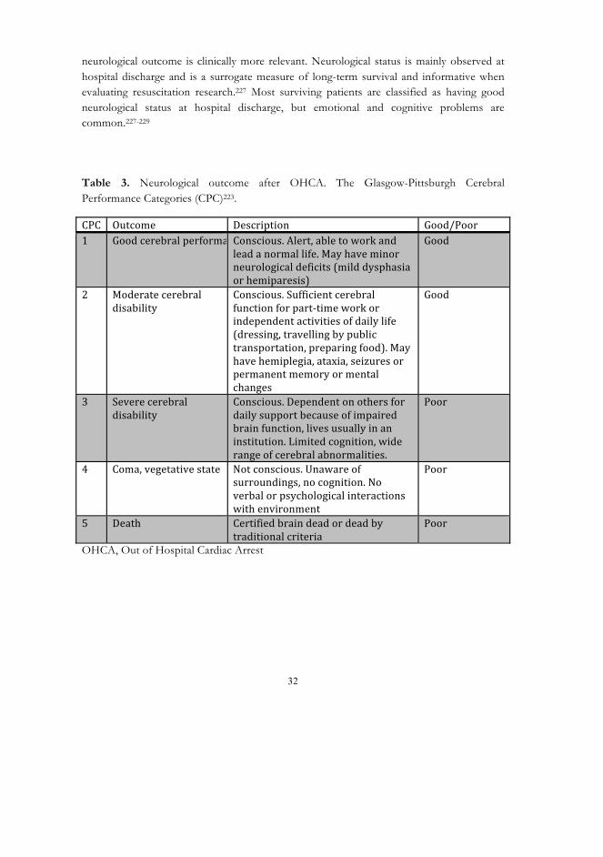
32
neurological outcome is clinically more relevant. Neurological status is mainly observed at hospital discharge and is a surrogate measure of long-term survival and informative when evaluating resuscitation research.227 Most surviving patients are classified as having good neurological status at hospital discharge, but emotional and cognitive problems are common.227-229
Table 3. Neurological outcome after OHCA. The Glasgow-Pittsburgh Cerebral Performance Categories (CPC)223.
OHCA, Out of Hospital Cardiac Arrest

33
2.5.3 Factors associated with outcome variation Good Survival rate is highly dependent on pre-hospital key predictors of survival (witnessed CA, bystander CPR, initial rhythm, ROSC) despite the quality or treatments in ICU care1. Reported survival vary between countries and there is also regional variation among hospitals treating CA patients230-233. Regional variation in CA patients and outcome is also reported in Finland5. There is some evidence that in larger hospitals that take care of more than 50 CA patients per year, survival rate would be higher than in hospitals admitting less than 20 patients per year233. In one study this difference between hospital size and survival disappeared when the results were adjusted for patient related factors234. Some studies also report an association between survival and transport and treatment in so called cardiac arrest centres234-238. There is no clear definition of a cardiac arrest centre and there is inconsistency in the services needed for the cardiac arrest centre status. General consensus is that facilities provide TTM and cardiac catheterisation laboratory accessible 24/7. Implementation of post resuscitation protocol, including post resuscitation care with TH and invasive coronary interventions, has shown to improve survival in historic control studies83,95,126,239. Improved survival has been reported in large hospitals with cardiac catheter facilities compared with smaller hospitals without these facilities233,235 and a combination of early coronary intervention and TH is also associated with favourable outcome240.
2.5.4 Outcome prediction The Sequential Organ Failure Assessment score (SOFA)241 and Acute Physiology and Chronic Health Evaluation II (APACHE II) 242 are widely used disease severity scorings in ICU care. The SOFA score describes the severity of organ failures (Table 4) and predicts mortality in medical or surgical ICU patients243. The SOFA score is not available at admission time and not useful or for CA patients, because a SOFA score includes points for level of consciousness, which gives maximal points for comatose CA patients. Some studies have used SOFA subscores244 and extra-cerebral SOFA scores, excluding the neurological component of SOFA for outcome and organ failure prediction70. Extra cerebral SOFA has been associated with hospital mortality mainly due haemodynamic instability70. APACHE II has been studied in CA patients, but it seems to be a poor predictor of outcome for OHCA patients245,246.

34
Table 4. The Sequential Organ Failure Assessment (SOFA) score.
μ
μ
* Not included in extra cerebral SOFA scores
Prognostic scoring systems, using data available at hospital admission for early outcome prediction have been also developed for OHCA patients247,248. These scoring systems use initial rhythm, age, time from collapse to BLS (no-flow time), time from BLS to ROSC (low-flow time), location of cardiac arrest, epinephrine dose, arterial pH, serum creatinine and lactate values for scoring calculations. The OHCA-score was developed using data from 130 patients mainly treated without temperature control but validated with 210 patients mainly treated with TH. The OHCA-score predicted poor neurological outcome with good accuracy (AUC 0.88), but later clinical studies could not repeat the results183,245. The OHCA-score has also been criticized for too low specificity to clinical use249. To ensure that all patients with potential favourable outcomes are treated, specificity should be close to 1 with a tight 95% CI. The recently published CAHP (Cardiac Arrest Hospital Prognosis)-score predicted poor outcome with good accuracy (AUC 0.93) and a CAHP-score over 200 points predicted poor neurological outcome with very high specificity (96-100%) for poor neurological outcome248. Brain injury is the leading cause of death in CA patients and prognosis is significantly related

35
to the severity of brain injury, but also a pessimistic prognosis leads to the withdrawal of treatment and death12,250. An ideal prognostication tool should predict poor outcome with 100% specificity and its false positive rate should be zero. But since such a prognostic tool for poor outcome is unavailable, multimodal prognostications, including clinical signs, biomarkers, imagination and neurological tests are recommended for all patients to predict poor outcome and possible withdrawal of intensive care178. The latest ERC guidelines adapted the prognostication strategy algorithm from this advisory statement178 and recommends an algorithm for all comatose CA patients after 72 hours from ROSC19.

36

37
3 AIMS OF THE STUDY
The main aims of this study were:
1. To evaluate the incidence of adult OHCA-patients treated in Finnish ICUs (I)
2. To evaluate the use of therapeutic hypothermia and its possible associations with one-year neurological outcome after OHCA (I)
3. To study the association between partial pressures of oxygen and carbon dioxide during the first 24 hours of intensive care and one-year neurological outcome after OHCA (II)
4. To evaluate the ability of biomarkers IL-6, hs-CRP and S-100B to predict subsequent severe organ dysfunction and one-year neurological outcome after OHCA from shockable rhythm (III)
5. To evaluate the ability of hs-TnT to predict mortality and one-year neurological outcome in OHCA patients with shockable rhythm (IV)
38
4 PATIENTS AND METHODS
4.1 Patients
This study included 548 adult patients resuscitated after out-of-hospital cardiac arrest and treated in 21 ICUs. All the patients were from the prospective, nationwide, observational FINNRESUSCI study, which included all adult patients treated in ICU after OHCA in Finland during the one-year study period, 1st March 2010 to 28th February 2011. In total 21 out of 22 ICUs treating adult cardiac arrest patients participated in this study. Approximately 98% of the whole Finnish adult population (4 290 980 at 31/12/2010) live in the referral areas of these ICUs. The study protocol was approved by the Ethics Committee of the Department of Surgery in Helsinki University Hospital and by the Ethics committee of each participating hospital when necessary. Written informed consent was given by the patient or next of kin for all patients for blood samples and for one-year neurological outcome.
The inclusion criteria for the FINNRESUSCI study were:
1. Out-of-hospital cardiac arrest 2. Successful resuscitation 3. Age over 18 years 4. Post-resuscitation care in ICU
Study I included all FINNRESUSCI study patients (n=548). Study II included a total of 409 patients. All these patients despite the initial rhythm were mechanically ventilated, comatose at ICU admission, ABG measured and recorded in the database and outcome data available 12 months after cardiac arrest. Study III included 186 patients resuscitated from shockable initial rhythm (VF/VT) and with blood samples available from the first 24 hours from ICU admission. Study IV included 155 patients resuscitated from VF/VT and with blood samples available from the first 6 hours from ICU admission. Table 5 presents the number of patients and inclusion criteria for each study and a flowchart of the study

39
patients is presented in Figure 3.
Table 5. Numbers of patients in Studies I-IV
ABG, Arterial Blood Gas; VF, Ventricular fibrillation; VT, Ventricular Tachycardia; ICU, Intensive Care Unit

40
Figure 3. Flowchart of FINNRESUSCI study patients, study populations and exclusion criteria for all the studies (I-IV).

41
Table 6. Characteristics of patients included in Studies I-IV
∗ ∗∗∗ ∗∗
Values are presented as numbers (percentages) or median (interquartile range, IQR); SAPS, Simplified Acute Physiology Score; APACHE, Acute Physiology and Chronic Health Evaluation; CPR, cardiopulmonary resuscitation; ROSC, Return Of Spontaneous Circulation; CA, Cardiac Arrest; ICU, Intensive Care Unit; PCI, Percutaneous Coronary Intervention; LOS, Length Of Stay, CPC; Cerebral Performance Categories; VF, Ventricular Fibrillation; VT, Ventricular Tachycardia; ASY, asystole, PEA, Pulseless Electrical Activity; *Neurological outcome (CPC) missing in 9 (1.8%) patients (3 VF/VT, 6 ASY/PEA)

42
4.2 Study design
4.2.1 Study I
This study included all patients from the FINNRESUSCI cohort. The main aims of the study were to report the incidence of OHCA patients in intensive care units and to evaluate the post-resuscitation care, the use and implementation of therapeutic hypothermia and one-year neurological outcome after OHCA in ICUs in Finland.
The number of Finnish adult population was obtained from Statistics Finland and the FICC database was used for ICU admissions and demographic data. All study patients were included in the incidence calculations, but only patients who were unconscious at ICU admission were included in the final outcome analysis.
4.2.2 Study II
This study assessed all arterial blood gas values during the first 24 hours after ICU admission and evaluated the possible associations of mean and time-weighted oxygen and carbon dioxide partial pressures to one-year neurological outcome during the first 24 hours in intensive care. Arterial blood gas values included in this study were collected only from comatose and mechanically ventilated patients. Pressure values of PaO2 and PaCO2 were defined into four different ranges prior to analysis. The mean 24-hour PaO2 and PaCO2 values and the proportion of time spent in different oxygen and carbon dioxide categories during the first 24-hour were calculated.
4.2.3 Study III
In this study magnitudes of inflammatory response and brain injury represented by plasma concentrations of IL-6, hs-CRP and protein S-100B were measured during ICU care. This study evaluated the levels of measured biomarkers and their associations with the duration of ischemia and ability to predict subsequent organ dysfunction and one-year neurological outcome. This study was designed to include only patients resuscitated from shockable initial rhythm to achieve a homogenous study cohort.

43
4.2.4 Study IV
This study investigated the prognostic value of hs-TnT levels according to hospital mortality and neurological status and mortality after 1 year from cardiac arrest. The correlations of admission hs-TnT value to time to ROSC, acute coronary occlusion and pre-existing diseases were also analysed. This study was also designed to include only patients resuscitated from shockable initial rhythm to achieve a homogenous study population due the cardiological aspect of the study design.
4.3 Data collection
Study data were collected from the Finnish Intensive Care Consortium (FICC) prospective routine database. The additional data of the study were collected prospectively by using Internet based study specific case report forms (CRFs) from three different time periods; pre-hospital event, daily during ICU care for the first five days and overall ICU care at the time of ICU discharge. Daily data collection was terminated if the patient was discharged from ICU before day five.
All the participated 21 ICUs belong to Finnish Intensive Care Consortium (FICC) and to the FICC database, which was originally used for benchmarking purposes and currently handled by Tieto Healthcare & Welfare Ltd. Data recorded in the routine database includes the reason for ICU admission, patient demographics, APACHE II admission diagnosis, International Classification of Diseases 10th revision diagnosis, ICU severity scores (SAPS II, SOFA, TISS), length of stay, and outcome measures (ICU- and hospital mortality). In addition to database routine records, the data from ventilators, patient monitors and laboratory systems were automatically transferred to the study database via the clinical information systems. 20 ICUs use electronic data management systems and the same data validation software (Web Validator, Tieto, Helsinki, Finland). All the participating ICUs kept logs of all CA patients admitted to ICU and these logbooks were used to crosscheck the number of patients, combine the data of patients treated in more than one ICU during the same hospitalization and separate in-hospital CA patients from OHCA study patients.
The study specific CRFs were developed to complete the data from the database. An ICU physician filled the pre-hospital form at ICU admission from the basis of pre-hospital medical report. The ICU physician and/or nurse filled the ICU CRFs daily forms for five days and at ICU discharge. Data collected with CRF comprised data from chronic health

44
status, pre-existing diagnoses of the heart, lungs or metabolic diseases, and present status of performance. These were obtained from the medical history of each patient.
Outcome data were collected from the FICC database, (ICU- and hospital mortality), and long-term mortality data up to 12 months from Statistics Finland. Neurologic outcome data according to the Pittsburgh Cerebral Performance Categories (CPC) classification were collected by structured telephone interview by one specialist in neurology after 12 months from cardiac arrest. Data from laboratory analyses were connected later to the data from the database to create different study cohorts.
4.4 Blood samples
After written informed consent from the patients or their next of kin, blood samples were collected from the study patients at ICU admission (0-6 h), 24 h, 48 h, and 96 h after ICU admission. Plasma samples collected to heparin tubes and serum samples to EDTA tubes, were kept in room temperature for 30-60 minutes before being centrifuged at 2200 G for 10 minutes. Samples were handled and frozen to a minimum of −20°C in the participating hospitals, and transferred in the frozen form to Kuopio University Hospital, where they were frozen to −70°C. All the samples were thawed at the same time before analysis.
Routine blood samples were obtained from each patient according to local ICUs standard operational protocols and the treating physicians’ decisions and part of the results such as for example, arterial blood gases were transferred to the study database via the clinical information system.
4.5 Measurements of biomarkers
Blood samples were collected at ICU admission (0-6 h) and 24 h, 48 h and 96 h after ICU admission. IL-6 and hs-CRP samples were analysed for all time points, despite missing admission samples, if the 24 h sample was available. Hs-TnT and S-100B samples in later time points were analysed only if admission samples were available. The analyst was blinded to patient information for all the laboratory measurements.
4.5.1 Interleukin 6 and hs-CRP
IL-6 was measured with a commercially available sandwich-type Enzyme Linked

45
Immunosorbent Assay (ELISA) following the manufacturer’s instructions (R&D Systems, Minneapolis. MN. USA). The sensitivity of the assay was 0.7 ng/l. Reference values for IL-6 is < 5.9 ng/l for all patients. The ELISA method had a measurement range of 0.5 to 300 ng/l. Concentrations above the upper limit were not diluted, but given a value of 300 ng/l in the statistical analyses. CRP was measured with a Cobas 6000 automated analyser with reagents (CRP and hs-CRP) from Roche Diagnostics (Penzberg, Germany). Reference values for hs-CRP are 0.05-2.5 mg/l for men and 0.05-3 mg/l for women. The analyses were performed at the routine laboratory of the Eastern Finland Laboratory Centre at the University Hospital of Kuopio, Finland.
4.5.2 Protein S-100B
Measurements of S-100B were performed with an automatic immune analyser (Cobas 8000, e602, Roche Diagnostics). Reference values for S-100B are < 0.11 μg/l for all patients. The total assay variation was less than 2.5%. The analyses were performed at the University Hospital in Uppsala, Sweden.
4.5.3 High-sensitivity troponin-T
Troponin T in serum samples was measured by the Elecsys TNT hs STAT assay (Roche Diagnostics, Penzberg, Germany). The hs-TnT assay has an analytical measurement range 3-10000 ng/L and the 99-percentile in the healthy population is 14 ng/L. Samples with concentrations above the upper limit were diluted before they were re-analysed. The analyses were performed at the Akershus University Hospital in Lørenskog, Norway.
4.6 Disease severity scorings and definitions
The severity of disease was described by APACHE II242 and SAPS II251 scores, which were calculated after 24 hours ICU admission. These scores were used mainly for demographic purposes to compare different subgroups in the ICU (I-IV). A modified APACHE II score, excluding points for oxygenation, was assessed for demographic purposes and statistical analysis (II). Sequential organ failure assessment (SOFA)241 scores (Table 4) were also calculated daily to define multiple organ dysfunctions (MODS), but only the first value (24 h) and SOFA-subscores were used for this purpose later in the analyses (II, III). An extra cerebral SOFA-score, excluding points for the neurological component, was also used to

46
define MODS (III)70. The infection status, aspiration, pneumonia and sepsis, during ICU care were defined by the treating physicians and local practice in every hospital (I-IV).
4.7 Outcome measures
4.7.1 Neurological outcome
Primary outcome in all studies were long-term neurological outcome determined 12 months after cardiac arrest according to the Pittsburgh Cerebral Performance Categories (CPC)223. One specialist in neurology, who was blinded to incident, treatment and pre-hospital management or during ICU care, contacted all surviving patients, who were not lost to follow up, by telephone and made a structured interview to determine the neurological outcome. Poor neurological outcome was defined as CPC 3-5 and good neurological outcome as CPC 1-2 (Table 3).
4.7.2 Mortality
ICU and hospital mortality data, which represent the short-term outcome, were obtained from the FICC database and 90-day and 12-month mortality from the Finnish Population Register Centre using the social security number of each study patient.
4.8 Statistical methods
Categorical data are presented as absolute numbers and percentages, and continuous data as median values with interquartile ranges (IQR, 25th-75th percentiles). Categorical variables were compared by Pearson´s chi-square test or Fisher´s exact test when appropriate. Kolmogorov-Smirnov was used to assess non-normal distribution of continuous variables, the Mann-Whitney U-test for comparing group differences and Kruskall-Wallis when comparing distributions between more than two groups. The first measured partial pressures of O2 and CO2 were considered to represent values from admission to the first measurement time point. Time intervals between PaO2 and PaCO2 were calculated and measured values assumed to remain constant until the next time point of measurement. These values and time intervals were used for calculations for time spent in different O2 and CO2 categories (II).
47
To evaluate the prognostic values of measured biomarkers, receiver-operating characteristics (ROC) curves and areas under curves (AUC) were calculated with 95% confidence intervals (III, IV). When comparing biomarker levels in different outcome groups, repeated measures analysis of ANOVA was used after adjusting for non-normal sphericity and the Spearman rank correlation factor was used for correlation calculations between variables (III). Multivariable logistic regression analysis were constructed and used to evaluate odds ratios for independent factors associated with neurologic outcome (I-IV), organ dysfunction (III) and mortality (IV) and to evaluate the additive predictive power of biomarkers, the category free net reclassification index (NRI) was calculated (III). The Wilcoxon matched-pairs signed-rank test was used to assess changes biomarker levels between different time points (IV). The Hosmer-Lemeshow goodness of fit test was used to assess the calibration of models created (II) and propensity analysis was performed and used to compare two separate groups (I). A p-value of <0.05 was considered significant in all analysis (I-IV). The statistical analysis was performed using IBM SPSS statistics version 19-20 (SPSS Chicago, Ill., USA), Graph Pad Prism version 6.0 and R version 3.0.1 for Windows (R Foundation for Statistical Computing, Vienna, Austria) and MedCalc for Windows, version 12.1.4.0 (MedCalc Software, Mariakerke, Belgium)

48
5 RESULTS
5.1 Incidence (I)
The total number of ICU admissions to the participating ICUs during the 1-year study period was 17 540. Cardiac arrest was recorded as the cause for ICU admission in 829 (4.7%) cases, which includes 548 individual adult OHCA patients (Fig 2). The population-based incidence of OHCA treated in ICU, using the numbers of adult inhabitants in the participating hospital districts as a reference population, was 13/100 000/year. Of those 548 patients, 311 patients (56.8%) were resuscitated from shockable initial rhythm (VF/VT) corresponding to an incidence of 7.4/100 000/year and 237 patients (43.2%) had a non-shockable initial rhythm, (asystole or PEA), corresponding to an incidence of 5.6/100 000/year. (I).
5.2 Temperature management (I)
Out of total 548 patients 44 (8.0%) patients were reported to be conscious and 504 (92.0%) were unconscious at ICU admission and therefore considered for therapeutic hypothermia (TH). TH was induced to a total of 311 patients comatose at ICU admission, 241/281 (85.8%) patients resuscitated from shockable rhythms and 70/223 (31.4%) patients resuscitated from non-shockable rhythms. The distribution of the patients divided by initial rhythms and use of TH is presented in (Fig 4).

49
Figure 4. Distribution of FINNRESUSCI patients and use of TH divided by initial rhythm.
OHCA, Out of Hospital Cardiac Arrest; VF, Ventricular Fibrillation; VT, Ventricular Tachycardia; ASY, asystole; PEA, Pulseless Electrical Activity; TH, Therapeutic Hypothermia

50
Endovascular cooling devices were used for hypothermia induction and maintenance in 247 (79.4%) and surface cooling devices in 58 (18.6%) cases. Five patients were cooled with other surface cooling methods and one patient with only ice-cold intravenous fluids. Ice-cold intravenous fluids were also used as a start of induction in the pre-hospital setting or as an additional method for cooling devices in 50 (16.1%) patients. The target temperature was set to 33ºC in 300 (97%) patients and temperatures of 32-34ºC were achieved in a median of 111 (70-180) minutes from the start of hypothermia induction. Targeted temperature maintenance time was 24 hours in 231(74.3%) patients (range 0-47 h) and the median (IQR) rewarming time from hypothermia to normal temperature was 9 (6-12) hours. Reasons for withholding TH in unconscious patients resuscitated from VF/VT (40) were based on clinical grounds and decisions according to the current guidelines (Table 7).
Table 7. Reported reasons from treating ICU physicians for withholding therapeutic hypothermia (TH) from out-of-hospital cardiac arrest (OHCA)-VF/VT patients who were comatose at ICU admission.
ICU, Intensive Care Unit; VF, Ventricular Fibrillation; VT, Ventricular Tachycardia; ROSC, Return Of Spontaneous Circulation; *Reporting multiple reasons was possible in individual patients.
The total number of patients across each participating study ICU varied between 2 and 74 patients/year and the use of TH varied between 32% and 100% of all OHCA patients and between 46% and 100% with OHCA-VF/VT (Table 8).

51
Table 8. The numbers of out-of-hospital cardiac arrest patients, distribution of initial rhythms and therapeutic hypothermia treatments in individual FINNRESUSCI study units (Vaahersalo et al., unpublished results).
ICU, Intensive Care Unit; VF, Ventricular Fibrillation; VT, Ventricular Tachycardia; TH, Therapeutic Hypothermia.

52
5.3 Blood gases (II)
Arterial blood gases (ABG) were measured, electronically recorded and validated in the database from a total of 468 patients. Of these, 409 patients fitted the inclusion criteria (comatose at admission, mechanically ventilated and one-year neurological outcome data available) and constituted the study cohort for ABG analysis during the first 24 hours of ICU care (Fig 2). The median amount of ABG measurements/patient was eight (IQR 6-11).
5.3.1 Oxygen
Hyperoxia exposure, defined by arterial oxygen (PaO2) values higher than 40 kPa (300mmHg), was found in 24 (6%) patients. Mean FiO2 was 46%, median (IQR) 41% (37-51), during the first 24 h of ICU care and there was no different in patient group with good or poor outcome. The proportions of time in different oxygen ranges are presented in Figure 5. PaO2 values were higher in patients with good outcome than those with poor neurological outcome (Table 9), but the mean PaO2 tension did not associate with better outcome OR 1.006 (95% CI, 0.998-1.014) and multivariable analysis showed no association between time spent in different PaO2 levels and outcome.

53
Figure 5. Proportion of mean times (SD) in different oxygen categories in mechanically ventilated OHCA patients during the first 24 h in ICU care. 1 mmHg = 7.5 kPa.

54
Table 9. Median values (IQR) of arterial PaO2 during the first 24 h in ICU after OHCA divided by one-year neurological outcome.
Values are presented in mmHg = 7.5 kPa; IQR, Interquartile range; ICU, Intensive Care Unit; OHCA, Out-of-Hospital Cardiac Arrest
5.3.2 Carbon dioxide
The mean arterial carbon dioxide (PaCO2) values during the first 24-hour did not differ between good and poor neurological outcome groups but the mean PaCO2 value predicted independently good outcome with OR 1.054 (95% CI; 1.006-1.104, p=0.027). Also the time spent over 6 kPa (45 mmHg) associated with good neurological outcome after 12 months in multivariable analysis OR 1.015 (95% CI; 1.002-1.029, p=0.024). This finding persisted in sensitivity analysis, when only TH treated patients were included in the analysis. Patients with the highest mean PaO2 and PaCO2 values in tertiles were found to have better outcome than predicted. The OR for good outcome was 3.2 (95% CI; 1.1-9.2, p=0.033) and 6.2 (95% CI; 1.3-27.5, p=0.019) when including only TH treated patients.

55
5.4 Biomarkers after cardiac arrest
5.4.1 IL-6 and hs-CRP (III)
Elevated levels of serum IL-6 and hs-CRP demonstrate the inflammatory response after cardiac arrest and levels changed over time in all patients. IL-6 was already elevated at ICU admission and reached the highest values at 48 hours, while hs-CRP elevated later and continued to increase until 96 hours after ICU admission (Fig 6). Admission values of IL-6 were higher in patients with poor neurologic outcome and associated over time with ROSC and subsequent organ dysfunction (p<0.001), while hs-CRP values were not. Admission IL-6 predicted poor neurological outcome with AUC 0.711 and extra cerebral organ dysfunction with AUC 0.679. Admission values of IL-6 and associations with extra cerebral SOFA scores and neurological outcome are presented in Figure 7.
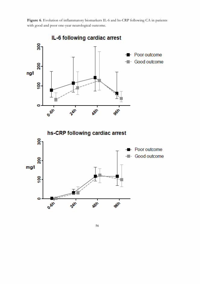
56
Figure 6. Evolution of inflammatory biomarkers IL-6 and hs-CRP following CA in patients with good and poor one-year neurological outcome.

57
Figure 7. Admission value of IL-6 according to one-year neurological outcome and extra-cerebral SOFA-score.
In multivariate logistic regression analysis, (including time to ROSC, age, history of coronary artery disease (yes/no), awake (yes/no), witnessed CA (yes/no), and admission values IL-6 and S-100B), only time to ROSC, age and admission IL-6 were independently associated with poor neurological outcome (Table 10). By adding IL-6 and S-100B to the basic variable model with AUC 0.820 (including age, time to ROSC, history of coronary artery disease (yes/no), awake (yes/no), the AUC improved to 0.842, but only adding IL-6 was statistically significant, NRI was 0.5846 (95% CI 0.2778-0.8914, p=0.0019).

58
Table 10. Independent predictors of poor neurological outcome (CPC 3-5) in multiple logistic regression analysis
CPC; Cerebral Performance Category, OR; Odds Ratio, CI; Confidence Interval, IL-6; Interleukin-6
5.4.2 S-100B (III)
S-100B values decreased over time in all OHCA patients, but the values were higher in patients with poor outcome (p<0.001). S-100B associated also with a longer time to ROSC (p=0.002), and the AUC to predict extra cerebral organ dysfunction for admission value of S-100B was 0.574. The corresponding AUC values for S-100B to predict poor neurological outcome were 0.699 at admission, 0.693 at 24h, 0.700 at 48h and 0.712 at 96h and S-100B did not predict outcome independently in the multivariable analysis (Table 10). By adding S-100B to the basic variable model with AUC 0.820 (including age, time to ROSC, history of coronary artery disease (yes/no), awake (yes/no), the AUC improved to 0.830, but it did not improve the model statistically significantly, NRI was 0.3 (95% CI -0.0017-0.6017, p=0.051).

59
Table 11. Values of S-100B on admission and at 24h divided by one-year neurologic outcome and organ dysfunction, defined by CPC extra cerebral SOFA score.
CPC; Cerebral Performance Category, SOFA; Sequential Organ Failure Assessment
5.4.3 Hs-TnT (IV)
Admission hs-TnT was analysed from 155 OHCA patients, 152 (98%) resuscitated from VF and 3 (2%) from VT. The numbers of samples analysed in later time points were 150 at 24h, 138 at 48h and 107 at 96h after ICU admission. The median (IQR) level of admission hs-TnT was 415 (199-916) ng/l, with a range of 18-17837 ng/l, indicating significant increase in all successfully resuscitated patients admitted to ICU. Admission hs-TnT levels correlated positively with time to ROSC (r=0.47, p<0.001) and the median value (IQR) of admission hs-TnT among patients with evidence of acute coronary occlusion (n=15) was significantly elevated 1.497 (753-8875) ng/l compared to 387 (182-815) for all other patients (p<0.001). Admission hs-TnT levels, median (IQR), were higher in patients with poor one-year neurological outcome vs. good neurological outcome 739 (191-1061) ng/l vs. 334 (195-716) ng/l (p=0.028) and one-year non-survivors compared to survivors, 747 (206-1061) ng/l vs. 345 (184-740) ng/l (p=0.023), but there was no statistical difference between hospital mortality groups. Reduction of hs-TnT levels from admission to later time points was demonstrated in most patients, but there were no differences in hs-TnT dynamics according to mortality or outcome. In the ROC curve analysis, admission hs-TnT is associated with one-year mortality (AUC 0.61) and one-year neurological outcome (AUC 0.60), but did not have the same influence on hospital mortality (AUC 0.59). Hs-TnT levels measured after 24 hours from ICU admission did not improve the predictive power of admission hs-TnT. After the first 24h when the Simplified Acute Physiology Score (SAPS) was available, SAPS II predicted outcome more accurately (Table 12). Admission hs-TnT levels were not associated with

60
hospital mortality OR 1.25 (95% CI 0.95-1.64, p=0.12), one-year mortality or neurological outcome OR 1.26 (95% CI 0.97-1.63, p=0.008) in the logistic regression analysis, while several other previously known risk variables (age, time to ROSC, history of coronary artery disease or heart failure) associated with mortality and neurological outcome. Table 12. Prognostic values of SAPS II score and hs-TnT values measured < 24 h from ICU admission as assessed by ROC analysis.
SAPS, Simplified Acute Physiology Score; ICU, Intensive Care Unit; ROC, Receiver Operating Characteristic; CPC, Cerebral Performance Categories; AUC, area under the curve; CI, confidence interval
5.5 Outcome (I)
Out of 548 ICU treated OHCA patients, 234 (42.7%) were alive at 12 months after CA. Out of comatose (504) patients, 12-month outcome data were not available for 9 (1.8%) patients and total of 13 (2.4%) patients were lost to follow up. Overall ICU mortality was 21.7%, hospital mortality 45.3% and 90-day mortality 51.3%. Median (IQR) length of stay (LOS) in ICU was 2.6 (1.5-4.2) for all patients and was significantly longer in patients treated with TH 3.3 (2.4-5.8) than in patients treated without TH 1.2 (0.8-1.9). Length of stay, mortality and neurological outcome of all FINNRESUSCI patients, comatose and conscious, are presented in Table 13.

61
Table 13. Outcome of FINNRESUSCI patients
∗∗∗∗
ICU, Intensive Care Unit, LOS, Length Of Stay; IQR, Interquartile Range; CPC, Cerebral Performance Categories; *Neurological outcome (CPC) missing in 13 (2.4%) patients
5.5.1 OHCA patients with shockable and non-shockable rhythms
Out of 281 OHCA patients resuscitated from VF/VT and unconscious at ICU admission, 147 (52.9%) had favourable one-year neurological outcomes (CPC 1-2). Of those patients 138 were treated with TH, representing favourable outcomes in 57.3% (138/241) patients. Of the comatose VF patients treated without TH, 22.5% (9/40) survived with good neurology. In patients resuscitated from ASY/PEA, the proportion of good neurological outcome was 19.4% (13/70) in patients treated with TH and 16.0% (24/153) without TH respectively. The proportion of good neurological outcomes in ASY/PEA patients was less than in VF/VT. TH treatment did not improve outcome in patients resuscitated from non-shockable rhythms. Only age and higher APACHE II score, but not the use of TH was not associated with neurological outcome in this group of OHCA patients. Length of stay, mortality and neurological outcomes of unconscious patients according to initial rhythm and TH treatment, are presented in Table 14.

62
Table 14. Outcome of unconscious OHCA patients (n=504) divided into initial rhythms and use of TH groups
Values are presented as numbers (percentages) or median (interquartile range, IQR); OHCA, Out-of-Hospital Cardiac Arrest; TH, Therapeutic Hypothermia; VF, Ventricular Fibrillation; VT, Ventricular Tachycardia; PEA, Pulseless Electrical Activity; LOS, Length of Stay; ICU, Intensive Care Unit; CPC; Cerebral Performance Categories, *Neurological outcome (CPC) missing in 9 (1.8%) patients. (3 VF/VT, 6 PEA/asystole),
The patients were treated in 21 different ICUs and the number of patients in each study ICU varied between 2 and 74 patients. Hospital mortality varied between 18 and 71% for individual study sites, but there was no correlation between ICU size or number of treated patients in ICU and hospital mortality or outcome. One-year neurological outcomes of each ICU are presented in Table 15.

63
Table 15. One-year neurological outcomes of OHCA patients divided by initial rhythms in individual FINNRESUSCI ICUs (Vaahersalo et al., unpublished results)
Values are presented as numbers (percentages); OHCA, Out-of-Hospital Cardiac Arrest; ICU, Intensive Care Unit; CPC, Cerebral Performance Categories; *Representing number of missing patient for one-year neurological outcome data

64
6 DISCUSSION
6.1 Incidence of OHCA (I)
The incidence of OHCA patients in ICU in this study (13/100 000/year) is in agreement
with a previous Finnish study by Oksanen at al. reporting an incidence of 15/100 000 inhabitants/year33. The incidence is also in agreement with previous Finnish studies reporting the overall incidence of OHCA5,26,30,31,252, while the reported rate of ROSC and survival to hospital is 30-35% in Scandinavia5,26,32. Of those OHCA patients who survive to hospital admission, only selected patients are treated in ICUs26,32. Local protocols, hospital resources and optional units for OCHA patients affect the selection of patients for ICU care, when intensive care is unlikely to benefit the patient. In some hospitals nearly all OHCA patients are treated in ICU despite pessimistic prognoses. In this study 56.8% of patients were resuscitated from shockable initial rhythm, but a variation between participating ICUs and portion of VF patients (0%-91%) existed. The number of OHCA-VF patients also varied between university hospital ICUs (50%-91%), which is comparable with other single hospital studies (65%-90%) reporting the results for OCHA patients treated with therapeutic hypothermia32,83,127.
The type of EMS system mostly explains the incidence of VF, because it is likely that
most CA patients have shockable rhythms at the time of collapse, but by the time of the first ECG, these rhythms have deteriorated to PEA or asystole2,253,254. It has been shown that if the rhythm is recorded soon after collapse, with public access defibrillators or EMS personnel on site, the proportion of VF can be as high as 76%42,255. So the time to first ECG recording and possible defibrillation, usually by EMS personnel, affects the VF incidence and also the portion of VF patients in ICU. In this study, the highest proportion of VF patients were in ICUs covering the largest cities and urban areas, and not hospitals in the rural area where response times of EMS systems can be assumed to be longer5. The proportion of VF as the initial rhythm has also decreased over the last few years28,30,256,257. Improved care of coronary artery disease and prevention have been suggested to explain this decrease258-261. VF decrease has been shown to be higher among patients with unwitnessed OHCA who collapsed at home257, which may also lead to differences in the numbers of VF patients between ICUs.

65
6.2 Temperature management (I)
Therapeutic hypothermia, temperature control or targeted temperature management has been in clinical practice in every ICU for over a decade in Finland33,95. ILCOR issued an advisory statement recommending TH for all OHCA patients resuscitated from VF already in 200316, and according the ERC Guidelines for Resuscitation, TH has been recommended for OHCA VF patients since 2005125. In this study nearly 86% of the comatose VF/VT patients were treated with TH, thus the usage of TH in Finland has increased significantly from 200795. The nationwide use of TH has not been reported in the literature, but the proportion of TH-treated VF patients is one of the highest compared to previous reports from single hospital or registry studies83,129,131,239,262. TH or TTM is administered to practically all OHCA-VF patients in Finland, while withdrawal of TH from VF patients is based on clinical judgment decisions (Table 7).
The ILCOR advisory statement also suggested TH for all comatose CA patients chosen to intensive care in 2003, and this recommendation was adopted in the European guidelines later, by the Scandinavian clinical practice guidelines in 2009103, and by ERC for all CA patients in 201018. In this study 61.7% of unconscious patients were treated with TH, and 31.8% of non-shockable patients, which is a reasonable result, considering the fact that the ERC guidelines were published in October 2010 during the study period. Adherence to the guidelines concerning the use of TH has been reported for 79% of ERC Hypothermia cases in a cardiac arrest registry study from 19 sites in Europe129 and 70% in a study by Lindner et al. from Norway, where cooling has been suggested for all CA patients since 2004262. Patients were treated to a target temperature of 33°C in 97% and for 24 hours in 74% cases, according to guidelines of the time. Previous RCTs of TH from the beginning of the 20th century have some weaknesses and limitations13,14. The HACA study has been criticized for patients temperature in normothermic control group, which seems to be well over 37°C, meaning their temperatures were hyperthermia not normothermia 13 and the study by Bernard et al. has been called pseudo-RCT because of methodological reasons14. A recent RCT study of 950 OHCA patients randomized the patients to 36 hours of temperature control either at 33°C or 36°C17. This Targeted Temperature Management (TTM) trial reported no difference in neurological outcome or mortality between the groups treated at 33°C or at 36°C. Hyperthermia was prevented for 72 hours in both groups. Despite these results, the optimal temperature and duration of TH or temperature control is still unknown263. According to the ILCOR’s latest recommendation104, temperature should maintain constant between 32° and 36° for at least 24 hours for adults after OHCA with initial shockable rhythm. This statement reflects the latest ERC and ESICM

66
recommendations and guidelines264. These guidelines suggested that TTM should also be given to comatose adults after OHCA resuscitated from non-shockable rhythm and IHCA patients, despite the evaluation of literature by the GRADE methodology ending up with very low-quality of evidence19,104.
Endovascular cooling devices were mainly (79%) used for induction and maintenance of hypothermia in Finnish ICUs, which is also recommended in the very latest ERC guidelines for controlling temperature during TTM19. The internal devices enable better control for temperature than other techniques153,154, but there is no evidence for better survival with internal devices. Ice-cold fluids were used as an additional cooling method and in pre-hospital setting for 50 (16%) patients, which according the ERC’s latest guidelines19 is no longer recommended. Increased risk of re-arrest was reported during transport to hospital in three studies148,149,151. The largest and the latest of these studies also reported increased risk of pulmonary oedema in a pre-hospital ice-cold fluid cooling group (RR, 1.34; 95% CI, 1.15-1.57)151, and the no benefit of pre-hospital cooling has so far been shown. The temperature management complied very well with the current international guidelines during the study period and, except for the minor use of ice-cold fluids, the temperature management was also in agreement with recent international recommendations and guidelines19,104.
6.3 Blood gases (II)
6.3.1 Oxygen
The findings of Study II indicated that hyperoxia exposure is rare (6%) after CA in Finland. Additionally, hyperoxia was not associated with poor outcome, instead a combination of moderately elevated oxygen and CO2 levels were associated with good neurological outcome.
In studies reporting the association of hyperoxia with mortality, the prevalence of hyperoxia has been between 10% and 40%158,160,265,266, significantly higher than the prevalence in Study II. The prevalence of hyperoxia was also high (41%) during the first 24 hours in ICU in one single centre study, which also reported hyperoxia association with longer times to ROSC and delays to ICU admission and cardiac arrest location163.

67
Hyperoxia after CA has been shown to associate with increased mortality in large observational studies, when only single values of PaO2 were analysed - either the highest or the first value158,159. These studies did not control the severity of ischemic insult, PCAS or the ICU care process, which is a major limitation to these results, because hyperoxia is associated with longer time to ROSC and longer delays for ICU admission163. In studies where these factors were controlled, no associations with hyperoxia and mortality were found160,266. In Study II the mean values of PaO2 during the first 24 hours in ICU were used for analyses instead of single values. The mean PaO2 values were higher for patients with good outcomes, but the values did not predict outcome, neither did time spent in different PaO2 levels have any influence on outcome. However, the prevalence of hyperoxia exposure was so low, that it is impossible to rule out a harmful effect of hyperoxia on outcome based on these study results.
A recent registry study from the Netherlands comprising over 5000 patients reported a U-shaped relationship between partial pressures of O2 and hospital mortality after adjustment for confounding factors. Only hypoxia was independently associated with hospital mortality with an OR of 1.34 (95% CI 1.08-1.66) but not hyperoxia, OR 1.13 (95% CI 0.81-1.57)267. A recent study also reported the disadvantages of severe hyperoxia, but it also reported an association with improved organ function (defined by a SOFA score at 24 h) and moderate hyperoxia268. According to this study, the relationship between hyperoxia and outcome seems not to be linear or dose-dependent, but poor outcome may occur only after oxygen tension exceeds a certain threshold. The results of Study II showed a combination of mild hyperoxia with mild hypercarbia to associate with better than predicted neurological outcome. These results are in close agreement with the recent studies by Elmer and colleagues268 and Helmerhorst and colleagues267. The number of hyperoxia studies with CA patients are limited and include several confusing factors, so the clear effect of hyperoxia or the optimal levels of oxygen partial pressures are still to be determined161. While the effects of hyperoxia are still unknown, hypoxia is definitely harmful for CA patients158,160,267, which indicates that there is a need for careful monitoring of oxygen levels to avoid possible hypoxia.
6.3.2 Carbon dioxide
In Study II it was showed that the mean 24-hour PaCO2 value was an independent predictor of favourable one-year neurological outcome. Also time spent over 6 kPa (45 mmHg) of PaCO2 was associated with good one-year neurological outcome in our multivariable model.

68
There are very limited data on the relationship between concentrations of CO2 and outcome after CA and the available data give conflicting results. The positive association of mild hypercapnia in Study II is in close agreement with a large observational study containing over 16 000 patients, which reported an independent association between hypocapnia and hospital mortality, but an increased rate of patients discharged in a hypercapnia group compared to a normocapnia group, (OR 1.16)92. A large registry study from the Netherlands found a U-shaped relationship between hospital mortality and PaCO2 levels, but when compared to normocapnia, only hypocapnia was a statistically significant predictor for hospital mortality (OR 1.37)) and not hypercapnia (OR 1.1)267. Both these studies used only single ABG values, the worst oxygenation time in the first 24 h, in the analyses and robust short term outcome variables as an outcome endpoint in contrast to Study II, where the mean value of PaCO2 and one-year neurological outcome were used to reduce concern about limitations observed269 in a study by Schneider and colleagues92
The positive association of mild hypercapnia is in contrast with a single centre study from the USA, which reported both hypocapnia and hypercapnia being independently associated with poor neurological function, OR 2.43 (95% CI, 1.04-5.65) and 2.20 (95% CI, 1.03-4.71), respectively91. This study also used one ABG value to define patients to hypocapnia or hypercapnia groups at any time point during the first 24 h and the majority of patients were IHCA patients, which is major difference compared to Study II, which included only OHCA patients.
There are several possible explanations for the positive association between mild hypercapnia and one-year neurological outcome. Elevated PaCO2 levels have been shown to be associated with less brain damage in animal models by increasing blood flow and diminishing local brain hypoxia in brain ischemia166. Cerebral blood flow could be decreased after CA in the early phases of PCAS270 due the impairment of autoregulation or an imbalance between local vasoconstrictors and vasodilators. CO2 is a well-known vasodilator of cerebral veins and mild hypercapnia might reverse these abnormalities. The upper normal range of PaCO2 increased cerebral blood flow and decreased cerebral lactate compared to the lower normal range of PaCO2271. CO2 also has anticonvulsant effects171 and possible alkalosis in brains induces seizures in an animal model272. In contrast hypercapnia is against the guidelines in neurocritical care for traumatic brain injury patients as it results in increased intracranial pressure (ICP) and brain oedema273, which correlate with unfavourable outcome with traumatic brain injury (TBI) patients274. ICP is not routinely monitored in CA patients and its role is unclear in PCAS, but also hypocapnia may be harmful for TBI patients, causing vasoconstriction and ischemia52,275.
Hypocapnia and hypercapnia are common (18%-22%, 35%-41%) after CA during ICU care92,267 and thus, a ventilation strategy is a relevant intervention after CA and it might have

69
a great influence on outcome. Hyperventilation leads to hypocapnia, which causes cerebral ischemia165,173, which can be controlled using a mechanical ventilation strategy. According to the international guidelines, all CA patients should be treated with temperature control or TTM in ICU19. Therapeutic hypothermia decreases metabolism and reduces the production of CO2, which easily leads to hyperventilation in patients treated with TH176. Hyperventilation has been shown to reduce cerebral tissue oxygenation significantly during TH174 and an association between hypocapnia and poor neurological outcome has been documented in observational registry studies91,92.
To date there are no prospective and randomized studies to clarify the optimal ventilation strategy and the ERC guidelines recommend normocapnia after cardiac arrest19, but according to results of Study II and the literature it seems that hypocapnia is more dangerous than mild hypercapnia, which may be even beneficial after CA.
6.4 Biomarkers (III-IV)
6.4.1 IL-6 and hs-CRP
The findings of Study III showed that admission values of IL-6, but not of hs-CRP are associated with subsequent organ dysfunction and predict one-year neurologic outcome with an OR of 1.006 (95% CI 1.00-1.01/ng/l, p=0.046).
Many inflammatory biomarkers have been investigated in critical care patients180. In these studies high levels of IL-6 have been shown to correlate with organ dysfunction and mortality in sepsis190,191. Cardiac arrest, global ischemia and successful ROSC lead to generalized activation of a systemic inflammatory response and release a profile of inflammatory biomarkers and clinical symptoms after CA that are similar to those of sepsis66. The magnitude of this inflammatory response is the determinant factor in developing PCAS and its severity7. The severity of PCAS is dependent on the severity of ischaemic insult but the ischemia and activation of inflammatory response increases the risk of multiple organ dysfunction. Development of organ dysfunction after CA will worsen the patient´s outcome8 and multiple organ dysfunction, defined by the extra-cerebral SOFA score, after cardiac arrest is an independent predictor of hospital mortality70.
The magnitude of inflammatory response was demonstrated in Study III by elevated levels of IL-6 and hs-CRP in all OHCA-VF patients, but only IL-6 values were higher in

70
patients with poor one-year neurological outcome (p<0.001). This result is in close agreement with one relatively small study which reported that elevated plasma concentrations of IL-6 and hs-CRP correlated with patients outcome196. A similar release profiles of CRP have been shown in one paediatric study, CRP levels were elevated in both survivors and nonsurvivors after CA193. In Study III hs-CRP levels did not differ between outcome groups, this is in contrast to a study by Samborska-Sablik196. This study also included IHCA patients (25/46), which may explain the difference in hs-CRP results compared to Study III. Similar release profiles, the late release of hs-CRP and CRP and earlier release of IL-6 or procalcitonin have been reported in previous studies142,244.
The findings in Study III, especially the results for admission IL-6 are in close agreement with a study by Annborn et al.244. In this study high levels of procalcitonin (PCT) at 12 h correlated strongly over time with ROSC, correlation coefficient 0.64, and associated with circulation SOFA subscore, surrogate markers of PCAS. Respectively the admission IL-6, correlation coefficient to time to ROSC was 0.387 and high values associated with circulation and renal SOFA subscores. When comparing IL-6 and PCT as predictors for neurological outcome in ROC analysis, PCT showed high accuracy at 12 h and at 24 h after cardiac arrest with AUCs of 0.88 and 0.86. The corresponding AUC values for IL-6 were lower, being 0.71 at admission and 0.64, 24 h after admission. On the other hand, admission IL-6 was independently associated with poor outcome (OR 1.006/ng/l) along with time to ROSC and age in multivariable analyses (Table 10).
Therapeutic hypothermia may cause bias in biomarker studies, because it influences inflammatory biomarker release, suppressing the release of CRP, but neither IL-6 nor PCT were unsuppressed142. The majority of the patients (91%) in Study III were treated with TH and only TH treated patients were included in the sensitivity analysis to remove any possible bias of TH. The association was non significant (p=0.07 with TH treated patients (OR 1.005 ng/l)).
6.4.2 S-100B
Study III showed that S-100B values were higher at all time points in patients with poor one-year neurological outcome. S-100B does not have adequate predictive value for neurological outcome (AUCs 0.693-0.712) or subsequent multiple organ dysfunction with an AUC of 0.574 for admission S-100B.
S-100B and NSE are the most studied neurological biomarkers for predicting outcome after CA177. Several studies have found high levels of NSE or S100B in patients with poor neurologic outcome before the time of TH treatment187,198,276,277. Clinical implementation

71
and usage of TH has changed to roles of these biomarkers as clinical tools for outcome prediction181.
The Study III results concerning S-100B contrast with a recent study that reported superior predictive value for S-100B when compared to other neurological biomarkers in TH treated patients188. This study reported a peak sensitivity of 87% at 24 h with 100% specificity. The results of Study III are supported by a registry study by Zellner and colleagues205. In this study S-100B predictive value was reported to be comparably poor for S-100B at admission (AUC 0.67, the value for Study III was AUC 0.699) and at 24 h (AUC 0.74) and (AUC 0.693), respectively. A previous study from Finland181 with OHCA-VF patients treated with TH also reported a comparable prognostic value for S-100B at 24 h (AUC 0.65) to that of Study III. Studies by Mörtberg and Zellner included IHCA patients (11%-13%) in their analyses and in the study by Mörtberg a relatively small number of patients in contrast to Study III included only OHCA-VF patients. Despite the fact that all the patients were treated with TH, the heterogeneous study population may have influenced the outcome data.
An interesting finding of Study III was that despite S-100B levels being associated with time to ROSC, the levels did not associate with subsequent organ dysfunction, while admission levels of S-100B were significantly higher in patients with poor neurological outcome. It is probable that multiple organ dysfunction correlates with the magnitude of inflammatory response and neurological injury associates with outcome independently.
6.4.3 Hs-TnT
Study IV showed that hs-TnT levels were above the 99th percentile of normal values, being 14 ng/l for all OHCA-VF patients on ICU admission (range 18 to 17837 ng/l). Hs-Tnt values were higher among patients with unfavourable one-year neurological outcome and non-survivors, but hs-TnT did not give additional value for prognostication compared to the traditional model of well-known risk factors.
Cardiac troponins are in clinical use for diagnostic purposes for acute myocardial infarction (AMI)211. Elevated troponin levels have been shown to correlate to the prognosis for AMI patients278, but the relevant clinical additional information for OHCA patients is still missing177. Previous studies with troponins for CA patients mainly report that troponins are markers that can be used to diagnose acute coronary occlusion220,221,279-281, but no relationship between this ability and patient outcome has been reported. A study220 on admission hs-TnT reported no difference in ICU mortality between patients with or without acute coronary occlusion, despite median hs-TnT at admission being reported to be three

72
times higher in the coronary occlusion group (1184 vs. 351 ng/l). The median hs-TnT was at admission 551 ng/l (IQR 203-2551) for all the OHCA patients. These results are in agreement with Study IV, which reported a median Hs-TnT at admission of 415 ng/l for all patients, respectively in patients with or without acute coronary occlusion (1497 ng/l vs. 387 ng/l).
In Study IV several factors, but not admission hs-TnT, were associated with mortality or one-year poor neurological outcome, but only age and time to ROSC were independent predictors of poor outcome in multivariate analysis. The limited prognostic value of hs-TnT can be explained in many ways, but probably the main reason is the aetiology of deaths after CA. Brain injury is a well established cause of deaths, especially during follow up periods, while myocardial dysfunction is a minor cause of deaths (25%-35%) and mainly responsible for very early deaths after CA11,12. Since troponin T has been shown to be elevated by several other factors other than coronary occlusion, like chest compressions and defibrillations217,218,281, it can be assumed that hs-TnT is similarly affected. So increased levels of hs-TnT do not represent only AMI, but may be elevated due to transient reasons with the levels decreasing quickly. The reduction in hs-TnT levels were clearly seen in most patients in Study IV from admission to 24 h, but the dynamics did not correlate with the outcome. It is also very probable that 34 (22%) patients went to coronary angiography and those treated with PCI 15 (10%) in Study IV, were patients with other acute clinical signs of AMI (ST-elevation on ECG) and their coronary occlusions had been treated. Early revascularization after CA may have been prevented the following of myocardial dysfunction, which has been shown to be beneficial in many observational studies78,85,221,240. It seems that despite the fact that AMI is the main cause for CA and that hs-TnT and other troponins are able to identify coronary occlusion and AMI, it has no prognostic value for outcome prediction.
6.5 Outcome (I-IV) In this study one-year neurological outcome was the main outcome variable measured in all (I-IV) independent studies. Mortality was used as an outcome endpoint in Studies I and IV, while multiple organ dysfunction was used for an additional endpoint in Study III. Overall mortality and neurological survival are highly dependent on the study population. Patient related factors (age, previous state of health, cardiac arrest location, in- or out-of hospital) and resuscitation key predictors witnessed CA, bystander CPR, initial rhythm and delays in response times are related to survival, which makes studies and results hard to compare. In the meta-analysis studies from 1984 to 2008 reporting factors associated with outcome,

73
pooled rate of survival for hospital discharge in all OCHA patients across various populations and EMS systems was reported to be 7.6% (95% CI 6.7-8.4)1. According to the same study successful resuscitation and survival from pre-hospital scene to hospital admission was reported to be 23.4% (95% CI 21.1-26.6). The proportion of VF as an initial rhythm and short delays in the EMS systems in study populations are highly dependent on reported survival rates, because OR for survival in locations with EMS with response times less than 8 minutes was 5.9, compared to 2.4 in locations with response times over 8 minutes1. Because of the more heterogeneous aetiology of cardiac arrest and generally worse outcome in patients resuscitated from non-shockable initial rhythms, outcome is usually reported in OHCA-VF and non-VF patients separately. Studies III and IV included only OHCA-VF patients in their analyses and reported results only for this group of patients.
6.5.1 Outcome of OHCA-shockable patients (I) In Study I the hospital mortality of unconscious OHCA-shockable patients (34%) is in
agreement with previous studies15,32,83. The great majority (86%) of comatose OHCA-VF patients were treated with TH in Study I. The outcome results of Study I for VF patients treated with TH are comparable or even better than in previous studies13,131,136. In RCTs good neurological outcome of OHCA patients have been reported to be between 49%-55%13,14,17. In two large registry studies good neurological outcomes were found to be 44% in a study by Dumas and colleagues131 and 56% in a study by Nielsen and colleagues136 respectively.
A recent TTM-study randomized 950 OHCA patients to 36 hours temperature control either at 33°C or 36°C 17. In this trial no difference was found in neurological outcome or mortality between the groups treated at 33°C or at 36°C, and they reported mortalities of 50% and 48% and good neurological outcomes (CPC 1-2) of 46% and 48% respectively. This well-known study changed the clinical practice later in many ICUs in Finland. The TTM study and Study I are not comparable because of different study settings, randomized vs. observational. But if comparing only the TH treated patients in Study I, good neurological outcomes (48%) are equal in both studies. The TTM study included all OHCA patients with presumed cardiac cause for arrest, and 20% had non-shockable rhythms, this compares with 22% of non-shockable patients from any cause for cardiac arrest in Study I. This difference may have had an influence on overall mortality and outcome results. Additionally in Study I, decisions to use TH was made by the clinicians, not randomization,

74
which may have had a beneficial effect on outcome due to the possible patient selection, despite the fact that reasons for withholding TH decisions were all based on clinical reasons.
The findings in Study I do not support the previously reported association of better survival in larger hospitals or cardiac arrest centres compared to smaller hospitals96,232,238. One study reported some evidence that OHCA patients’ survival is better in ICUs treating more than 50 CA patients compared to ICUs with less than 20 CA admissions233. Great variation was found in Study I in the use of TH and number of treated patients, but patient outcomes did not associate with ICU size, number of treated patients or use of TH among participating ICUs. The variation in outcome and TH use between ICUs may be explained by variations in ICU and hospital resources across Finland together with variations in individual patients.
6.5.2 Outcome of OHCA-non-VF-patients (I)
The findings in Study I, are comparable to previous registry studies reporting outcomes of OHCA patients resuscitated from non-shockable initial rhythms, which has been shown to be significantly lower than patients with shockable initial rhythms129,131,136. These studies report good neurological outcomes (CPC 1-2) from 15% to 28%, respectively; the equivalent percentage was 17% in Study I. Some smaller studies have been reported good neurological outcomes127 of up to 50% and also an association between TH and improved outcome compared to patients treated without TH127,282. The results of Study I do not support the use of TH in OHCA-non-shockable patients, but this observational study cannot rule out any possible benefit. The lack of benefit of TH in this group of patients is in agreement with meta-analysis reporting TH association to reduced in-hospital mortality, but not to neurological outcome134. Two large registry studies, not included in the meta-analysis, also reported no significant association with neurological outcome and TH in patients resuscitated from non-shockable rhythm131,283.
6.6 Strengths and limitations
The main strength of this study is that all the individual studies were conducted on the same study population, which was prospectively collected and covered practically all OHCA patients treated in ICUs in Finland over one year. Due the observational nature of this multicentre study including 21 out of 22 ICUs, the study well represents the real life and

75
clinical practices in ICUs and its results are can be generalized to other populations. The majority of OHCA-VF-patients were treated with TH with comparable targets, which provided a fairly large and homogenous study cohort (III, IV). In addition, clinically relevant one-year neurological outcome was the primary endpoint for all studies. Furthermore, a specialist in neurology, who was blinded to resuscitation variables or treatments in ICU, contacted the patients and used a structured phone interview to determine neurological outcome (I-IV).
However, several limitations in these studies (I-IV) should be addressed. First the FINNRESUSCI study was an observational study and only associations can be shown, not causality. In Study I, even though the study population can be considered representative, only patients treated in the ICU were included in the study. Therefore, successfully resuscitated patients treated outside the ICUs, either with very good or very poor prognosis, are not included in the study population. Thus, the study population does not represent all OCHA patients in Finland, only those treated in the ICUs.
An obvious strength of Study II is that all ABG values were collected and analysed for the first 24 hours in ICU care, but there are also some important limitations. First, ABG tensions were assumed to be constant between the ABG measurements and changes in minute ventilation or oxygen fraction were not taken into account at the time changes were made. Second, the first measured ABG values were used to represent admission values, because it was not possible to include ABG measurements prior to ICU admission in the analysis. Third, there was no individual control whether temperature correction was used or not in individual measurements of PaCO2 during TH, only the standard practice at each site (yes/no) for temperature correction was reported.
In Studies III and IV the main limitation is that blood samples were not obtained for all patients, because written consent from a relative was required before the first blood samples were taken. However, in both studies patients with blood samples were comparable to whole FINNRESUSCI OHCA-VF study population in terms of baseline characteristics, resuscitation variables, treatment and outcome. Study populations in these studies (III and IV) were comparable, thus the studied populations are considered representative. Second, there is slight variation in ROSC and actual time delay for the first blood sampling, because admission samples were drawn between 0 to 6 hours from ICU admission and the time from ROSC to ICU admission varies. In Study IV only a relatively small proportion of the patients were examined by angiography, which may have given additional information of missed coronary occlusions and the need for revascularization.

76
6.7 Clinical implications
The FINNRESUSCI study shows that the international guidelines for post resuscitation care were well implemented in clinical practice and even the newest guidelines were part of clinical practice in Finnish ICUs rapidly after their publication. The use of TH has still increased over the last years95 and nearly all OHCA-VF and also a significant portion of OHCA-non-VF patients are treated with TH as recommended in the latest international guidelines19. The guidelines also recommend avoiding hyperoxia and aiming at 94-98% oxygen saturation after CA19, which has also been well observed in Finnish clinical practice. The Finnish ICU society seems to follow guidelines and the challenges of their implementation is probably harder in some other countries284-286. Outcome for patients, especially TH treated OHCA-VF patients, was very good and it was not related to the size or location of the treating hospital. This finding indicates that the individuals in the Finnish population have the same possibilities for post resuscitation care no matter where they live. The FINNRESUSCI study results are included in a recent ILCOR advisory statement104, where ILCOR evaluated, and summarized the current evidence of Temperature Management After Cardiac Arrest by using the GRADE methodology. The FINNRESUSCI study results were also noted in the recent ERC guidelines for post-resuscitation care19. The great majority of surviving patients were classified as having good neurological outcome (CPC 1-2) one-year after CA, which means that the numbers of surviving patients with poor neurologic performance is limited in Finland.
Most treatment goals for CA patients in ICU have been extrapolated from other groups of patients with critical illness, like sepsis or TBI. The majority of CA patients remain comatose after ROSC and are treated with mechanical ventilation in ICU. Mild hypercapnia is associated with good neurological outcome after OHCA while hypocapnia has been suggested to be harmful. The results of Study II were noted in the latest international guidelines 19 and they recommended that practitioners should aim for normal values of O2 and CO2 after CA. Ventilation strategy has a great influence on the partial pressures of O2 and CO2 in comatose OHCA patients treated with TH and arterial blood pressures should be monitored carefully, because it seems that avoiding hypocapnia is more critical in OHCA patients than mild hypercapnia.
None of the measured biomarkers S-100B, hs-TnT, hs-CRP or IL-6 provide significant
additional assistance for clinicians in mortality or neurological outcome prediction, but admission IL-6 was associated with subsequent multiple organ dysfunctions. Development of organ dysfunction after CA will worsen the patients’ outcome, so IL-6 may help

77
clinicians to prepare for the support of hemodynamics and tailor other treatment strategies to optimize the patients’ care.
Hs-TnT is very capable of detecting acute coronary occlusion also after CA, so it could be a valuable tool, at least in theory, for selecting patients for early revascularisation. The latest guidelines for post resuscitation care recommend immediate coronary angiography and possible PCI for all OHCA patients of suspected cardiac origin with ST-elevation and consider early angiography in other patients without obvious non-cardiac cause for arrest19. However, the clinical importance of hs-TnT is considered limited in AMI and after OHCA according to the recent guidelines19,87.
6.8 Future perspectives
Despite the improved outcome in ICU treated OHCA patients, mortality is still high. In many countries, including Finland, ICU resources are limited. Therefore, ICU care should be focused on patients with a reasonable chance for a favourable outcome. Optimally, patients should return to the same state of health as before ICU care. Early prognostication of outcome in CA population would help clinicians to make decisions in patient selection for ICU care or withdrawal of care in patients without a reasonable chance of recovery. So far, there is no reliable single factor that can be used to predict outcome, even though this area has been studied extensively. The main problem has been that a high specificity is needed to avoid withholding of ICU care to patients with a plausible possibility of survival. There is great variation among the CA patients, but the most powerful factors associated to outcome are related to pre-hospital scenarios, common to all patients. There are only robust data (yes/no) data available on some important factors (CPR provided by bystander, witnessed CA), or data are based on rough estimations (time delays to start of CPR, time of CA, ROSC). New available technologies will enable the collection of more precise data from actual resuscitation, quality of CPR, and “no-flow” and “low-flow” times. It is very probable that these factors have more influence on outcome than inaccurate binomial (yes/no) data. These data associations to outcome should be studied, and combining these data to other available factors associated to outcome could improve the precision of prognostication. It should be possible to routinely collect these data in the future with the newest devices without any additional costs or interventions.
Prediction of outcome after CA is very hard for individual patients in ICU care. The outcome for patients treated in ICUs has been improved significantly over the last years. Although implementation of therapeutic hypothermia in clinical practice has been the most
78
important single intervention that has improved outcome, intensive care has also developed. TH or TTM has improved the outcome of OHCA-VF patients, but the optimal temperature to use and the duration of temperature control are still unknown. According to latest recommendations temperature should be maintained constant between 32°C and 36°C for at least 24 hours for adults after OHCA with initial shockable rhythm19. It is possible that there is no single target temperature or time for all CA patients, but some patients might benefit from lower or a longer target temperature control, while some patients might get more side effects or do not any benefit from lower or longer temperature control. The most recent guidelines reflected by ERC and ESICM suggest that TTM should also be given to comatose OHCA patients resuscitated from non-shockable rhythm and IHCA patients19, despite evaluation of the literature by the GRADE methodology and the very low-quality of evidence104. These questions concerning the use of TTM should be clarified in appropriate study settings in further studies. Coronary artery disease and AMI are the most common reasons for cardiac arrest. Early or even immediate angiography combined with PCI (if required), is recommended for all CA patients with a presumed cardiac cause for arrest according to the latest guidelines. There are no randomized studies confirming the benefit of early coronary intervention for CA patients, thus these recommendations are based only on observational studies. While hypoxic brain injury is the most common cause of death in CA patients, including VF patients, the main future focus should be on preventing or limiting the brain damage. It is possible that early coronary interventions may delay or even prevent other treatments essential for brain damage. Targeted temperature control, optimizing haemodynamics and ventilation are definitely more complex in a cardiac laboratory than in an ICU environment. It is possible to administer all these interventions with cardiac interventions, but it is demanding. Therefore, it is highly probable that invasive management is beneficial in OHCA patients with classic AMI manifestations, but this should be confirmed in further randomized studies.
Another interesting area for preventing brain damage is drug therapy after cardiac arrest. Several drugs have been shown to have neuroprotective features, but so far none of these have been shown to increase neurological survival in cardiac arrest patients treated with or without temperature control. Inhaled xenon after cardiac arrest has been shown to be feasible in ICU circumstances and potentially has positive effects also for the heart287. OHCA patients treated with inhaled xenon had less white matter damage in magnetic resonance imaging (MRI) but no differences in mortality or neurological outcome have been found288. Xenon and other potential drugs should be studied in relevant study settings to confirm or rule out any possible benefits. Further studies in this area should focus on therapies, which could be combined easily in clinical practice.

79
7 CONCLUSIONS
1. The incidence of adult OHCA-patients was 13/100 000/year in Finnish ICUs. The incidence of VF/VT patients was 7.4/100 000/year and PEA/ASY 5.6/100 000/year.
2. Therapeutic hypothermia was used in 62% of all comatose OHCA patients: in 86%
of patients with VF/VT, and in 31% of patients PEA/ASY as their initial rhythm. One-year good neurological outcome of TH treated VF/VT patients (58%) is equal or higher than in most previous studies. The worse outcome of patients with PEA/ASY as an initial rhythm was in agreement with previous studies and TH did not associate with better neurological outcome in these patients.
3. The prevalence of hyperoxia was rare in this study population and was not associated
with one-year neurological outcome. Mild hypercapnia was associated with good one-year neurological outcome and the outcome of patients with a combination of mild hypercapnia and mild hyperoxia was associated with improved one-year neurological outcome.
4. High values of admission IL-6, but not hs-CRP or S-100B, are associated with
subsequent organ dysfunction. High admission IL-6 and S-100B were associated with one-year poor neurological outcome, but only IL-6 predicted poor neurological outcome independently in OHCA-VF patients, as did age and time to ROSC.
5. Admission hs-TnT was associated with one-year mortality and one-year poor
neurological outcome but not hospital mortality. Hs-Tnt did not produce new prognostic information for OHCA-VF patients.

80
8 ACKNOWLEDGEMENTS
This study was mainly carried out at the Department of Anaesthesiology and Intensive Care Medicine, Helsinki University Hospital during 2010-2016. This thesis is based on the national FINNRESUSCI study, which was conducted at 21 Finnish ICUs during 2010-2011. I have received financial support for this work from the Finnish Society of intensive Care and HUS EVO grants, which allowed me to concentrate on scientific work. My scientific FINNRESUSCI journey started unexpectedly in 2009 when I got a telephone call from Docent Tom Silfvast. He asked me to book a meeting room for a short study planning session from the HEMS base Vantaa and join the meeting if I was interested. Before I even actually realised the nature of that session, I found my name as one of the responsible investigators of the FINNRESUSCI study together with my colleague Pamela Hiltunen, MD from Kuopio. Now when the journey is almost over, I have confused and surreal feelings as I write this page for my thesis. As most of you know, I am a rather practical man and more a clinician than a scientist. Pure science has never been one of my passions, but it has been fabulous to be a part of the scientific world and learn to enjoy these challenges. Sometimes the scientific world has been quite hard to understand and combine with my world, but it has also given me a lot. It took over six years, but somehow I managed to finish this study. It would have not been possible without the great help and support from all the people involved with this study and I am very grateful to all of you. I want to express my sincere gratitude to my supervisors Professor Ville Pettilä and Docent Tero Varpula for understandable and supportive guidance throughout this project. You were the best combination I could have wished for myself during all these years. Professor Ville Pettilä’s contribution to this study started slowly, as so-called “long distance guidance” from Australia, but he has been the guiding force throughout this project. His enthusiasm, experience and vision are admirable and his supportive attitude is never ending. Every time when I reached some milestone, he was already heading for the next one. Your never resting attitude was needed to finish the project. Docent Tero Varpula has been a very important resource for me during these years. You have always found time for me and answered my questions despite your busy schedule. You have also the great ability to sense

81
when it is the right time to discuss science or something totally different. I will never forget our scientific meetings at the Master Golf Club, which brought humanity into this hectic time of my life. I also want to thank Docent Markus Skrifvars. You have been like a third supervisor for me. You helped me over the most desperate and difficult moments with your calm and constructive words. Your supportive attitude and contribution to this project have been priceless and has made the FINNRESUSCI continuation possible. I want to thank the official reviewers of this thesis, Docent Mika Valtonen and Docent Mika Laine for your supportive and kind comments that certainly improved this work. I was very pleased about the private scientific conversations I have had with both of you. I owe my deepest gratitude to the whole Finnish Intensive Care community, all the doctors and nurses in the 21 participating ICUs. Being a part of the inspiring and committed ICU network has been a privilege and now I understand how fortunate I have been ending up doing this project with you. I really enjoyed the five years I worked in the ICU and I appreciate that you haven’t forgot me, even after I left the ICU for the Emergency Department almost four years ago. I want to thank all of my co-authors and co-researchers around Finland and Norway. You all did a great job. I especially want to thank Helge Rosjo, MD, PhD for his contribution as the main author of Study IV and for help with the laboratory analyses in Norway. This kind of collaboration is really needed. My special thanks go to Matti Reinikainen, MD, PhD for his encouraging words over all these years and his sharp comments and fresh eyes when reading my manuscripts at times when I was totally blind to my own text. Marjaana Tiainen, MD, PhD for her committed attitude in evaluating the neurological outcomes of all FINNRESUSCI patients. That was amazing. This project included lots of blood samples and study papers and I would have been totally lost without the great help of our excellent study nurses in participating ICUs, especially Elina Halonen in Kuopio and Sari Sutinen and Leena Pettilä in Helsinki. You kept the papers, data and blood samples in precise order and pushed me forward towards the unknown. I couldn’t imagine the amount of work behind the scenes in advance, so your contributions were essential. I am also very grateful to Docent Tom Silfvast. We have been known each other since I was a medical student. You taught me how to take the first steps in emergency care and afterwards asked me to join the Medi-Heli emergency doctor team in 2005. Ten years in the pre-hospital HEMS unit gave me the perfect place to learn and understand the out-of-hospital cardiac arrest patient issue. Resuscitation guidelines inspired me and finally you got me hooked to the world of science after quite many attempts over the years. I owe a special

82
thanks to my FINNRESUSCI research partner and co-author Pamela Hiltunen, MD, who is defending her thesis 4 weeks before me. I had the possibility to share all the anxiety involved in a study project with a person who was exactly in the same situation. I believe that many of those long phone calls were necessary for both of us. My sincerest thanks go to my colleague and very good friend Mikko Rantasalo, MD. Since day one in medical school we have experienced a lot together, discussed everything without exception and also tried to heal the world during many sessions, which have been very important to me. I am also truly grateful to all my friends for all the joyful moments that reminded me of the importance of friendship and life outside work. Regular dinners, traditional crayfish weekends in Hanko, summer cottage weekends, pre-Christmas parties and the many of rounds of golf in Finnish and European golf courses have been unforgettable. I owe my deepest gratitude to my wonderful parents, Eija and Jorma for your continuous love and support. You have always believed in my skills, encouraged me to go forward, trusted my choices and never forced me to do anything that against my wishes and opinions. You have never refused any of my requests for help, no matter what. I also appreciate my parents-in-law Riitta and Heikki for their support and help in taking care of our kids whenever needed. All four of you are also exceptionally good grandparents. I would like to thank my brother Mikko, my sisters-in-law Marja and Kaisa and their families for enjoyable family moments together and for reminding me of what is really important in life. You can always be trusted. Finally, I dedicate my love and gratitude to my wife Hanna and our children Siiri, Lauri and Eero. Hanna, I will forever be grateful for your endless love, patience and understanding during our life together. You have never complained about your workload in daily routines, even when I was away or only physically present. I never felt any pressure from you in this project, except during the last week, when the deadline for the application was closing and you pushed me over the last fence, thank you. Siiri, Lauri and Eero, no matter what I do now, or might achieve later in life, the fact will not change that nothing can be more important to me than you. You are the best. Espoo, April 2016 Jukka Vaahersalo

83
9 REFERENCES
1. Sasson C, Rogers MAM, Dahl J, Kellermann AL. Predictors of survival from out-of-hospital cardiac arrest: a systematic review and meta-analysis. Circ Cardiovasc Qual Outcomes 2010;3(1):63–81.
2. Waalewijn RA, Nijpels MA, Tijssen JG, Koster RW. Prevention of deterioration of ventricular fibrillation by basic life support during out-of-hospital cardiac arrest. Resuscitation 2002;54(1):31–6.
3. Nichol G, Thomas E, Callaway CW, et al. Regional variation in out-of-hospital cardiac arrest incidence and outcome. JAMA 2008;300(12):1423–31.
4. Berdowski J, Berg RA, Tijssen JGP, Koster RW. Global incidences of out-of-hospital cardiac arrest and survival rates: Systematic review of 67 prospective studies. Resuscitation 2010;81(11):1479–87.
5. Hiltunen P, Kuisma M, Silfvast T, et al. Regional variation and outcome of out-of-hospital cardiac arrest (ohca) in finland -- the finnresusci study. Scand J Trauma Resusc Emerg Med 2012;20(1):80.
6. Negovsky VA. Postresuscitation disease. Crit Care Med 1988;16(10):942–6.
7. Nolan JP, Neumar RW, Adrie C, et al. Post-cardiac arrest syndrome: epidemiology, pathophysiology, treatment, and prognostication. A Scientific Statement from the International Liaison Committee on Resuscitation; the American Heart Association Emergency Cardiovascular Care Committee; the Council on Cardiovascular Surgery and Anesthesia; the Council on Cardiopulmonary, Perioperative, and Critical Care; the Council on Clinical Cardiology; the Council on Stroke. 2008. p. 350–79.
8. Neumar RW, Nolan JP, Adrie C, et al. Post-cardiac arrest syndrome: epidemiology, pathophysiology, treatment, and prognostication. A consensus statement from the International Liaison Committee on Resuscitation (American Heart Association, Australian and New Zealand Council on Resuscitation, European Resuscitation Council, Heart and Stroke Foundation of Canada, InterAmerican Heart Foundation, Resuscitation Council of Asia, and the Resuscitation Council of
84
Southern Africa); the American Heart Association Emergency Cardiovascular Care Committee; the Council on Cardiovascular Surgery and Anesthesia; the Council on Cardiopulmonary, Perioperative, and Critical Care; the Council on Clinical Cardiology; and the Stroke Council. 2008. p. 2452–83.
9. Mongardon N, Dumas F, Ricome S, et al. Postcardiac arrest syndrome: from immediate resuscitation to long-term outcome. Ann Intensive Care 2011;1(1):45.
10. Stub D, Bernard S, Duffy SJ, Kaye DM. Post cardiac arrest syndrome: a review of therapeutic strategies. Circulation 2011;123(13):1428–35.
11. Laver S, Farrow C, Turner D, Nolan J. Mode of death after admission to an intensive care unit following cardiac arrest. Intensive Care Med 2004;30(11):2126–8.
12. Lemiale V, Dumas F, Mongardon N, et al. Intensive care unit mortality after cardiac arrest: the relative contribution of shock and brain injury in a large cohort. Intensive Care Med 2013;39(11):1972–80.
13. Hypothermia after Cardiac Arrest Study Group. Mild therapeutic hypothermia to improve the neurologic outcome after cardiac arrest. N Engl J Med 2002;346(8):549–56.
14. Bernard SA, Gray TW, Buist MD, et al. Treatment of comatose survivors of out-of-hospital cardiac arrest with induced hypothermia. N Engl J Med 2002;346(8):557–63.
15. Holzer M, Bernard SA, Hachimi-Idrissi S, et al. Hypothermia for neuroprotection after cardiac arrest: systematic review and individual patient data meta-analysis. Crit Care Med 2005;33(2):414–8.
16. Nolan JP, Morley PT, Vanden Hoek TL, et al. Therapeutic hypothermia after cardiac arrest: an advisory statement by the advanced life support task force of the International Liaison Committee on Resuscitation. Circulation. 2003;108(1):118–21.
17. Nielsen N, Wetterslev J, Cronberg T, et al. Targeted temperature management at 33°C versus 36°C after cardiac arrest. N Engl J Med 2013;369(23):2197–206.
18. Nolan JP, Soar J, Zideman DA, et al. European Resuscitation Council Guidelines for Resuscitation 2010 Section 1. Executive summary. Resuscitation 2010;81(10):1219–76.
19. Nolan JP, Soar J, Cariou A, et al. European Resuscitation Council and European

85
Society of Intensive Care Medicine Guidelines for Post-resuscitation Care 2015: Section 5 of the European Resuscitation Council Guidelines for Resuscitation 2015. Resuscitation 2015;95:202–22.
20. Zheng ZJ, Croft JB, Giles WH, Mensah GA. Sudden cardiac death in the United States, 1989 to 1998. Circulation 2001;104(18):2158–63.
21. Pell JP, Sirel JM, Marsden AK, Ford I, Walker NL, Cobbe SM. Presentation, management, and outcome of out of hospital cardiopulmonary arrest: comparison by underlying aetiology. Heart 2003;89(8):839–42.
22. Myerburg RJ, Kessler KM, Castellanos A. Sudden cardiac death. Structure, function, and time-dependence of risk. Circulation 1992;85(1 Suppl):I2–10.
23. Atwood C, Eisenberg MS, Herlitz J, Rea TD. Incidence of EMS-treated out-of-hospital cardiac arrest in Europe. Resuscitation 2005;67(1):75–80.
24. Herlitz J, Bahr J, Fischer M, Kuisma M, Lexow K, Thorgeirsson G. Resuscitation in Europe: a tale of five European regions. Resuscitation 1999;41(2):121–31.
25. Rea TD, Pearce RM, Raghunathan TE, et al. Incidence of out-of-hospital cardiac arrest. Am J Cardiol 2004;93(12):1455–60.
26. Kuisma M, Määttä T. Out-of-hospital cardiac arrests in Helsinki: Utstein style reporting. Heart 1996;76(1):18–23.
27. Rosamond W, Flegal K, Furie K, et al. Heart disease and stroke statistics--2008 update: a report from the American Heart Association Statistics Committee and Stroke Statistics Subcommittee. Circulation 2008;117(4):e25–146.
28. Cobb LA, Fahrenbruch CE, Olsufka M, Copass MK. Changing incidence of out-of-hospital ventricular fibrillation, 1980-2000. JAMA 2002;288(23):3008–13.
29. Ringh M, Herlitz J, Hollenberg J, Rosenqvist M, Svensson L. Out of hospital cardiac arrest outside home in Sweden, change in characteristics, outcome and availability for public access defibrillation. Scand J Trauma Resusc Emerg Med 2009;17:18.
30. Kuisma M, Repo J, Alaspää A. The incidence of out-of-hospital ventricular fibrillation in Helsinki, Finland, from 1994 to 1999. Lancet 2001;358(9280):473–4.
31. Väyrynen T, Boyd J, Sorsa M, Määttä T, Kuisma M. Long-term changes in the incidence of out-of-hospital ventricular fibrillation. Resuscitation 2011;82(7):825–9.

86
32. Bro-Jeppesen J, Kjaergaard J, Horsted TI, et al. The impact of therapeutic hypothermia on neurological function and quality of life after cardiac arrest. Resuscitation 2009;80(2):171–6.
33. Oksanen T, Pettilä V, Hynynen M, Varpula T, Intensium Consortium study group. Therapeutic hypothermia after cardiac arrest: implementation and outcome in Finnish intensive care units. Acta Anaesthesiol Scand 2007;51(7):866–71.
34. Daya MR, Schmicker RH, Zive DM, et al. Out-of-hospital cardiac arrest survival improving over time: Results from the Resuscitation Outcomes Consortium (ROC). Resuscitation 2015;91:108–15.
35. McNally B, Robb R, Mehta M, et al. Out-of-hospital cardiac arrest surveillance --- Cardiac Arrest Registry to Enhance Survival (CARES), United States, October 1, 2005--December 31, 2010. MMWR Surveill Summ 2011;60(8):1–19.
36. Waalewijn RA, de Vos R, Tijssen JG, Koster RW. Survival models for out-of-hospital cardiopulmonary resuscitation from the perspectives of the bystander, the first responder, and the paramedic. Resuscitation 2001;51(2):113–22.
37. Weaver WD, Cobb LA, Hallstrom AP, et al. Considerations for improving survival from out-of-hospital cardiac arrest. Ann Emerg Med 1986;15(10):1181–6.
38. Valenzuela TD, Roe DJ, Cretin S, Spaite DW, Larsen MP. Estimating effectiveness of cardiac arrest interventions: a logistic regression survival model. Circulation 1997;96(10):3308–13.
39. Holmberg M, Holmberg S, Herlitz J, Swedish Cardiac Arrest Registry. Factors modifying the effect of bystander cardiopulmonary resuscitation on survival in out-of-hospital cardiac arrest patients in Sweden. Eur Heart J 2001;22(6):511–9.
40. Herlitz J, Bång A, Gunnarsson J, et al. Factors associated with survival to hospital discharge among patients hospitalised alive after out of hospital cardiac arrest: change in outcome over 20 years in the community of Göteborg, Sweden. Heart 2003;89(1):25–30.
41. Dumas F, Rea TD. Long-term prognosis following resuscitation from out-of-hospital cardiac arrest: role of aetiology and presenting arrest rhythm. Resuscitation 2012;83(8):1001–5.
42. Weisfeldt ML, Sitlani CM, Ornato JP, et al. Survival after application of automatic external defibrillators before arrival of the emergency medical system: evaluation in the resuscitation outcomes consortium population of 21 million. J Am Coll Cardiol 2010;55(16):1713–20.

87
43. Nichol G, Stiell IG, Laupacis A, Pham B, De Maio VJ, Wells GA. A cumulative meta-analysis of the effectiveness of defibrillator-capable emergency medical services for victims of out-of-hospital cardiac arrest. Ann Emerg Med 1999;34(4 Pt 1):517–25.
44. Negovsky VA. The second step in resuscitation--the treatment of the 'post-resuscitation disease'. Resuscitation 1972;1(1):1–7.
45. Opie LH. Reperfusion injury and its pharmacologic modification. Circulation 1989;80(4):1049–62.
46. White BC, Grossman LI, Krause GS. Brain injury by global ischemia and reperfusion: a theoretical perspective on membrane damage and repair. Neurology 1993;43(9):1656–65.
47. Nishizawa H, Kudoh I. Cerebral autoregulation is impaired in patients resuscitated after cardiac arrest. Acta Anaesthesiol Scand 1996;40(9):1149–53.
48. Sundgreen C, Larsen FS, Herzog TM, Knudsen GM, Boesgaard S, Aldershvile J. Autoregulation of cerebral blood flow in patients resuscitated from cardiac arrest. Stroke 2001;32(1):128–32.
49. Wolfson SK, Safar P, Reich H, et al. Dynamic heterogeneity of cerebral hypoperfusion after prolonged cardiac arrest in dogs measured by the stable xenon/CT technique: a preliminary study. Resuscitation 1992;23(1):1–20.
50. Böttiger BW, Krumnikl JJ, Gass P, Schmitz B, Motsch J, Martin E. The cerebral “no-reflow” phenomenon after cardiac arrest in rats--influence of low-flow reperfusion. Resuscitation 1997;34(1):79–87.
51. Müllner M, Sterz F, Binder M, et al. Arterial blood pressure after human cardiac arrest and neurological recovery. Stroke 1996;27(1):59–62.
52. Muizelaar JP, Marmarou A, Ward JD, et al. Adverse effects of prolonged hyperventilation in patients with severe head injury: a randomized clinical trial. J Neurosurg 1991;75(5):731–9.
53. Douzinas EE, Patsouris E, Kypriades EM, et al. Hypoxaemic reperfusion ameliorates the histopathological changes in the pig brain after a severe global cerebral ischaemic insult. Intensive Care Med 2001;27(5):905–10.
54. Vereczki V, Martin E, Rosenthal RE, Hof PR, Fiskum G. Normoxic resuscitation after cardiac arrest protects against hippocampal oxidative stress, metabolic dysfunction, and neuronal death. J Cereb Blood Flow Metab 2006;26(6):821–35.

88
55. Richards EM, Fiskum G, Rosenthal RE, Hopkins I, McKenna MC. Hyperoxic reperfusion after global ischemia decreases hippocampal energy metabolism. Stroke 2007;38(5):1578–84.
56. Pilcher J, Weatherall M, Shirtcliffe P, Bellomo R, Young P, Beasley R. The effect of hyperoxia following cardiac arrest - A systematic review and meta-analysis of animal trials. Resuscitation 2012;83(4):417–22.
57. Johnston AJ, Steiner LA, Gupta AK, Menon DK. Cerebral oxygen vasoreactivity and cerebral tissue oxygen reactivity. Br J Anaesth 2003;90(6):774–86.
58. Prengel AW, Lindner KH, Ensinger H, Grünert A. Plasma catecholamine concentrations after successful resuscitation in patients. Crit Care Med 1992;20(5):609–14.
59. Kern KB, Hilwig RW, Berg RA, et al. Postresuscitation left ventricular systolic and diastolic dysfunction. Treatment with dobutamine. Circulation 1997;95(12):2610–3.
60. Kern KB, Hilwig RW, Rhee KH, Berg RA. Myocardial dysfunction after resuscitation from cardiac arrest: an example of global myocardial stunning. J Am Coll Cardiol 1996;28(1):232–40.
61. Laurent I, Monchi M, Chiche J-D, et al. Reversible myocardial dysfunction in survivors of out-of-hospital cardiac arrest. J Am Coll Cardiol 2002;40(12):2110–6.
62. El-Menyar AA. The resuscitation outcome: revisit the story of the stony heart. Chest 2005;128(4):2835–46.
63. Chalkias A, Xanthos T. Pathophysiology and pathogenesis of post-resuscitation myocardial stunning. Heart Fail Rev 2012;17(1):117–28.
64. Karimova A, Pinsky DJ. The endothelial response to oxygen deprivation: biology and clinical implications. Intensive Care Med 2001;27(1):19–31.
65. Shoemaker WC, Appel PL, Kram HB. Role of oxygen debt in the development of organ failure sepsis, and death in high-risk surgical patients. Chest 1992;102(1):208–15.
66. Adrie C, Adib-Conquy M, Laurent I, et al. Successful cardiopulmonary resuscitation after cardiac arrest as a “sepsis-like” syndrome. Circulation 2002;106(5):562–8.

89
67. Cerchiari EL, Safar P, Klein E, Diven W. Visceral, hematologic and bacteriologic changes and neurologic outcome after cardiac arrest in dogs. The visceral post-resuscitation syndrome. Resuscitation 1993;25(2):119–36.
68. Cerchiari EL, Safar P, Klein E, Cantadore R, Pinsky M. Cardiovascular function and neurologic outcome after cardiac arrest in dogs. The cardiovascular post-resuscitation syndrome. Resuscitation 1993;25(1):9–33.
69. Adams JA. Endothelium and cardiopulmonary resuscitation. Crit Care Med 2006;34(12 Suppl):S458–65.
70. Roberts BW, Kilgannon JH, Chansky ME, et al. Multiple organ dysfunction after return of spontaneous circulation in postcardiac arrest syndrome. Crit Care Med 2013;41(6):1492–501.
71. Rivers E, Nguyen B, Havstad S, et al. Early goal-directed therapy in the treatment of severe sepsis and septic shock. N Engl J Med 2001;345(19):1368–77.
72. Pearse R, Dawson D, Fawcett J, Rhodes A, Grounds RM, Bennett ED. Early goal-directed therapy after major surgery reduces complications and duration of hospital stay. A randomised, controlled trial [ISRCTN38797445]. Crit Care 2005;9(6):R687–93.
73. Peake SL, Bailey M, Bellomo R, et al. Australasian resuscitation of sepsis evaluation (ARISE): A multi-centre, prospective, inception cohort study. Resuscitation 2009;80(7):811–8.
74. Bro-Jeppesen J, Kjaergaard J, Wanscher M, et al. Systemic Inflammatory Response and Potential Prognostic Implications After Out-of-Hospital Cardiac Arrest: A Substudy of the Target Temperature Management Trial. Crit Care Med 2015;43(6):1223–32.
75. Huikuri HV, Castellanos A, Myerburg RJ. Sudden death due to cardiac arrhythmias. N Engl J Med 2001;345(20):1473–82.
76. Spaulding CM, Joly LM, Rosenberg A, et al. Immediate coronary angiography in survivors of out-of-hospital cardiac arrest. N Engl J Med 1997;336(23):1629–33.
77. Larsen JM, Ravkilde J. Acute coronary angiography in patients resuscitated from out-of-hospital cardiac arrest--a systematic review and meta-analysis. Resuscitation 2012;83(12):1427–33.
78. Garcia-Tejada J, Jurado-Román A, Rodríguez J, et al. Post-resuscitation electrocardiograms, acute coronary findings and in-hospital prognosis of survivors
90
of out-of-hospital cardiac arrest. Resuscitation 2014;85(9):1245–50.
79. Dumas F, Cariou A, Manzo-Silberman S, et al. Immediate percutaneous coronary intervention is associated with better survival after out-of-hospital cardiac arrest: insights from the PROCAT (Parisian Region Out of hospital Cardiac ArresT) registry. Circ Cardiovasc Interv 2010;3(3):200–7.
80. Radsel P, Knafelj R, Kocjancic S, Noc M. Angiographic characteristics of coronary disease and postresuscitation electrocardiograms in patients with aborted cardiac arrest outside a hospital. Am J Cardiol 2011;108(5):634–8.
81. Hollenbeck RD, McPherson JA, Mooney MR, et al. Early cardiac catheterization is associated with improved survival in comatose survivors of cardiac arrest without STEMI. Resuscitation 2014;85(1):88–95.
82. Redfors B, Råmunddal T, Angerås O, et al. Angiographic findings and survival in patients undergoing coronary angiography due to sudden cardiac arrest in western Sweden. Resuscitation 2015;90:13–20.
83. Sunde K, Pytte M, Jacobsen D, et al. Implementation of a standardised treatment protocol for post resuscitation care after out-of-hospital cardiac arrest. Resuscitation 2007;73(1):29–39.
84. Knafelj R, Radsel P, Ploj T, Noc M. Primary percutaneous coronary intervention and mild induced hypothermia in comatose survivors of ventricular fibrillation with ST-elevation acute myocardial infarction. Resuscitation 2007;74(2):227–34.
85. Gräsner J-T, Meybohm P, Caliebe A, et al. Postresuscitation care with mild therapeutic hypothermia and coronary intervention after out-of-hospital cardiopulmonary resuscitation: a prospective registry analysis. Crit Care 2011;15(1):R61.
86. Camuglia AC, Randhawa VK, Lavi S, Walters DL. Cardiac catheterization is associated with superior outcomes for survivors of out of hospital cardiac arrest: review and meta-analysis. Resuscitation 2014;85(11):1533–40.
87. Nikolaou NI, Arntz H-R, Bellou A, et al. European Resuscitation Council Guidelines for Resuscitation 2015 Section 8. Initial management of acute coronary syndromes. Resuscitation 2015;95:264–77.
88. Nadkarni VM, Larkin GL, Peberdy MA, et al. First documented rhythm and clinical outcome from in-hospital cardiac arrest among children and adults. JAMA 2006;295(1):50–7.

91
89. Soar J, Nolan JP, Böttiger BW, et al. European Resuscitation Council Guidelines for Resuscitation 2015: Section 3. Adult advanced life support. Resuscitation 2015;95:100–47.
90. Deakin CD, Fothergill R, Moore F, Watson L, Whitbread M. Level of consciousness on admission to a Heart Attack Centre is a predictor of survival from out-of-hospital cardiac arrest. Resuscitation 2014;85(7):905–9.
91. Roberts BW, Kilgannon JH, Chansky ME, Mittal N, Wooden J, Trzeciak S. Association between postresuscitation partial pressure of arterial carbon dioxide and neurological outcome in patients with post-cardiac arrest syndrome. Circulation 2013;127(21):2107–13.
92. Schneider A, Eastwood GM, Bellomo R, et al. Arterial carbon dioxide tension and outcome in patients admitted to the intensive care unit after cardiac arrest. Resuscitation 2013;84:927-34.
93. Padkin A. Glucose control after cardiac arrest. Resuscitation 2009;80(6):611–2.
94. Niskanen M, Reinikainen M, Kurola J. Outcome from intensive care after cardiac arrest: comparison between two patient samples treated in 1986-87 and 1999-2001 in Finnish ICUs. Acta Anaesthesiol Scand 2007;51(2):151–7.
95. Reinikainen M, Oksanen T, Leppänen P, et al. Mortality in out-of-hospital cardiac arrest patients has decreased in the era of therapeutic hypothermia. Acta Anaesthesiol Scand 2012;56(1):110–5.
96. Søholm H, Wachtell K, Nielsen SL, et al. Tertiary centres have improved survival compared to other hospitals in the Copenhagen area after out-of-hospital cardiac arrest. Resuscitation 2013;84(2):162–7.
97. Takino M, Okada Y. Hyperthermia following cardiopulmonary resuscitation. Intensive Care Med 1991;17(7):419–20.
98. Hickey RW, Kochanek PM, Ferimer H, Graham SH, Safar P. Hypothermia and hyperthermia in children after resuscitation from cardiac arrest. Pediatrics 2000;106(1 Pt 1):118–22.
99. Zeiner A, Holzer M, Sterz F, et al. Hyperthermia after cardiac arrest is associated with an unfavorable neurologic outcome. Arch Intern Med 2001;161(16):2007–12.
100. Diringer MN, Reaven NL, Funk SE, Uman GC. Elevated body temperature independently contributes to increased length of stay in neurologic intensive care unit patients. Crit Care Med 2004;32(7):1489–95.

92
101. Bro-Jeppesen J, Hassager C, Wanscher M, et al. Post-hypothermia fever is associated with increased mortality after out-of-hospital cardiac arrest. Resuscitation 2013;84(12):1734–40.
102. Leary M, Grossestreuer AV, Iannacone S, et al. Pyrexia and neurologic outcomes after therapeutic hypothermia for cardiac arrest. Resuscitation 2013;84(8):1056–61.
103. Castrén M, Silfvast T, Rubertsson S, et al. Scandinavian clinical practice guidelines for therapeutic hypothermia and post-resuscitation care after cardiac arrest. Acta Anaesthesiol Scand. 2009;53(3):280–8.
104. Donnino MW, Andersen LW, Berg KM, et al. Temperature Management After Cardiac Arrest: An Advisory Statement by the Advanced Life Support Task Force of the International Liaison Committee on Resuscitation and the American Heart Association Emergency Cardiovascular Care Committee and the Council on Cardiopulmonary, Critical Care, Perioperative and Resuscitation. Circulation 2015;132:2448-56
105. Kammersgaard LP, Jørgensen HS, Rungby JA, et al. Admission body temperature predicts long-term mortality after acute stroke: the Copenhagen Stroke Study. Stroke 2002;33(7):1759–62.
106. Gunn AJ, Thoresen M. Hypothermic neuroprotection. NeuroRx 2006;3(2):154–69.
107. Froehler MT, Geocadin RG. Hypothermia for neuroprotection after cardiac arrest: mechanisms, clinical trials and patient care. J Neurol Sci 2007;261(1-2):118–26.
108. Milde LN. Clinical use of mild hypothermia for brain protection: a dream revisited. J Neurosurg Anesthesiol 1992;4(3):211–5.
109. HEGNAUER AH, D'AMATO HE. Oxygen consumption and cardiac output in the hypothermic dog. Am J Physiol 1954;178(1):138–42.
110. Mezrow CK, Sadeghi AM, Gandsas A, et al. Cerebral blood flow and metabolism in hypothermic circulatory arrest. Ann Thorac Surg 1992;54(4):609–16.
111. Polderman KH. Application of therapeutic hypothermia in the ICU: opportunities and pitfalls of a promising treatment modality. Part 1: Indications and evidence. Intensive Care Med 2004;30(4):556–75.
112. Povlishock JT, Buki A, Koiziumi H, Stone J, Okonkwo DO. Initiating mechanisms involved in the pathobiology of traumatically induced axonal injury and interventions targeted at blunting their progression. Acta Neurochir Suppl 1999;73:15–20.

93
113. Xu L, Yenari MA, Steinberg GK, Giffard RG. Mild hypothermia reduces apoptosis of mouse neurons in vitro early in the cascade. J Cereb Blood Flow Metab 2002;22(1):21–8.
114. Ning X-H, Chen S-H, Xu C-S, et al. Hypothermic protection of the ischemic heart via alterations in apoptotic pathways as assessed by gene array analysis. J Appl Physiol 2002;92(5):2200–7.
115. Siesjö BK, Bengtsson F, Grampp W, Theander S. Calcium, excitotoxins, and neuronal death in the brain. Ann N Y Acad Sci 1989;568:234–51.
116. Globus MY, Busto R, Lin B, Schnippering H, Ginsberg MD. Detection of free radical activity during transient global ischemia and recirculation: effects of intraischemic brain temperature modulation. J Neurochem 1995;65(3):1250–6.
117. Globus MY, Alonso O, Dietrich WD, Busto R, Ginsberg MD. Glutamate release and free radical production following brain injury: effects of posttraumatic hypothermia. J Neurochem 1995;65(4):1704–11.
118. Kimura A, Sakurada S, Ohkuni H, Todome Y, Kurata K. Moderate hypothermia delays proinflammatory cytokine production of human peripheral blood mononuclear cells. Crit Care Med 2002;30(7):1499–502.
119. Dempsey RJ, Combs DJ, Maley ME, Cowen DE, Roy MW, Donaldson DL. Moderate hypothermia reduces postischemic edema development and leukotriene production. Neurosurgery 1987;21(2):177–81.
120. Leonov Y, Sterz F, Safar P, et al. Mild cerebral hypothermia during and after cardiac arrest improves neurologic outcome in dogs. J Cereb Blood Flow Metab 1990;10(1):57–70.
121. Leonov Y, Sterz F, Safar P, Radovsky A. Moderate hypothermia after cardiac arrest of 17 minutes in dogs. Effect on cerebral and cardiac outcome. Stroke 1990;21(11):1600–6.
122. Sterz F, Safar P, Tisherman S, Radovsky A, Kuboyama K, Oku K. Mild hypothermic cardiopulmonary resuscitation improves outcome after prolonged cardiac arrest in dogs. Crit Care Med 1991;19(3):379–89.
123. Kuboyama K, Safar P, Radovsky A, Tisherman SA, Stezoski SW, Alexander H. Delay in cooling negates the beneficial effect of mild resuscitative cerebral hypothermia after cardiac arrest in dogs: a prospective, randomized study. Crit Care Med 1993;21(9):1348–58.

94
124. Safar P, Xiao F, Radovsky A, et al. Improved cerebral resuscitation from cardiac arrest in dogs with mild hypothermia plus blood flow promotion. Stroke 1996;27(1):105–13.
125. Nolan JP, Deakin CD, Soar J, Böttiger BW, Smith G, European Resuscitation Council. European Resuscitation Council guidelines for resuscitation 2005. Section 4. Adult advanced life support. Resuscitation. 2005;67 Suppl 1:S39–86.
126. Oddo M, Schaller M-D, Feihl F, Ribordy V, Liaudet L. From evidence to clinical practice: effective implementation of therapeutic hypothermia to improve patient outcome after cardiac arrest. Crit Care Med 2006;34(7):1865–73.
127. Storm C, Steffen I, Schefold JC, et al. Mild therapeutic hypothermia shortens intensive care unit stay of survivors after out-of-hospital cardiac arrest compared to historical controls. Crit Care 2008;12(3):R78.
128. Walters JH, Morley PT, Nolan JP. The role of hypothermia in post-cardiac arrest patients with return of spontaneous circulation: a systematic review. Resuscitation 2011;82(5):508–16.
129. Arrich J, European Resuscitation Council Hypothermia After Cardiac Arrest Registry Study Group. Clinical application of mild therapeutic hypothermia after cardiac arrest. Crit Care Med 2007;35(4):1041–7.
130. Oddo M, Ribordy V, Feihl F, et al. Early predictors of outcome in comatose survivors of ventricular fibrillation and non-ventricular fibrillation cardiac arrest treated with hypothermia: a prospective study. Crit Care Med 2008;36(8):2296–301.
131. Dumas F, Grimaldi D, Zuber B, et al. Is hypothermia after cardiac arrest effective in both shockable and nonshockable patients?: insights from a large registry. Circulation 2011;123(8):877–86.
132. Testori C, Sterz F, Behringer W, et al. Mild therapeutic hypothermia is associated with favourable outcome in patients after cardiac arrest with non-shockable rhythms. Resuscitation 2011;82(9):1162–7.
133. Storm C, Nee J, Roser M, Jörres A, Hasper D. Mild hypothermia treatment in patients resuscitated from non-shockable cardiac arrest. Emerg Med J 2012;29(2):100–3.
134. Kim YM, Yim H-W, Jeong S-H, Klem ML, Callaway CW. Does therapeutic hypothermia benefit adult cardiac arrest patients presenting with non-shockable initial rhythms?: A systematic review and meta-analysis of randomized and non-randomized studies. Resuscitation 2012;83(2):188–96.

95
135. Nichol G, Huszti E, Kim F, et al. Does induction of hypothermia improve outcomes after in-hospital cardiac arrest? Resuscitation 2013;84(5):620–5.
136. Nielsen N, Hovdenes J, Nilsson F, et al. Outcome, timing and adverse events in therapeutic hypothermia after out-of-hospital cardiac arrest. Acta Anaesthesiol Scand 2009;53(7):926–34.
137. Polderman KH, Herold I. Therapeutic hypothermia and controlled normothermia in the intensive care unit: practical considerations, side effects, and cooling methods. Crit Care Med 2009;37(3):1101–20.
138. Holzer M, Müllner M, Sterz F, et al. Efficacy and safety of endovascular cooling after cardiac arrest: cohort study and Bayesian approach. Stroke 2006;37(7):1792–7.
139. Daviaud F, Dumas F, Demars N, et al. Blood glucose level and outcome after cardiac arrest: insights from a large registry in the hypothermia era. Intensive Care Med 2014;40(6):855–62.
140. Brinkman ACM, Tusscher Ten BL, de Waard MC, de Man FR, Girbes ARJ, Beishuizen A. Minimal effects on ex vivo coagulation during mild therapeutic hypothermia in post cardiac arrest patients. Resuscitation 2014;85(10):1359–63.
141. Yanagawa Y, Ishihara S, Norio H, et al. Preliminary clinical outcome study of mild resuscitative hypothermia after out-of-hospital cardiopulmonary arrest. Resuscitation 1998;39(1-2):61–6.
142. Fries M, Stoppe C, Brücken D, Rossaint R, Kuhlen R. Influence of mild therapeutic hypothermia on the inflammatory response after successful resuscitation from cardiac arrest. J Crit Care 2009;24(3):453–7.
143. Mongardon N, Perbet S, Lemiale V, et al. Infectious complications in out-of-hospital cardiac arrest patients in the therapeutic hypothermia era. Crit Care Med 2011;39(6):1359–64.
144. Perbet S, Mongardon N, Dumas F, et al. Early-onset pneumonia after cardiac arrest: characteristics, risk factors and influence on prognosis. Am J Respir Crit Care Med 2011;184(9):1048–54.
145. Davies KJ, Walters JH, Kerslake IM, Greenwood R, Thomas MJC. Early antibiotics improve survival following out-of hospital cardiac arrest. Resuscitation 2013;84(5):616–9.
146. Stær-Jensen H, Sunde K, Olasveengen TM, et al. Bradycardia during therapeutic hypothermia is associated with good neurologic outcome in comatose survivors of

96
out-of-hospital cardiac arrest. Crit Care Med 2014;42(11):2401–8.
147. Thomsen JH, Hassager C, Bro-Jeppesen J, et al. Sinus bradycardia during hypothermia in comatose survivors of out-of-hospital cardiac arrest - a new early marker of favorable outcome? Resuscitation 2015;89:36–42.
148. Kim F, Olsufka M, Longstreth WT, et al. Pilot randomized clinical trial of prehospital induction of mild hypothermia in out-of-hospital cardiac arrest patients with a rapid infusion of 4 degrees C normal saline. Circulation 2007;115(24):3064–70.
149. Kämäräinen A, Virkkunen I, Tenhunen J, Yli-Hankala A, Silfvast T. Prehospital therapeutic hypothermia for comatose survivors of cardiac arrest: a randomized controlled trial. Acta Anaesthesiol Scand 2009;53(7):900–7.
150. Bernard SA, Smith K, Cameron P, et al. Induction of therapeutic hypothermia by paramedics after resuscitation from out-of-hospital ventricular fibrillation cardiac arrest: a randomized controlled trial. Circulation 2010;122(7):737–42.
151. Kim F, Nichol G, Maynard C, et al. Effect of prehospital induction of mild hypothermia on survival and neurological status among adults with cardiac arrest: a randomized clinical trial. JAMA 2014;311(1):45–52.
152. Castrén M, Nordberg P, Svensson L, et al. Intra-arrest transnasal evaporative cooling: a randomized, prehospital, multicenter study (PRINCE: Pre-ROSC IntraNasal Cooling Effectiveness). Circulation 2010;122(7):729–36.
153. Hoedemaekers CW, Ezzahti M, Gerritsen A, van der Hoeven JG. Comparison of cooling methods to induce and maintain normo- and hypothermia in intensive care unit patients: a prospective intervention study. Crit Care 2007;11(4):R91.
154. Gillies MA, Pratt R, Whiteley C, Borg J, Beale RJ, Tibby SM. Therapeutic hypothermia after cardiac arrest: a retrospective comparison of surface and endovascular cooling techniques. Resuscitation 2010;81(9):1117–22.
155. Winters SA, Wolf KH, Kettinger SA, Seif EK, Jones JS, Bacon-Baguley T. Assessment of risk factors for post-rewarming “rebound hyperthermia” in cardiac arrest patients undergoing therapeutic hypothermia. Resuscitation 2013;84(9):1245–9.
156. Eastwood GM, Young PJ, Bellomo R. The impact of oxygen and carbon dioxide management on outcome after cardiac arrest. Curr Opin Crit Care 2014;20(3):266–72.

97
157. Kuisma M, Boyd J, Voipio V, Alaspää A, Roine RO, Rosenberg P. Comparison of 30 and the 100% inspired oxygen concentrations during early post-resuscitation period: a randomised controlled pilot study. Resuscitation 2006;69(2):199–206.
158. Kilgannon JH, Jones AE, Shapiro NI, et al. Association between arterial hyperoxia following resuscitation from cardiac arrest and in-hospital mortality. JAMA 2010;303(21):2165–71.
159. Kilgannon JH, Jones AE, Parrillo JE, et al. Relationship Between Supranormal Oxygen Tension and Outcome After Resuscitation From Cardiac Arrest. Circulation 2011;123(23):2717–22.
160. Bellomo R, Bailey M, Eastwood GM, et al. Arterial hyperoxia and in-hospital mortality after resuscitation from cardiac arrest. Crit Care 2011;15(2):R90.
161. Wang C-H, Chang W-T, Huang C-H, et al. The effect of hyperoxia on survival following adult cardiac arrest: a systematic review and meta-analysis of observational studies. Resuscitation 2014;85(9):1142–8.
162. Young P, Bailey M, Bellomo R, et al. HyperOxic Therapy OR NormOxic Therapy after out-of-hospital cardiac arrest (HOT OR NOT): a randomised controlled feasibility trial. Resuscitation 2014;85(12):1686–91.
163. Nelskylä A, Parr MJ, Skrifvars MB. Prevalence and factors correlating with hyperoxia exposure following cardiac arrest--an observational single centre study. Scand J Trauma Resusc Emerg Med 2013;21:35.
164. Buunk G, van der Hoeven JG, Frölich M, Meinders AE. Cerebral vasoconstriction in comatose patients resuscitated from a cardiac arrest? Intensive Care Med 1996;22(11):1191–6.
165. Buunk G, van der Hoeven JG, Meinders AE. Cerebrovascular reactivity in comatose patients resuscitated from a cardiac arrest. Stroke 1997;28(8):1569–73.
166. Vannucci RC, Towfighi J, Heitjan DF, Brucklacher RM. Carbon dioxide protects the perinatal brain from hypoxic-ischemic damage: an experimental study in the immature rat. Pediatrics 1995;95(6):868–74.
167. Ohyu J, Endo A, Itoh M, Takashima S. Hypocapnia under hypotension induces apoptotic neuronal cell death in the hippocampus of newborn rabbits. Pediatr Res 2000;48(1):24–9.

98
168. Caulfield EV, Dutton RP, Floccare DJ, Stansbury LG, Scalea TM. Prehospital hypocapnia and poor outcome after severe traumatic brain injury. J Trauma 2009;66(6):1577–82–discussion1583.
169. Curley G, Kavanagh BP, Laffey JG. Hypocapnia and the injured brain: Evidence for harm. Crit Care Med 2011;39(1):229–30.
170. Carrera E, Kim D-J, Castellani G, et al. Effect of hyper- and hypocapnia on cerebral arterial compliance in normal subjects. J Neuroimaging 2011;21(2):121–5.
171. POLLOCK GH, STEIN SN, GYARFAS K. Central inhibitory effects of carbon dioxide; man. Proc Soc Exp Biol Med 1949;70(2):291.
172. Shoja MM, Tubbs RS, Shokouhi G, Loukas M, Ghabili K, Ansarin K. The potential role of carbon dioxide in the neuroimmunoendocrine changes following cerebral ischemia. Life Sci 2008;83(11-12):381–7.
173. Roine RO, Launes J, Nikkinen P, Lindroth L, Kaste M. Regional cerebral blood flow after human cardiac arrest. A hexamethylpropyleneamine oxime single photon emission computed tomographic study. Arch Neurol 1991;48(6):625–9.
174. Bouzat P, Suys T, Sala N, Oddo M. Effect of moderate hyperventilation and induced hypertension on cerebral tissue oxygenation after cardiac arrest and therapeutic hypothermia. Resuscitation 2013;84(11):1540–5.
175. Curley G, Laffey JG, Kavanagh BP. Bench-to-bedside review: carbon dioxide. Crit Care 2010;14(2):220.
176. Falkenbach P, Kämäräinen A, Mäkelä A, et al. Incidence of iatrogenic dyscarbia during mild therapeutic hypothermia after successful resuscitation from out-of-hospital cardiac arrest. Resuscitation 2009;80(9):990–3.
177. Scolletta S, Donadello K, Santonocito C, Franchi F, Taccone FS. Biomarkers as predictors of outcome after cardiac arrest. Expert Rev Clin Pharmacol 2012;5(6):687–99.
178. Sandroni C, Cariou A, Cavallaro F, et al. Prognostication in comatose survivors of cardiac arrest: an advisory statement from the European Resuscitation Council and the European Society of Intensive Care Medicine. Resuscitation 2014;85(12):1779–89.
179. Rivers EP, Kruse JA, Jacobsen G, et al. The influence of early hemodynamic optimization on biomarker patterns of severe sepsis and septic shock. Crit Care Med 2007;35(9):2016–24.

99
180. Pierrakos C, Vincent J-L. Sepsis biomarkers: a review. Crit Care 2010;14(1):R15.
181. Tiainen M, Roine RO, Pettilä V, Takkunen O. Serum neuron-specific enolase and S-100B protein in cardiac arrest patients treated with hypothermia. Stroke 2003;34(12):2881–6.
182. Reisinger J, Höllinger K, Lang W, et al. Prediction of neurological outcome after cardiopulmonary resuscitation by serial determination of serum neuron-specific enolase. Eur Heart J 2007;28(1):52–8.
183. Oksanen T, Tiainen M, Skrifvars MB, et al. Predictive power of serum NSE and OHCA score regarding 6-month neurologic outcome after out-of-hospital ventricular fibrillation and therapeutic hypothermia. Resuscitation 2009;80(2):165–70.
184. Cronberg T, Rundgren M, Westhall E, et al. Neuron-specific enolase correlates with other prognostic markers after cardiac arrest. Neurology 2011;77(7):623–30.
185. Rundgren M, Karlsson T, Nielsen N, Cronberg T, Johnsson P, Friberg H. Neuron specific enolase and S-100B as predictors of outcome after cardiac arrest and induced hypothermia. Resuscitation 2009;80(7):784–9.
186. Storm C, Nee J, Jörres A, Leithner C, Hasper D, Ploner CJ. Serial measurement of neuron specific enolase improves prognostication in cardiac arrest patients treated with hypothermia: a prospective study. Scand J Trauma Resusc Emerg Med 2012;20:6.
187. Rosén H, Sunnerhagen KS, Herlitz J, Blomstrand C, Rosengren L. Serum levels of the brain-derived proteins S-100 and NSE predict long-term outcome after cardiac arrest. Resuscitation 2001;49(2):183–91.
188. Mörtberg E, Zetterberg H, Nordmark J, Blennow K, Rosengren L, Rubertsson S. S-100B is superior to NSE, BDNF and GFAP in predicting outcome of resuscitation from cardiac arrest with hypothermia treatment. Resuscitation 2011;82(1):26–31.
189. Gabay C, Kushner I. Acute-phase proteins and other systemic responses to inflammation. N Engl J Med 1999;340(6):448–54.
190. Osuchowski MF, Connett J, Welch K, Granger J, Remick DG. Stratification is the key: inflammatory biomarkers accurately direct immunomodulatory therapy in experimental sepsis. Crit Care Med 2009;37(5):1567–73.

100
191. Pettilä V, Hynninen M, Takkunen O, Kuusela P, Valtonen M. Predictive value of procalcitonin and interleukin 6 in critically ill patients with suspected sepsis. Intensive Care Med 2002;28(9):1220–5.
192. Oppert M, Gleiter CH, Müller C, et al. Kinetics and characteristics of an acute phase response following cardiac arrest. Intensive Care Med 1999;25(12):1386–94.
193. Los Arcos M, Rey C, Concha A, Medina A, Prieto B. Acute-phase reactants after paediatric cardiac arrest. Procalcitonin as marker of immediate outcome. BMC Pediatr 2008;8:18.
194. Schuetz P, Affolter B, Hunziker S, et al. Serum procalcitonin, C-reactive protein and white blood cell levels following hypothermia after cardiac arrest: a retrospective cohort study. Eur J Clin Invest 2010;40(4):376–81.
195. Oda Y, Tsuruta R, Kasaoka S, Inoue T, Maekawa T. The cutoff values of intrathecal interleukin 8 and 6 for predicting the neurological outcome in cardiac arrest victims. Resuscitation 2009;80(2):189–93.
196. Samborska-Sablik A, Sablik Z, Gaszynski W. The role of the immuno-inflammatory response in patients after cardiac arrest. Arch Med Sci 2011;7(4):619–26.
197. Kaca-Oryńska M, Tomasiuk R, Friedman A. Neuron-specific enolase and S 100B protein as predictors of outcome in ischaemic stroke. Neurol Neurochir Pol 2010;44(5):459–63.
198. Martens P, Raabe A, Johnsson P. Serum S-100 and neuron-specific enolase for prediction of regaining consciousness after global cerebral ischemia. Stroke 1998;29(11):2363–6.
199. Grubb NR, Simpson C, Sherwood RA, et al. Prediction of cognitive dysfunction after resuscitation from out-of-hospital cardiac arrest using serum neuron-specific enolase and protein S-100. Heart 2007;93(10):1268–73.
200. Pfeifer R, Börner A, Krack A, Sigusch HH, Surber R, Figulla HR. Outcome after cardiac arrest: predictive values and limitations of the neuroproteins neuron-specific enolase and protein S-100 and the Glasgow Coma Scale. Resuscitation 2005;65(1):49–55.
201. Prohl J, Röther J, Kluge S, et al. Prediction of short-term and long-term outcomes after cardiac arrest: a prospective multivariate approach combining biochemical, clinical, electrophysiological, and neuropsychological investigations. Crit Care Med 2007;35(5):1230–7.

101
202. Piazza O, Cotena S, Esposito G, De Robertis E, Tufano R. S100B is a sensitive but not specific prognostic index in comatose patients after cardiac arrest. Minerva Chir 2005;60(6):477–80.
203. Hachimi-Idrissi S, Van der Auwera M, Schiettecatte J, Ebinger G, Michotte Y, Huyghens L. S-100 protein as early predictor of regaining consciousness after out of hospital cardiac arrest. Resuscitation 2002;53(3):251–7.
204. Prohl J, Bodenburg S, Rustenbach SJ. Early prediction of long-term cognitive impairment after cardiac arrest. J Int Neuropsychol Soc 2009;15(3):344–53.
205. Zellner T, Gärtner R, Schopohl J, Angstwurm M. NSE and S-100B are not sufficiently predictive of neurologic outcome after therapeutic hypothermia for cardiac arrest. Resuscitation 2013;84(10):1382–6.
206. Huntgeburth M, Adler C, Rosenkranz S, et al. Changes in neuron-specific enolase are more suitable than its absolute serum levels for the prediction of neurologic outcome in hypothermia-treated patients with out-of-hospital cardiac arrest. Neurocrit Care 2014;20(3):358–66.
207. Stammet P, Collignon O, Hassager C, et al. Neuron-Specific Enolase as a Predictor of Death or Poor Neurological Outcome After Out-of-Hospital Cardiac Arrest and Targeted Temperature Management at 33°C and 36°C. J Am Coll Cardiol 2015;65(19):2104–14.
208. Omland T. New features of troponin testing in different clinical settings. J Intern Med 2010;268(3):207–17.
209. Morrow DA, Cannon CP, Jesse RL, et al. National Academy of Clinical Biochemistry Laboratory Medicine Practice Guidelines: Clinical characteristics and utilization of biochemical markers in acute coronary syndromes. Circulation. 2007;115(13):e356–75.
210. Gupta S, de Lemos JA. Use and misuse of cardiac troponins in clinical practice. Prog Cardiovasc Dis 2007;50(2):151–65.
211. Thygesen K, Alpert JS, Jaffe AS, et al. Third universal definition of myocardial infarction. 2012. p. 2020–35.
212. Giannitsis E, Kurz K, Hallermayer K, Jarausch J, Jaffe AS, Katus HA. Analytical validation of a high-sensitivity cardiac troponin T assay. Clin Chem 2010;56(2):254–61.

102
213. Omland T, de Lemos JA, Sabatine MS, et al. A sensitive cardiac troponin T assay in stable coronary artery disease. N Engl J Med 2009;361(26):2538–47.
214. Reichlin T, Hochholzer W, Bassetti, et al. Early diagnosis of myocardial infarction with sensitive cardiac troponin assays. N Engl J Med 2009;361(9):858–67.
215. de Lemos JA, Drazner MH, Omland T, et al. Association of troponin T detected with a highly sensitive assay and cardiac structure and mortality risk in the general population. JAMA 2010;304(22):2503–12.
216. Røsjø H, Andreassen J, Edvardsen T, Omland T. Prognostic usefulness of circulating high-sensitivity troponin T in aortic stenosis and relation to echocardiographic indexes of cardiac function and anatomy. Am J Cardiol 2011;108(1):88–91.
217. Grubb NR, Fox KA, Cawood P. Resuscitation from out-of-hospital cardiac arrest: implications for cardiac enzyme estimation. Resuscitation 1996;33(1):35–41.
218. Müllner M, Oschatz E, Sterz F, et al. The influence of chest compressions and external defibrillation on the release of creatine kinase-MB and cardiac troponin T in patients resuscitated from out-of-hospital cardiac arrest. Resuscitation 1998;38(2):99–105.
219. Lin C-C, Chiu T-F, Fang J-Y, Kuan J-T, Chen J-C. The influence of cardiopulmonary resuscitation without defibrillation on serum levels of cardiac enzymes: a time course study of out-of-hospital cardiac arrest survivors. Resuscitation 2006;68(3):343–9.
220. Geri G, Mongardon N, Dumas F, et al. Diagnosis performance of high sensitivity troponin assay in out-of-hospital cardiac arrest patients. Int J Cardiol 2013;169(6):449–54.
221. Dumas F, Manzo-Silberman S, Fichet J, et al. Can early cardiac troponin I measurement help to predict recent coronary occlusion in out-of-hospital cardiac arrest survivors? Crit Care Med 2012;40(6):1777–84.
222. Kruse JM, Enghard P, Schröder T, et al. Weak diagnostic performance of troponin, creatine kinase and creatine kinase-MB to diagnose or exclude myocardial infarction after successful resuscitation. Int J Cardiol 2014;173(2):216–21.
223. Cummins RO, Chamberlain DA, Abramson NS, et al. Recommended guidelines for uniform reporting of data from out-of-hospital cardiac arrest: the Utstein Style. A statement for health professionals from a task force of the American Heart Association, the European Resuscitation Council, the Heart and Stroke

103
Foundation of Canada, and the Australian Resuscitation Council. 1991. p. 960–75.
224. Dunne RB, Compton S, Zalenski RJ, Swor R, Welch R, Bock BF. Outcomes from out-of-hospital cardiac arrest in Detroit. Resuscitation 2007;72(1):59–65.
225. Grmec S, Kupnik D. Does the Mainz Emergency Evaluation Scoring (MEES) in combination with capnometry (MEESc) help in the prognosis of outcome from cardiopulmonary resuscitation in a prehospital setting? Resuscitation 2003;58(1):89–96.
226. Jacobs I, Nadkarni V, Bahr J, et al. Cardiac arrest and cardiopulmonary resuscitation outcome reports: update and simplification of the Utstein templates for resuscitation registries. A statement for healthcare professionals from a task force of the international liaison committee on resuscitation (American Heart Association, European Resuscitation Council, Australian Resuscitation Council, New Zealand Resuscitation Council, Heart and Stroke Foundation of Canada, InterAmerican Heart Foundation, Resuscitation Council of Southern Africa). Resuscitation. 2004;63(3):233–49.
227. Phelps R, Dumas F, Maynard C, Silver J, Rea T. Cerebral Performance Category and long-term prognosis following out-of-hospital cardiac arrest. Crit Care Med 2013;41(5):1252–7.
228. Cronberg T, Lilja G, Rundgren M, Friberg H, Widner H. Long-term neurological outcome after cardiac arrest and therapeutic hypothermia. Resuscitation 2009;80(10):1119–23.
229. Wachelder EM, Moulaert VRMP, van Heugten C, Verbunt JA, Bekkers SCAM, Wade DT. Life after survival: long-term daily functioning and quality of life after an out-of-hospital cardiac arrest. Resuscitation 2009;80(5):517–22.
230. Langhelle A, Tyvold SS, Lexow K, Hapnes SA, Sunde K, Steen PA. In-hospital factors associated with improved outcome after out-of-hospital cardiac arrest. A comparison between four regions in Norway. Resuscitation 2003;56(3):247–63.
231. Herlitz J, Engdahl J, Svensson L, Angquist K-A, Silfverstolpe J, Holmberg S. Major differences in 1-month survival between hospitals in Sweden among initial survivors of out-of-hospital cardiac arrest. Resuscitation 2006;70(3):404–9.
232. Carr BG, Goyal M, Band RA, et al. A national analysis of the relationship between hospital factors and post-cardiac arrest mortality. Intensive Care Med 2009;35(3):505–11.

104
233. Carr BG, Kahn JM, Merchant RM, Kramer AA, Neumar RW. Inter-hospital variability in post-cardiac arrest mortality. Resuscitation 2009;80(1):30–4.
234. Callaway CW, Schmicker R, Kampmeyer M, et al. Receiving hospital characteristics associated with survival after out-of-hospital cardiac arrest. Resuscitation 2010;81(5):524–9.
235. Stub D, Smith K, Bray JE, Bernard S, Duffy SJ, Kaye DM. Hospital characteristics are associated with patient outcomes following out-of-hospital cardiac arrest. Heart 2011;97(18):1489–94.
236. Spaite DW, Bobrow BJ, Vadeboncoeur TF, et al. The impact of prehospital transport interval on survival in out-of-hospital cardiac arrest: implications for regionalization of post-resuscitation care. Resuscitation 2008;79(1):61–6.
237. Spaite DW, Stiell IG, Bobrow BJ, et al. Effect of transport interval on out-of-hospital cardiac arrest survival in the OPALS study: implications for triaging patients to specialized cardiac arrest centers. Ann Emerg Med 2009;54(2):248–55.
238. Spaite DW, Bobrow BJ, Stolz U, et al. Statewide regionalization of postarrest care for out-of-hospital cardiac arrest: association with survival and neurologic outcome. Ann Emerg Med 2014;64(5):496–506.e1.
239. Martinell L, Larsson M, Bång A, et al. Survival in out-of-hospital cardiac arrest before and after use of advanced postresuscitation care: a survey focusing on incidence, patient characteristics, survival, and estimated cerebral function after postresuscitation care. Am J Emerg Med 2010;28(5):543–51.
240. Callaway CW, Schmicker RH, Brown SP, et al. Early coronary angiography and induced hypothermia are associated with survival and functional recovery after out-of-hospital cardiac arrest. Resuscitation 2014;85(5):657–63.
241. Vincent JL, Moreno R, Takala J, et al. The SOFA (Sepsis-related Organ Failure Assessment) score to describe organ dysfunction/failure. On behalf of the Working Group on Sepsis-Related Problems of the European Society of Intensive Care Medicine. 1996. p. 707–10.
242. Knaus WA, Draper EA, Wagner DP, Zimmerman JE. APACHE II: a severity of disease classification system. Crit Care Med 1985;13(10):818–29.
243. Minne L, Abu-Hanna A, de Jonge E. Evaluation of SOFA-based models for predicting mortality in the ICU: A systematic review. Crit Care 2008;12(6):R161.

105
244. Annborn M, Dankiewicz J, Erlinge D, et al. Procalcitonin after cardiac arrest - an indicator of severity of illness, ischemia-reperfusion injury and outcome. Resuscitation 2013;84(6):782–7.
245. Skrifvars MB, Varghese B, Parr MJ. Survival and outcome prediction using the Apache III and the out-of-hospital cardiac arrest (OHCA) score in patients treated in the intensive care unit (ICU) following out-of-hospital, in-hospital or ICU cardiac arrest. Resuscitation 2012;83(6):728–33.
246. Donnino MW, Salciccioli JD, Dejam A, et al. APACHE II scoring to predict outcome in post-cardiac arrest. Resuscitation 2013;84(5):651–6.
247. Adrie C, Cariou A, Mourvillier B, et al. Predicting survival with good neurological recovery at hospital admission after successful resuscitation of out-of-hospital cardiac arrest: the OHCA score. Eur Heart J 2006;27(23):2840–5.
248. Maupain C, Bougouin W, Lamhaut L, et al. The CAHP (Cardiac Arrest Hospital Prognosis) score: a tool for risk stratification after out-of-hospital cardiac arrest. Eur Heart J 2015;
249. Sunde K, Kramer-Johansen J, Pytte M, Steen PA. Predicting survival with good neurologic recovery at hospital admission after successful resuscitation of out-of-hospital cardiac arrest: the OHCA score. Eur Heart J 2007;28(6):773–4.
250. Dragancea I, Rundgren M, Englund E, Friberg H, Cronberg T. The influence of induced hypothermia and delayed prognostication on the mode of death after cardiac arrest. Resuscitation 2013;84(3):337–42.
251. Le Gall JR, Lemeshow S, Saulnier F. A new Simplified Acute Physiology Score (SAPS II) based on a European/North American multicenter study. JAMA 1993;270(24):2957–63.
252. Silfvast T. Prehospital resuscitation in Helsinki, Finland. Am J Emerg Med 1990;8(4):359–64.
253. Cummins RO, Thies W. Encouraging early defibrillation: the American Heart Association and automated external defibrillators. Ann Emerg Med. 1990;19(11):1245–8.
254. Automated external defibrillators and ACLS: a new initiative from the American Heart Association. Am J Emerg Med. 1991;9(1):91–4.

106
255. Berdowski J, Blom MT, Bardai A, Tan HL, Tijssen JGP, Koster RW. Impact of onsite or dispatched automated external defibrillator use on survival after out-of-hospital cardiac arrest. Circulation 2011;124(20):2225–32.
256. Polentini MS, Pirrallo RG, McGill W. The changing incidence of ventricular fibrillation in Milwaukee, Wisconsin (1992-2002). Prehosp Emerg Care 2006;10(1):52–60.
257. Hulleman M, Zijlstra JA, Beesems SG, et al. Causes for the declining proportion of ventricular fibrillation in out-of-hospital cardiac arrest. Resuscitation 2015;96:23–9.
258. Youngquist ST, Kaji AH, Niemann JT. Beta-blocker use and the changing epidemiology of out-of-hospital cardiac arrest rhythms. Resuscitation 2008;76(3):376–80.
259. Wang H, Tang W, Ristagno G, et al. The potential mechanisms of reduced incidence of ventricular fibrillation as the presenting rhythm in sudden cardiac arrest. Crit Care Med 2009;37(1):26–31.
260. Bunch TJ, White RD, Friedman PA, Kottke TE, Wu LA, Packer DL. Trends in treated ventricular fibrillation out-of-hospital cardiac arrest: a 17-year population-based study. Heart Rhythm 2004;1(3):255–9.
261. Hulleman M, Berdowski J, de Groot JR, et al. Implantable cardioverter-defibrillators have reduced the incidence of resuscitation for out-of-hospital cardiac arrest caused by lethal arrhythmias. Circulation 2012;126(7):815–21.
262. Lindner TW, Langørgen J, Sunde K, et al. Factors predicting the use of therapeutic hypothermia and survival in unconscious out-of-hospital cardiac arrest patients admitted to the ICU. Crit Care 2013;17(4):R147.
263. Nielsen N, Friberg H. Temperature management after cardiac arrest. Curr Opin Crit Care 2015;21(3):202–8.
264. Nolan JP, Cariou A. Post-resuscitation care: ERC-ESICM guidelines 2015. Intensive Care Med 2015;41(12):2204-06.
265. Janz DR, Hollenbeck RD, Pollock JS, McPherson JA, Rice TW. Hyperoxia is associated with increased mortality in patients treated with mild therapeutic hypothermia after sudden cardiac arrest. Crit Care Med 2012;40(12):3135–9.
266. Ihle JF, Bernard S, Bailey MJ, Pilcher DV, Smith K, Scheinkestel CD. Hyperoxia in the intensive care unit and outcome after out-of-hospital ventricular fibrillation cardiac arrest. Crit Care Resusc 2013;15(3):186–90.

107
267. Helmerhorst HJF, Roos-Blom M-J, van Westerloo DJ, Abu-Hanna A, de Keizer NF, de Jonge E. Associations of arterial carbon dioxide and arterial oxygen concentrations with hospital mortality after resuscitation from cardiac arrest. Crit Care 2015;19:348.
268. Elmer J, Scutella M, Pullalarevu R, et al. The association between hyperoxia and patient outcomes after cardiac arrest: analysis of a high-resolution database. Intensive Care Med 2015;41(1):49–57.
269. Cortés DO, Creteur J, Vincent J-L, Taccone FS. Carbon dioxide management after cardiac arrest: Quite a complex issue. Resuscitation 2013.
270. Lemiale V, Huet O, Vigué B, et al. Changes in cerebral blood flow and oxygen extraction during post-resuscitation syndrome. Resuscitation 2008;76(1):17–24.
271. Pynnönen L, Falkenbach P, Kämäräinen A, Lönnrot K, Yli-Hankala A, Tenhunen J. Therapeutic hypothermia after cardiac arrest - cerebral perfusion and metabolism during upper and lower threshold normocapnia. Resuscitation 2011;82(9):1174–9.
272. Helmy MM, Tolner EA, Vanhatalo S, Voipio J, Kaila K. Brain alkalosis causes birth asphyxia seizures, suggesting therapeutic strategy. Ann Neurol 2011;69(3):493–500.
273. Brain Trauma Foundation, American Association of Neurological Surgeons, Congress of Neurological Surgeons. Guidelines for the management of severe traumatic brain injury. J. Neurotrauma. 2007;24 Suppl 1:S1–106.
274. Badri S, Chen J, Barber J, et al. Mortality and long-term functional outcome associated with intracranial pressure after traumatic brain injury. Intensive Care Med 2012;38(11):1800–9.
275. Menon DK, Coles JP, Gupta AK, et al. Diffusion limited oxygen delivery following head injury. Crit Care Med 2004;32(6):1384–90.
276. Fogel W, Krieger D, Veith M, et al. Serum neuron-specific enolase as early predictor of outcome after cardiac arrest. Crit Care Med 1997;25(7):1133–8.
277. Böttiger BW, Möbes S, Glätzer R, et al. Astroglial protein S-100 is an early and sensitive marker of hypoxic brain damage and outcome after cardiac arrest in humans. Circulation 2001;103(22):2694–8.
278. Anderson JL, Adams CD, Antman EM, et al. 2012 ACCF/AHA focused update incorporated into the ACCF/AHA 2007 guidelines for the management of patients with unstable angina/non-ST-elevation myocardial infarction: a report of the

108
American College of Cardiology Foundation/American Heart Association Task Force on Practice Guidelines. Circulation. 2013;127(23):e663–828.
279. Kontos MC, Ornato JP, Kurz MC, et al. Prevalence of troponin elevations in patients with cardiac arrest and implications for assessing quality of care in hypothermia centers. Am J Cardiol 2013;112(7):933–7.
280. Oh SH, Kim YM, Kim HJ, et al. Implication of cardiac marker elevation in patients who resuscitated from out-of-hospital cardiac arrest. Am J Emerg Med 2012;30(3):464–71.
281. Voicu S, Sideris G, Deye N, et al. Role of cardiac troponin in the diagnosis of acute myocardial infarction in comatose patients resuscitated from out-of-hospital cardiac arrest. Resuscitation 2012;83(4):452–8.
282. Lundbye JB, Rai M, Ramu B, et al. Therapeutic hypothermia is associated with improved neurologic outcome and survival in cardiac arrest survivors of non-shockable rhythms. Resuscitation 2012;83(2):202–7.
283. Mader TJ, Nathanson BH, Soares WE, Coute RA, McNally BF. Comparative Effectiveness of Therapeutic Hypothermia After Out-of-Hospital Cardiac Arrest: Insight from a Large Data Registry. Ther Hypothermia Temp Manag 2014;4(1):21–31.
284. Binks AC, Murphy RE, Prout RE, et al. Therapeutic hypothermia after cardiac arrest - implementation in UK intensive care units. Anaesthesia 2010;65(3):260–5.
285. Wolfrum S, Radke PW, Pischon T, Willich SN, Schunkert H, Kurowski V. Mild therapeutic hypothermia after cardiac arrest - a nationwide survey on the implementation of the ILCOR guidelines in German intensive care units. Resuscitation 2007;72(2):207–13.
286. Gasparetto N, Scarpa D, Rossi S, et al. Therapeutic hypothermia in Italian Intensive Care Units after 2010 resuscitation guidelines: still a lot to do. Resuscitation 2014;85(3):376–80.
287. Arola OJ, Laitio RM, Roine RO, et al. Feasibility and cardiac safety of inhaled xenon in combination with therapeutic hypothermia following out-of-hospital cardiac arrest. Crit Care Med 2013;41(9):2116–24.
288. Laitio R, Hynninen M, Arola O, et al. Effect of Inhaled Xenon on Cerebral White Matter Damage in Comatose Survivors of Out-of-Hospital Cardiac Arrest: A Randomized Clinical Trial. JAMA 2016;315(11):1120–8.