inborn error of metabolism - med study...
TRANSCRIPT

Inborn Error Of Metabolism
Mohammed El-Khateeb
MGL-9
July 6th 2014

2
Genetic diseases
Single gene disorders Caused by individual
mutant gene
Example : Inborn
errors of metabolism
Chromosomal disorders Numerical disorders
Structural disorders
Multifactorial disorders
Inborn Error Of
Metabolism
Definition of IEM
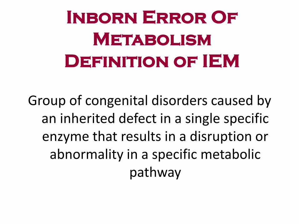
Group of congenital disorders caused by an inherited defect in a single specific enzyme that results in a disruption or
abnormality in a specific metabolic pathway

What Are Inborn Errors
of Metabolism? Genetic Disorders that affect
the metabolism of food. There are missing or defective
enzymes necessary to metabolize the
food eaten
Generally they are autosomal
recessive traits
Food not broken down properly may
produce chemicals that build up in
various parts of the body causing
medical problems and learning disorders.
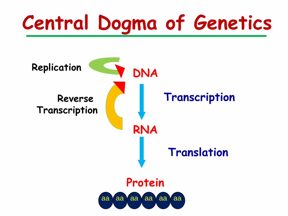
Central Dogma of Genetics
DNA
RNA
Protein
Replication
Transcription
Translation
Reverse Transcription
aa aa aa aa aa aa

Chemical Individuality
Garrod 20th Century
Developed “Inborne
Error of Metabolism”
Beadle & Tatum
Developed one gene
one enzyme concept.

7
Inborn Errors of Metabolism
a genetic disease also known as biochemical genetics
Gene-level Gene mutation
Protein-level Abnormal protein
Transpor Other Enzyme protein protein Metabolic-level Abnormal metabolites

Inborn Errors Overview
General mechanism of problems
Substrate accumulates to toxic levels
Toxic byproducts produced from shunting
of accumulated substrate
Deficiency of end product
Poor regulation results in overproduction
of intermediates to toxic level

Lipids Sugar Protein
BASIC IDEA,,,
Need factors to break them
Need close interactions
Excess is like deficiency

Complex compound
( Glycogen)
Intermediate substance
( tyrosine)
Simple molecules
( propionic A)
Energy
( Glucose )
Accumulate Accumulate Accumulate Deficiency
Enzyme
Co-Enzyme
Enzyme
Co-Enzyme
Enzyme
Co-Enzyme
Organomegaly
Storage diseases Energy defects
BASIC IDEA,,,
Toxic Toxic
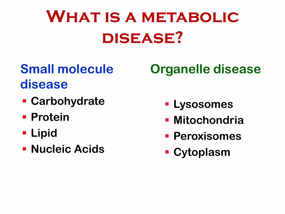
What is a metabolic
disease?
Small molecule
disease
Carbohydrate
Protein
Lipid
Nucleic Acids
Organelle disease
Lysosomes
Mitochondria
Peroxisomes
Cytoplasm

• Protein Disorders Amino Acid
Organic
Urea Cycle
• Carbohydrate Disorders Galactose, Glucose transport, Glycogen, Fructose
• Fatty Acid Disorders Medium chain acyl-CoA dehydrogenase def.
Long chain 3 hydroxycayl-CoA dehydrogenase def.
Types of Inborn Errors
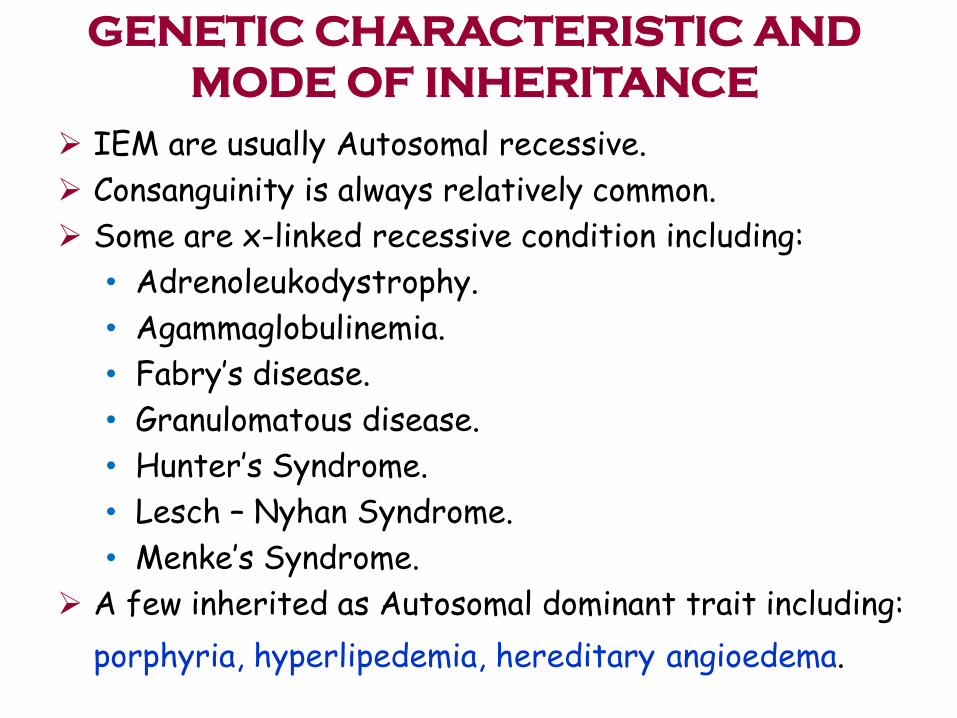
IEM are usually Autosomal recessive.
Consanguinity is always relatively common.
Some are x-linked recessive condition including:
• Adrenoleukodystrophy.
• Agammaglobulinemia.
• Fabry’s disease.
• Granulomatous disease.
• Hunter’s Syndrome.
• Lesch – Nyhan Syndrome.
• Menke’s Syndrome.
A few inherited as Autosomal dominant trait including:
porphyria, hyperlipedemia, hereditary angioedema.
GENETIC CHARACTERISTIC AND
MODE OF INHERITANCE

Inborn Errors of
Carbohydrate Metabolism Carbohydrates are important energy stores, fuels and metabolic
intermediates
Routine biochemistry tests e.g. lactate, glucose and second-line metabolic tests e.g. amino acids are essential for the investigation of disorders of carbohydrate metabolism. However, definitive diagnosis is usually achieved by measurement of the activity of the affected enzyme.
The easiest sample type to obtain is blood (erythrocytes,
leucocytes, lymphocytes) but the choice of tissue depends on the pattern of expression of the enzyme in question. For some assays, cultured skin fibroblasts (from a punch biopsy) or liver/muscle biopsies are required.

Inborn Errors of
Carbohydrate Metabolism
Galactosaemia
Glycogen storage diseases
Pyruvate carboxylase deficiency
Fructose-1,6-bisphophatase deficiency
Hereditary fructose intolerance
Glucose-6-phosphate dehydrogenase deficiency

Disorders of Carbohydrate
Metabolism
Galactosemia
Results from a disturbance in the conversion of galactose to glucose
The enzyme deficiency causes an accumulation of galactose in body tissues.
Classic type lacks Galactose-1-phosphate uridyl transferase (GALT)
Two types:
Galactokinase (GALK) deficiency results in infantile cataracts from accumulation of galacticol
Galactose epimerase (GALE) deficiency mostly confined to blood cells and most appear normal
Estimated incidence 1/50,000 births

Lactose
Galactose
Galactose-1phosphate
Galactose -1 phosphate
Uridyl transferase
Glucose
Lactase
Galactokinase
Metabolism of Galactose
Brain MR
Liver Jaundice
Hepatomegaly
Cirrhosis
Eyes Chataract

This presents with lactic acidosis, neurological dysfunction (seizures, hypotonia, coma)
It is a defect in the first step of gluconeogenesis which is the production of oxaloacetate from pyruvate. In addition to the effect on gluconeogenesis, lack of oxaloacetate affects the function of the Krebs cycle and the synthesis of aspartate (required for urea cycle function).
In the acute neonatal form the lactic acidosis is severe, there is moderately raised plasma ammonia, citrulline (& alanine, lysine, proline) and ketones. Fasting results in hypoglycaemia with a worsening lactic acidosis.
The diagnosis can be confirmed by assay of pyruvate carboxylase activity in cultured skin fibroblasts
Patients rarely survive >3 months in the severe form
Glycogen Storage Diseases

Uridine-Diphosphoglucose
Glycogen
Straight chains
Glycogen
Branched structure
Limit dextrin+ Glucose-1-PO4 Glucose-1-PO4
Glycogen ( normal branch) + Glucose
3
1
1
2
Glycogen synthetase
Brancher enzyme (GSD-IV)
Debrancher enzyme (GSD-III)
Glucose-6-phosphatase (GSD-1)
2
3
4
4
Glycogen Storage Diseases

Glycogen Storage Diseases
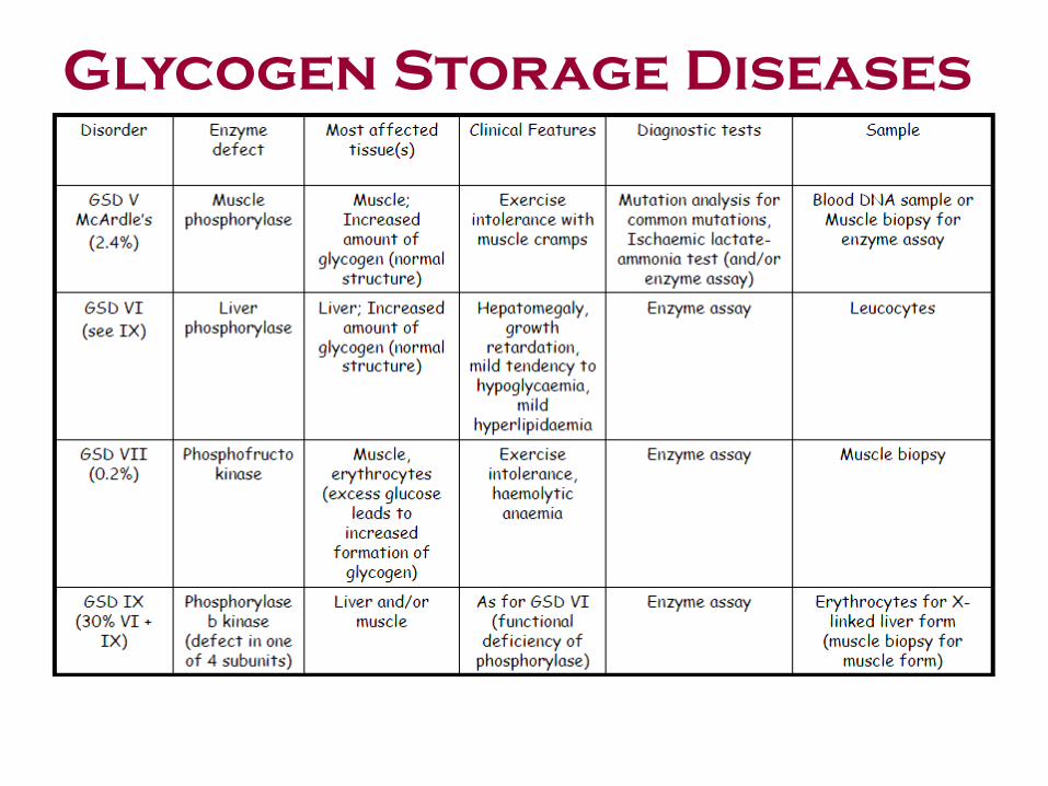
Glycogen Storage Diseases
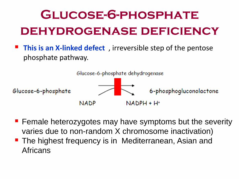
This is an X-linked defect , irreversible step of the pentose phosphate pathway.
Female heterozygotes may have symptoms but the severity
varies due to non-random X chromosome inactivation)
The highest frequency is in Mediterranean, Asian and
Africans
Glucose-6-phosphate
dehydrogenase deficiency
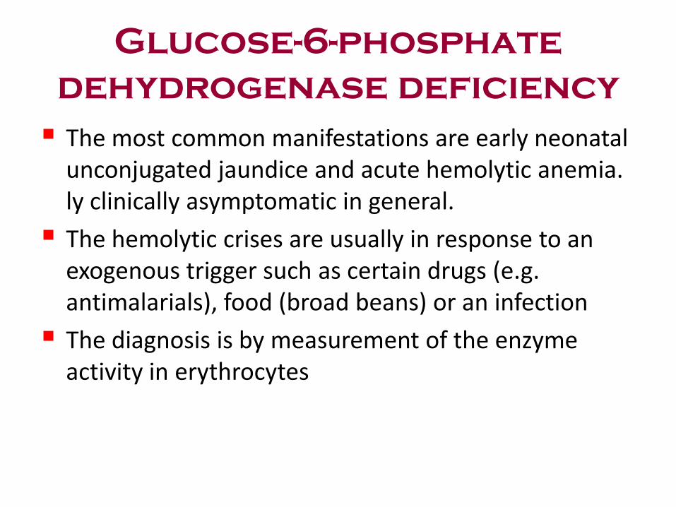
The most common manifestations are early neonatal unconjugated jaundice and acute hemolytic anemia. ly clinically asymptomatic in general.
The hemolytic crises are usually in response to an exogenous trigger such as certain drugs (e.g. antimalarials), food (broad beans) or an infection
The diagnosis is by measurement of the enzyme activity in erythrocytes
Glucose-6-phosphate
dehydrogenase deficiency

DISORDERS OF CH METABOLISM
• t
HEREDITARY FRUCTOSE INTOLERANCE: Fructose 1 phosphate aldolase deficiency
• Diagnosis: Fructose in Urine + Enzyme in the intestine mucosa and liver bx
• Clinical: Mild to sever • Treatment:Diet restriction

DISORDERS OF AA
METABOLISM
• PHENYLKETONURIA
• ALKAPTONURIA
• OCULOCUTANEOUS ALBINIS
• HOMOCYSTINURIA
• BRANCHED AMINOACIDS
History and Diagnosis
• PKU was discovered in 1934 by Dr. A
Folling in Sweden by identifying
phenylpyruvic acid in the urine of two
siblings who were mentally retarded.
• 1950’s Jervis discovered a deficiency of the
enzyme phenylalanine dehydrogenase in
the liver tissue of an affected patient.
• 1955- Bickel demonstrated that restricting
dietary phenylalanine lowers the blood concentration of phenylalanine.

Phenylketonuria (PKU):
• Clinical features: Development delay in infancy, ? neurological manifestations such as seizures. hyper activity, behavioral disturbances, hyperpigmentation and MR.
• Incidence: 1/5000 -1/16000.
• Genetics: AR, 12q22-q24, >70 mutations
• Basic Defect: Mutation in the gene of PA hydroxylase.
• Pathophysiology: PA or derivatives cause damage in the developing brain
• Treatment: Dietary reduction of phenylalanine within 4W
• Significance: Inborn Metabolic disorder, The first Dietary restriction treatment. Mass screening of newborns
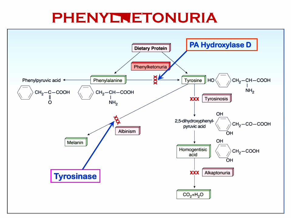
PA Hydroxylase D
Tyrosinase
PHENYLKETONURIA

Phenylalanine Metabolism
• Phenylalanine
• Essential AA
• Major interconversions through tyrosine
PHE
TYR
Body Protein
Melanin
DOPA
NE / EPI
Food Catabolism
50%
7/8/2014 30
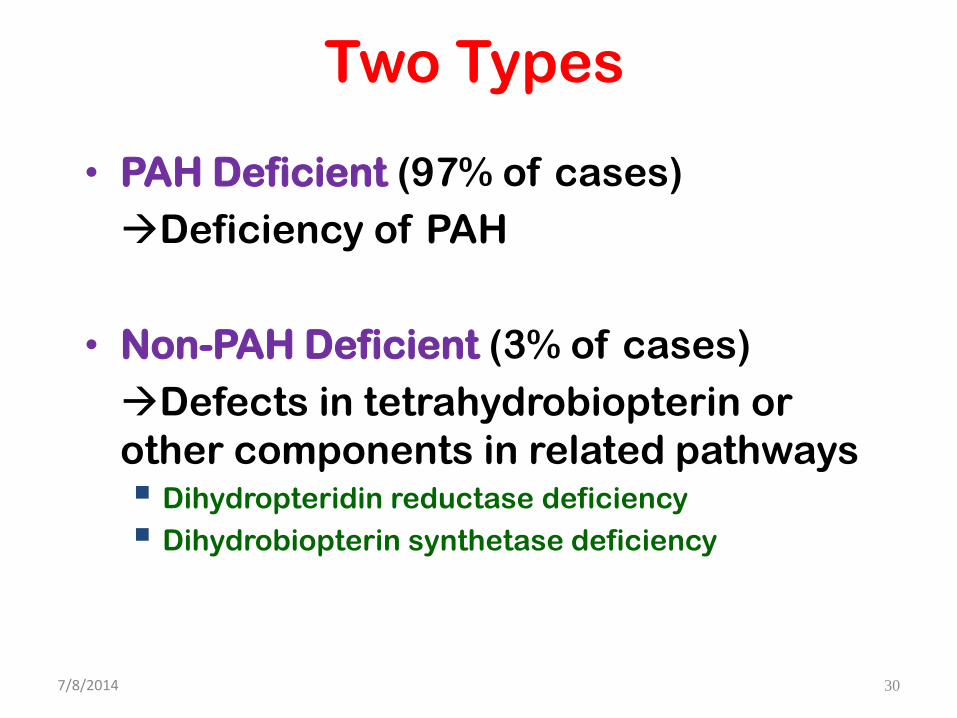
Two Types
• PAH Deficient (97% of cases)
Deficiency of PAH
• Non-PAH Deficient (3% of cases)
Defects in tetrahydrobiopterin or
other components in related pathways Dihydropteridin reductase deficiency
Dihydrobiopterin synthetase deficiency

7/8/2014 31
Diagnostic Criteria
• Normal: 120 – 360 umol/L
• PAH Deficient:
– Mild: 600 – 1200 umol/L
– Classical: > 1200 umol/L
• Non-PAH Deficient:
– < 600 umol/L
• Guthrie Bacterial Inhibition Assay
• Confirmation of diagnosis

Guthrie Test--1961
1965 - Screening for PKU was mandated
legislatively in most of the states in US

Low tyrosine High phe
Low tyrosine
High phe
Plasma Amino Acid
Profile, PKU
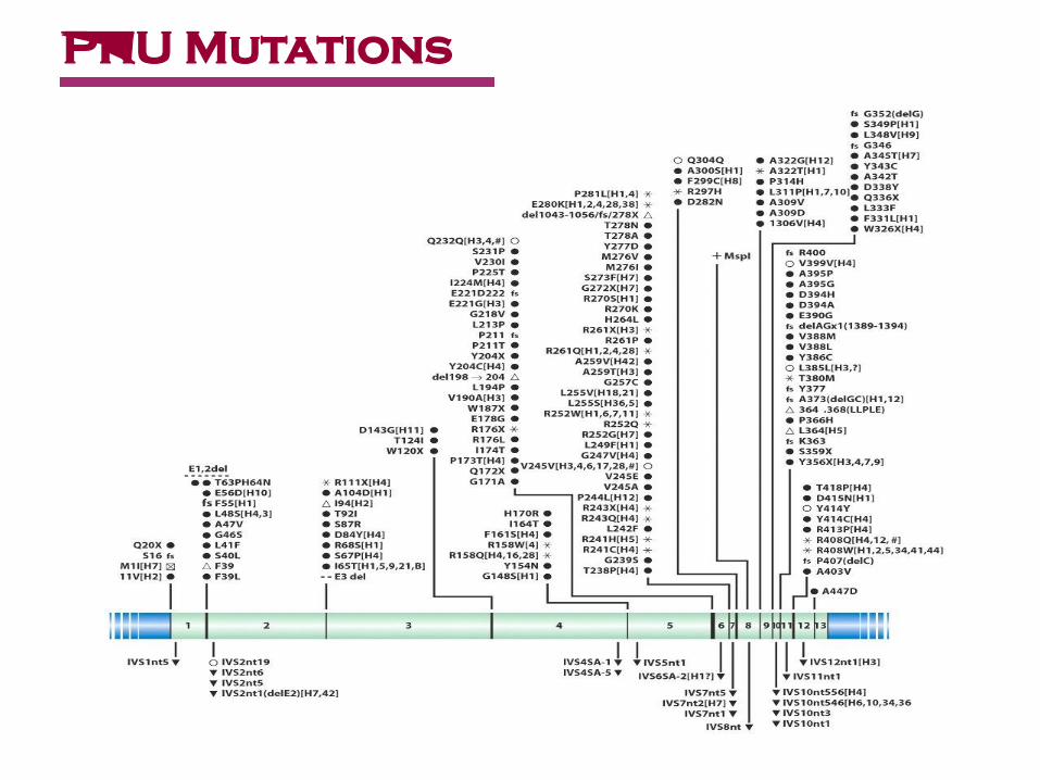
PKU Mutations

Treatment
Low phenylalanine diet
• requires careful monitoring
• risk of tyrosine insufficiency
• risk vitamin and trace element deficiencies
? biopterin supplementation (sapropterin)
Large Neutral Amino Acids (val, leu, ileu) supplements
Diet for life
Management of PKU pregnancies
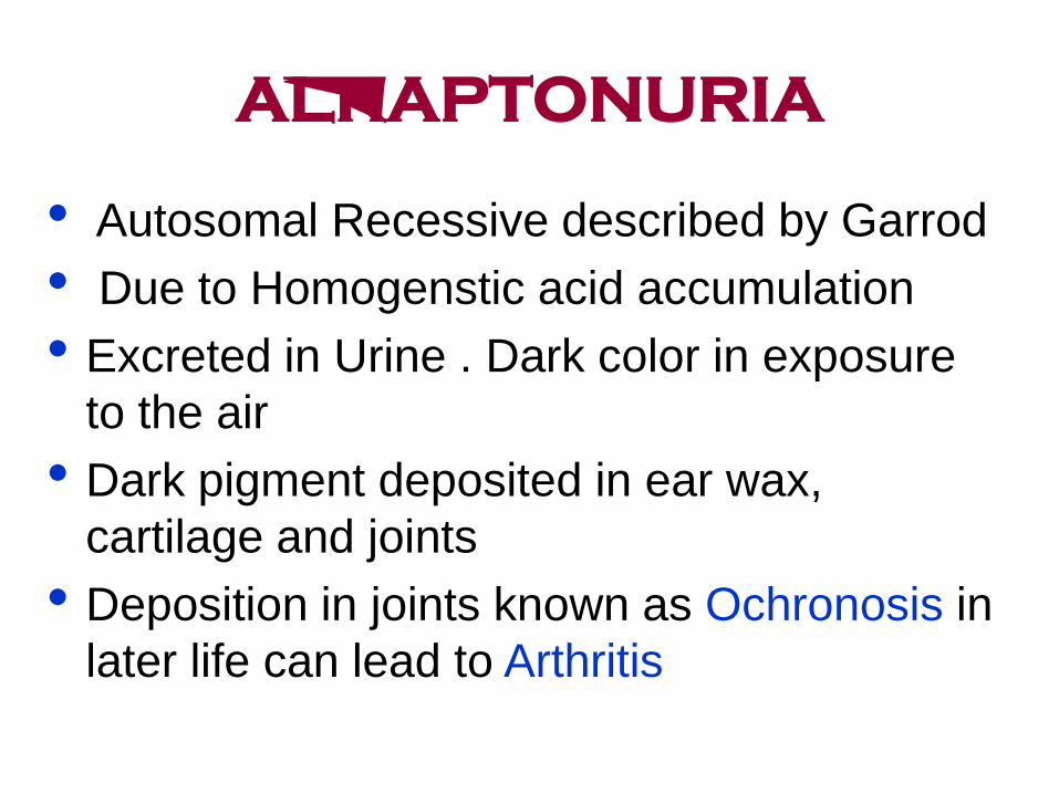
ALKAPTONURIA
• Autosomal Recessive described by Garrod
• Due to Homogenstic acid accumulation
• Excreted in Urine . Dark color in exposure
to the air
• Dark pigment deposited in ear wax,
cartilage and joints
• Deposition in joints known as Ochronosis in
later life can lead to Arthritis

Normal urine
Urine from patients with
alkaptonuria
Symptoms of alkaptonuria
Patients may display painless bluish darkening of the outer ears,
nose and whites of the eyes. Longer term arthritis often occurs.

Alkaptonuria - Biochemistry • Alkaptonuria reflects the absence of homogentisic acid oxidase activity.

OCULOCUTANEOUS
ALBINISM
• OCA is AR due to tyrosinase deficiency no
melanine formation
• No pigment in skin, hair, iris and ocular fundus
• Nystagmus
• Genetically and bichemically heterogeneous
Classical tyrosinase negative
Tyrosinase positive, reduced enzyme level (type 1) OCA 1
located on chromosome11q.
OCA 2 on chromosome 15q (pink-eye)
Third loci OCA-3 not related to above mentioned
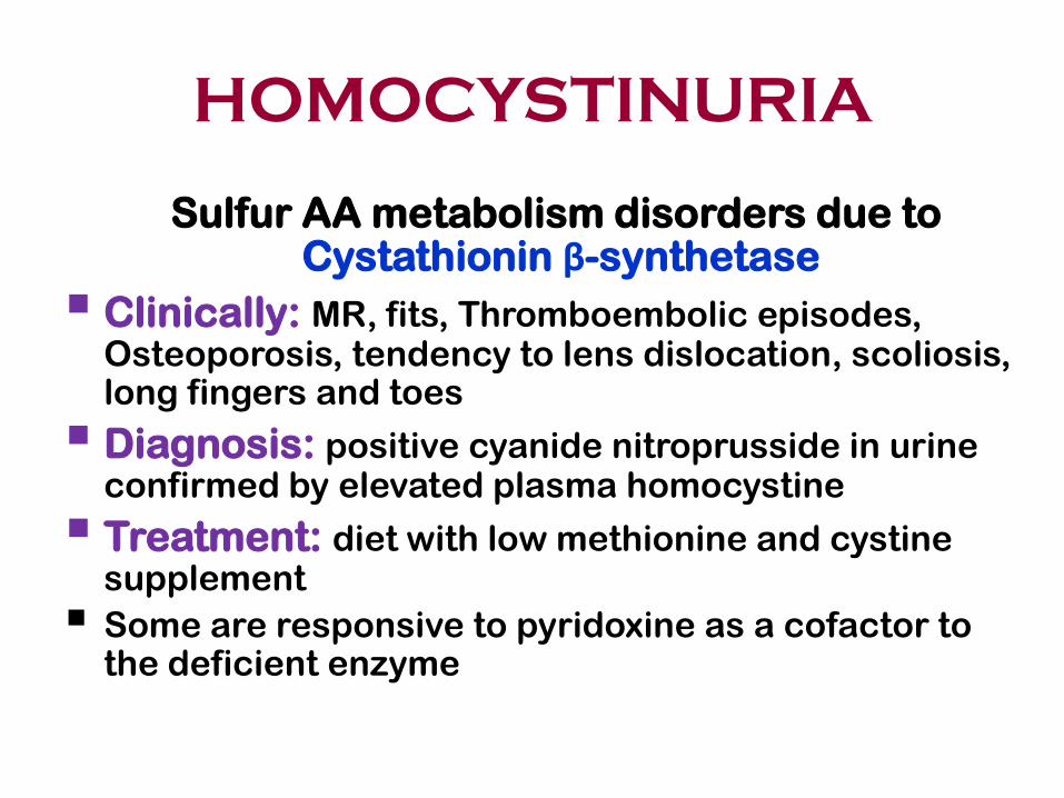
HOMOCYSTINURIA
Sulfur AA metabolism disorders due to Cystathionin β-synthetase
Clinically: MR, fits, Thromboembolic episodes, Osteoporosis, tendency to lens dislocation, scoliosis, long fingers and toes
Diagnosis: positive cyanide nitroprusside in urine confirmed by elevated plasma homocystine
Treatment: diet with low methionine and cystine supplement
Some are responsive to pyridoxine as a cofactor to the deficient enzyme
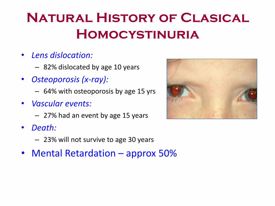
Natural History of Clasical
Homocystinuria
• Lens dislocation: – 82% dislocated by age 10 years
• Osteoporosis (x-ray): – 64% with osteoporosis by age 15 yrs
• Vascular events: – 27% had an event by age 15 years
• Death: – 23% will not survive to age 30 years
• Mental Retardation – approx 50%
Branched Chain Amino
Acids
• 40% of preformed AA used by mammalians are BCAA
Valine, Leucine, Isoleuchin
• Energy supply through -ketoacid decarboylase enzyme
• BCAA disease composed of 3 catalytic and 2 regulatory
enzyme and encoded by 6 loci
• Deficiency in any one of these enzymes cause MSUD
• Untreated patients, accumulation of BCAAs cause
neurodegeneration leads to death in the first few months
of life
• Treatment BCAAs restriction diet
• Early detection
• Gene therapy ?????

Maple Syrup Urine Disease
(MSUD) AR
• Involves the Branch-chain amino acids:
• Leucine
• Iso-leucine
• Valine
• Incidence is 1:200,000
• Infants appear normal at birth. By four days of age they
demonstrate poor feeding, vomiting and lethargy.
• Urine has a characteristic sweet, malty odor toward the
end of the first week of life
• Treatment: Formulas low in the branch chain amino
acids

UREA CYCLE DISORDERS
• UC main function to prevent accumulation of N2 waste as urea
• UC responsible for de novo arginine synthesis
• UC consists of 5 major biochemical reactions, defects in humans: Carpamyl phosphate synthetase (CPS), AR
Ornithin transcarbamylase (OTC), X-linked
Argininosuccinic acid synthatase (ASA),AR
Argininosuccinase (AS), AR
N-acetyl glutamate synthetae (NAGS).AR

UREA CYCLE DISORDERS
Carbmyle Phophtase Deicfiency
Ornithine Transcarmylase D
Argininosuccinic A Synthetase A AS aciduria
Hyperarginemia
UREA CYCLE DISORDERS
Characteristics
• Neonatal period or anytime
• Wide inter and intra familial variations in the
severity of the disease,
• Lethargy, coma. Arginase deficiency cause
progressive spastic quadriplegia and Mental
retardation
• No acidosis (respiratory alkalosis)
• No ketones (unlike organic acidemia)
• No hypoglycemia
• But there is hyperammonemia
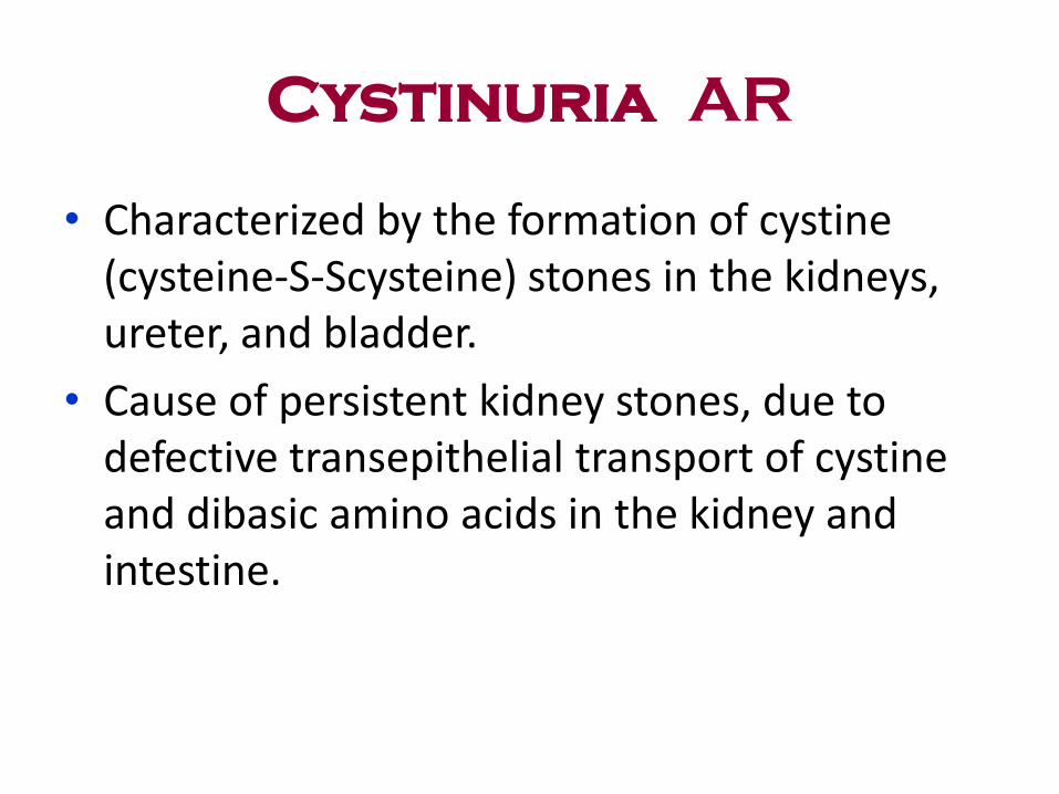
Cystinuria AR
• Characterized by the formation of cystine (cysteine-S-Scysteine) stones in the kidneys, ureter, and bladder.
• Cause of persistent kidney stones, due to defective transepithelial transport of cystine and dibasic amino acids in the kidney and intestine.

Lipid Metabolism
• Backbone of phosopholipide and sphingolipids = biological membranes and hormones
• Intracellular messengers and energy substrate
• Hyperlipidemia, due to defective in lipid transport
• Fatty Acidemias is less common (fatty acid oxidation)
• FA mobilization from adipose tissue to cell = energy substrate in liver, skeletal and cardiac muscles
• FA transport across outer and inner mitochondrial membrane and entry into mitochondrial matrix
• Defects in any of these steps cause disease (Short, Medium & Long chain fatty acidemias)
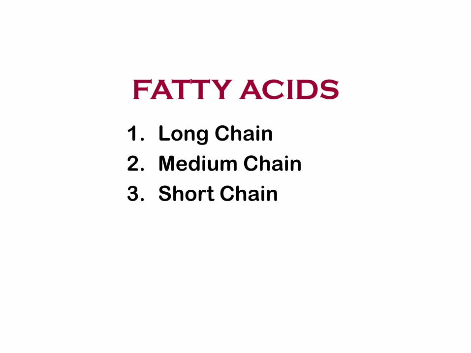
FATTY ACIDS
1. Long Chain
2. Medium Chain
3. Short Chain
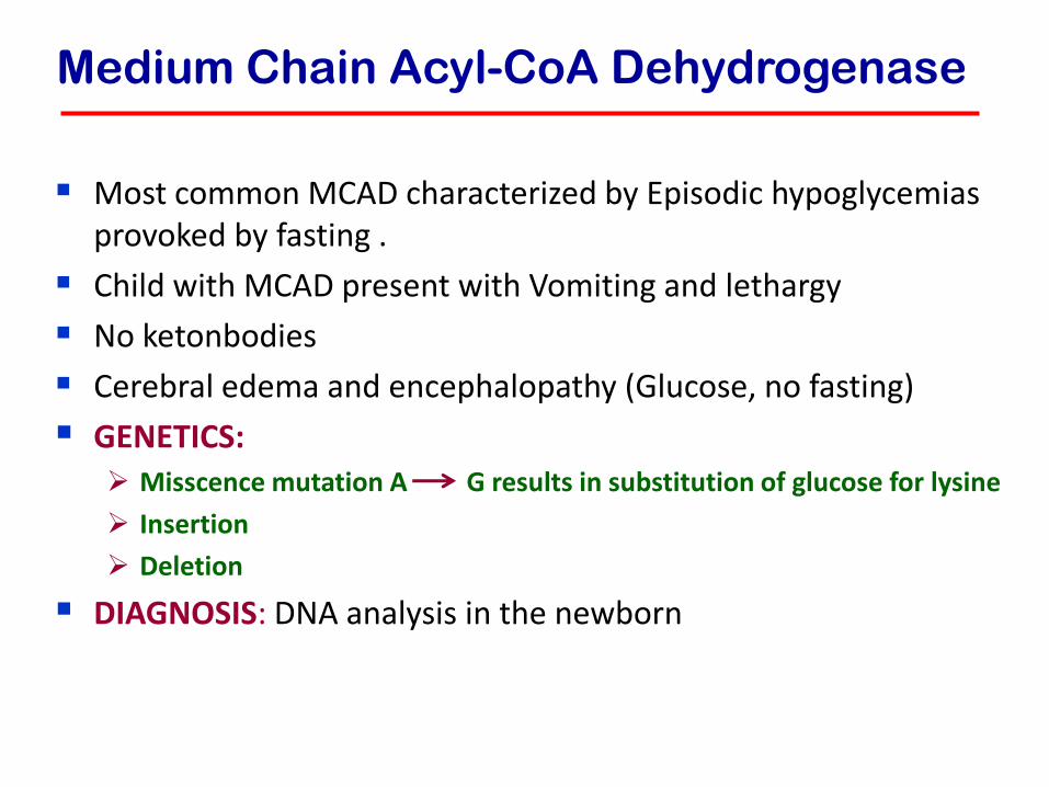
Medium Chain Acyl-CoA Dehydrogenase
Most common MCAD characterized by Episodic hypoglycemias provoked by fasting .
Child with MCAD present with Vomiting and lethargy
No ketonbodies
Cerebral edema and encephalopathy (Glucose, no fasting)
GENETICS: Misscence mutation A G results in substitution of glucose for lysine
Insertion
Deletion
DIAGNOSIS: DNA analysis in the newborn screening

Long Chain Acyl-CoA
Dehydrogenase
LCAD patients are presented with Fasting induced coma Hepatomegaly Cardiomegaly Muscle weakness Hypotonia Peripheral neuropathy
Clinical and biochemical characteristics can be differentiated from each others
SCAD: Very few case are reported with variable
presentation

LDL Receptor
Pathway and
Regulation
of Cholesterol
Metabolism

Organic Acidemia (OA)
The term "organic acidemia" or "aciduria" applies to a group of disorders characterized by the excretion of non-amino organic acids in urine at birth and for the first few days of life.
Toxic encephalopathy.
Difficult to differentiate in acute presentation
All are autosomal recessive, the commonest Methylmalonic acidemia MMA,,,,
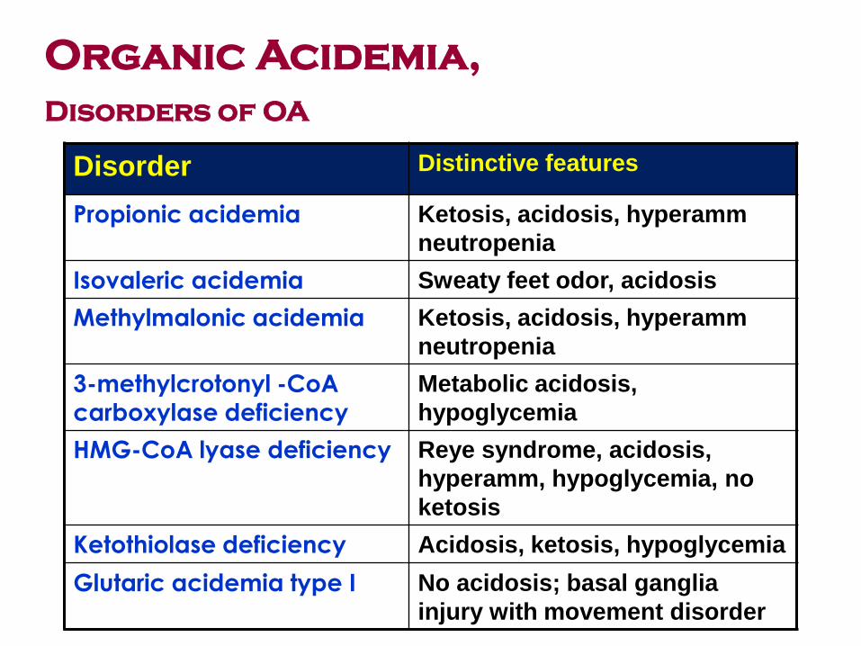
Disorder Distinctive features
Propionic acidemia Ketosis, acidosis, hyperamm
neutropenia
Isovaleric acidemia Sweaty feet odor, acidosis
Methylmalonic acidemia Ketosis, acidosis, hyperamm
neutropenia
3-methylcrotonyl -CoA carboxylase deficiency
Metabolic acidosis,
hypoglycemia
HMG-CoA lyase deficiency Reye syndrome, acidosis,
hyperamm, hypoglycemia, no
ketosis
Ketothiolase deficiency Acidosis, ketosis, hypoglycemia
Glutaric acidemia type I No acidosis; basal ganglia
injury with movement disorder
Organic Acidemia,
Disorders of OA

Organic acidemia
Clinically:
• Healthy NB rapidly ill,
Ketoacidosis, poor
feeding
• Vomiting, dehydration
• Hypotonia, lethargy
• Tachypnea, seizures
• Coma, unusual odors
• Pancreatitis,
cardiomyopathy, infection
( recurrent).
Lab diagnosis
• Metabolic acidosis
• Hyperammonemia
• Hypoglycemia
• Lactic acidosis
• Anemia, ± thrombocytopenia ± neutropenia
• Definite diagnosis, Tandem MS & Urine organic acid analysis

LYSOSOMAL STORAGE
DISEASE
• The hydrolytic enzymes within lysosomes are involved in the breakdown of sphingolipids, glycoproteins, and mucopolysaccharides into products.
• These molecular complexes can derive from the turnover of
intracellular organelles or enter the cell by phagocytosis, • A number of genetic diseases lacking Iysosomal enzymes
result in the progressive accumulation within the cell of partially degraded insoluble products, This condition leads to clinical conditions known as:
Iysosomal storage disorders.

Mucoploysaccharides
(glycosaminoglycans
Bone, connective tissue,
skin, cornea,joints etc
Cell membranes,
organelles
Bacteria,
viruses
Lysosome
Sphingolipids,
glycolipids etc
Food
particles
Glycoproteins
Acid hydrolases
“The cells wrecking
crew”
Glycogen
Abnormal
lysosomal
storage leads to developmental regression

Lysosomal Storage
Disorders
• Resulted from accumulation of substrate • Deficiency or inability to activate or to transport
the Enzymes within lysosomes that catalyses stepwise the degradation of: Glycosaminoglycans (MPS) Sphingolipids Glycoproteins Glycolipids
• May be it is a result of genetic drift and natural selection
• Children normal at birth, downhill course of differing duration

LIPIDOSES
Enzyme Disease
- galactosidase GM1 Gangliosidosis.
Hexosamindase A GM2 Tay –Sach.
Hexosamindase A+B Sandhoff disease.
Sphingomylinase Niemann – Pick disease.
Acidic – – Glucosidase
Gaucher’s disease.
Arylsulfatase A Neuronal ceroid lipofuscinosis
Metachromatic Leukodystrophy.

Sphingolipidoses
• Tay-Sachs disease AR Hexosaminidase -A
– Developmental regression, Blindness, – Cherry-red spot, Deafness
• Gaucher' s disease AR Glucosylcerarnide Type l – Joint and limb pains, Splenomegaly
β- Glucosidase Type II – Spasticity, fits; death
• Niemann-Pick disease AR Sphingomyelinase – Failure to thrive, Hepatomegaly – Cherry-red spot, Developmental regression
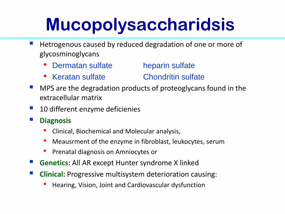
Mucopolysaccharidsis Hetrogenous caused by reduced degradation of one or more of
glycosminoglycans
• Dermatan sulfate heparin sulfate
• Keratan sulfate Chondritin sulfate
MPS are the degradation products of proteoglycans found in the extracellular matrix
10 different enzyme deficienies disorders
Diagnosis
• Clinical, Biochemical and Molecular analysis,
• Meausrment of the enzyme in fibroblast, leukocytes, serum
• Prenatal diagnosis on Amniocytes or CVS
Genetics: All AR except Hunter syndrome X linked
Clinical: Progressive multisystem deterioration causing:
• Hearing, Vision, Joint and Cardiovascular dysfunction

62
Examples
• Hunter syndrome • Hurler syndrome • Scheie syndrome • Sanfilippo syndrome • Morquio disease • Maroteaux-Lamy syndrome

CYSTINOSIS AR
• 1/200,000 births
• Lysosomal storage disease due to impaired transport of cystine out of lysosomes.
• High intracellular cystine content Crystals in many tissues. Clinical Manifestations are age dependent include renal tubular Fanconi syndrome, growth retardation(Infancy syndrome), Renal failure develops by 10 year of year( Late childhood) and cerebral calcification( adolescence period).

Purine/pyrimidine metabolism Lesch-Nyhan disease XR
• Hypoxanthine Guanine Phosphoribosyltransferase Deficiency • Mental retardation, • uncontrolled movements, } Uric Acid Crystals in CNS • S64}elf-mutilation
Adenosine deaminase deficiency AR
• Adenosine deaminase Deficiency • Severe combined immunodeficiency
Purine nucleoside phosphorylase AR
• Purine nucleoside Phosphorylase deficiency • Severe viral infections due to impaired cell function
Hereditary orotic aciduria AR Orotate phosphoribosy ltransferase , Deficiency • Orotidine 5'-phosphate Decarboxylase Deficiency • Megaloblastic anaemia in the first year of life, • Failure to thrive, – Developmental delay

Copper Metabolism
• Wilson AR ATPase
membrane copper
Spasticity , Rigidity, Dysphagia, Cirrhosis
Transport protein ;
• Menkes' disease XR ATPase
membrane copper
Failure to thrive, Neurological deterioration
Transport protein
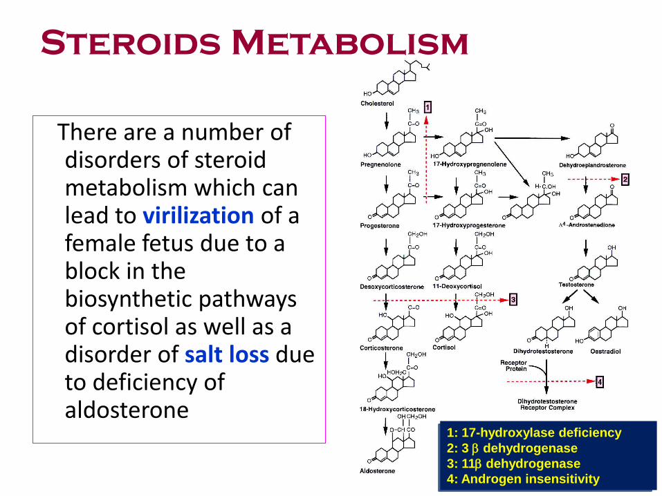
Steroids Metabolism
There are a number of disorders of steroid metabolism which can lead to virilization of a female fetus due to a block in the biosynthetic pathways of cortisol as well as a disorder of salt loss due to deficiency of aldosterone
1: 17-hydroxylase deficiency
2: 3 dehydrogenase
3: 11 dehydrogenase
4: Androgen insensitivity

Steroid Metabolism
• CongenitaI adrenal hyperplasia AR
• Virilization ( any new born female with ambiguous genitalia )
• Salt-Iosing 21-hydroxylase Most common (90%)
11,13-hydroxy!ase,
3 13-dehydrogenase
17a-hydroxylase, very rare
17,20-lyase. Very rare
• Testicular feminization XR Androgen receptor
Female external genitalia,
Male internal genitalia,
Male chromosomes

Every child with unexplained . . . Neurological deterioration
Metabolic acidosis
Hypoglycemia
Inappropriate ketosis
Hypotonia
Cardiomyopathy
Hepatocellular dysfunction
Failure to thrive
. . . should be suspected of having a metabolic disorder
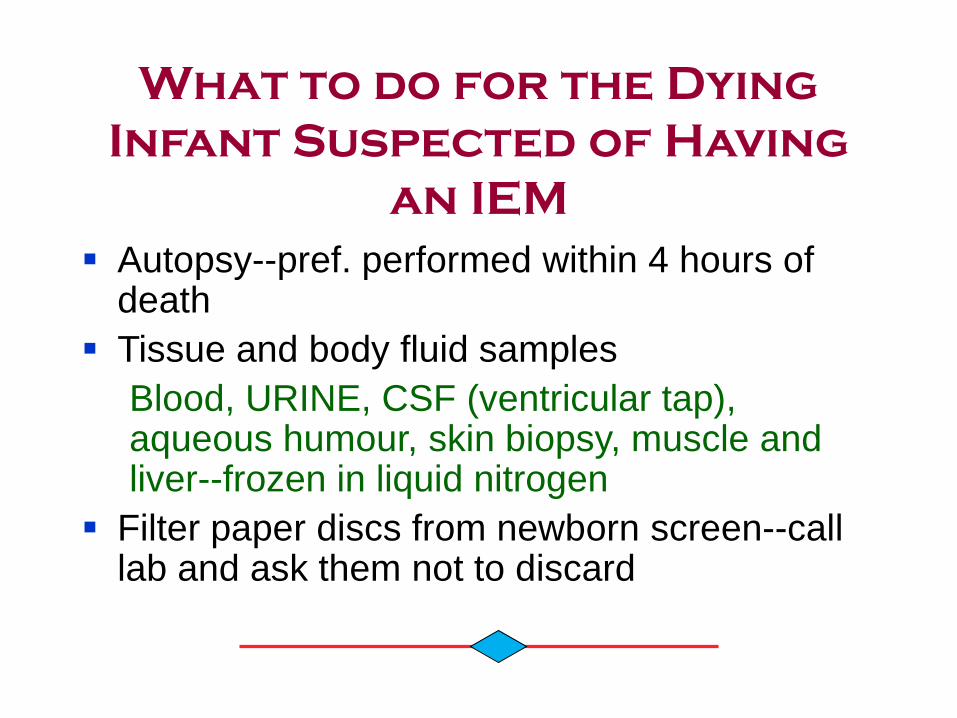
What to do for the Dying
Infant Suspected of Having
an IEM
Autopsy--pref. performed within 4 hours of death
Tissue and body fluid samples
Blood, URINE, CSF (ventricular tap), aqueous humour, skin biopsy, muscle and liver--frozen in liquid nitrogen
Filter paper discs from newborn screen--call lab and ask them not to discard
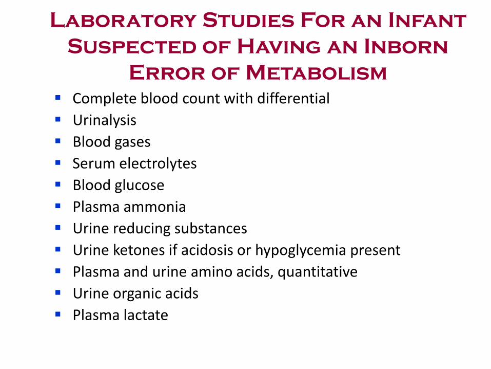
Laboratory Studies For an Infant
Suspected of Having an Inborn
Error of Metabolism
Complete blood count with differential
Urinalysis
Blood gases
Serum electrolytes
Blood glucose
Plasma ammonia
Urine reducing substances
Urine ketones if acidosis or hypoglycemia present
Plasma and urine amino acids, quantitative
Urine organic acids
Plasma lactate

SUMMARY
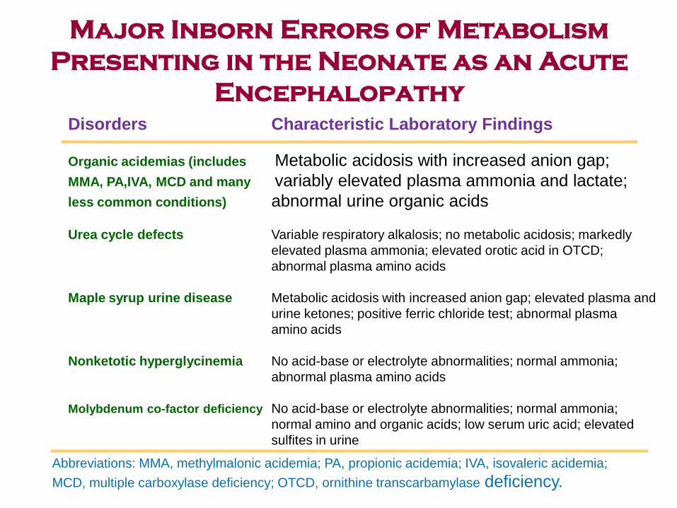
Major Inborn Errors of Metabolism
Presenting in the Neonate as an Acute
Encephalopathy
Disorders Characteristic Laboratory Findings
Organic acidemias (includes Metabolic acidosis with increased anion gap;
MMA, PA,IVA, MCD and many variably elevated plasma ammonia and lactate;
less common conditions) abnormal urine organic acids
Urea cycle defects Variable respiratory alkalosis; no metabolic acidosis; markedly
elevated plasma ammonia; elevated orotic acid in OTCD;
abnormal plasma amino acids
Maple syrup urine disease Metabolic acidosis with increased anion gap; elevated plasma and
urine ketones; positive ferric chloride test; abnormal plasma
amino acids
Nonketotic hyperglycinemia No acid-base or electrolyte abnormalities; normal ammonia;
abnormal plasma amino acids
Molybdenum co-factor deficiency No acid-base or electrolyte abnormalities; normal ammonia;
normal amino and organic acids; low serum uric acid; elevated
sulfites in urine
Abbreviations: MMA, methylmalonic acidemia; PA, propionic acidemia; IVA, isovaleric acidemia;
MCD, multiple carboxylase deficiency; OTCD, ornithine transcarbamylase deficiency.

Group I . Disorders involving COMPLEX molecules .
Glycoproteinosis , MPS, Sphingolipidosis . Lysosomal disorders.
Zellweger syndrome & Variants , Refsum disease,. Peroxisomal disorders .
NPD-type C Disorders of intracellular trafficking & processing .
Wolman disease Disorders of Cholesterol synthesis
Group II . Disorders that give rise to INTOXICATION .
PKU, MSUD. Homocysteinuria, Tyrosinemia . Aminoacidopathies .
CPT, OTC, Citrullinaemia, ASA.
Arginase, NAGS deficiency . Congenital Urea Cycle Defects .
Methylmalonic acidemia .Propionic acidemia .
Isovaleric acidemia .Glutaric aciduria type I . Organic acidemias .
Galactosemia .Heredietary Fructose intolerance . Sugar intolerances .
Group III . Disorders involving ENERGY METABOLISM
Glycogenoses (glycogen storage disease ) .
Fructose 1,6-diphosphatase deficiency .
Phosphoenolpyruvate carboxykinase . Gluconeogesis defects .
Pyruvate Carboxylase deficiency .
Pyruvate Dehydrogenase deficiency . Congenital Lactic Acidemia .
VLCAD, MCAD , etc Fatty Acid Oxidation defects .
Mitochondrial respiratory-chain disorders .

Inborn Errors of Metabolism
Associated With Neonatal Liver Disease
and Laboratory Studies Useful in
Diagnosis
Disorder Laboratory Studies
Galactosemia Urine reducing substances; RBC galactose-1-
phosphate uridyl transferase
Hereditary tyrosinemia Plasma quantitative amino acids; urine
succinylacetone a1-Antitrypsin deficiency
Quantitative serum a1-antitrypsin; protease inhibitor
typing
Neonatal hemochromatosis Serum ferritin; liver biopsy
Zellweger syndrome Plasma very long-chain fatty acids
N-Pick disease type C Skin biopsy for fibroblast culture; studies of
cholesterol esterification and accumulation
GSD type IV Liver biopsy for histology and biochemical
(brancher deficiency) analysis or skin biopsy with assay
of branching enzyme in cultured fibroblasts