inactivation of cryptosporidium across the wastewater
TRANSCRIPT

SWF2M-7103
February 2012
Inactivation of Cryptosporidium across the wastewater treatment train for water recycling
Phase 1: Optimization of an in vitro cell culture infectivity assay
Prepared by
Dr Brendon King and Dr Alexandra Keegan, Australian Water Quality Centre
Final Report
February 2012 © Copyright Smart Water Fund 2012 – Inactivation of Cryptosporidium across the
wastewater treatment train for water recycling. Phase 1: Optimization of an in vitro cell
culture infectivity assay.
Page 2
1.1 Copyright and Intellectual Property
This publication is copyright. Other than for the purposes of and subject to the conditions prescribed on the Copyright Act 1968, no part of any Material in this Report may in any form or by any means (including optical, magnetic, electronic, mechanical, microcopying, photocopying or recording) be reproduced, broadcast, published, transmitted, adapted, or stored without the express written permission of the copyright owner. All other rights are reserved. “Smart Water Fund” is a registered trademark, jointly owned by the Smart Water Fund participants, and is protected by laws governing intellectual property. The Smart Water Fund trademark and logo must not be used except as part of any authorised reproduction of the Report as set out above. The Smart Water Fund logo must not be modified in any way.
1.2 Disclaimer
The material contained in this Report has been developed for the Smart Water Fund. The views and opinions expressed in the Report do not necessarily reflect the views, or have the endorsement of the Victorian Water Utilities or the Department of Sustainability and Environment, or indicate the Victorian Water Utilities or the Department of Sustainability and Environment commitment to a particular course of action.
1.3 Enquiries
For enquiries or copies of this report please contact: Smart Water Fund Knowledge Transfer Manager Email: [email protected] Phone: 1800 882 432 (freecall) Quote Project SW72M-7103 © Copyright Smart Water Fund, 2012

February 2012 © Copyright Smart Water Fund 2012 – Inactivation of Cryptosporidium across the
wastewater treatment train for water recycling. Phase 1: Optimization of an in vitro cell
culture infectivity assay.
Page 3
Executive Summary
Quantification of Cryptosporidium removal through wastewater treatment processes
currently relies on the enumeration of oocysts present in samples but has not considered
their potential inactivation or the loss of infectivity through treatment processes. The lack
of inactivation data has been due to the absence of a satisfactory tool capable of measuring
oocyst infectivity from environmental samples. The work detailed within this report
describes our research on the optimization of a cell culture infectivity test termed the
“Focus Detection Method”. The ultimate aim of this work has been the development of a
robust and sensitive measure of Cryptosporidium infectivity that can be applied to
investigate the inactivation of Cryptosporidium across the wastewater treatment train.
In this report, we examined factors affecting the timing of contact between excysted
sporozoites and target host cells and the subsequent impact of this upon the establishment
of infection. We demonstrated that excystation rate impacts upon establishment of
infection. The consequence is that in standard assay formats the majority of sporozoites are
not close enough to the cell monolayer when they are released from the oocyst to
successfully establish infection. However, this can be easily overcome by centrifugation of
oocysts onto the cell monolayer, resulting in approximately 4 fold increases in sporozoite
attachment and subsequent infection.
Further to this we demonstrated that excystation procedures can be tailored to control the
excystation rate and that this needs to be taken into account as this can significantly
influence data interpretation. By modification of initial acid pre-treatment temperature
(from 37°C to 30°C) we were able to successfully control oocyst excystation rate, so that
pre-treated oocysts could be centrifuged onto the cell monolayer before they excyst,
thereby giving us greater confidence that an individual focus is the result of a single
infectious oocyst. This reduces the possibility that multiple foci may develop from the four
sporozoites from a single oocyst, potentially inflating the enumeration of foci.
Previous analyses undertaken in our laboratory using the standard method demonstrated
that the method of recovery of Cryptosporidium had a significant effect on the infectivity
assay (data not shown), with great importance placed on clean up of the oocysts after the
initial concentration process. The addition of a washing step post sporozoite attachment
may be appropriate depending on the water matrix of the samples. A washing step at 4 h
post infection results in improved in vitro culture. Therefore for wastewater matrices a
wash step can be incorporated into the modified assay to give improved Cryptosporidium in
vitro growth.

February 2012 © Copyright Smart Water Fund 2012 – Inactivation of Cryptosporidium across the
wastewater treatment train for water recycling. Phase 1: Optimization of an in vitro cell
culture infectivity assay.
Page 4
A series of time course experiments were conducted in order to examine when foci
development could be first accurately detected. An opportunity exists to enumerate the
foci 24 h earlier if the turnaround time of results is paramount. However as foci increase in
area with further incubation, 48 h is the most suitable incubation period because slides can
be more rapidly scanned.
Bile has been reported to be an essential supplement needed for oocyst excystation,
sporozoite infection and subsequent amplification of parasite lifecycles within in vitro
culture. However the incorporation of bile or bile salts into in vitro assays is not universal
and recent work by some authors has reported negative effects of bile or bile salt inclusion
on in vitro parasite growth. Our current assay includes bovine bile in the infection medium
at a concentration of 0.02% wt/vol. We investigated the effect of bile and the bile salt
sodium taurocholate on both excystation and infection in our in vitro system. The current
concentration of bile within the infection medium is optimal, with higher concentrations
resulting in cell cytotoxicity and possible assay failure. The omission of bile from the
infection medium results in very poor in vitro infection. The bile salt sodium taurocholate
does not appear to be a complete replacement for total bovine bile as its incorporation in
the medium results in slightly reduced excystation and a considerable reduction in infection.
To finalize the sensitivity of this modified assay, low numbers of oocysts were applied to cell
culture and the level of infection quantified. For the optimized cell culture infectivity assay,
the limit of detection was determined to be approximately 2-3 infectious oocysts.
Furthermore we examined whether the new assay is capable of in vitro infection by C.
hominis oocysts as the optimization work had been carried out using only C. parvum. Our
modified assay is capable of supporting C. hominis in vitro infections, and the foci which
develop are typical of those observed for in vitro culture of C. parvum.
We now have a greatly improved Cryptosporidium cell culture infectivity assay, with a 4 fold
increase in assay sensitivity, improved synchronicity of infection and a modified excystation
methodology allowing greater precision in defining an infective focus formed as a result of a
single oocyst infection. Therefore as a consequence of this research project, we now have
the capability to accurately quantify not only the removal of oocysts from various stages of
the wastewater treatment train but also their inactivation, thereby accurately quantifying
the “true effect” of the treatment train on oocyst risk reduction. By quantifying oocyst
inactivation instead of just removal, a more complete assessment of Cryptosporidium oocyst
risk will be obtained for water utilities, enabling cost reduction for provision of “fit for
purpose” recycled water without incorporation of further, costly and unnecessary treatment
steps.
February 2012 © Copyright Smart Water Fund 2012 – Inactivation of Cryptosporidium across the
wastewater treatment train for water recycling. Phase 1: Optimization of an in vitro cell
culture infectivity assay.
Page 5
Table of contents
Page Contents
3-4 Executive Summary
7 Acknowledgements
8-34 8-10
11
12-13
13-15
15-16
17-26
17
17-19
19-20
20-21
21-22
22-24
24-25
25
26
27-30
27
27
27-30
30
31-32
32-33
33-34
Chapter 1: Review of the Literature and Key Findings 1.1 Overview
1.2 Cryptosporidium species present in the Australian community
1.3 Infection of Cryptosporidium in humans
1.4 Current methods for isolation and detection for Cryptosporidium - determination of
presumptive and confirmed oocysts
1.5 In vitro assays for assessing viability of Cryptosporidium
1.6 Methods for assessing infectivity of Cryptosporidium
1.6.1 Human infectivity assay
1.6.2 Animal models
1.6.3 Infection of Cell Culture with C. parvum
1.6.4 Host cell type 1.6.5 Culture media supplementation and formulation
1.6.6 Focus detection method
1.6.7 Definition of foci
1.6.8 Alternative Detection Methods
1.6.9 The influence of oocyst age and temperature on infectivity
1.7 Factors affecting the success of Cryptosporidium in cell culture
1.7.1 The role of excystation in Cryptosporidium infection
1.7.2 Pre-treatment of oocysts with sodium hypochlorite
1.7.3 Host triggers temperature, pH, trypsin and age of oocysts
1.7.4 Role of centrifugation in improving infectivity
1.8 Potential modifications to the focus detection method
1.9 Key Findings
1.10 Summary of The Current Utilized Focus Detection Method
35-40 35
35
35-36
36
36-37
37
37-38
38
38
39
39
39
Chapter 2: Methodology 2.1 Source of Oocysts
2.2 Oocyst excystation protocol
2.3 Excystation measurements using flow cytometry
2.4 Cell culture-Taqman infectivity assay
2.5 Cell culture focus detection method
2.6 Bile time-course excystation experiments
2.7 Bile and sodium taurocholate dosage excystation experiments
2.8 Bile and sodium taurocholate infectivity experiments
2.9 Bile and sodium tauorcholate cell monolayer cytotoxcity experiments
2.10 Sensitivity of the revised cell culture focus detection method
2.11 Purification of C. hominis oocysts and species confirmation
2.12 Cell culture infections with Cryptosporidium hominis oocysts

February 2012 © Copyright Smart Water Fund 2012 – Inactivation of Cryptosporidium across the
wastewater treatment train for water recycling. Phase 1: Optimization of an in vitro cell
culture infectivity assay.
Page 6
40 2.13 Statistics
41-68
41-42
42-45
45-48
48
48-51
52-55
56-57
57-63
63-65
66-68
Chapter 3: Results & Discussion 3.1 Excystation rate for the standard excystation technique
3.2 Variation in medium volume for application of pretreated oocysts and the
incorporation of centrifugation of oocysts onto the monolayer
3.3 Washing of the infected cell monolayer
3.4 The relationship between oocyst numbers applied to cell culture monolayers and the
number of foci and sporozoite equivalents enumerated
3.5 Timing of excystation and the effect on foci number and size
3.6 Parasite amplification in vitro and Growth phases of the foci
3.7 Excystation rate for aged oocysts using the modified excystation technique
3.8 The addition/absence of bile in the supplemented RPMI Maintenance Medium
(Infection medium)
3.9 Sensitivity of the revised cell culture focus detection method with low number of
inocula
3.10 Cell culture infections with C. hominis oocysts
69-70 Conclusions
71-72 The Revised Focus Detection Method
73-81 References

February 2012 © Copyright Smart Water Fund 2012 – Inactivation of Cryptosporidium across the
wastewater treatment train for water recycling. Phase 1: Optimization of an in vitro cell
culture infectivity assay.
Page 7
Acknowledgements
The authors wish to thank the Smart Water Fund of Victoria for funding this project work.
We especially acknowledge our project sponsors Dr Judy Blackbeard of Melbourne Water
and Michelle Carsen of South East Water. The authors wish to express gratitude to the
South Australian Water Corporation and Water Quality Research Australia (WQRA) for both
in-kind and cash support of this project. Finally the authors also thank Bret Robinson, Renae
Phillips, Stella Fanok, Daniel Inglis and Shaila Pannersilvam for their invaluable technical
support.

February 2012 © Copyright Smart Water Fund 2012 – Inactivation of Cryptosporidium across the
wastewater treatment train for water recycling. Phase 1: Optimization of an in vitro cell
culture infectivity assay.
Page 8
Chapter 1: Review of the Literature and Key Findings
1.1 Overview
Cryptosporidium remains a problematic organism for the water industry, having a significant
impact on treatment processes for both drinking water and wastewater targeted for
recycling. Where wastewater is recycled there is potential exposure of the community
through the third pipe system and via irrigation of agricultural crops. Increased safety
requirements for recycled water has required the implementation of further treatment
processes. As a result wastewater targeted for recycling now frequently utilizes alternative
disinfection methods such as ultraviolet light (UV) and ozone. These alternative methods are
utilized due to the innate resistance of oocysts to chlorine based disinfection. Further to
this, the Australian Guidelines for Water Recycling (AGWR, 2008) require the removal of 4.8
log10 of Cryptosporidium for commercial crops, 4.4 log10 for garden irrigation, 4.9 log10 for
residential garden and internal use and 5.0 log10 for dual reticulation systems (Table 1). This
requirement enhances the need for the multiple barrier approach and hazard analysis and
the use of HACCP principles.
The removal of Cryptosporidium through treatment processes has been demonstrated on a
number of occasions to be variable and is often plant dependant with the range of removals
for individual treatment processes reported in the AGWR (2008) (Table 2). This relies on the
enumeration of oocysts present in samples but has not considered their potential
inactivation or the loss of infectivity of oocysts through wastewater treatment processes.
The lack of inactivation data has been due to the absence of a satisfactory tool capable of
measuring the infectivity of oocysts from environmental samples.
More recently the development of methods for the assessment of Cryptosporidium
infectivity such as the focus detection method (Slifko et al. 1997) has enabled a wider range
of environmental samples with low oocyst numbers to be analyzed for oocyst infectivity.
The assay utilizes a cultured cell monolayer and excysted oocysts are applied to the cultured
cells and incubated to allow development of lifecycle stages. The stages are detected using
a fluorescently labeled monoclonal antibody which detects all lifecycle stages of
Cryptosporidium. These areas of infection are termed foci of infection and the percentage
of infectious oocysts can be determined by applying known numbers of oocysts.
However, in vitro cell culture infectivity assays have been hampered by a number of factors,
including low levels of infectivity which affects assay sensitivity (Rochelle et al. 2001;
Arrowood 2002; Schets et al. 2005), as well as delayed life-cycle development and poor
synchronicity affecting the precision and interpretation of data (Weir et al. 2001). Recent

February 2012 © Copyright Smart Water Fund 2012 – Inactivation of Cryptosporidium across the
wastewater treatment train for water recycling. Phase 1: Optimization of an in vitro cell
culture infectivity assay.
Page 9
research has however identified that sporozoites need to be in close proximity or intimate
contact with a compatible cell for successful infection to be established (King et al. 2009).
Therefore excysted sporozoites have a limited opportunity to infect a compatible cell (Upton
et al. 1994; Widmer et al. 2007). Importantly, oocysts may release their sporozoites before
settling on a monolayer and possibly perish and in vitro assays have not necessarily been
optimized bearing this in mind.
Table 1: Australian Guidelines for Water Recycling (2008) log reductions required for priority uses of
recycled water from treated sewage.

February 2012 © Copyright Smart Water Fund 2012 – Inactivation of Cryptosporidium across the
wastewater treatment train for water recycling. Phase 1: Optimization of an in vitro cell
culture infectivity assay.
Page 10
Table 2: Australian Guidelines for Water Recycling (2008) indicative log removals by treatment
processes of enteric pathogens and indicator organisms.
The purpose of the literature review was to summarise findings of national and
international studies of relevance to this Smart Water Fund project. The literature was
considered when developing and progressing the detailed project experimental plan for this
project. Areas of focus for this review were:
Current methods of detection of Cryptosporidium
Methods of assessing viability and infectivity of Cryptosporidium
Potential modifications to the existing assay for improvement in detection of
infectious oocysts

February 2012 © Copyright Smart Water Fund 2012 – Inactivation of Cryptosporidium across the
wastewater treatment train for water recycling. Phase 1: Optimization of an in vitro cell
culture infectivity assay.
Page 11
1.2 Cryptosporidium species present in the Australian community
Critical to developing an appropriate assay for determining Cryptosporidium oocyst
infectivity, is determining which species are most frequently detected in the human
population of Australia. As such, the species present in humans will be the species most
prevalent in human sewage with a potentially smaller load from animal faeces flushed from
within the home. A comprehensive study investigating the prevalence of Cryptosporidium
species occurring in Australia was reported by Jex et al. (2008) where Cryptosporidium
oocysts were collected from 98 faecal samples from human patients with clinical
cryptosporidiosis in Victoria between 1999 and 2006. Species genotyping was based on the
18Si primers (Morgan et al. 1998) and mutation analysis was performed using GP60 primers
(Strong et al. 2000; Mallon et al. 2003). Sequencing of the amplicons from the 98 samples
revealed three Cryptosporidium species: C. hominis (n=74, 75.5%), C. parvum (n= 23, 23.5%)
and C. meleagridis (n=1, 1.0%). This demonstrated the predominating species causing
cryptosporidiosis in Victoria is C. hominis. A second study found that the majority of
Australian human isolates (n=50) were C. hominis (92%) while the remainder were C.
parvum (8%) (O'Brien et al. 2008). Of the Australian neonatal dairy calves tested, 100% were
infected with C. parvum although the sample size was limited (n=7). Furthermore, the
distribution of C. hominis and C. parvum in immune-competent patients was 83% and 17%
respectively when analyzed by Morgan et al. (2000).
Different considerations are required when considering the species present in catchments
for drinking water as Cryptosporidium species have been identified in over 140 vertebrates
including humans, dogs, cats, mice, deer, cattle, kangaroo, fish, birds, snakes and other
reptiles. These species are generally host specific but may be encountered in surface waters
and would not be anticipated in wastewaters.
Around the world, C. hominis predominates in most surveys of human cryptosporidiosis,
accounting for approximately 70% of infections (Xiao et al. 2001). Other species detected in
humans include C. meleagridis (Xiao et al. 2001) and C. felis (Morgan et al. 2000). Infections
with the dog genotype, C. canis (Pieniazek et al. 1999) and cervine (deer) genotype (Ong et
al. 2002) have also been reported in both children and immunocompromised patients,
although these have not been detected in Australia. As this project is focussed on
wastewater sources in Australia, the main focus for Cryptosporidium will be C. parvum and
C. hominis.

February 2012 © Copyright Smart Water Fund 2012 – Inactivation of Cryptosporidium across the
wastewater treatment train for water recycling. Phase 1: Optimization of an in vitro cell
culture infectivity assay.
Page 12
1.3 Infection of Cryptosporidium in humans
The Cryptosporidium lifecycle has been described by Smith et al. (2005) and is depicted in
Figure 1. The infectious stage is the sporulated oocyst, which contains four naked, motile
and potentially infectious sporozoites. The sporozoites are released through the suture in
the oocyst wall following exposure to body temperature, acid, trypsin and bile salts, and
attach themselves to the surface of adjacent enterocytes (the epithelial cells that line the
gastrointestinal tract). Sporozoites invade enterocytes to initiate the asexual cycle of
development. Sporozoites and all subsequent endogenous asexual and sexual stages
develop within a parasitophorus vacuole that is intracellular but extracytoplasmic. Once
encapsulated by the parasitophorus vacuole, sporozoites form a trophozoite stage before
differentiating into 6-8 merozoites. Merozoites from type 1 schizonts can either infect
neighbouring cells, where they undergo a further asexual multiplication cycle similar to
that described for the trophozoite stage, and produce further type 1 merozoite progeny, or
develop into type II schizonts. Each maturing type II schizont develops into 4 type II
merozoites that are thought to initiate the sexual cycle. In sexual multiplication, individual
merozoites produce either microgamonts or macrogamonts. Nuclear division in the
microgamonts leads to the production of numerous microgametes that are released from
the parasitophorous vacuole and are capable of fertilizing a macrogamont. The product of
fertilization, the zygote, developes into an oocyst. The zygote differentiates into four
sporozoites within the oocyst and fully sporulated oocysts (containing 4 sporozoites) are
released into the lumen of the intestine and passed out of the body in faeces, where they
are infectious for other susceptible hosts. Some of the oocysts released in the lumen of the
gut have been reported to cause autoinfection within the already infected host by
liberating their sporozoites in the gut lumen. The released sporozoites undergo the already
described developmental processes of schizogony, gametogony and sporogony in the
enterocytes. In this life cycle, both the recycling of merozoites to produce further type I
generations of schizonts and endogenous re-infections from the thin-walled oocysts ensure
that large numbers of infective, thick walled, oocysts are excreted in the faeces. The
intracellular reproductive stages interfere with fluid and nutrient absorption. The oocysts
of C. parvum are spherical and their modal size measurement is 4.5µm x 5.5µm (Smith et
al. 2005).

February 2012 © Copyright Smart Water Fund 2012 – Inactivation of Cryptosporidium across the
wastewater treatment train for water recycling. Phase 1: Optimization of an in vitro cell
culture infectivity assay.
Page 13
Fig 1. Cryptosporidium lifecycle. (Reproduced from Smith et al. (2005), Trends in
Parasitology).
1.4 Current methods for isolation and detection for Cryptosporidium – determination of
presumptive and confirmed oocysts.
The majority of laboratories in Australia utilize an adapted form of the United States
Environment Protection Agency immunofluorescent assay (USEPA 1622 and 1623) for the
detection of the protozoan parasites Cryptosporidium and Giardia. The method is used in
various forms depending on the source of the water under analysis. When raw, primary or
secondary treated wastewater is analyzed, volumes of 20mL to 10L are processed, while
cleaner waters such as tertiary treated wastewaters and waters destined for reuse may
have 10-50L or greater processed for analysis. The volume of sample processed depends
on the anticipated level of contamination of the water (i.e. raw wastewater has much higher
numbers of Cryptosporidium and Giardia than tertiary treated wastewater), and the
difficulty in the processing of raw wastewaters.

February 2012 © Copyright Smart Water Fund 2012 – Inactivation of Cryptosporidium across the
wastewater treatment train for water recycling. Phase 1: Optimization of an in vitro cell
culture infectivity assay.
Page 14
Methods utilized typically concentrate the parasites from large volumes of water using
filtration, calcium carbonate flocculation or direct centrifugation. Due to the amount of
debris and extraneous material concentrated during the process, an additional purification
step using immunomagnetic separation (IMS) is incorporated. IMS utilizes monoclonal
antibodies specific for Cryptosporidium and Giardia that are attached to paramagnetic
beads (Dynal, Oslo, Norway). The (oo)cyst surface antigens bind to the antibody-bead
complex. The beads are collected using a magnetic strip and the oocysts are disassociated
from the beads using hydrochloric acid. The sample concentrate is stained with a FITC
(fluorescein-iso-thiocyanate) conjugated monoclonal antibody and the DNA binding
fluorochrome 4',6-diamidino-2-phenylindole (DAPI). Any Cryptosporidium oocysts that are
present are detected by fluorescence microscopy, with confirmation of their internal
structure from DAPI staining of the sporozoite nuclei. Many laboratories incorporate a
recovery control in the form of Colorseed™ (BTF, Sydney, Australia) (Colorseed™ is a
commercially available product which contains a known number of gamma-irradiated and
Texas Red dyed Cryptosporidium oocysts which can be added to samples to act as an
internal standard).
General staining characteristics determine that the organism is spherical in nature, 4 to 6
microns in diameter, and fluoresces green under excitation by blue light when using FITC
labelled Cryptosporidium specific monoclonal antibody (Fig. 2). Under ultraviolet light (UV),
oocysts with substantially intact nuclear material show a characteristic response to DAPI,
namely bright blue staining of the 4 sporozoite nuclei (Fig. 2). Under bright field or
differential interference contrast (DIC) microscopy, intact oocysts contain discernable
sporozoites, with their elongated shape easily recognizable. DAPI negative organisms may
be empty or contain only granular or condensed material and are recorded as
‘presumptive’, while intact oocysts with internal material are designated ‘confirmed’ (Fig. 2).
In summary, current methods enumerate the number of oocysts present within a sample
and define whether internal contents are present for a proportion of the oocysts, providing
a presumptive number (total number of oocysts observed) and a confirmed number
(number of oocysts with identifiable sporozoite internal contents). Oocysts that contain
sporozoites are considered potentially viable, while empty oocysts are not and therefore
pose no infectious risk. However, the use of the term viable is confusing, as a viable oocyst
may be metabolically active (alive), but incapable of infection. This therefore provides an
extremely conservative measure of risk, which can be too conservative for oocysts
inactivated by various stresses such as UV-C, which do not kill the sporozoites, but render
them non-infectious.

February 2012 © Copyright Smart Water Fund 2012 – Inactivation of Cryptosporidium across the
wastewater treatment train for water recycling. Phase 1: Optimization of an in vitro cell
culture infectivity assay.
Page 15
Fig 2. Cryptosporidium oocysts stained with Cryptosporidium specific FITC labelled
monoclonal antibody under blue light, DAPI under UV light and under bright field/DIC with
sporozoites and intact oocysts.
1.5 In vitro assays for assessing viability of Cryptosporidium
In vitro surrogate assays were developed as a convenient and user friendly alternatives to
the gold standard mouse infectivity assays for determining C. parvum oocyst viability.
These viability assays were used to determine oocyst inactivation following chemical,
physical or environmental stresses prior to the development of effective cell culture based
infectivity methods. The methods determine viability using indicators such as cellular
integrity or the presence of cellular components (e.g. ribosomal RNA and messenger RNA)
that are associated with living cells.
One such viability assay is the vital dye staining method which allows the determination of
the intactness of an oocyst and whether internal contents (sporozoites) are present. The
method does not assess the infectivity of the oocyst. A number of fluorescent nucleic acid
binding dyes are used as indicators of C. parvum oocyst viability. The inclusion or exclusion
of DAPI (4´,6 amidino-2-phenylindole) fluorescent dye was developed to determine C.
parvum oocyst viability in water and environmental samples (Campbell et al. 1992) (as
discussed in section 3.0). Intact oocysts are said to be permeable to DAPI. Occasionally,
freshly excreted or cultured oocysts are capable of excluding the dye, making it appear
non-viable. Correlation with DAPI staining and in vitro excystation was demonstrated to be
highly statistically significant (Campbell et al. 1992). However, when this was compared to

February 2012 © Copyright Smart Water Fund 2012 – Inactivation of Cryptosporidium across the
wastewater treatment train for water recycling. Phase 1: Optimization of an in vitro cell
culture infectivity assay.
Page 16
animal infectivity it was demonstrated that in vitro excystation significantly over estimated
infectivity (Black et al. 1996; Belosevic et al. 1997)
Alternative nucleic acid stains used for oocyst viability are the live/dead stains, in particular
SYTO-59, which stains dead oocysts red with live oocysts remaining unstained. Belosevic et
al., (1997) demonstrated that for untreated, heat treated and chemically inactivated C.
parvum oocysts SYTO-59 staining related well with animal infectivity using neonatal CD-1
mice, but not with in vitro excystation. The assay was used to determine log10 inactivation
levels similar to that of animal infectivity. Further investigations by Neumann et al. (2000)
showed a correlation between mouse infectivity and nucleic acid staining in conjunction
with flow cytometry, although the method is only useful for relatively high numbers of
oocysts.
A fluorescence in situ hybridization (FISH) technique was developed for the species-specific
labelling of C. parvum oocysts in water samples (Vesey et al. 1998). Using the standard
recovery method based on the USEPA method 1623, samples are enumerated on a
microscope slide. After enumeration, FISH is performed by fixing the oocysts to the slide
and applying a probe which targets a specific sequence in the Cryptosporidium RNA. Vesey
et al. (1998), developed a FISH targeting the 18S ribosomal RNA (rRNA) of C. parvum. The
specific probe is conjugated to a fluorochrome that allows for easy detection of FISH
positive oocysts by fluorescence under specific wavelengths of UV light with a fluorescence
microscope. This method relies on the effective degradation of rRNA in oocysts that have
been inactivated. Although the method correlated well with excystation methods, and
allows species specific detection and determination of viability of Cryptosporidium target
species, the method does not directly address infectivity.
RT-PCR is a variation of the standard PCR that amplifies a fragment of DNA specific for a
certain species or strains of species. RT-PCR involves enzymatic activity to convert
messenger RNA (mRNA) into DNA and then applies the standard PCR methodology to
amplify the DNA. RNA is shorter lived and only present in live cells, while DNA is present in
dead and live cells alike. RT-PCR can therefore be used as an indication of oocyst viability.
The detection of viable C. parvum oocysts using this technique was developed by Stinear et
al. (1996) targeting the heat shock protein hsp70 messenger RNA. The assessment of
mRNA as a marker of oocyst viability using RT-PCR found that the stability of the target
mRNA was critical. However RT-PCR methods have been poorly adopted by the water
industry as methods for assessment of viability due to issues with reproducibility between
laboratories and sensitivity.
The in vitro methods offer a number of advantages as they are relatively cheap and do not
involve animals. The disadvantage in most instances is that the assays do not correlate well

February 2012 © Copyright Smart Water Fund 2012 – Inactivation of Cryptosporidium across the
wastewater treatment train for water recycling. Phase 1: Optimization of an in vitro cell
culture infectivity assay.
Page 17
with infectivity as they are not a direct measure of infectivity. Further issues are
encountered when oocysts are inactivated by stresses such as exposure to ultraviolet light
or various chemical challenges , as oocysts may remain intact with internal contents still
observed or detected but the oocyst is in fact no longer infectious. Infectivity assays using
cell culture which determine whether an oocyst is capable of infecting a host cell and
replicating within the in vitro system are potentially the closest to the mouse assay and
most readily amendable techniques for determination of oocyst infectious risk.
1.6 Methods for assessing infectivity of Cryptosporidium
Three methods to assess Cryptosporidium infectivity are human volunteer studies, animal
models and in vitro cell culture. Each method has its advantages and disadvantages and
these are discussed.
1.6.1 Human infectivity assay
Human infectivity studies have demonstrated variable infection rates with the 50%
infectious dose (ID50) of 132 oocysts in healthy, serologically negative (via ELISA) human
volunteers, while some individuals became infected with as few as 34+3 oocysts, the
lowest challenge dose (DuPont et al. 1995) using the C. parvum Iowa isolate. In a second
study the ID50 for humans ranged from 9 to 1042 oocysts for three different C. parvum
(genotype 2) isolates (Okhuysen et al. 1999). The variation in ID50 is attributable to the
virulence of individual isolates as some isolates such as the TAMU isolate (ID50 = 9 oocysts)
have a greater level of virulence (fewer oocysts are required to infect an individual)
(Okhuysen et al. 1999). Other isolates tested in the same human trials were Iowa (ID50=87
oocysts), and UCP (ID50=1042 oocysts) with the results lower than observed by DuPont et
al. (1995). A trend towards a longer duration of diarrhoea was observed for the TAMU
isolate (94.5h) versus UCP (81.6 h) and Iowa (64.2hr) isolates. C. parvum isolates therefore
appear to differ in their infectivity for humans (Okhuysen et al. 1999).
Human infectivity trials, although advantageous in determining the infectious dose of a
range of isolates, are not practical on a routine basis for a number of ethical and practical
reasons. When considering the low numbers of oocysts encountered in treated
wastewaters, the human infectivity model is not useful. Human volunteer studies offer the
most reliable information regarding the potential of oocyst infectivity, but have many
inherent difficulties including medical screening of potential volunteers, ethical
considerations, the costs involved and the low number of oocysts generally isolated from
environmental water samples (Rochelle et al. 2002).
1.6.2 Animal models
Animal bioassays have been considered as the “Gold standard” for assessing
Cryptosporidium oocyst infectivity and the neonatal mouse model has been utilized

February 2012 © Copyright Smart Water Fund 2012 – Inactivation of Cryptosporidium across the
wastewater treatment train for water recycling. Phase 1: Optimization of an in vitro cell
culture infectivity assay.
Page 18
extensively in the assessment of oocyst disinfection. A wide range of animal models
including hamsters, macaques, pigs, lambs and opossums have been used for
Cryptosporidium infectivity assays with the most common model using adult suckling mice.
The assay is based on determining the level of infection using a most probable number
(MPN) style enumeration of infection by inoculating mice with oocysts of a range of
dilutions. The assay has also been effectively applied to assessing disinfection technologies
using C. parvum as the model oocyst species.
The ID50 of the Iowa isolate in CD-1 mice (mice lacking the group 1 cluster of differentiation
glycoproteins expressed on the surface of antigen presenting cells instead having two
copies of CD-1d) has been reported to be 60-87 oocysts (Korich et al. 2000), while ID50 in
humans was 87-132 oocysts. Other research has demonstrated the ID50 to be higher e.g.
Rochelle et al. (2002) demonstrated the ID50 for the C. parvum oocysts (genotype 2), Iowa
isolate in mice was 347 oocysts, while the TAMU isolate was 23 oocysts, Moredun 16
oocysts, Maine 21 oocysts and Glasgow, 21 oocysts. The ID50 of the Iowa isolate for CD-1
mice was considerably higher than those typically obtained for the isolate and strain of
mouse. The use of the CD-1 mouse has demonstrated reasonable correlation for the ID50’s
(summarized in Table 3). The study by Okhuysen et al. (1999) utilized GKO mice. The GKO
mice were selected based on being the most susceptible to infection with Cryptosporidium.
In this case, the mice are significantly more susceptible to infection with the Moredun
isolate than humans and thereby provide a conservative model for determining ID50.
Table 3: Summary of Human and mouse Cryptosporidium infection trials.
GKO= gamma interferon knockout mice
Oocyst
isolate
Human
ID50
Reference Mouse
ID50
Mouse
model
Reference
Iowa 132
87
DuPont et al. 1995
Okhuysen et al. 1999
60-87
347
CD-1
CD-1
Korich et al. 2000
Rochelle et al. 2002
TAMU 9 Okhuysen et al. 1999 23 CD-1 Rochelle et al. 2002
UCP 1042 Okhuysen et al. 1999 na
Moredun 300 Okhuysen et al. 2002 1
16
GKO
CD-1
Okhuysen et al. 2002
Rochelle et al. 2002

February 2012 © Copyright Smart Water Fund 2012 – Inactivation of Cryptosporidium across the
wastewater treatment train for water recycling. Phase 1: Optimization of an in vitro cell
culture infectivity assay.
Page 19
Maine na 21 CD-1 Rochelle et al. 2002
Glasgow na 21 CD-1 Rochelle et al. 2002
na = not applicable
The use of animals in scientific research encounters a number of ethical issues as well as
the expense of maintaining and operating an animal care facility. There is also
considerable variation in the disinfection data generated using the mouse model, although
much of this is to do with the experimental design rather than the failing of the model
(Rochelle et al. 2002). Relatively high numbers of oocysts are required to adequately utilize
the animal models. Although this model has provided significant information regarding
ID50 and oocyst inactivation with a variety of disinfectants, and the development of
therapeutic agents to treat cryptosporidiosis, the link between human infection and mouse
infection is not fully understood.
However, this model has limited applications for the assessment of waterborne oocysts
because of the limited number of oocysts present in the water (generally requiring > 30
oocysts to cause infection). Secondly a range of Cryptosporidium species have been found
to cause infection in humans and the mouse model cannot support the growth of type 1
(human) C. hominis, while type 2 oocysts (C. parvum) can infect mice. Once again, this is a
limitation of the assay for determining the infectivity of Cryptosporidium isolated from
environmental sources where recovery is variable and most often in recycled waters the
numbers are low. Similar problems exist for disinfection assays with the majority of testing
performed using the neonatal mouse model.
The data generated from mouse infectivity assays can be subject to large variations and
this can be due to variation in mouse litters, and the resulting susceptibility of infection of
the individuals. The human genotype can be cultured in gnotobiotic pigs (Widmer et al.
2000) and this model has been used to assess drug efficacy (Theodos et al. 1998). The
majority of research performed on Cryptosporidium disinfection has focussed on the C.
parvum oocysts as they are more easily obtained from infected calves, with a small area of
research devoted to the human genotype of C. hominis. C. hominis displayed similar levels
of infectivity and had the same sensitivity to UV light as C. parvum (Johnson et al. 2005)
when tested in cell culture based systems.
1.6.3 Infection of Cell Culture with C. parvum
Significant developments in determining oocyst infectivity have been made with the
development of cell culture assays for C. hominis oocysts (Widmer et al. 1998; Hijjawi et al.
2001) and for C. parvum oocysts (Current & Haynes 1984; Upton et al. 1994; Rochelle et al.

February 2012 © Copyright Smart Water Fund 2012 – Inactivation of Cryptosporidium across the
wastewater treatment train for water recycling. Phase 1: Optimization of an in vitro cell
culture infectivity assay.
Page 20
1996; Di Giovanni et al. 1999; Slifko et al. 1999; Hijjawi et al. 2001). Cell culture has been
used previously to study the lifecycle and metabolic growth requirements of
Cryptosporidium in vitro (Upton et al. 1994). These methods have been further developed to
use cell culture with polymerase chain reaction (PCR) and applied to determining
disinfection efficiency (Keegan et al. 2003; Johnson et al. 2005) where large numbers of
oocysts are inoculated onto cultured cells after treatment to determine disinfection kinetics.
Studies were performed to determine the correlation between the ‘Gold standard’ animal
models and cell culture based methods. Rochelle et al. (2002) compared in vitro cell culture
to neonatal mice for infectivity of five genotype 2 isolates of C. parvum (Table 4). The ID50
for mice was 347 oocysts. This was higher than the 60-87 oocysts observed by Korich et al.
(2000). The variation is attributed to inherent variability in infectivity assays even when the
gold standard animal model was used. The result fitted within the 80% prediction limits of
the earlier study and was considered valid. Correlations for infection in the 2 models were
R2=0.85 for CD-1 mice and HCT8 cells for untreated oocysts and R2=0.89 for treated oocysts
(UV and ozone) demonstrating the in vitro culture was equivalent to the ‘gold standard’
mouse infectivity assay and should be used as a practical and accurate alternative for
assessing oocyst infectivity (Rochelle et al. 2002). Other reports demonstrate comparable
results between the two techniques (Upton et al. 1994; Jenkins et al. 2003; Joachim et al.
2003). These results support the view that in vitro cell culture can replace the neonatal
mouse model for certain studies, being more suited to high throughput testing and the
ability to detect waterborne C. hominis and C. parvum oocysts (Di Giovanni et al. 1999;
Tzipori & Widmer 2008).
Table 4: Comparison of ID50s for five genotype 2 isolates of C. parvum in mouse and cell culture
based infectivity models (Rochelle et al. 2002).

February 2012 © Copyright Smart Water Fund 2012 – Inactivation of Cryptosporidium across the
wastewater treatment train for water recycling. Phase 1: Optimization of an in vitro cell
culture infectivity assay.
Page 21
1.6.4 Host cell type
The choice of host cell is an important parameter which can influence the growth of
Cryptosporidium in cell culture. Ideally the cell line should support the complete
development of the parasite, and be morphologically and functionally as close as possible
to enterocytes (the cells in the human body where infection takes place) (Favennec 1997).
In a study by Upton et al. (1994), HCT-8 cells were found to be superior to 11 other cell
lines for their ability to support the growth of C. parvum. Parasite development was
assessed by counting parasite numbers atop monolayers. Results revealed that the human
illeocaecal adenocarcinoma (HCT-8) cell line supported nearly twice the number of parasite
developmental stages than MDBK cells or any other host cell types and was more prolific in
a conventional 5% CO2-95% air incubator (compared with 10 other cell lines and 6
atmospheres). Rochelle et al. (2002) also investigated a range of cell lines including HCT-8,
Caco2 and MDCK. The combinations were HCT-8 cells with reverse transcription PCR (RT-
PCR), Caco-2 cells with RT-PCR and, MDCK cells with immunofluorescence microscopy.
Results showed that HCT-8 cells correlated most closely with the CD-1 mouse model (R2=
0.85). It is therefore recommended that HCT-8 cells be utilized for the cell culture based
assays.
Host cells are usually infected with Cryptosporidium oocysts when the monolayer is
confluent. However, in one study (Widmer et al. 2006), C. parvum lifecycle stages were
more prevalent in cells in the mitotic cycle than in the non-dividing cells, suggesting that
dividing cells may be more favourable to parasite development. Cell culture in the HCT-8
cells supports the growth of C. parvum lifecycle stages from the initial infection of
sporozoites into the monolayer to form trophozoites, followed by differentiation into
meront stages, the autoinfection cycle of releasing merozoites to reinfect the surrounding
monolayer, differentiation of meronts into micro and macrogametocyte and the
production of thin walled oocysts (Fig. 1.1). The cell culture method does not complete the
lifecycle by producing thick walled oocysts, the type generally encountered in the
environment. It does however assess the ability of sporozoites to infect the monolayer and
replicate within.
pH also plays an important role in successfully maintaining the growth of C. parvum in vitro.
The optimum pH range of HCT-8 cells is pH 7.2-7.6, which is likely to be important for both
maintenance of host cells, invasion, development and release of Cryptosporidium lifecycle
stages (Upton et al. 1995; Meloni & Thompson 1996; Hijjawi et al. 2001). Sloughing of the
host cell monolayer results following sudden drops or rises in pH. This demonstrates the
need to ensure the methods for recovery of oocysts from environmental samples are

February 2012 © Copyright Smart Water Fund 2012 – Inactivation of Cryptosporidium across the
wastewater treatment train for water recycling. Phase 1: Optimization of an in vitro cell
culture infectivity assay.
Page 22
delivering oocysts within an appropriate pH range that will not adversely affect the host
cell line.
1.6.5 Culture media supplementation and formulation
Supplementing the culture medium with foetal bovine serum (FBS), vitamins and sugars
can greatly enhance the development of Cryptosporidium in vitro. Upton et al. (1995)
developed a complex growth medium incorporating 5 to 10% FBS which resulted in
increased parasite lifecycle stage development but excessive proliferation of the host cell
line. Meloni and Thompson (1996) found improved detection of lifecycle stages and only a
slight deterioration of the cell line with 1% FBS. Vitamins such as ascorbic acid, calcium
pantothenate, folic acid and para-aminobenzoic acid had a positive effect on the growth of
C. parvum in vitro (Upton et al. 1995; Meloni & Thompson 1996). Based on observations by
Upton et al. 1995, a medium was developed that incorporated these vitamins that
increased the in vitro growth of C. parvum 10-fold. The media incorporates RPMI 1640
containing L-glutamine, further supplemented with 2mM L-glutamine, 15mM HEPES buffer,
50mM glucose, 35 µg/mL ascorbic acid, 4.0 µg/mL para-aminobenzoic acid, 2.0 µg/mL
calcium pantothenate, 1.0 µg/mL folic acid, 10% FBS, 100U/mL penicillin and 100 µg/mL
streptomycin. Meloni and Thompson (1996) used the same formulation with the following
modifications: the concentration of the vitamins was reduced to half, FBS to 1% and the
addition of bile salts to the maintenance medium at a concentration of 0.02 g/100mL. This
modified medium (infection medium) has been used to reduce the overgrowth of the host
cells and improved detection of lifecycle stages, and is therefore recommended for use in
the cell culture assays.
1.6.6 Focus detection method
Cell culture technology has developed into a tool that can be used to study the
microorganism in an environment similar to the in vivo situation avoiding the need for
animal models. The original focus detection method was developed for low level detection
of infectious oocysts by using the HCT-8 cells in culture as hosts to C. parvum reproductive
stages (Slifko et al. 1997). In vitro culture in the HCT-8 cell line was optimized by Upton et
al. (1995), to optimize the development of C. parvum lifecycle stages. Alternative cell lines
have been tested but the most reliable, reproducible and statistically similar results were
observed using the HCT-8 cell line.
The original focus detection method described by Slifko et al. (1997) follows these steps:
pre-treatment of oocysts with a bleach solution (5.25%) to excyst the oocysts,
followed by washing (to remove the bleach) and centrifugation to concentrate the
oocysts.

February 2012 © Copyright Smart Water Fund 2012 – Inactivation of Cryptosporidium across the
wastewater treatment train for water recycling. Phase 1: Optimization of an in vitro cell
culture infectivity assay.
Page 23
resuspension of oocysts in maintenance medium and application to confluent
monolayers of HCT-8 cells using growth medium described by Upton et al. (1995)
for optimal growth of lifecycle stages.
Samples are incubated at 37°C, 5% CO2 in a humidified air incubator for 90 min to
allow attachment of sporozoites to cells.
Wash away unattached sporozoites and cell debris from cell culture and apply fresh
maintenance medium and incubate at 37°C, 5% CO2 for 48 h.
Growth media was aspirated, rinsing with phosphate buffered saline and fixing the
cells with 100% methanol for 10 min. Cell monolayers were blocked for 30 min with
blocking buffer containing rabbit serum, Tween 20 and PBS to rehydrate the cells.
Detection used the indirect antibody method with primary polyclonal rat anti-
sporozoite and – merozoite antiserum and utilized a secondary antibody with a
FITC label, raised against the primary rat antibody for detection.
Enumeration of infection using epifluorescence microscopy and Nomarski (DIC)
microscopy. The infectious foci indicated at least one of the four sporozoites
released from a viable cell had infected a cell.
A range of modifications have been made to the assay including the excystation method,
removal of the wash step with no deleterious effects, the introduction of Sporo-Glo™
polyclonal antibody (Waterborne, New Orleans, USA). These modifications have made the
assay useful for disinfection experiments by the elimination of a chlorine disinfection step
and easier to process. AWQC has utilized the modified method which incorporates the
following modifications;
Remove the pre-treatment step with bleach solution (when investigating
disinfection methods, this step interferes with the results as oocysts have been
exposed to a second disinfectant).
Pre-treatment step using acidified water (pH 2.5) and trypsin (Meloni & Thompson
1996) incubated at 37°C for 20 min with mixing by inversion every 5 min,
centrifugation to concentrate the oocysts and remove the excystation medium.
Apply to HCT-8 cells in 400µL maintenance medium per Labtek II slide well. Labtek
slides were trialled by Griffiths et al. (1994) and have removable wells allowing
improved microscopic examination of the infected monolayers.
The washing step has been eliminated to optimize time for sporozoites to attach to
host cells, without deleterious effects.

February 2012 © Copyright Smart Water Fund 2012 – Inactivation of Cryptosporidium across the
wastewater treatment train for water recycling. Phase 1: Optimization of an in vitro cell
culture infectivity assay.
Page 24
Detection using the Sporo-Glo™ polyclonal antibody, without deleterious
background fluorescence.
Enumeration of individual infectious clusters (i.e. foci).
Early in the development of the technique, some researchers observed high levels of
background fluorescence with the use of fluorescent antibody based assays. This was due
to antigenic material deposited by motile stages of the parasite (Arrowood et al. 1991) and
high background with some cell lines (Tzipori 1988) that interfered with quantitation of
infections. Throughout use of this assay in the AWQC laboratory, we have utilized the
Sporo-Glo™ polyclonal antibody as reported by Weir et al. (2001) and combined this with a
non-quenching mounting medium and HCT-8 cell line. We have not encountered any issues
with background fluorescence, except under sub-optimal conditions where the pH change
caused damage to the host cells.
1.6.7 Definition of foci
The Slifko et al. (1997) method defined a foci or focus as an individual reproductive
lifecycle stage. It was anticipated that if all 4 sporozoites were infectious then a ratio of foci
to oocysts of 4:1 would be seen. At the 24 h time point, the focus to oocyst ratio ranged
from 0.5:1 to 1.5:1. After 48 h, the focus-to-oocyst ratios ranged from 9.3:1 to 29.6:1. Each
area of infection was observed for different lifecycle stages including trophozoites,
meronts, microgametocytes and macrogametocytes. The variability in number of foci was
potentially due to initial nonsynchronous excystation, infection, and development of the
oocysts, sporozoites and subsequent developmental stages in cell culture. For enumeration
of infection, Slifko et al. (1997) utilized an MPN format where a cell culture well is scored
positive where infection was observed. Where large numbers of oocysts are present, an
MPN format is useful, but where the inoculum is restricted, the MPN format introduces
greater variability.
This definition has been addressed by a number of researchers. Infections developed
focally (around the central point of where the oocyst/sporozoites landed and attached to
the host cells) and the term infectious focus was applied to each cluster of developmental
stages analogous to a virus plaque-forming unit or a bacterial colony forming unit (Rochelle
et al. 2001). The boundary of a single focus was delineated as a cluster of developmental
stages in closer proximity to each other than to other foci. Foci that developed following
incubation of infected monolayers for 24 h typically contained less than 8 developmental
stages, whereas following incubation for 48 h, the number of stages generally increased to
greater than 20 (Rochelle et al. 2001). For the purpose of enumerating infections, it was
assumed that each infectious focus arose from the sporozoites in a single oocyst. However,
it is possible that sporozoites from more than one oocyst attach in the same area of the cell
February 2012 © Copyright Smart Water Fund 2012 – Inactivation of Cryptosporidium across the
wastewater treatment train for water recycling. Phase 1: Optimization of an in vitro cell
culture infectivity assay.
Page 25
monolayer. Consequently some foci, particularly the larger ones, may actually be
overlapping foci originating from separate oocysts. Focal development of C. parvum
infections in cell culture is a result of the finite migration distance of merozoites released
from meronts. Based on the maximum size of observed foci this distance is 143.5 microns.
Focal development is also affected by the lower susceptibility to infection of regions of the
cell monolayer that contain older cells (Upton, 1997).
There were no significant differences in the number of infectious foci following 24 h or 48 h
incubation. However, 24 h and 48 h dose response curves were significantly different when
infectivity was measured as the number of developmental stages per monolayer instead of
infectious foci, with an average of 5.3-fold more stages following 48 h incubation (Rochelle
et al. 2001). Most foci were irregularly shaped, and the number of developmental stages
per focus ranged between 5 and 20. The incubation period for the assay at AWQC was set
at 48 h. By utilizing the 24 h time point, improvements in turn-around-time would be
possible.
1.6.8 Alternative Detection Methods
A range of alternative detection methods have been developed starting with an enzyme-
linked immunosorbent assay that involved a polyclonal antibody raised against C. parvum
sporozoites. Other detection methods have been used in conjunction with the cell culture
assay including RT-PCR (Rochelle et al. 2002) and in situ hybridization (Rochelle et al. 2001).
These methods were developed to overcome the difficulties experienced when using
fluorescent antibodies as the original antibody, used by Griffiths et al. (1994), reacted with
the epitopes of both oocysts and subsequent reproductive stages. The method has been
further modified to incorporate the ‘Sporo-Glo’ antibody (which detects sporozoites and
reproductive stages only (Lanancette et al. 2010) thus avoiding the need for use of a
secondary antibody for detection, along with improved washing and blocking of the cell
monolayer and has thus successfully resolved the issue. Variation in the number of
developmental stages has been observed within the cell culture system. The number of
developmental stages were enumerated and demonstrated a yield of 17.9:1 infectious foci
per oocyst (Slifko et al. 1997), while Rochelle et al. (2001) reported in the range of 2-50
developmental stages per focus. The foci were irregularly shaped. The number of stages
per foci increased with increasing incubation period, with invasive motile stages
(merozoites) migrating from the original point of invasion before re-infecting cells.
The use of an MPN style method where samples are diluted and applied across a number
of replicate wells, is useful where large numbers of oocysts are observed. Where low
numbers of oocysts are encountered, such as recycled waters, the method would increase
the likelihood of large variations in the results due to the large confidence intervals
generated through MPN.

February 2012 © Copyright Smart Water Fund 2012 – Inactivation of Cryptosporidium across the
wastewater treatment train for water recycling. Phase 1: Optimization of an in vitro cell
culture infectivity assay.
Page 26
The PCR assays are most useful when assessing disinfection methods where large numbers
of oocysts are present. Comparisons of infection before and after treatment can be made
and the level of inactivation assessed (Keegan et al. 2003). Where low numbers of
environmental oocysts are encountered, a method that offers greater sensitivity is
required for the detection of infectivity of individual oocysts within the cell monolayer,
such as the focus of infectivity method (Slifko et al. 1997).
1.6.9 The influence of oocyst age and temperature on infectivity
Rochelle et al. (2001) investigated the influence of ageing on oocyst infectivity in cell
culture. Rather than using the total number of infectious foci or developmental stages
observed in each monolayer, the infectivity rate was expressed as the number of infectious
foci divided by the number of oocysts that were originally inoculated onto the monolayer.
Multiplying this figure by 100 produced a value that was termed percent infectivity. Using
this measure of infectivity, it was clearly demonstrated that the rate of infection decreased
as oocyst age increased. Oocysts that were 7-10 days old (post shedding) demonstrated an
average percent infectivity of 7.8±2.4% with a range of 5-14%. At 21-28 days post shedding,
infectivity had decreased to 4.2±0.8% and at 42-70 days post shedding, infectivity had
decreased further to 1.4±1.3% (Rochelle et al. 2001).
Measurements were made of the size of the infectious focus that developed with aged and
fresh oocysts. The foci were at least as large and contained as many stages as foci
generated by fresh oocysts. This suggests that sporozoites that were released by aged
oocysts were just as infectious as those from fresh oocysts, but the aged populations
contained fewer infectious oocysts. Therefore the effect of ageing on oocysts appears to be
a reduction in the overall number of infectious oocysts in a population rather than a
gradual decrease in the infectivity of each individual oocyst or sporozoite. Oocysts that age
naturally in the environment may lose infectivity at a different rate.
King et al. (2005) conducted replicate in vitro microcosm experiments in reagent-grade and
raw waters to assess oocyst inactivation rates at temperatures ranging between 4 and
25°C. Infectivity for each time-point was measured using a cell culture TaqMan assay, with
inactivation determined by comparison with the level of infectivity observed in the 4°C
control. Temperature survival studies showed that oocysts incubated at 4°C and 15°C
remained infective for at least a 12-week period, but a 4 log10 reduction in infectivity was
observed for both 20 and 25°C after 12 and 8 weeks, respectively, for all water types
examined. Additional experiments quantifying oocyst ATP concentrations identified
reductions in oocyst ATP content were found to closely parallel decreases in oocyst
infectivity for all temperatures investigated. Importantly, the rate of decrease in ATP
content and oocyst infectivity became more rapid as incubation temperatures approached

February 2012 © Copyright Smart Water Fund 2012 – Inactivation of Cryptosporidium across the
wastewater treatment train for water recycling. Phase 1: Optimization of an in vitro cell
culture infectivity assay.
Page 27
37°C, indicating that temperature-dependent inactivation of oocysts is indeed a function of
oocyst metabolic activity.
In summary, the assay will require investigation with fresh oocysts for optimising the
process, and also laboratory and temperature aged (metabolic exhaustion) oocysts to
ensure adequate detection of oocysts that have been treated through a range of
wastewater treatment processes.
1.7 Factors affecting the success of Cryptosporidium in cell culture
1.7.1 The role of excystation in Cryptosporidium infection
Successful excystation of viable sporozoites from Cryptosporidium oocysts is a critical step
that is required for the initiation of a successful in vitro culture. The timing of excystation
(emergence) of sporozoites from the oocyst may be critical for in vitro assays as excessive
exposure to acidic conditions prior to application to the host cells may be deleterious to
lifecycle development. Several excystation protocols have been developed by different
research groups for the achievement of high excystation rates. Several factors that affect
the excystation rate have been postulated and include reducing conditions, carbon dioxide,
temperature, pancreatic enzymes and bile salts (Fayer & Leek 1984; Reduker et al. 1985;
Woodmansee 1987; Current 1990; Robertson et al. 1993; Pezzani et al. 1998; Gut & Nelson
1999; Smith et al. 2005).
Studies have shown that excystation is possibly induced by proteolytic enzymes secreted
by sporozoites or by the alteration of the structure of the oocyst wall or suture by external
chemical or physical factors (Nesterenko et al. 1995; Forney et al. 1996; Forney et al. 1998).
Choudhry et al. (2008) has also suggested that host mediated factors may affect the
excystation process.
1.7.2 Pre-treatment of oocysts with sodium hypochlorite
In many excystation studies, pre-treatment of C. parvum oocysts with sodium hypochlorite
is often recommended in order to prevent microbial contamination and also had potential
to aid in the separation of the inner and outer walls (Reduker et al. 1985; Meloni &
Thompson 1996). This has been overcome to some degree by improved concentration and
isolation methods and the incorporation of a range of antibiotics into the maintenance
medium. In contrast, other studies have reported that pre-treatment with sodium
hypochlorite was unnecessary and may be harmful to the sensitive sporozoites which are
released during excystation and may affect the outcome of the Cryptosporidium culturing
(Arrowood 2002; Karanis et al. 2008). When assessing disinfection methods for the
inactivation of Cryptosporidium, pre-treatment of oocysts with sodium hypochlorite is best
avoided as this may complicate the disinfection process. Effective excystation and culture
of C. parvum in HCT-8 cells has been achieved without the need for pre-treatment of
February 2012 © Copyright Smart Water Fund 2012 – Inactivation of Cryptosporidium across the
wastewater treatment train for water recycling. Phase 1: Optimization of an in vitro cell
culture infectivity assay.
Page 28
oocysts with sodium hypochlorite (Keegan et al. 2003; King et al. 2005) and will be used
in this project.
1.7.3 Host triggers temperature, pH, trypsin and age of oocysts
Excystation of C. parvum is characterised by the release of infective sporozoites following
ingestion of the viable oocyst by a susceptible host. The excystation stage of
Cryptosporidium can be mimicked in vitro by providing similar conditions to those
experienced in the gut of the host – an acidic environment in the stomach through to the
alkaline small intestine where excystation occurs and infection begins, pancreatic enzymes
(trypsin), and metabolic host temperature (Fayer & Leek 1984). Acid treatment was found
to alter oocyst walls, making them permeable to trypsin and bile salts, which in turn
activate the sporozoites by increasing motility and facilitating their escape from the oocyst.
In most protocols for excystation, oocysts are incubated in an acidified balanced salt
solution or acidified water (pH~2) containing trypsin followed by incubation at 37°C in an
excystation fluid containing bile salts and sodium bicarbonate (Robertson et al. 1993;
Meloni & Thompson 1996; Hijjawi et al. 2001; Hijjawi et al. 2002; Hijjawi et al. 2004).
Reduced oocyst excystation rates at 4°C support the hypothesis that increasing
temperature activates excystation, even in the absence of other triggers. Oocyst walls
become more permeable to DAPI during prolonged incubation at increased temperatures
(37°C), but not at lower temperatures (Campbell et al. 1992). Apart from temperature and
pH, knowledge is limited about the roles played by other triggers, including bile salts,
reducing agents, proteases and time. There is insufficient comprehension of the hierarchy
or synergism of specific triggers because of a lack of standardized methods. The inclusion
of trypsin into the excystation medium does not increase the number of sporozoites that
excyst (Robertson et al. 1993), but it has a clear effect on both oocysts and sporozoites.
Trypsin increased the translucency of thick walled oocysts and increased the motility of
unexcysted and excysted sporozoites (Robertson et al. 1993). Increased translucency may
be due to degradation, modification or rearrangement of oocyst wall molecules, whereas
increased motility is probably a consequence of triggering sporozoite gliding motility and
/or invasion mechanisms (Smith et al. 2005).
Different host triggers are proposed to exist for different species dependant on the
digestive system of the animal. Cryptosporidium muris excysts in the stomach, where as C.
parvum can excyst in either the small intestine or, less frequently, extra-intestinally, in
susceptible hosts. There does not appear to be a fixed hierarchy of triggers, although
temperature elevation appears to be fundamental (Smith et al. 2005). Further research is
required to fully understand the host triggers and chronological events in the host-parasite
relationship. On release of the sporozoites, gliding motility, orientation, attachment and

February 2012 © Copyright Smart Water Fund 2012 – Inactivation of Cryptosporidium across the
wastewater treatment train for water recycling. Phase 1: Optimization of an in vitro cell
culture infectivity assay.
Page 29
invasion are sequential events that enable extracellular sporozoites to become internalised
and the contents of the sporozoite organelles can participate in attachment, invasion and
formation of the parasitic vacuole (Smith et al. 2005). Sporozoite attachment occurs within
the first hour of incubation with human billiary cells (Huang et al. 2004).
In vitro excystation can be partially inhibited by oocyst exposure to saliva or faecal fats
(Robertson et al. 1993). This interferes with signalling triggers for excystation, and is
reversible following exposure to sodium hypochlorite or acidified Hanks Balanced Salt
Solution (aHBSS) (Robertson et al. 1993). Such treatments re-expose surface expressed
receptors that are required for signalling the induction of excystation. Hypochlorite
treatment results in the oocyst surface unravelling in sheets (Smith and Ronald 2002) and
results in thinning, perforation or removal of the outer and inner layers of the oocyst wall
(Reduker et al. 1985). This may lead to the exposure of other surface receptors which may
require acid and bile stimulation before they become exposed in vivo (Smith et al. 2005)
although this pre-treatment step is not necessary.
Many of the excystation assays were developed to optimize the excystation of sporozoites
from the oocyst over extended incubation times of up to 2 hours. Like other
Apicomplexans, Cryptosporidium sporozoites contain a number of vesicular secretory
organelles and apical organelle discharge has been identified as essential in sporozoite
motility, secretion and infection. Significantly, recent work has demonstrated that once
excysted, sporozoites rapidly undergo distinct physiological changes including apical
organelle discharge of secretory organelles (Chen et al. 2004) accompanied by changes in
membrane depolarization, increased sporozoite intracellular calcium and rapid reductions
in internal energy resources (King et al. 2009; Matsubayashi et al. 2010). The timing of this
discharge is critical and needs to occur in close proximity or intimate contact with a
compatible cell if successful infection is to be established. While Cryptosporidium oocysts
are long lived and resilient to numerous environmental and water treatments stresses,
excysted sporozoites have a limited opportunity to infect a compatible cell, and will perish
within a short time post excystation. Importantly, oocysts may release their sporozoites
before settling on a monolayer and in vitro assays have not necessarily been optimized
with this in mind. The age of oocysts, purification procedures and storage conditions have
also been suggested as factors that influence excystation.
The majority of C. parvum sporozoites (>90%) are released from pre-acidified oocysts 30-90
min after the induction of excystation. However, not all sporozoites are released at the
same time or rate. The variability in excystation time course, particularly in field isolates, is
probably a result of differential responsiveness to the host triggers supplied (Robertson et
al. 1993). The excystation method plays an important role in the Cryptosporidium
infectivity assay as it is necessary for the lifecycle stages (sporozoites) to be released from

February 2012 © Copyright Smart Water Fund 2012 – Inactivation of Cryptosporidium across the
wastewater treatment train for water recycling. Phase 1: Optimization of an in vitro cell
culture infectivity assay.
Page 30
the oocyst, but it is not directly linked to infectivity rates. However, improved infection
was observed when oocysts were pre-treated in sodium taurocholate prior to inoculation
of cell monolayers (Rochelle et al. 2002).
Further investigation by Di Giovanni and LeChevallier (2005) revealed that the infectivity of
0.1N HCl pre-treated oocysts was only 36% of the infectivity obtained with oocysts treated
with acidified Hanks’ Balanced Salt Solution containing 1% trypsin. Therefore, the aHBSS-
trypsin or acidified water-trypsin method is recommended over the over the 0.1N HCl
treatment for C. parvum culture based assays that rely upon IMS purification of naturally
occurring oocysts from environmental samples.
Although excystation has been utilized as a standalone assay to estimate oocyst viability, it
is also used as a measure of oocyst viability for oocyst stocks. Much of the reported
literature incorporates the excystation rate of the oocyst batches as a method of
standardizing across publications. Oocysts with an excystation rate of less than 80% are
often no longer used in experimental work for determining disinfection rates due to the
variability in infection. This is independent of the time since recovery from the animal
source.
The excystation method incorporating acidified water and trypsin will be investigated for
optimizing the pre-treatment of oocysts prior to application to the host cells. The ideal
treatment would open the oocyst suture but leave the sporozoites contained within the
oocyst until contact with the host cells was made. This would allow localization of the
sporozoites in the host cells and more accurately determine an individual focus of infection.
1.7.4 Role of centrifugation in improving infectivity
It has been suggested that delayed development of some foci may be due to the relatively
slow initial rate of contact between the infectious particles and the cell monolayer. Weir et
al. (2001) investigated the use of centrifugation with the focus infectivity assay to overcome
this issue. Centrifugation is often used in a clinical setting to optimize the contact between
the microorganism and the cell monolayer such as viruses and bacteria. It also has the
potential to reduce incubation time allowing faster results. The time for foci development
can be significantly reduced by centrifugation of C. parvum oocysts onto cell monolayers.
Plates were centrifuged at 228 g for 10 min and incubated at 37°C for 5, 12, 18, 24 and 48 h.
Results were compared to non-centrifuged controls. Foci in centrifuged samples could be
enumerated after 18 h. According to MPN analysis, the number of infectious oocysts
estimated at 48 h was reached by 18 h in centrifuged samples. After 48 h there was no
significant difference (P<0.05) between centrifuged and non-centrifuged samples
enumerated by number of foci or MPN of infectious oocysts.

February 2012 © Copyright Smart Water Fund 2012 – Inactivation of Cryptosporidium across the
wastewater treatment train for water recycling. Phase 1: Optimization of an in vitro cell
culture infectivity assay.
Page 31
1.8 Potential modifications to the focus detection method
From the review of the literature a number of areas within the focus infectivity assay were
identified as targets for potential modification. The following areas described below were
designated for further investigation within this project in an attempt to optimize the
infection rate, increase precision and improve assay turn-around time without reducing the
quality and sensitivity of the assay.
Potential modifications
o Centrifugation. The current assay undertaken by AWQC does not incorporate
centrifugation in the standard protocol. By incorporating the centrifugation
step and assessing the infectivity, improved infection rates may be observed
and improved turn-around times may be achievable.
o Smaller inoculation volume. Currently the AWQC inoculates the monolayer
with a 400 µL volume. Reducing the final volume of oocyst suspension,
would increase the potential for contact between the oocysts and host cells.
The pH of the medium however must be maintained between pH 7.2 and 7.6
and, by using a smaller volume of media, the potential to exceed the
buffering capacity is likely to be reduced.
o Efficiency and timing of excystation. Release of the sporozoites from the
oocyst is a critical step in the infection process. If oocysts are released from
the oocyst too early, there may be a degree of degradation of the sporozoite
in the acidified medium. The same applies where the 4 sporozoites are
released and land in different areas of the monolayer artificially increasing
the percentage of infectious oocysts observed (one focus is attributed to one
infectious oocyst). Ideally sporozoites should remain within the oocyst until
contact with the cells, thus releasing the sporozoites onto the local area and
forming a single focus. Investigating a number of excystation parameters
including time, temperature and medium composition to delay release of the
sporozoites until close contact is obtained with the host cells may improve
both infectivity and assay precision.
o Incubation period and growth phases of the foci. The incubation period was
identified as an important parameter for investigation in an attempt to
decrease the total time required for the assay. The time taken for contact
between oocysts and host cells can affect the rate of development of the
individual foci. By standardising the centrifugation step to enhance contact,

February 2012 © Copyright Smart Water Fund 2012 – Inactivation of Cryptosporidium across the
wastewater treatment train for water recycling. Phase 1: Optimization of an in vitro cell
culture infectivity assay.
Page 32
the development of individual foci can be assessed, including their growth
phases.
o Limit of detection. Small numbers of oocysts are typically encountered in
recycled waters and it is therefore essential to determine the limit of
detection for any modified assay. Low levels of C. parvum inoculums would
need to be inoculated onto HCT-8 cells using any optimized assay to
determine the limit of detection.
o In vitro growth of C. hominis. In recycled waters, the likely species of
Cryptosporidium are C. parvum and C. hominis, with C. hominis being
predominant. Ideally C. hominis would also be used for the experimental
work. However it is extremely difficult to obtain from medical laboratories
and control of the sample holding conditions and collection cannot be
guaranteed, thus all experimental work would need to use C. parvum as the
model organism. However if C . hominis samples became available it would
be invaluable to inoculate cell monolayers using any optimized assay to
confirm that the assay was capable of supporting in vitro growth of C.
hominis.
1.9 Key Findings
The review of literature has highlighted some of the limitations within the current
Cryptosporidium infectivity assay and methods that have been developed for the
assessment of Cryptosporidium infectivity. A summary of the major findings have been
listed below.
C. hominis is the most prevalent human infectious species of Cryptosporidium in
Australia followed by C. parvum.
Human infectivity trials have determined ID50’s for a range of Cryptosporidium
isolates and demonstrate variable virulence.
The mouse infectivity model has been demonstrated to be the equivalent of human
infectivity trials.
Cell culture has been demonstrated to be equivalent to the mouse model using HCT-
8 cells.
HCT-8 cells are recommended as the host cell of choice for the cell culture model
because they support an increased number of lifecycle stages.

February 2012 © Copyright Smart Water Fund 2012 – Inactivation of Cryptosporidium across the
wastewater treatment train for water recycling. Phase 1: Optimization of an in vitro cell
culture infectivity assay.
Page 33
Media formulation for optimal infection requires the incorporation of vitamins,
antibiotics, sugars and foetal bovine serum.
Excystation with acidified water and trypsin results in improved infectivity rates in
cell culture. Carefully timed excystation may aid in improving the infection rate by
allowing faster interaction between the sporozoites and host cells.
Host triggers for Cryptosporidium include pH, bile salts and temperature and these
are mimicked in the in vitro assay.
A focus is now defined as a cluster of lifecycle stages and it is assumed that each
focus derives from the sporozoites from a single oocyst.
Centrifugation has the potential to increase infectivity rates and decrease incubation
time.
1.10 Summary of The Current Utilized Focus Detection Method
The current infectivity assay has been developed within the Microbiology Research team at
AWQC based on the published method of Slifko et al. (1997) and has been utilized to
analyze oocysts recovered from wastewater and drinking water samples. The method
comprises of the following steps:
The HCT-8 cells are cultured in growth medium within a humidified 37°C incubator
with 5% CO2 and split every 2 to 3 days until the cell monolayer is >85% confluent as
previously described (Meloni & Thompson 1996).
Split cells, are diluted in growth medium and seeded onto LabTek II slides at 4×10⁵
cells/well and grown until confluent over a 48 h incubation period before infection
with oocysts.
Oocysts are pre-treated using acidified water (pH 2.4) with trypsin (0.025% [wt/vol]
incubated at 37°C for 20 min with mixing by inversion every 5 min.
Oocysts are then concentrated by centrifugation (10 min at 1800 g) before the
supernatant (excystation solution) is removed down to 50 µL.
The pre-treated oocysts are then resuspended in a final volume of 400 µL warm
maintenance medium (infection medium) (Meloni & Thompson 1996).
The growth medium is removed from the LabTek II slide chamber and the pre-
treated oocysts are applied to HCT-8 cells in Labtek II slide well/s.

February 2012 © Copyright Smart Water Fund 2012 – Inactivation of Cryptosporidium across the
wastewater treatment train for water recycling. Phase 1: Optimization of an in vitro cell
culture infectivity assay.
Page 34
Inoculated monolayers are then incubated at 37°C, 5% CO2 in a humidified air
incubator for 48 h.
After incubation the medium is aspirated from the slides, followed by rinsing three
times with phosphate buffered saline (PBS) before fixing the cells with 200 µl of
absolute methanol for 10 min at RT.
The methanol is then removed by aspiration and the cell monolayer is blocked for 30
min with 200 µl blocking buffer (containing 1 x Dulbecco’s PBS, Tween 20 and 0.5%
BSA) to rehydrate the cells.
The buffer is then removed and 100 µl of 1 x solution Sporo-Glo™ polyclonal
antibody is applied before incubation at room temperature for 30 min (in the dark)
on an orbital shaker.
The antibody solution is then removed and the cell monolayer washed 4 times with
PBS.
The Labtek well partitions are then removed from the slide before the addition of
5µl non-quenching mounting medium per well and coverslip applied to seal the
wells.
Slides are then analysed by fluorescence microscopy and individual infectious
clusters (i.e. foci) enumerated using blue light to observe FITC labelled lifecycle
stages.

February 2012 © Copyright Smart Water Fund 2012 – Inactivation of Cryptosporidium across the
wastewater treatment train for water recycling. Phase 1: Optimization of an in vitro cell
culture infectivity assay.
Page 35
Chapter 2: Methodology
2.1 Source of Oocysts
Both C. parvum Swiss cattle C26 and C. parvum Iowa isolates were used in these studies. C.
parvum cattle isolate (Swiss cattle C26) oocysts, originally obtained from the Institute of
Parasitology, Zurich, were purchased from the Department of Veterinary and Biomedical
Sciences, Murdoch University, Perth, Australia. C. parvum cattle isolate (Iowa) oocysts were
obtained from Waterborne Inc. New Orleans, LA, USA. Both isolates were used for all
centrifugation and excystation experiments. On receipt, infectivity of each oocyst batch was
determined using a cell culture-Taqman PCR assay (Keegan et al. 2003) and unless where
otherwise stated all experiments using oocysts were carried out within 12 weeks of receipt.
Oocyst counts were performed using both flow cytometry (Vesey et al. 1997) and
fluorescence microscopy (Keegan et al. 2003). An Olympus BX60 microscope was used for
examination of samples stained with EasyStain (BTF, Sydney, New South Wales, Australia) at
200 or 400 times magnification.
2.2 Oocyst excystation protocol
C. parvum oocysts were pretreated to promote excystation using a standard procedure.
Oocysts were incubated in 1 ml of acidified water (pH 2.4)-trypsin (0.025% [wt/vol]) for 20
min at 37C, centrifuged (10 min at 1800 g) and re-suspended in 1 ml of supplemented
RPMI 1640 medium (Infection medium) (37C) (Sigma, Australia) (Keegan et al. 2003) ready
for subsequent experimentation. Excystation rates were determined for each oocyst batch
using flow cytometry (Vesey et al. 1997). In a number of experiments a modified
excystation technique was substituted which involved oocysts being incubated in the same
solution for 20 min at 30C, followed by centrifugation (10 min at 1800 g) and re-suspension
in 1 ml of supplemented RPMI 1640 medium (infection medium) at room temperature
before in vitro infection and flow cytometric studies.
2.3 Excystation measurements using flow cytometry
Oocysts pretreated using both excystation procedures were re-suspended in 1ml of
maintenance medium (infection medium) at 37°C for time periods ranging from 0 through
to 2 h. Oocysts were centrifuged (10 min at 1800 g), supernatant removed down to 100 µl
and resuspended in 390 µl of PBS with 10 µl of monoclonal antibody EasyStain (BTF, Sydney,
New South Wales, Australia). The monoclonal antibody detects only the oocyst and does
not react with the sporozoites. Samples were incubated for 15 min in the dark at RT.
Stained oocysts were then analyzed by flow cytometry for excystation using a procedure

February 2012 © Copyright Smart Water Fund 2012 – Inactivation of Cryptosporidium across the
wastewater treatment train for water recycling. Phase 1: Optimization of an in vitro cell
culture infectivity assay.
Page 36
described previously (Vesey et al. 1997). Briefly, flow cytometry was performed using a
Becton Dickinson (San Jose, USA) FACSCalibur flow cytometer. Logarithmic signals were
used for all parameters and the forward light scatter detector set to E00, the side light
scatter detector set to 335 V, and the green fluorescent detector set to 350 V. Each sample
was collected for a defined time-period of 2 min with a minimum of 3000 events collected.
To ensure that non-oocyst particles were removed from the data analysis,
immunofluorescently stained particles were gated on a plot of side scatter versus green
fluorescence data. That region was subsequently analyzed by a scatter plot of forward
versus side scatter data. Those excysted oocysts were identified by their relative decrease in
forward light scatter compared to intact oocysts (Vesey et al. 1997).
2.4 Cell culture-Taqman infectivity assay
A cell culture-Taqman assay developed in our laboratory (Keegan et al. 2003) was also
utilized to help corroborate results from our experiments employing the focus detection
method. This assay has been previously used to generate valuable data on the impact of
environmental parameters on oocyst longevity, as well being used in the investigation of
water treatment processes on oocyst inactivation. While the assay is quantitative for total
parasite lifecycle stages, the format however is not sensitive enough or has the required
specificity for the detection of environmentally relevant numbers of oocysts from the
environment. It is also unable to quantify the infectivity of individual oocysts. However the
cell culture-Taqman assay provides an excellent research tool and was thus used in
conjunction with the focus detection method for this purpose.
For the cell culture-Taqman infectivity assay, oocyst concentrations were determined by
microscopy and flow cytometry. For the cell culture Taqman assay ten thousand oocysts
were used for infection of each cell monolayer. The HCT-8 cell line was seeded into 24 well
plates (Greiner Bio-One CELLSTAR® 662 160) at 1×10⁶ cells/well and grown until confluent
over a 48 h incubation period before infection. In vitro culturing, cell culture infection of the
HCT-8 line, and DNA extraction from the infected monolayer were as previously described
(Keegan et al. 2003). Real-time PCR and the preparation of DNA standards for the
quantification of cell culture infection were as previously described (King et al. 2005).
Normalized infectivity was calculated using the equation;
Infectivity of the sample = (Taqman PCR result of the modified treatment / Taqman PCR
result of the standard treatment) × 100.
2.5 Cell culture focus detection method
Oocysts were applied to the cell culture monolayer after pre-treatment using either the
standard or modified excystation protocol. Unless stated otherwise, 100 oocysts were
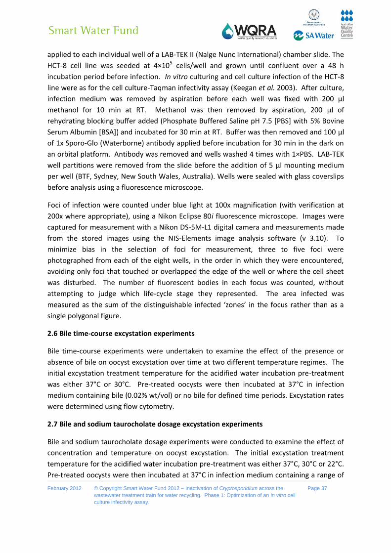
February 2012 © Copyright Smart Water Fund 2012 – Inactivation of Cryptosporidium across the
wastewater treatment train for water recycling. Phase 1: Optimization of an in vitro cell
culture infectivity assay.
Page 37
applied to each individual well of a LAB-TEK II (Nalge Nunc International) chamber slide. The
HCT-8 cell line was seeded at 4×10⁵ cells/well and grown until confluent over a 48 h
incubation period before infection. In vitro culturing and cell culture infection of the HCT-8
line were as for the cell culture-Taqman infectivity assay (Keegan et al. 2003). After culture,
infection medium was removed by aspiration before each well was fixed with 200 µl
methanol for 10 min at RT. Methanol was then removed by aspiration, 200 µl of
rehydrating blocking buffer added (Phosphate Buffered Saline pH 7.5 [PBS] with 5% Bovine
Serum Albumin [BSA]) and incubated for 30 min at RT. Buffer was then removed and 100 µl
of 1x Sporo-Glo (Waterborne) antibody applied before incubation for 30 min in the dark on
an orbital platform. Antibody was removed and wells washed 4 times with 1×PBS. LAB-TEK
well partitions were removed from the slide before the addition of 5 µl mounting medium
per well (BTF, Sydney, New South Wales, Australia). Wells were sealed with glass coverslips
before analysis using a fluorescence microscope.
Foci of infection were counted under blue light at 100x magnification (with verification at
200x where appropriate), using a Nikon Eclipse 80i fluorescence microscope. Images were
captured for measurement with a Nikon DS-5M-L1 digital camera and measurements made
from the stored images using the NIS-Elements image analysis software (v 3.10). To
minimize bias in the selection of foci for measurement, three to five foci were
photographed from each of the eight wells, in the order in which they were encountered,
avoiding only foci that touched or overlapped the edge of the well or where the cell sheet
was disturbed. The number of fluorescent bodies in each focus was counted, without
attempting to judge which life-cycle stage they represented. The area infected was
measured as the sum of the distinguishable infected ‘zones’ in the focus rather than as a
single polygonal figure.
2.6 Bile time-course excystation experiments
Bile time-course experiments were undertaken to examine the effect of the presence or
absence of bile on oocyst excystation over time at two different temperature regimes. The
initial excystation treatment temperature for the acidified water incubation pre-treatment
was either 37°C or 30°C. Pre-treated oocysts were then incubated at 37°C in infection
medium containing bile (0.02% wt/vol) or no bile for defined time periods. Excystation rates
were determined using flow cytometry.
2.7 Bile and sodium taurocholate dosage excystation experiments
Bile and sodium taurocholate dosage experiments were conducted to examine the effect of
concentration and temperature on oocyst excystation. The initial excystation treatment
temperature for the acidified water incubation pre-treatment was either 37°C, 30°C or 22°C.
Pre-treated oocysts were then incubated at 37°C in infection medium containing a range of

February 2012 © Copyright Smart Water Fund 2012 – Inactivation of Cryptosporidium across the
wastewater treatment train for water recycling. Phase 1: Optimization of an in vitro cell
culture infectivity assay.
Page 38
bile (0.00, 0.00125, 0.0025, 0.005, 0.01, 0.02, 0.04, 0.08 % [wt/vol]) and sodium
taurocholate (0.00, 0.02, 0.04, 0.08, 0.16 % [wt/vol]) concentrations for 30 min at 37°C.
Excystation rates were determined using flow cytometry.
2.8 Bile and sodium taurocholate infectivity experiments
Bile and sodium taurocholate dosage experiments were performed to examine the effect of
concentration on sporozoite infectivity. Two infectivity assays were utilized, a cell culture-
Taqman infectivity assay and the focus detection method. For the cell culture-Taqman
assay, oocysts were taken through the standard excystation methodology before being
applied to cell monolayers in infection medium containing a range of bile (0.00, 0.01, 0.02,
0.04, % [wt/vol]) and sodium taurocholate (0.00, 0.02, 0.04, 0.08 % [wt/vol]) concentrations.
For the focus detection method, oocysts were taken through a 30°C excystation in acidified
water pre-treatment before application to cell monolayers in infection medium containing
either bile (0.02, % [wt/vol]), sodium taurocholate (0.08 % [wt/vol]) or neither of these two.
2.9 Bile and sodium tauorcholate cell monolayer cytotoxcity experiments
The HCT-8 cell line used for cell culture infections was cultured in vitro as previously
described (Keegan et al. 2003). The cell line was seeded into 24 well plates (Greiner Bio-One
CELLSTAR® 662 160) at 5×10⁵ cells/well and grown until confluent over a 48 h incubation
period, after which bile and sodium taurocholate dosage experiments were conducted. Bile
and sodium taurocholate dosage experiments were performed to examine the effect of
concentration on cell cytotoxicity. A range of bile (0.00, 0.00125, 0.0025, 0.005, 0.01, 0.015,
0.02, 0.025, 0.03, 0.035, 0.04, 0.045, 0.050, 0.055, 0.060, 0.065, 0.070, 0.075, 0.08 %
[wt/vol]) and sodium taurocholate (0.00, 0.02, 0.04, 0.08, 0.16 % [wt/vol]) concentrations
were applied in cell monolayers in infection medium and incubated for a further 48 h. Cells
were then screened by light microscopy for cytotoxicity before a resazurin reduction assay
was performed.
Resazurin (Sigma-Aldrich, MO, USA) is a nonfluorescent dye, which is metabolized to a
highly fluorescent product, resorufin, by viable cells. The level of fluorescence produced is
directly proportional to the number of viable cells. Briefly, 50 µl resazurin medium (100
µg/ml resazurin in Vogel-Bonner medium), was mixed and incubated at 37°C for 1 h.
Fluorescence (RFU) at 530/580 (excitation/emission) was measured using a Wallac VICTOR3
1420 Multilabel Counter, PerkinElmer™. Cytotoxicity was expressed as percentage
reduction of cell viability compared with the no-treatment control.

February 2012 © Copyright Smart Water Fund 2012 – Inactivation of Cryptosporidium across the
wastewater treatment train for water recycling. Phase 1: Optimization of an in vitro cell
culture infectivity assay.
Page 39
2.10 Sensitivity of the revised cell culture focus detection method
A fresh batch of C. parvum oocysts (< than 3 weeks in age) were used to inoculate cell
culture monolayers. Low numbers of oocysts (5, 10, 15, 20, 25, and 30) were taken through
the modified excystation pre-treatment and inoculated onto monolayers to determine the
sensitivity of the revised technique. For each treatment, 4 replicate in vitro cell infections
were performed, while in parallel, a set of oocysts which were taken through the complete
excystation pre-treatment steps were counted at the end of the process (just before
application to the monolayer) in order to account for any losses during the pre-treatment.
This was done in order to precisely quantify the sensitivity of the cell culture component of
the modified assay.
2.11 Purification of C. hominis oocysts and species confirmation
Five human fecal samples identified as positive for Cryptosporidium were gifted from two
separate clinical diagnostic laboratories, Healthscope and IMVS (Institute of Medial and
Veterinary Sciences). Samples were processed by the Microbiology Analytical Unit (courtesy
Phil Dobson, AWQC SA Water) using a Fecal parasite concentrator (Evergreen) according to
the manufacturer’s instructions with the exception that sterile water was used instead of
formalin during step 2 to maximize oocyst infectivity after sample processing. Processed
samples were then analyzed and enumerated by microscopy as described in section 2.1.
Oocysts from each sample were then processed by FISH (Fluorescent in situ hybridization)
(courtesy Phil Dobson, AWQC SA Water) to confirm whether samples were C. hominis
or/and C. parvum positive. The two FISH probes utilized are specific to either C. parvum or
C. hominis and not other Cryptosporidium species. The FISH protocol and FISH probes
utilized are proprietary sensitive information the Microbiology Analytical Unit, AWQC, SA
Water. Therefore protocols, probes sequences and fluorochrome labels relating to the FISH
technique are not presented within this report.
2.12 Cell culture infections with Cryptosporidium hominis oocysts
All samples identified as positive by FISH for C. hominis as well as a single sample which was
neither positive for C. hominis nor C. parvum were inoculated onto cell culture monolayers
using the revised cell culture focus detection method. Briefly, oocysts were excysted and
applied to cell monolayers as described in section 2.2. For each sample, 8 replicate in vitro
cell infections were undertaken.

February 2012 © Copyright Smart Water Fund 2012 – Inactivation of Cryptosporidium across the
wastewater treatment train for water recycling. Phase 1: Optimization of an in vitro cell
culture infectivity assay.
Page 40
2.13 Statistics
Unless otherwise stated the statistical analysis conducted was a Student’s t-test and the
level of significance was stated at a P value of either <0.05 or <0.01. The mean and standard
deviation were also determined.

February 2012 © Copyright Smart Water Fund 2012 – Inactivation of Cryptosporidium across the
wastewater treatment train for water recycling. Phase 1: Optimization of an in vitro cell
culture infectivity assay.
Page 41
Chapter 3: Results & Discussion
3.1 Excystation rate for the standard excystation technique
To investigate the rate of Cryptosporidium excystation using our standard excystation
procedure, oocysts were pre-treated using the standard acidified water–trypsin pre-
treatment. Pre-treated oocysts were then subjected to varying time-courses in infection
medium before labeling with an antibody specific to an oocyst wall protein. Stained oocysts
were then subjected to flow cytometric analysis (Fig. 3), which revealed the majority of
oocysts excysting by 30 min after the initial excystation treatment. Excysting and excysted
oocysts (R2) were identified by their relative decrease in forward light scatter compared to
the intact oocysts (R1) (Fig. 1 plots A1-A3) (Vesey et al. 1997).
Fig 3. Time-course experiments of C. parvum excystation using a standard protocol.
Representative dot plots of the flow cytometric analysis of unexcysted Cryptosporidium
oocysts (A1) and excysted oocysts (A2 & A3) stained with an antibody specific to an oocyst
wall protein which does react with the sporozoites. Region 1 (R1) has previously been
identified as the region representing unexcysted oocysts, while region 2 (R2) consists of
excysted and excysting oocysts. Intact oocysts not taken through the standard excystation
method were used as non-excysted controls. Oocysts taken through the standard excystation
method (acidified water treatment at 37°C, ACH20) were incubated at 37°C in infection medium
for defined time-periods (30 min, 60 min and 120 min) before cytometric analysis (B). For the
ACH20 treatment, oocysts were taken through acidified water treatment and then placed in
infection medium before immediate flow cytometric analysis. Each data point is the mean
value from three independent experiments and the error bars indicate the standard deviations
between experiments.
A1 A2 A3
R2 R1 R2 R1 R2 R1
96%
4%
6%
94%
5%
95%
Non-excysted 30 min 120 min
B
0
10
20
30
40
50
60
70
80
90
100
Control AC H₂O 30min 60min 120min
Oo
cysts
Excyste
d (
%)

February 2012 © Copyright Smart Water Fund 2012 – Inactivation of Cryptosporidium across the
wastewater treatment train for water recycling. Phase 1: Optimization of an in vitro cell
culture infectivity assay.
Page 42
Oocysts have a low settling velocity, resulting from their small size and low specific gravity.
This rate has been reported in the literature to range between 0.22 µm/sec to 1 µm/sec
(Kim & Corapcioglu 2002; Searcy et al. 2005). Our standard 24 well plate Taqman assay uses
a total oocyst suspension volume of 1 ml which results in a medium depth of 5 mm. For the
focus detection method the medium depth within wells is 4mm. Based on the above
settling rates, oocysts near the top of the suspension could take between 1 and 5 h to reach
the monolayer. Our flow cytometric studies indicated that, using our standard excystation
assay, the majority of sporozoites excyst within 30 min post excystation treatment. Once
excysted, sporozoites go through rapid physiological and biochemical changes (apical
organelle discharge) that can dramatically reduce their infectivity (King et al. 2009;
Matsubayashi et al. 2010). We therefore postulated that in the current assay format, a
majority of the excysted sporozoites in the infection medium may not be close enough to
the cell monolayer to infect. While excysted sporozoites can actively move by helical gliding
at a maximum speed of 5.1 µm/sec (Wetzel et al. 2005), this movement is substrate
dependent and any excysted sporozoites in the medium and not in intimate contact with
the monolayer would have limited opportunity to find and infect a compatible cell before
extensive apical organelle discharge ensues (Chen et al. 2004; King et al. 2009).
3.2 Variation in medium volume for application of pretreated oocysts and the
incorporation of centrifugation of oocysts onto the monolayer
The rapid excystation of sporozoites from Cryptosporidium oocysts prompted the
investigatation parameters that may enhance the timing of sporozoite contact with a target
cell, particularly variation in the volume of medium in which pre-treated oocysts were
applied to the monolayer and addition of centrifugation of oocysts onto the monolayer.
Reducing the initial volume of the infection medium from 1 ml to 500 µl in which the pre-
treated oocysts were applied to cell monolayers resulted in increased in vitro infectivity as
determined by the Taqman cell culture assay (Single factor ANOVA, P <0.01) (Fig. 4A).
However, centrifugation of pre-treated oocysts onto the cell monolayer resulted in superior
increases in infectivity in comparison to the no-centrifuge control (approx. 5 fold) (P <0.01)
(n=6) as determined by the Taqman cell culture assay (Fig. 4B). When we attempted to
reduce the volume of the medium in which oocyst were applied to the monolayer below
500 µl, the cell monolayer lost viability and often lifted from the plate. Therefore it was
concluded that centrifugation was superior over variation of the amount of medium in
which pre-treated oocysts were applied.
To confirm that the increases measured by quantitative PCR (QPCR) were indeed reflective
of increased infectivity and not increased QPCR signal from embedded oocysts on the HCT-8
monolayer that were not removed during the washing steps, the experiment was repeated
with an additional control, consisting of oocysts inactivated by UV-C (40 mJ/cm2 dose) (Fig.

February 2012 © Copyright Smart Water Fund 2012 – Inactivation of Cryptosporidium across the
wastewater treatment train for water recycling. Phase 1: Optimization of an in vitro cell
culture infectivity assay.
Page 43
4C). The centrifuged treatment again exhibited a comparable increase in infectivity (approx.
4 fold) in comparison to our standard assay format (P <0.01). However, no significant QPCR
signal was evident for the UV treatments, supporting the hypothesis that increases in QPCR
resulted from genuine increases in cell culture infectivity. Analysis of infected monolayers
at 2 h post infection confirmed that this increase in infectivity resulted from an increase in
initial sporozoite attachment which was comparable to increases witnessed in infectivity
(approx 3-4 fold) (P <0.01) (Fig. 4D).
Fig 4. Representative experiments of variations in the volume of medium containing oocysts
applied to the HCT-8 monolayer and the incorporation of centrifugation for C. parvum in vitro
infection. Oocysts were pre-treated using standard excystation protocols before application
to HCT-8 cell monolayers. For variation in addition of medium (A), oocysts were applied to the
monolayer in varying amounts of infection medium (500-1000 µl). After 2 h incubation at 37°C,
the volume of medium in individual infected monolayer wells was made up to a total volume of
2 ml and plates incubated for a further 2 days. Infectivity was expressed as a percentage of
the standard assay format (1 ml). For the centrifugation treatments (B) & (C), pre-treated
oocysts were spun onto the monolayer at 408 g for 5 min. Control treatments without
centrifugation were left on the bench for the duration of the centrifugation spins. For the UV-C
(254 nm) treatments (C), a 40 mJ/cm2 dose was applied to the oocysts using a collimated
beam. Infectivity was calculated using a cell-culture-Taqman PCR assay and expressed as a
percentage of the no-centrifuge treatments (B-D). With the exception of the sporozoite
attachment assay, infected monolayers were incubated for 48 h at 37°C. Sporozoite
attachment/invasion was calculated using a cell-culture-Taqman PCR assay and expressed as
a percentage of the no-centrifuge control after a 2 h incubation period at 37°C. Error bars
indicate standard deviations (n=6).
0
100
200
300
400
500
600
700
No Centrifuge Centrifuge
No
rmalized
Infe
cti
on
0
100
200
300
400
500
600
700
No Centrifuge Centrifuge
Sp
oro
zo
ites d
ete
cte
d
(No
rmalized
)
0
100
200
300
400
500
600
700
Centrifuge UV Centrifuge
No Centrifuge
UV No Centrifuge
No
rmalized
Infe
cti
on
0
50
100
150
200
250
300
350
500 750 1000 1500 2000
No
rmalized
Infe
cti
on
Volume of Medium (µL)
BA
C D

February 2012 © Copyright Smart Water Fund 2012 – Inactivation of Cryptosporidium across the
wastewater treatment train for water recycling. Phase 1: Optimization of an in vitro cell
culture infectivity assay.
Page 44
To further confirm the positive affect of centrifugation on parasite in vitro culture we next
applied the step to the focus detection method. Four individual experiments were
performed with centrifugation treatments, each demonstrating an approximately 4 fold
increase in infectivity (number of foci observed) above the standard no centrifugation
controls regardless of oocyst age (P <0.01) (Table 5). This confirmed that increases in
parasite amplification were as a result of increased oocyst infections. The energy-intensive
nature of helical gliding, the finite energy reserves available to sporozoites once excysted
and their ensuing apical organelle discharge reinforce the importance of sporozoites
excysting in close proximity to a compatible host cell (King et al. 2009).
Table 5. Infectious foci detected for centrifuge and no-centrifuge treatments
Mean Foci Detected Standard Deviation
No Centrifugation Centrifugation Increase in Infectivity
Exp 1 15 2 64 16 426 %
Exp 2 14 3 49 9 350 %
Exp 3 12 4 47 10 390 %
Exp 4* 6 2 23 2 383 %
*Experiment 4 used oocysts greater than 6 months old, while for experiments 1-3 oocysts were less than 3
months old. For each experiment a minimum of 8 cell culture replicates were undertaken for each treatment.
For each cell culture well 100 oocysts were applied.
A previous report by Weir et al. (2001) identified that centrifugation of oocysts onto the
monolayer increased the rate of infection. While those authors identified more rapid
development of foci, they reported no increase in the number of infective foci by 48 h, in
contrast to the work presented here. However the authors did demonstrate significant and
large differences in foci number between non-centrifuged and centrifuged treatments for
the earlier 18 and 24 h incubation periods. Notably the authors reported that they replaced
the media at 24 h for the 48 h incubation period, possibly and inadvertently resulting in a
loss of invasive lifecycle stages. Secondly, the authors used a different excystation protocol
where oocysts were exposed to sodium hypochlorite before incubation within pre-warmed

February 2012 © Copyright Smart Water Fund 2012 – Inactivation of Cryptosporidium across the
wastewater treatment train for water recycling. Phase 1: Optimization of an in vitro cell
culture infectivity assay.
Page 45
media and centrifugation onto the monolayer, possibly delaying the excystation of oocysts
and allowing those oocysts in the non-centrifuged treatments more time to settle onto the
monolayer before excystation and subsequent infection.
Previous studies employing the focus detection method to determine in vitro infectivity and
defining foci by the criteria of Rochelle et al. (2001) have reported low infectivity of 4-22%
even for freshly excreted oocysts (Rochelle et al. 2001; Connelly et al. 2007). These results
are within the range reported when using our standard assay format, which does not
include centrifugation of oocysts onto the monolayer. The work presented here
demonstrates that oocyst excystation rate in concert with the format of the in vitro assay
utilized, notably parameters such as medium volume/depth in which oocyst inoculum is
applied, can dramatically affect assay sensitivity. The incorporation of centrifugation of
oocysts onto the monolayer in the focus detection assay format will therefore help to
maximize in vitro infectivity and assay sensitivity.
3.3 Washing of the infected cell monolayer
The length of the centrifugation period was increased to examine if all potentially infectious
oocysts were in close enough contact to infect the monolayer (Fig. 5A). However, increasing
the length of the centrifugation period did not appear to further increase infectivity,
suggesting that a short centrifugation period was sufficient to bring oocysts and sporozoites
in close enough proximity to the cell monolayer to maximize infection. A washing step was
incorporated in an attempt to increase the synchronicity of early infection by removing
those sporozoites unable to successfully attach/invade the cell monolayer. Sporozoites can
quickly attach to and invade compatible cells; the sporozoite attaches to the membrane of
a host epithelial cell by its apical end and is subsequently encapsulated by a parasitophorous
vacuole (PV) membrane (Chen et al. 2004; Borowski et al. 2008). Previous work has
reported maximal attachment/invasion of sporozoites to a cell monolayer occurs by 2 h post
infection (Chen et al. 2004; Feng et al. 2006), and this was chosen as the initial time-point in
our investigation.
The incorporation of washing the monolayer at 2 h post infection resulted in a further
positive increase in infection above that for centrifugation alone (P <0.01) (Fig. 5B).
Additionally, the time at which the monolayer was washed after initial infection was
investigated as this step could result in sporozoites which were successfully attached, but
not yet encapsulated by the host cell membrane, being accidently removed during washing
steps, thus negatively impacting in vitro infectivity. Therefore an experiment to examine the
effect of washing the monolayer at various time-points post infection was conducted. The
optimal time for washing the monolayer was determined to be between 2 and 4 h post
infection (Fig. 5C), confirming that maximal attachment occurs between by 2 h and 4 h post
February 2012 © Copyright Smart Water Fund 2012 – Inactivation of Cryptosporidium across the
wastewater treatment train for water recycling. Phase 1: Optimization of an in vitro cell
culture infectivity assay.
Page 46
infection of the monolayer. Washing the monolayer earlier reduced infection, suggesting
sporozoites that would otherwise go on to successfully infect the monolayer, were being
washed away. However, not washing the monolayer until 48 h post infection (standardassay
format) also resulted in inferior infection in comparison to the 2 and 4 h time-points (P
<0.01).
Fig 5. Representative experiments of the effect of washing and centrifugation on C. parvum in
vitro infectivity. C. parvum oocysts were pre-treated using standard excystation protocols
before application in infection medium to a HCT-8 cell monolayer. For the centrifugation
treatments, pre-treated oocysts were spun onto the monolayer at 408 g for either 5 or 20 min
(A). Paired no-centrifuge treatments were left on the bench for the duration of the
centrifugation. Pre-treated oocysts were also centrifuged onto the monolayer at 408 g for
either 5 or 20 min and infected monolayers were either washed (W) with PBS at 2 h post
infection and medium replaced, or not washed (NW) (B). Pre-treated oocysts were also
centrifuged onto the monolayer at 408 g for 5 min and infected monolayers were subjected to
various incubation periods at 37°C before washing with PBS and replacement of medium.
Infected monolayers were subsequently incubated until a total incubation period of 48 h (A-C).
Infectivity was calculated using a cell-culture-Taqman PCR assay and expressed as a
percentage of the either the no centrifuge control (5 min) (A), 5 min NW control (B) or the 48 h
incubation period no wash control (C). Error bars indicate standard deviations (n=6).
0
50
100
150
200
250
300
30min 1h 2h 4h 48h
No
rmalized
Infe
cti
on
Incubation time before washing
0
100
200
300
400
No Centrifuge
(5 min)
Centrifuge (5 min)
No Centrifuge
(20 min)
Centrifuge (20 min)
No
rmalized
Infe
cti
on
0
50
100
150
200
5 min NW 5 min W 20 min NW 20 min W
No
rmalized
Infe
cti
on
A
B
C

February 2012 © Copyright Smart Water Fund 2012 – Inactivation of Cryptosporidium across the
wastewater treatment train for water recycling. Phase 1: Optimization of an in vitro cell
culture infectivity assay.
Page 47
This increased infectivity for when the cell monolayer was washed at 2 and 4 h post
infection compared to the standard assay format was unexpected. We can think of a
number of possible explanations for this increased infectivity. Firstly, sporozoites that do
not successfully infect the monolayer may lyse and release inhibitory metabolic products
that affect the growth of other successfully infecting sporozoites and subsequent lifecycle
stages. Removing these sporozoites by washing could possibly have a beneficial effect on in
vitro infectivity. Secondly, sporozoites able to attach to a host cell but not successfully
invade may prevent subsequent lifecycles stages from infecting that cell by blocking the
binding sites for infection. Any such sporozoites are attached to the monolayer would
greatly reduce infection by other lifecycle stages. However, washing may remove these
sporozoites before the emergence of other lifecycle stages, freeing such attachment sites.
Finally, washing the monolayer after maximal sporozoite attachment/invasion could remove
other organisms such as bacteria and fungi present in the oocyst suspension but not killed
by antibiotics in the medium. Such organisms may inhibit parasite replication by the
production of inhibitory compounds and/or use of available resources in the infection
medium needed for healthy maintenance of the cell monolayer.
Our results suggest that washing the monolayer as early as 2 h post infection had no
detrimental effect on infectivity and was beneficial to in vitro parasite growth. This is in
agreement with other reports in the literature demonstrating that maximal sporozoite
attachment/invasion occurs around 2 h post inoculation of the monolayer (Chen et al. 2004;
Feng et al. 2006). A number of authors have reported washing the monolayer after initial
inoculation of sporozoites; however it does not appear to have been universally adopted. In
a number of experiments subsequently conducted using different oocyst batches (data not
presented) we have found that increases in infectivity due to washing, while always positive,
can vary considerably, signifying that the reasons behind increases in infectivity may vary as
well. This suggests the presence of other contaminating organisms/chemicals in the oocyst
suspension may be a key factor; however it is probable that increased infectivity is a
combination of the factors above, and others we have yet to consider.
While washing the cell monolayer may be a prudent practice for experiments aimed at
trying to synchronize early lifecycle stage development, we are yet to be convinced that it is
appropriate for all applications, especially the detection and enumeration of infectious
oocysts from the environment, where slowly excysting oocysts may be washed away at 2 h
post inoculation. We therefore decided to further examine this by investigating the effect of
oocyst age on oocyst excystation and this work is presented in section “3.7 Excystation rate
for aged oocysts using the modified excystation technique”. However, in extremely dirty
samples from wastewater, a strong case may be argued to include a washing step to ensure

February 2012 © Copyright Smart Water Fund 2012 – Inactivation of Cryptosporidium across the
wastewater treatment train for water recycling. Phase 1: Optimization of an in vitro cell
culture infectivity assay.
Page 48
that contaminants in the sample do not affect in vitro cell culture, while maximizing oocyst
infection and parasite replication.
3.4 The relationship between oocyst numbers applied to cell culture monolayers and the
number of foci and sporozoite equivalents enumerated
While centrifugation of oocysts onto the monolayer resulted in positive increases for in vitro
culture infectivity, it was necessary to establish that the cell monolayer did not become
saturated with lifecycle stages by effectively increasing the number of infectious oocysts in
contact with the monolayer. Therefore, standard curves to examine the relationship
between applied oocysts and infectivity were constructed for both the focus detection
method (Fig. 6A) and the cell culture Taqman assay (Fig 6B). A linear relationship was
observed between foci detected and oocyst number over a range of 5-160 oocysts
(R2=0.98). While for the Taqman assay which was less sensitive, the range was more
dynamic with a strong linear relationship demonstrated (R2=0.99) between 10-10,000
oocysts when applied to an individual monolayer.
Fig 6. Establishment of the relationship between oocyst numbers applied to cell culture
monolayers and the number of foci and sporozoite equivalents enumerated. C. parvum
oocysts were pre-treated using standard excystation protocols before centrifugation in
infection medium onto a HCT-8 cell monolayer. Varying numbers of oocysts were applied to
cell monolayers for the focus detection method (A) and the cell-culture-Taqman PCR assay
(B). Error bars indicate standard deviations (focus detection n=8) (Taqman PCR assay n=6).
3.5 Timing of excystation and the effect on foci number and size
As previously discussed, oocyst excystation rate can influence assay sensitivity and may also
influence data interpretation, depending on the assay format employed. For the focus
detection method, an individual focus of infection is theoretically defined as the area
BA
0
20
40
60
80
100
120
140
0 50 100 150 200
Fo
ci D
ete
cte
d
Oocysts Applied
10
100
1000
10000
100000
1000000
1 10 100 1000 10000 100000
Sp
oro
zo
ite
Eq
uiv
ale
nts
D
ete
cte
d
Oocysts Applied

February 2012 © Copyright Smart Water Fund 2012 – Inactivation of Cryptosporidium across the
wastewater treatment train for water recycling. Phase 1: Optimization of an in vitro cell
culture infectivity assay.
Page 49
infected by sporozoites that excyst from an individual oocyst, which then form a cluster of
intracellular life-cycle stages (Rochelle et al. 2001). Our standard excystation technique,
however, results in a considerable percentage of oocysts releasing their sporozoites before
coming into contact with the cell monolayer. This suggested the possibility that multiple
foci may develop from the sporozoites from a single oocyst, potentially confusing the
enumeration of foci. We therefore examined the possibility of slowing the initial rate of
oocyst excystation to limit the likelihood of sporozoite release before oocysts are in contact
with the monolayer. A number of different variables were trialed for their effect on the rate
of oocyst excystation; however modification of excystation temperature proved to be the
most effective parameter for variation. Reducing the temperature of the acidified water
pre-treatment of oocysts from 37°C to 30°C in concert with the addition of RT infection
medium instead of warm media greatly reduced the rate of excystation (Fig. 7A). However,
once incubated at 37°C in infection medium, a rapid rate of excystation was re-established
and by the end of a 2 h period the majority of oocysts had excysted (>85%) (Fig. 7B).
Fig 7. Time-course experiments of C. parvum excystation, using a modified excystation
protocol with alternative subsequent incubation temperatures. Intact oocysts not taken
through the excystation method were used as controls. Oocysts taken through the modified
excystation method (acidified water treatment [AC H20] at 30°C) were incubated at room
temperature in infection medium for defined time-periods (20 min, 40 min and 60 min) before
flow cytometric analysis (A). Oocysts taken through the modified excystation method were
also incubated at 37°C in infection medium for defined time-periods (30 min, 60 min and 120
min) before flow cytometric analysis (B). Each data point is the mean value from three
independent experiments and the error bars indicate the standard deviations between
experiments.
0
10
20
30
40
50
60
70
80
90
100
CONTROL AC H₂O 30min 60min 120min
Oo
cysts
Excyste
d (
%)
0
10
20
30
40
50
60
70
80
90
100
CONTROL AC H₂O 20min 40min 60min
Oo
oc
ys
ts E
xc
ys
ted
(%
)
A
B
RT Incubation
37 C Incubation

February 2012 © Copyright Smart Water Fund 2012 – Inactivation of Cryptosporidium across the
wastewater treatment train for water recycling. Phase 1: Optimization of an in vitro cell
culture infectivity assay.
Page 50
The modified excystation method resulted in significantly fewer foci (P <0.01) for both
experiments 1 and 3, and fewer (though not statistically significant) in experiment 2 (Table
6). Foci resulting from both excystation protocols consisted typically of discrete ‘zones’ of
infection (commonly two, occasionally three or four), which were interpreted as initiated
separately by sporozoites released from a single oocyst. Foci were variable in size,
consisting of fewer than 40 to more than 350 fluorescent bodies at 48 h (standard protocol -
mean 182 105, median 174; modified protocol mean 188 95, median 182; n = 25). The
area of HCT-8 cells infected correlated positively with the number of fluorescent bodies, and
was greater for the standard protocol (median 4.9 x 104 μm2, r = 0.9059) than for the
modified protocol (median 2.5 x 104 μm2, r = 0.8909) (Fig. 8). Foci of infection resulting from
the modified protocol were therefore considerably more compact (density of lifecycles
stages per μm2) than those resulting from the standard protocol (P <0.01).
Table 6. Infectious foci detected using the standard and modified excystation treatments
Mean Foci Detected Standard Deviation
Standard Excystation Modified Excystation Decrease in observed
foci
Exp 1 54 8 31 5 43%
Exp 2 42 12 34 7 19%
Exp 3* 27 4 16 4 41%
*Experiment 3 used oocysts that were 2 months old, while for experiments 1 & 2 oocysts were less than 1
month old. For each experiment a minimum of 8 cell culture replicates were undertaken for each treatment.
For each cell culture well 100 oocysts were applied.
By tailoring the excystation rate through varying the initial acidified water treatment
temperature, we were able to more precisely control excystation rate thereby increasing
the probability that a single focus of infection would be a reflection of the sporozoites from
an individual oocyst. Importantly, we demonstrated that the use of a particular excystation
methodology can significantly overestimate the number of infective oocysts present in a
sample. Therefore, if the focus detection method is to be utilized the probability of
sporozoites from an individual oocyst infecting a more circumscribed area of the cell
monolayer can be maximized by the use of centrifugation in combination with an
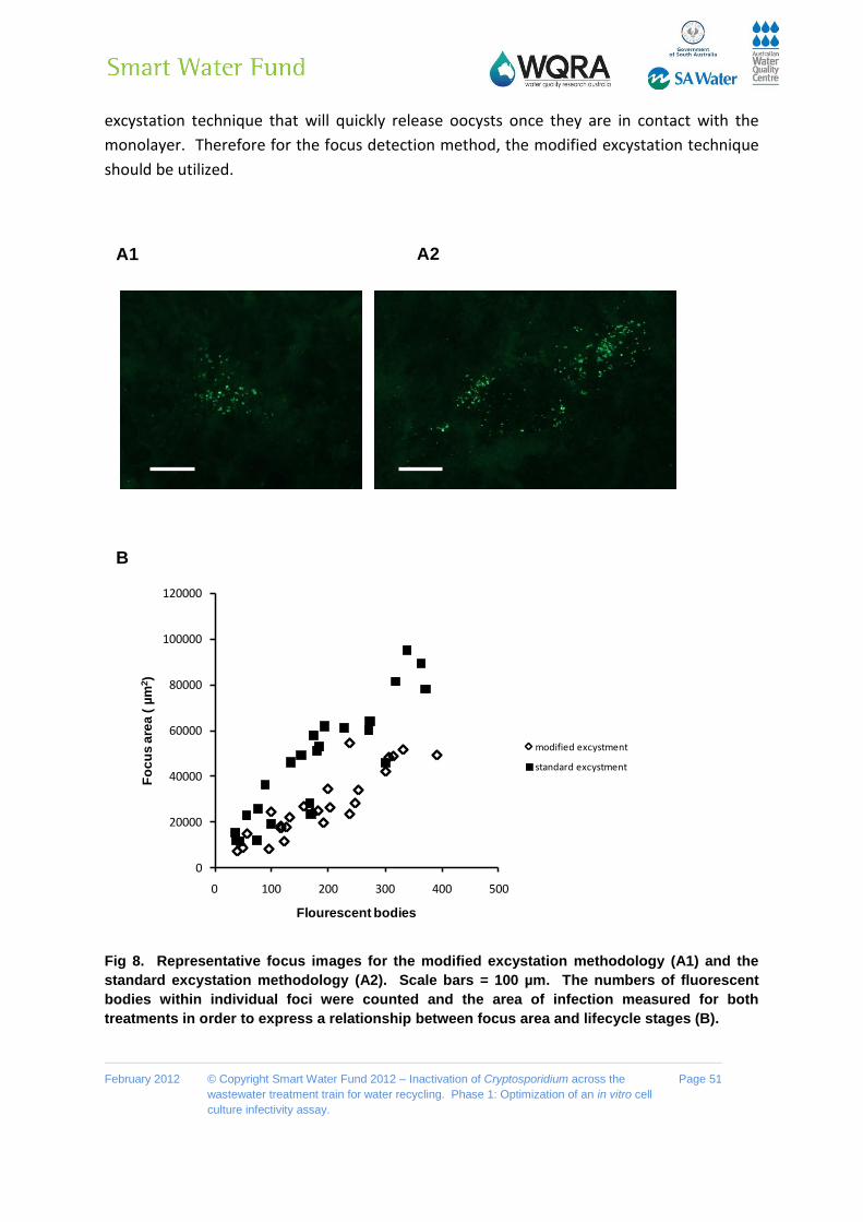
February 2012 © Copyright Smart Water Fund 2012 – Inactivation of Cryptosporidium across the
wastewater treatment train for water recycling. Phase 1: Optimization of an in vitro cell
culture infectivity assay.
Page 51
excystation technique that will quickly release oocysts once they are in contact with the
monolayer. Therefore for the focus detection method, the modified excystation technique
should be utilized.
Fig 8. Representative focus images for the modified excystation methodology (A1) and the
standard excystation methodology (A2). Scale bars = 100 µm. The numbers of fluorescent
bodies within individual foci were counted and the area of infection measured for both
treatments in order to express a relationship between focus area and lifecycle stages (B).
A1 A2
B
0
20000
40000
60000
80000
100000
120000
0 100 200 300 400 500
Fo
cu
s a
rea
( µ
m2)
Flourescent bodies
modified excystment
standard excystment

February 2012 © Copyright Smart Water Fund 2012 – Inactivation of Cryptosporidium across the
wastewater treatment train for water recycling. Phase 1: Optimization of an in vitro cell
culture infectivity assay.
Page 52
3.6 Parasite amplification in vitro and Growth phases of the foci
Centrifugation and washing of the cell monolayer was incorporated into our standard
Taqman assay format in our attempt to identify in vitro stages of parasite amplification
using qPCR. A series of 5 independent experiments was undertaken in which the cell
monolayer was infected with oocysts, the monolayer washed at 2 h post infection and then
infected monolayers recovered at different time-points during a 48 h incubation period. No
amplification in parasite numbers was detectable until 12-14 h post infection (Fig. 9). The
increase in amplification was a 2-8 fold increase above the initial numbers of sporozoites
quantified as attaching/invading the cell monolayer at 2 h. This suggested the formation of
merozoites in our in vitro cell culture system within this time frame, is in agreement with
previous work reported (Hijjawi et al. 2001). However by 16 h post infection, substantial
increases in parasite amplification in cell culture were apparent (Fig. 9). Further large
increases accompanied by longer incubation periods, suggesting that the generation time
between merozoites and subsequent parasite lifecycles is greatly reduced compared with
the initial time required between attachment/invasion of sporozoites and generation of the
first merozoites.
Fig 9. Time-course of C. parvum amplification in cell culture over 48 h. Oocysts were pre-
treated using standard excystation protocols before centrifugation in infection medium onto a
HCT-8 cell monolayer. Infected monolayers were washed with PBS at 2 h, the media replaced,
then incubated at 37°C for defined time periods before the infected monolayer was harvested.
Infectivity was calculated using a cell-culture-Taqman PCR assay and expressed as a
percentage, relative to the sporozoites that had attached/invaded the cell monolayer at 2 h
post infection. The figure represents data generated from 5 separate experiments. A 2 h time-
point was included for each experiment. Each data point represents the mean infectivity from
an individual experiment and error bars indicate standard deviations for cell culture infectivity
of that experiment (n=6).
A
10
100
1000
10000
100000
1 2 4 8 16 32 64
No
rmalized
Infe
cti
on
Incubation time (h)

February 2012 © Copyright Smart Water Fund 2012 – Inactivation of Cryptosporidium across the
wastewater treatment train for water recycling. Phase 1: Optimization of an in vitro cell
culture infectivity assay.
Page 53
Utilizing the parasite amplification data generated from the above experiments, a more
defined series of time course experiments were conducted in order to examine when we
could first accurately detect foci development. The focus detection method was therefore
employed, utilizing both the modified excystation methodology and centrifugation of
oocysts onto the monolayer in order to increase both sensitivity and synchronicity of the
assay. A series of 3 independent experiments were undertaken in which the cell monolayer
was inoculated with oocysts, infected monolayers were then incubated at 37°C for defined
time periods (16 h, 20 h, 24 h, 48 h) before being fixed, washed, stained and foci
enumerated (Fig. 10).
A
Fig 10. Time-course of C. parvum foci development in vitro over 48 h. Oocysts were pre-
treated using the modified excystation protocol before centrifugation in infection medium onto
a HCT-8 cell monolayer. Infected monolayers were then incubated at 37°C for defined time
periods (16 h, 20 h, 24 h, 48 h) before the infected monolayer was fixed, washed, stained and
foci enumerated. The figure represents data generated from 3 separate experiments. Each data
point represents the mean number of foci from an individual experiment and error bars
indicate standard deviations (n=8). Experiment 1 used oocysts that were >3 months old, while
for experiments 2 & 3 oocysts were less than 1 month old.
0
5
10
15
20
25
30
35
40
45
50
0 20 40 60
Nu
mb
er
of
Foci
Incubation time (h)
Foci v incubation time, Expt-1
Foci v incubation time, Expt-2
Foci v incubation time, Expt-3

February 2012 © Copyright Smart Water Fund 2012 – Inactivation of Cryptosporidium across the
wastewater treatment train for water recycling. Phase 1: Optimization of an in vitro cell
culture infectivity assay.
Page 54
While small with considerably less fluorescent bodies, individual foci were visible as early as
16 h post inoculation. However for each of the three experiments, the 16 h incubation
period had significantly less foci (P <0.05) compared to the standard 48 h incubation period.
The number of enumerated foci for the 20 h incubation period resulted in significantly fewer
foci (P <0.01) for experiment 3 and fewer (though not statistically significant) in both
experiments 1 & 2 in comparison to the 48 h incubation period. The number of foci for the
24 h incubation however was not significantly less than the 48 h incubation period for any of
the experiments conducted, suggesting that if needed foci could be analysed 24 h earlier.
Possible reasons for fewer foci being enumerated from the 16 h and 20 h incubations
periods may include slower excystation of some infective sporozoites and/or delayed
development of subsequent lifecycle stages after initial sporozoite infection. However foci
were quite difficult to count at both 16 & 20 h post infection due to their small size (Fig. 11),
and it was possible that a small number of foci were missed during scanning. Foci became
considerably larger for the longer incubation periods (24 h & 48 h) (Fig. 11), making counting
more accurate and efficient when scanning the slides, thereby reducing the possibility of an
individual focus of infection being missed.
Importantly oocyst age did not affect our ability to accurately enumerate foci at the earlier
incubation periods, suggesting that if required, environmentally aged infective oocysts may
be able to scanned 24 h earlier than usual after inoculation of the monolayer. While a more
rapid turnaround time for the focus detection method would be useful for a number of
applications requiring more timely information for decision making, we deemed the 48 h
incubation period superior when turnaround time of test results is not of utmost priority.
Our rationale for this was that foci were considerably larger and able to be more rapidly
scanned by both experienced and inexperienced operators at this time and hence counting
accuracy was improved.

February 2012 © Copyright Smart Water Fund 2012 – Inactivation of Cryptosporidium across the
wastewater treatment train for water recycling. Phase 1: Optimization of an in vitro cell
culture infectivity assay.
Page 55
Fig 11. Cryptosporidium foci of infection after incubation in HCT-8 cells for 16 h (a-d), 20 h (e,
f), 24 h (g, h) and 48 h (i). Scale bar = 10 μm.

February 2012 © Copyright Smart Water Fund 2012 – Inactivation of Cryptosporidium across the
wastewater treatment train for water recycling. Phase 1: Optimization of an in vitro cell
culture infectivity assay.
Page 56
3.7 Excystation rate for aged oocysts using the modified excystation technique
During our preceding investigations we identified the possibility of washing the cell
monolayer in order to improve in vitro cell culture by reducing the presence of
microbiological or chemical contaminants in the sample. Secondly we also identified the
possibility for enumerating foci at 24 h post inoculation of the monolayer instead of 48 h. In
an attempt to further assess the suitability of incorporating these steps into the focus
detection protocol we examined the effect of the modified excystation technique on
excystation of aged oocysts. Three different batches of aged oocysts, (1 month, 4 months
and 14 months) were utilized for these experiments (Fig. 12).
Fig 12. Time-course experiments of C. parvum excystation using the modified excystation
protocol for three batches of aged oocysts. Intact oocysts not taken through the standard
excystation method were used as non-excysted controls. Oocysts taken through the modified
excystation method (acidified water treatment at 30°C, ACH20) were then incubated at 37°C in
infection medium for defined time-periods (2 h, 4 h and 20 h) before cytometric analysis. Each
data point is the mean value of at least three independent experiments and the error bars
indicate the standard deviations between experiments.
For the 1 & 4 month aged oocyst batches, a significant difference in the percentage of
excysted oocysts between the batches was only evident for the unexcysted control and AC
water treatment (P <0.01), however there was no significant difference in excystation for
the 2, 4, and 20 h incubation periods. This suggested that while a number of oocysts may
have become more fragile during the ageing process, the excystation rate for the remaining
oocyst population was similar for these batches. Even though these oocysts differed in age
0
10
20
30
40
50
60
70
80
90
100
Control ACH2O 2hr 4hr 20hr
Oo
cyst
ed
Exc
yste
d (%
)
1 month age
4 months age
14 months age
February 2012 © Copyright Smart Water Fund 2012 – Inactivation of Cryptosporidium across the
wastewater treatment train for water recycling. Phase 1: Optimization of an in vitro cell
culture infectivity assay.
Page 57
by approx 3 months, they showed similar number of foci when analysed using the focus
detection method. As expected, the 14 month aged oocyst batch had an increased
population of fragile and broken oocysts with a significantly higher proportion excysting
after the initial acidified water treatment (P <0.01) compared to that of the other aged
oocyst batches. The 14 month aged oocyst batch also had a significantly slower excystation
rate over the entire incubation period when compared at each time point with either of the
other oocyst batches (P <0.01). When this batch was analysed for infectivity, no foci were
detected.
These excystation results suggested that if the cell monolayer was washed at 2 or 4 h post
inoculation, considerably aged oocysts with significantly slower excystation rates may be
removed before they have a chance to excyst and their sporozoites able to infect the
monolayer. Secondly it also suggested that if the inoculated monolayer was analysed for
foci 24 h earlier, it may be possible that slowly excysting oocysts which result in
considerably smaller foci may be missed during scanning or misinterpreted as being part of
a closely neighbouring focus. However when analysed for infectivity, no foci were detected
for the 14 month oocyst batch, suggesting that none of these sporozoites, either rapidly or
slowly excysting, were infective. Thus the incorporation of a washing step into the assay
format for certain circumstances, such as wastewater influent with heavy microbiological
and/or chemical contaminants, may be a prudent option.
While the total percentage of excysted oocysts for the 2 & 4 h incubations periods was
similar to the 20 h period for both 1 & 4 month aged oocyst batches , the difference, while
small (< approx 5%) between each of these time-points was significant (P <0.05). This again
raises the possibility that a small number of potentially infective oocysts could be lost by a
washing step. Therefore a recommendation from this work would be that washing steps
only be incorporated when samples are heavily contaminated heavy microbiological and/or
chemical contaminants that negatively affect cell monolayer growth.
3.8 The addition/absence of bile in the supplemented RPMI Maintenance Medium
(Infection medium)
While a number of triggers have been identified for oocyst excystation, including metabolic
temperature, pH fluctuations, bile salts, reducing agents and time, a lack of comprehension
of the hierarchy or synergism of specific triggers involved in Cryptosporidium excystation
still exist. Those triggers responsible for sporozoite apical organelle discharge (essential for
infection) are even more poorly understood, however this discharge has been demonstrated
to be temperature, intracellular and cytoskeleton dependant. In addition to being identified
as a trigger for excystation, bile salts have also been recognized to play a role increasing the
invasiveness of sporozoites. While the mechanism through this enhanced invasion is not yet

February 2012 © Copyright Smart Water Fund 2012 – Inactivation of Cryptosporidium across the
wastewater treatment train for water recycling. Phase 1: Optimization of an in vitro cell
culture infectivity assay.
Page 58
understood, it has been suggested that this may be via modification of the secretory
pathway, possibly involving calcium signalling. However the incorporation of bile or bile
salts into in vitro assays is not universal and recent work by some authors has reported
negative effects on in vitro parasite growth by its inclusion. Our current assay includes
bovine bile in the infection medium at a concentration of 0.02% wt/vol. We decided to
further examine this dichotomy and investigate the effect of bile and the bile salt sodium
taurocholate on both excystation and infection in our in vitro system. Time-course
experiments were conducted to examine the effect of bile on oocyst excystation using both
our standard and modified excystation technique (Fig. 12).
Fig 13. Time-course experiments of C. parvum excystation using the standard excystation (A)
and the modified excystation protocol (B) with and without the presence of bile in the infection
medium after pre-treatment at two different temperatures. Intact oocysts not taken through
the excystation method were used as controls. Oocysts were taken through either the
standard or modified excystation procedure (acidified water treatment [AC] @ 37°C or 30°C
respectively) before incubation in infection medium (+/- bile) for defined time-periods (30 min,
60 min and 120 min at 37°C) before flow cytometric analysis. Each data point is the mean
value from three independent experiments and the error bars indicate the standard deviations
between experiments.
0
10
20
30
40
50
60
70
80
90
100
CONTROL AC 30min 60min 120min
Ex
cy
ste
d O
oc
ys
ts (
%)
BILE
No Bile
0
10
20
30
40
50
60
70
80
90
100
CONTROL AC 30min 60min 120min
Ex
cy
ste
d O
oc
ys
ts (
%)
BILE
No Bile
A
B
37°C pre-treatment
30°C pre-treatment

February 2012 © Copyright Smart Water Fund 2012 – Inactivation of Cryptosporidium across the
wastewater treatment train for water recycling. Phase 1: Optimization of an in vitro cell
culture infectivity assay.
Page 59
The absence of bile in the infection medium (supplemented RPMI medium) resulted in
significance reductions (P <0.01) in excystation rate at each of the time-points analysed for
the standard excystation methodology (Fig. 13A). This effect was further exacerbated when
oocysts were pre-treated at 30°C using the modified excystation protocol (P <0.01; Fig. 13B).
These results confirm the importance of bile in the infection medium when trying to achieve
maximum excystation soon after oocysts contact the cell monolayer. This becomes even
more critical when utilizing the modified technique, as without bile, less than 50% of oocysts
had excysted by 2 h post excystation pre-treatment. These results suggested that the
efficacy of bile induced oocyst excystation is temperature dependent. Therefore to further
investigate these parameters and to examine if the concentration of bile in our infection
medium was optimal we undertook a series of experiments examining the synergism
between bile concentration and temperature of the pre-treatment on oocyst excystation.
The bile salt sodium taurocholate was also included as it had been previously reported in
the literature to achieve similar effects to bovine bile and may be suitable as a replacement.
We demonstrated the effect of bile concentration on excystation was positively associated
with excystation pre-treatment temperature for both bovine bile and the bile salt sodium
taurocholate (Fig. 14). By lowering the excystation pre-treatment temperature, an
increasing concentration of either bile or sodium taurocholate was required to obtain
similar excystation rates. However when the pre-treatment excystation temperature was
lowered to room temperature, the highest bile/sodium taurocholate concentration resulted
in only approximately 50 % excystation 30 min post pre-treatment, indicating that bile was
not a sufficient gastrointestinal signal in itself to overcoming a reduction in metabolic
temperature.
The minimum concentration of bile for optimal excystation at 37°C was 0.00125 % wt/vl,
while for 30°C it was 0.02 % wt/vl, the later concentration currently used in our infection
medium. For the bile salt sodium taurocholate the optimal concentration was 0.02% wt/vl
(lowest concentration tested) at both 30°C and 37°C. However for all sodium taurocholate
treated oocysts, total excysted oocysts at 30 min ranged between ranged 70-75 %. In
comparison, all bile concentrations at and above 0.02 % wt/vl achieved excystation rates
greater than 80 %. This suggested the possibility that a number of other components within
bovine bile may be important for excystation, not just the bile salt sodium taurocholate.

February 2012 © Copyright Smart Water Fund 2012 – Inactivation of Cryptosporidium across the
wastewater treatment train for water recycling. Phase 1: Optimization of an in vitro cell
culture infectivity assay.
Page 60
Fig 14. The effect of temperature excystation pre-treatment in combination with varying
concentrations of bile (A) and sodium taurocholate (NaTC) (B) on oocyst excystation. The
initial excystation treatment temperature for the acidified water incubation pre-treatment was
either 37°C, 30°C or 22°C. Pre-treated oocysts were then incubated at 37°C in infection medium
containing a range of bile and NaTC [wt/vol]) concentrations for 30 min at 37°C before flow
cytometric analysis. Each data point is the mean value from three independent experiments
and the error bars indicate the standard deviations between experiments.
0102030405060708090
100
0 0.02 0.04 0.06 0.08
Ex
cy
ste
d O
oc
ys
ts (
%)
Bile (% wt/vl)
37°C
30°C
22°C
B
0102030405060708090
100
0 0.05 0.1 0.15
Ex
cy
ste
d O
oc
ys
ts (
%)
NaTC (%wt/vl)
37°C
30°C
22°C
A

February 2012 © Copyright Smart Water Fund 2012 – Inactivation of Cryptosporidium across the
wastewater treatment train for water recycling. Phase 1: Optimization of an in vitro cell
culture infectivity assay.
Page 61
In addition to oocyst excystation, bile and bile salts have been suggested to be essential for
sporozoite attachment and subsequent infection. Therefore varying concentrations of both
bile and sodium taurocholate were also examined for their effect on in vitro culture of
Cryptosporidium (Fig. 15).
Fig 15. An experiment illustrating the effect of varying concentrations of bile and sodium
taurocholate (NaTC) on C. parvum in vitro infectivity. Pre-treated oocysts were suspended in
infection medium with varying amounts of bile or sodium taurocholate before centrifugation
onto cell monolayers. Infected monolayers were subsequently incubated for a period of 48 h.
Infectivity was calculated using a cell-culture-Taqman PCR assay and expressed as a
percentage of the standard treatment (bile 0.02% wt/vl). Error bars indicate standard
deviations (n=6).
The absence of bile or sodium taurocholate resulted in considerably lower in vitro infectivity
(Fig. 15), while increasing the concentration of either bile or sodium taurocholate resulted in
substantial improvements in infectivity. However replicate experiments conducted
demonstrated inconsistent results for infection for the bile concentrations greater than the
standard treatment of 0.02% wt/vol. When the cell monolayer was examined it appeared
that bile may be possibly cytotoxic at these increased concentrations (above the standard
treatment of 0.02% wt/vol), resulting in increased cell death, and occasionally total loss of
the cell monolayer.
0
50
100
150
200
250
No Bile Bile 0.01% (wt/vl)
Bile 0.02% (wt/vl)
Bile 0.04% (wt/vl)
No NaTC
NaTC 0.02% (wt/vl)
NaTC 0.04% (wt/vl)
NaTC 0.08% (wt/vl)
No
rma
lize
d In
fec
tiv
ity
(%
)

February 2012 © Copyright Smart Water Fund 2012 – Inactivation of Cryptosporidium across the
wastewater treatment train for water recycling. Phase 1: Optimization of an in vitro cell
culture infectivity assay.
Page 62
A series of experiments was therefore undertaken to examine the potential cytotoxicity of
bile and sodium taurocholate on the HCT-8 cell line (Fig. 16). For bovine bile, a gradual
cytotoxic response was observed at concentrations of 0.04 %wt/vol and greater, with strong
visual cytotoxicty observed at concentrations of 0.055 %wt/vol and greater. For sodium
taurocholate typical cytotoxic cell morphology was not observed over the range of
concentrations tested. However the resazurin reduction assay indicated decreased
metabolic activity below 70 % of the control with concentrations of sodium taurocholate
above 0.08 % wt/vol.
Fig 16. Bile and sodium tauorocholate cell cytotoxcity experiments. Confluent HCT-8 cell
monolayers were treated with a variety of bile (A) and sodium tauorocholate (B)
concentrations in the infection medium and incubated for 48 h before a resazurin reduction
assay was performed. Each data point is the mean value from four independent experiments
and the error bars indicate the standard deviations between experiments.
Based on the above information we incorporated both bile and sodium taurocholate in the
focus detection method at concentrations that should increase in vitro infectivity but not
result in a cytotoxic response. Both bile (0.02% wt/vl) and sodium taurocholate (0.08%
wt/vl) treatments resulted in a significantly (p<0.01) greater number of foci than treatments
without either bile or sodium taurocholate for each experiment conducted (Table 7). These
foci were also considerably larger with more lifecycle stages. While there was no significant
difference between the number of foci from the bile and sodium taurocholate treatments, it
was observed that foci from the sodium taurocholate treatment were smaller with fewer
lifecycle stages, suggesting that other components within bovine bile may be important for
in vitro infection. Therefore we concluded that bile was an essential supplement for both
A B
0
20
40
60
80
100
120
0 0.05 0.1 0.15
No
rma
lize
d R
es
azu
rin
A
cti
vit
y
NaTC (%wt/vl)
0
20
40
60
80
100
120
0 0.02 0.04 0.06 0.08
No
rma
lize
d R
es
azu
rin
A
cti
vit
y
Bile (%wt/vl)

February 2012 © Copyright Smart Water Fund 2012 – Inactivation of Cryptosporidium across the
wastewater treatment train for water recycling. Phase 1: Optimization of an in vitro cell
culture infectivity assay.
Page 63
oocyst excystation and in vitro cell culture, and that sodium taurocholate was not an
adequate substitute.
Table 7. Infectious foci detected for Bile and Sodium Tauorocholate (NaTC) treatments
Mean Foci Detected Standard Deviation
No Bile Bile NaTC
Exp 1 5 2 31 5 * n.p.
Exp 2 8 5 27 4 28 4
Exp 3 9 5 23 6 22 5
For each experiment a minimum of 8 cell culture replicates were undertaken for each treatment. *This
treatment was not performed for experiment 1. For each cell culture well 100 oocysts were applied.
3.9 Sensitivity of the revised cell culture focus detection method with low number of
inocula
Low numbers of oocysts (5, 10, 15, 20, 25, and 30) were taken through the modified
excystation pre-treatment and inoculated onto monolayers to determine the sensitivity of
the revised technique. In parallel, a set of oocysts which were taken through the complete
excystation pre-treatment steps were counted at the end of the process in order to account
for any losses during the pre-treatment before oocyst addition to the monolayer. The
parallel oocyst counts confirmed that there was no significant loss in oocyst numbers after
the excystation pre-treatment steps, and oocysts numbers were within expectations of the
predicted number (Table 8).

February 2012 © Copyright Smart Water Fund 2012 – Inactivation of Cryptosporidium across the
wastewater treatment train for water recycling. Phase 1: Optimization of an in vitro cell
culture infectivity assay.
Page 64
Table 8. Infectious foci detected using the revised focus detection after application of low
numbers of oocysts (n=4).
Predicted number of
Oocysts applied to
cell culture
Mean oocyst
numbers after pre-
treatment Standard
Deviation
Mean Foci Standard
Deviation
Infection rate
5 5 1.8 3 1.4 60%
10 11 3 2.7 0.6 25%
15 13 6.4 5 1.4 33%
20 29 1.2 9 2.6 31%
25 25 6.1 10.8 3.1 43%
30 36 7.0 16.3 2.2 45%
A strong linear relationship was found to exist between foci observed and the number of
oocysts applied to in vitro cell culture (mean oocysts count after pre-treatment) (Fig. 17).
The number of foci observed for oocysts applied was consistent with results generated
when higher levels of inocula were used for in vitro culture.
Fig. 17 The relationship between foci detected and low numbers of oocyst inocula
y = 0.4165x - 0.4819R² = 0.8959
0
2
4
6
8
10
12
14
16
18
20
0 10 20 30 40
Fo
ci D
ete
cte
d
Oocysts Applied to the Monolayer

February 2012 © Copyright Smart Water Fund 2012 – Inactivation of Cryptosporidium across the
wastewater treatment train for water recycling. Phase 1: Optimization of an in vitro cell
culture infectivity assay.
Page 65
Using the linear relationship established between foci detected and oocysts applied to the
monolayer, the in vitro infection rate for a fresh oocyst batch was determined to be
approximately 40%. This is a significantly large increase in sensitivity considering previously
reported in vitro cell culture infection rates range between 5-20%. Importantly this suggests
that the detection limit for the modified assay may be approximately 2-3 infectious oocysts.
The current approach for Cryptosporidium is to assume that all confirmed Cryptosporidium
(Cryptosporidium detected with internal contents) are infectious. This is a very conservative
approach to determining a detection limit, because it infers that the 60% of oocysts that do
not infect the cell line are actually infectious and are not capable of infecting within our in
vitro system due to unknown limitations of the assay. However, it is much more likely that a
proportion of these oocysts are not infectious to humans and unable to infect the cell
monolayer. Though, the proportion of oocysts which are either dead or non-infectious is
unknown. For example, a small proportion of freshly excreted oocysts (< 5%) never excyst
because they are dead, empty or defective, thereby contributing to those oocysts which
cannot infect the monolayer.
Despite the fact that an unknown proportion of oocysts are non-infective, we find it
counterintuitive from an evolutionary perspective (fitness/metabolic cost) that this could be
as high as 60%. It is more likely that limitations to infection still exist for our in vitro culture
system. These may include incompatibility of some cells within the monolayer to infection.
We have observed that foci infection does not appear to be randomly distributed across the
cell monolayer and that there are areas of cells that appear not to support infection and
areas that appear to support a higher density of foci. The compatibility of cells to infection
is complex, as a compatible cell must express the correct array of ligands as well as exhibit
other critical host cell determinants allowing the sporozoite to attach and successfully infect
the cell. Those host factors required for successful infection are becoming better
understood, however their expression levels and patterns in cell types used for cell infection
is still poorly understood. Further investigation into these parameters may be warranted if
future assays with a detection limit of 1 infectious oocyst are to be developed. However it
must still be considered that it is a possibility that a large percentage of freshly excreted are
indeed non-infectious, and that the detection limit of our assay may indeed be 1 infectious
oocyst. Our ability to be able to effectively detect down to 2-3 infectious oocysts (possibly
lower) has been a strident improvement over existing assays.

February 2012 © Copyright Smart Water Fund 2012 – Inactivation of Cryptosporidium across the
wastewater treatment train for water recycling. Phase 1: Optimization of an in vitro cell
culture infectivity assay.
Page 66
3.10 Cell culture infections with C. hominis oocysts
Unlike C. parvum, C. hominis is not zoonotic, instead it is human specific. Therefore this
makes C. hominis oocysts not readily available as they are dependent on human
cryptosporidiosis infections and access to the human fecal samples from those patients.
However, due to a timely increase in the incidence of cryptosporidiosis in Adelaide towards
the end of this project, we were able to source five positive human fecal samples for
Cryptosporidium from two clinical laboratories (Table 9).
Table 9. Infectious foci detected using the revised focus detection method from C. hominis
identified oocysts
Sample
ID.
Origin Sample
Description
FISH Probe
Hybridization
Results
Infectivity
(Total foci
observed
over 8 reps)
Infectivity Mean
Foci/100 oocysts
and standard
deviation
P1 Healthscope Dry smear C. homins (-ve)
C. parvum (-ve)
3 0.4/100 0.5
P2 Healthscope Fresh/Not
Watery
C. hominis (+ve)
C. parvum (-ve)
8 1/100 1.0
P3 Healthscope Fresh/Not
Watery
C. hominis (+ve)
C. parvum (-ve)
143 18/100 3.6
P4 IMVS Fresh/Not
Watery
C. hominis (+ve)
C. parvum (-ve)
21 2.7/100 1.9
P5 IMVS Fresh/Not
Watery
C. hominis (+ve)
C. parvum (-ve)
31 3.9/100 1.5

February 2012 © Copyright Smart Water Fund 2012 – Inactivation of Cryptosporidium across the
wastewater treatment train for water recycling. Phase 1: Optimization of an in vitro cell
culture infectivity assay.
Page 67
Of the five positive samples, four (P2-P5) were determined to be C. hominis infections by
FISH. These four samples also did not react with the C. parvum probe suggesting that the
infection was not mixed. A fifth sample (P1) positive for Cryptosporidium did not react with
either the C. parvum or C. hominis probe, suggesting that that either the viability of these
oocysts was compromised or that this may be an unusual species infection.
All samples were inoculated onto cell culture. For sample P1, only three infectious foci were
observed out of a total of 800 oocysts inoculated onto the cell culture monolayers. This
sample was a dry smear when received and therefore the viability of these oocysts was
most likely compromised, a possibility why neither FISH probe hybridized strongly to the
oocysts, and that these oocysts were most likely not another Cryptosporidium species
infection.
While cell monolayers were inoculated with the same number of oocysts for the remaining
4 samples, there was a large degree of variability between samples for the number of foci
observed. Sample P3, had a level of infection comparable to infection levels seen with some
C. parum oocyst batches received by our laboratory. However, all other samples were
substantially lower in infectivity. The foci observed for all the samples analyzed were typical
of foci we have observed using the modified technique for C. parvum infection. The
variation within a sample was also typical of that seen in vitro for C. parvum infections.
One of the aims of this work was to determine if our modified in vitro cell culture assay was
capable of supporting C. hominis infection, as all preliminary experimental work for assay
optimization had been undertaken using the model organism C. parvum. Our results
suggest that our modified assay is capable of supporting C. hominis in vitro infections, and
that those foci which develop are typical of those observed for in vitro culture of C. parvum.
It was noteworthy that not all C. hominis samples had equivalent levels of in vitro infectivity,
and generally infection levels were lower than seen in C. parvum. However only a limited
amount of information can be inferred about comparative levels of infection between the
two species as there is a limited level of sample control for the human fecal samples when
compared to the available certified sources of C. parvum. Samples were all greater than 4
weeks of age when they were inoculated onto cell monolayers with the exception of sample
P3, which was less than 4 weeks and exhibited infectivity of similar levels to that of C.
parvum. Length of storage and how samples are stored are parameters well known to
influence oocyst infectivity. While all samples were stored at 4°C when received, the history
of storage conditions before receipt is unknown, especially around the initial collection from
patient to health service provider is considered to be very influential.
It was noted that none of the samples received could be described as watery diarrhea, a
common descriptor of the disease of cryptosporidiosis. While symptoms appear from 2 to

February 2012 © Copyright Smart Water Fund 2012 – Inactivation of Cryptosporidium across the
wastewater treatment train for water recycling. Phase 1: Optimization of an in vitro cell
culture infectivity assay.
Page 68
10 days after infection, with an average of 7 days, and lasting usually for up to 2 weeks, the
disease is usually self-limiting. It is therefore possible when fecal samples were collected by
patients the disease was towards the end of the infection cycle and diarrhea had ceased.
This is important as watery diarrhea is often associated with severe cramps and rapid bowel
movements resulting in rapid shedding off oocysts from the host. When patients become
asymptomatic or are between watery diarrhea episodes, oocysts will still be shed but the
passage through the host will have a significantly increased residence time. An increased
residence time at high metabolic temperatures may therefore significantly reduce oocyst
infectivity.
Finally to purify the Cryptosporidium oocysts a fecal parasite concentrator kit was utilized.
This kit and similar kits employ ethyl acetate to breakdown fats within the sample, however
we are uncertain whether this may potentially affect oocyst infectivity and were unable to
find any references in the literature in regards to this with the exception of one reference
suggesting that there no affect on viability. However the parameter of viability cannot
necessarily be accurately extended to oocyst infectivity.

February 2012 © Copyright Smart Water Fund 2012 – Inactivation of Cryptosporidium across the
wastewater treatment train for water recycling. Phase 1: Optimization of an in vitro cell
culture infectivity assay.
Page 69
Conclusions
In this work we examined factors affecting the timing of contact between excysted
sporozoites and target host cells and the subsequent impact of this upon the
establishment of infection. We demonstrate that excystation rate impacts upon
establishment of infection. In our standard assay format the majority of sporozoites
are not close enough to the cell monolayer when they are released from the oocyst
to successfully establish infection. However, this can be easily overcome by
centrifugation of oocysts onto the cell monolayer, resulting in approximately 4 fold
increases in sporozoite attachment and subsequent infection.
We further demonstrate that excystation procedures can be tailored to control
excystation rate to match the assay end purpose and that excystation rate can
influence data interpretation. By reduction of the initial acid pre-treatment
temperature (from 37°C to 30°C) we were able to successfully control oocyst
excystation rate, so that pre-treated oocysts could be centrifuged onto the cell
monolayer before they excyst. This gives greater confidence that an individual focus
is the result of a single infectious oocyst because it reduces the possibility that
multiple foci may develop from the sporozoites released by a single oocyst.
The addition of a washing step post sporozoite attachment may be appropriate
when considering the water matrix from which the samples have originated. It may
be worthwhile to conduct some additional experiment where oocysts are spiked into
different water matrices (eg. wastewater influent, filtered water, reagent water) to
determine how beneficial a washing step may be when incorporated into the
modified assay. Previous analyses using the standard method demonstrated that the
method of recovery of Cryptosporidium had a significant effect on the infectivity
assay (data not shown), with great importance placed on the cleanup of the oocysts
after the initial concentration process
Opportunity exists to enumerate the foci 24 h earlier if reduced turnaround time of
results is paramount. However, as foci become larger with increasing incubation
time, 48 h is deemed to be the most suitable incubation period as it permits slides to
be scanned more rapidly.
Bile is an essential supplement needed for oocyst excystation, sporozoite infection
and subsequent amplification of the parasite lifecycle within in vitro culture. The
current concentration of bile within the infection medium is optimal, with higher
concentrations resulting in cell cytotoxicity and possible assay failure. The bile salt

February 2012 © Copyright Smart Water Fund 2012 – Inactivation of Cryptosporidium across the
wastewater treatment train for water recycling. Phase 1: Optimization of an in vitro cell
culture infectivity assay.
Page 70
sodium taurocholate does not appear to be a complete replacement for total bovine
bile as its incorporation in the medium results in slightly reduced excystation and a
considerable reduction in focus size.
Our modified assay is capable of supporting C. hominis in vitro infections, and the
foci which develop are typical of those observed for in vitro culture of C. parvum. At
present we have an optimized cell culture infectivity assay, with a 4 fold increase in
assay sensitivity, improved synchronicity of infection and a modified excystation
methodology allowing greater precision in defining an infective focus formed as a
result of a single oocyst infection. Our limit of detection is approximately 2-3
infectious oocysts. Therefore this assay should be useful to accurately quantify not
only the removal of oocysts from various stages of the wastewater treatment train
but also their inactivation, thereby accurately quantifying the “true effect” of the
treatment train on oocyst risk reduction.

February 2012 © Copyright Smart Water Fund 2012 – Inactivation of Cryptosporidium across the
wastewater treatment train for water recycling. Phase 1: Optimization of an in vitro cell
culture infectivity assay.
Page 71
The Revised Focus Detection Method
Our revised infectivity assay is presented below with the new steps underlined;
The HCT-8 cells are cultured in growth medium within a humidified 37°C incubator
with 5% CO2 and split every 2 to 3 days until the cell monolayer is >85% confluent as
previously described (Meloni and Thompson 1996).
Split cells, are diluted in growth medium and seeded onto LabTek II slides at 4×10⁵
cells/well and grown until confluent over a 48 h incubation period before infection.
Oocysts are pre-treated using acidified water (pH 2.4) with trypsin (0.025% [wt/vol]
and incubated at 30°C for 20 min.
Oocysts are then concentrated by centrifugation (10 min at 1800 g) before the
supernatant (excystation solution) is removed down to 50 µL.
The pre-treated oocysts are then resuspended in a final volume of 400µL room
temperature (RT) maintenance medium (infection medium) (Meloni and Thompson,
1996).
The growth medium is removed from the LabTek II slide chamber and the pre-
treated oocysts are applied to HCT-8 cells in Labtek II slide well/s.
Inoculated LabTek slides are then centrifuged at 408 g for 5 min to spin oocysts onto
the cell monolayer.
Inoculated monolayers are then incubated at 37°C, 5% CO2 in a humidified air
incubator for 48 h. If a rapid turnaround in results is required, inoculated
monolayers can be incubated for a minimum of 24 h.
If samples are from water matrices heavily contaminated with other microorganisms
and /or contaminants which have been problematic for in vitro cell culture (eg.
cytotoxic) a washing step can be incorporated at 4 h post inoculation of oocysts. The
maintenance medium (infection medium) is carefully aspirated from the Labtek II
slide well/s and replaced with fresh warm maintenance medium (infection medium)
before the inoculated monolayers are then incubated at 37°C, 5% CO2 in a
humidified air incubator for a further 44 h.

February 2012 © Copyright Smart Water Fund 2012 – Inactivation of Cryptosporidium across the
wastewater treatment train for water recycling. Phase 1: Optimization of an in vitro cell
culture infectivity assay.
Page 72
After incubation the medium is aspirated from the slides, followed by rinsing three
times with phosphate buffered saline (PBS) before fixing the cells with 200 µl of
absolute methanol for 10 min at RT.
The methanol is then removed by aspiration and the cell monolayer is blocked for 30
min with 200 µl blocking buffer (containing 1 x Dulbecco’s PBS, Tween 20 and 0.5%
BSA) to rehydrate the cells.
The buffer is then removed and 100 µl of 1 x solution Sporo-Glo™ polyclonal
antibody applied before incubation at room temperature for 30 min (in the dark) on
an orbital shaker
The antibody solution is then removed and the cell monolayer washed 4 x with PBS
The labtek well partitions are then removed from the slide before the addition of 5
µl non-quenching mounting medium per well and coverslip applied to seal wells.
Slides are then analysed by fluorescence microscopy and individual infectious
clusters (i.e. foci) enumerated using blue light to observe FITC labelled lifecycle
stages.

February 2012 © Copyright Smart Water Fund 2012 – Inactivation of Cryptosporidium across the
wastewater treatment train for water recycling. Phase 1: Optimization of an in vitro cell
culture infectivity assay.
Page 73
References
AGWR. (2008). Australian Guidelines for Water Recycling, Managing Health and
Environmental risks. Natural Resource Management Ministerial Council,
Environmental Protection and Heritage Council and Australian Health Ministers
Conference.
Arrowood, M. J., Sterling, C. R. and Healey, M. C. (1991). Immunofluorescent microscopical
visualization of trails left by gliding Cryptosporidium parvum sporozoites. J Parasitol
77, 315-317.
Arrowood, M. J. (2002). In vitro cultivation of Cryptosporidium species. Clin Microbiol Rev
15, 390-400.
Belosevic, M., Guy, R. A., Taghi-Kilani, R., Neumann, N. F., Gyurek, L. L., Liyanage, L. R.,
Millard, P. J. and Finch, G. R. (1997). Nucleic acid stains as indicators of
Cryptosporidium parvum oocyst viability. Int J Parasitol 27, 787-798.
Black, E. K., Finch, G. R., Taghi-Kilani, R. and Belosevic, M. (1996). Comparison of assays for
Cryptosporidium parvum oocysts viability after chemical disinfection. FEMS Microbiol
Lett 135, 187-189.
Borowski, H., Clode, P. L. and Thompson, R. C. (2008). Active invasion and/or
encapsulation? A reappraisal of host-cell parasitism by Cryptosporidium. Trends
Parasitol 24, 509-516.
Campbell, A. T., Robertson, L. J. and Smith, H. V. (1992). Viability of Cryptosporidium
parvum oocysts: correlation of in vitro excystation with inclusion or exclusion of
fluorogenic vital dyes. Appl Environ Microbiol 58, 3488-3493.
Chen, X. M., O'Hara, S. P., Huang, B. Q., Nelson, J. B., Lin, J. J., Zhu, G., Ward, H. D. and
Larusso, N. F. (2004). Apical organelle discharge by Cryptosporidium parvum is
temperature, cytoskeleton, and intracellular calcium dependent and required for
host cell invasion. Infection and Immunity 72, 6806-6816.

February 2012 © Copyright Smart Water Fund 2012 – Inactivation of Cryptosporidium across the
wastewater treatment train for water recycling. Phase 1: Optimization of an in vitro cell
culture infectivity assay.
Page 74
Choudhry, N., Bajaj-Elliott, M. and Mcdonald, V. (2008). The terminal sialic acid of
glycoconjugates on the surface of intestinal epithelial cells activates excystation of
Cryptosporidium parvum. Infect Immun 76, 3735-3741.
Connelly, S. J., Wolyniak, E. A., Williamson, C. E. and Jellison, K. L. (2007). Artificial UV-B
and solar radiation reduce in vitro infectivity of the human pathogen
Cryptosporidium parvum. Environ Sci Technol 41, 7101-7106.
Current, W. L. and Haynes, T. B. (1984). Complete development of Cryptosporidium in cell
culture. Science 224, 603-605.
Current, W.L. (1990). Technique and laboratory maintenance of Cryptosporidium. In: Fayer,
R., Dubey, J.P, Speer C.A. (eds.) Cryptosporidiosis of man and animals. CRC Press,
Florida, pp 31-49.
Di Giovanni, G. D., Hashemi, F. H., Shaw, N. J., Abrams, F. A., Lechevallier, M. W. and
Abbaszadegan, M. (1999). Detection of infectious Cryptosporidium parvum oocysts
in surface and filter backwash water samples by immunomagnetic separation and
integrated cell culture-PCR. Appl Environ Microbiol 65, 3427-3432.
Di Giovanni, G. D. and Lechevallier, M. W. (2005). Quantitative-PCR assessment of
Cryptosporidium parvum cell culture infection. Appl Environ Microbiol 71, 1495-1500.
Dupont, H. L., Chappell, C. L., Sterling, C. R., Okhuysen, P. C., Rose, J. B. and Jakubowski,
W. (1995). The infectivity of Cryptosporidium parvum in healthy volunteers. N Engl J
Med 332, 855-859.
Favennec, L. (1997). Physiopathologic and therapeutic studies in in vitro and in vivo models
of Cryptosporidium parvum infection. J Eukaryot Microbiol 44, 69S-70S.
Fayer, R. and Leek, R. G. (1984). The effects of reducing conditions, medium, pH,
temperature, and time on in vitro excystation of Cryptosporidium. J Protozool 31,
567-569.
Feng, H., Nie, W., Sheoran, A., Zhang, Q. and Tzipori, S. (2006). Bile acids enhance
invasiveness of Cryptosporidium spp. into cultured cells. Infect Immun 74, 3342-
3346.

February 2012 © Copyright Smart Water Fund 2012 – Inactivation of Cryptosporidium across the
wastewater treatment train for water recycling. Phase 1: Optimization of an in vitro cell
culture infectivity assay.
Page 75
Forney, J. R., Yang, S. and Healey, M. C. (1996). Protease activity associated with
excystation of Cryptosporidium parvum oocysts. J Parasitol 82, 889-892.
Forney, J. R., Vaughan, D. K., Yang, S. and Healey, M. C. (1998). Actin-dependent motility in
Cryptosporidium parvum sporozoites. J Parasitol 84, 908-913.
Griffiths, J. K., Moore, R., Dooley, S., Keusch, G. T. and Tzipori, S. (1994). Cryptosporidium
parvum infection of Caco-2 cell monolayers induces an apical monolayer defect,
selectively increases transmonolayer permeability, and causes epithelial cell death.
Infect Immun 62, 4506-4514.
Gut, J. and Nelson, R. G. (1999). Cryptosporidium parvum: synchronized excystation in vitro
and evaluation of sporozoite infectivity with a new lectin-based assay. J Eukaryot
Microbiol 46, 56S-57S.
Hijjawi, N. S., Meloni, B. P., Morgan, U. M. and Thompson, R. C. A. (2001). Complete
development and long-term maintenance of Cryptosporidium parvum human and
cattle genotypes in cell culture. Int J Parasitol 31, 1048-1055.
Hijjawi, N. S., Meloni, B. P., Ryan, U. M., Olson, M. E. and Thompson, R. C. (2002).
Successful in vitro cultivation of Cryptosporidium andersoni: evidence for the
existence of novel extracellular stages in the life cycle and implications for the
classification of Cryptosporidium. Int J Parasitol 32, 1719-1726.
Hijjawi, N. S., Meloni, B. P., Ng'anzo, M., Ryan, U. M., Olson, M. E., Cox, P. T., Monis, P. T.
and Thompson, R. C. (2004). Complete development of Cryptosporidium parvum in
host cell-free culture. Int J Parasitol 34, 769-777.
Huang, B. Q., Chen, X. M. and Larusso, N. F. (2004). Cryptosporidium parvum attachment to
and internalization by human biliary epithelia in vitro: A morphologic study. Journal
of Parasitology 90, 212-221.
Jenkins, M., Trout, J. M., Higgins, J., Dorsch, M., Veal, D. and Fayer, R. (2003). Comparison
of tests for viable and infectious Cryptosporidium parvum oocysts. Parasitol Res 89,
1-5.
Jex, A. R., Pangasa, A., Campbell, B. E., Whipp, M., Hogg, G., Sinclair, M. I., Stevens, M. and
Gasser, R. B. (2008). Classification of Cryptosporidium species from patients with

February 2012 © Copyright Smart Water Fund 2012 – Inactivation of Cryptosporidium across the
wastewater treatment train for water recycling. Phase 1: Optimization of an in vitro cell
culture infectivity assay.
Page 76
sporadic cryptosporidiosis by use of sequence-based multilocus analysis following
mutation scanning. J Clin Microbiol 46, 2252-2262.
Joachim, A., Eckert, E., Petry, F., Bialek, R. and Daugschies, A. (2003). Comparison of
viability assays for Cryptosporidium parvum oocysts after disinfection. Vet Parasitol
111, 47-57.
Johnson, A. M., Linden, K., Ciociola, K. M., De Leon, R., Widmer, G. and Rochelle, P. A.
(2005). UV inactivation of Cryptosporidium hominis as measured in cell culture. Appl
Environ Microbiol 71, 2800-2802.
Karanis, P., Kimura, A., Nagasawa, H., Igarashi, I. and Suzuki, N. (2008). Observations on
Cryptosporidium life cycle stages during excystation. J Parasitol 94, 298-300.
Keegan, A. R., Fanok, S., Monis, P. T. and Saint, C. P. (2003). Cell culture-Taqman PCR assay
for evaluation of Cryptosporidium parvum disinfection. Appl Environ Microbiol 69,
2505-2511.
Kim, S. B. and Corapcioglu, M. Y. (2002). Vertical transport of Cryptosporidium parvum
oocysts through sediments. Environ Technol 23, 1435-1446.
King, B. J., Keegan, A. R., Monis, P. T. and Saint, C. P. (2005). Environmental temperature
controls Cryptosporidium oocyst metabolic rate and associated retention of
infectivity. Appl Environ Microbiol 71, 3848-3857.
King, B. J., Hoefel, D., Lim, S. P., Robinson, B. S. and Monis, P. T. (2009). Flow cytometric
assessment of distinct physiological stages within Cryptosporidium parvum
sporozoites post-excystation. Parasitology 136, 953-966.
Korich, D. G., Marshall, M. M., Smith, H. V., O'grady, J., Bukhari, Z., Fricker, C. R., Rosen, J.
P. and Clancy, J. L. (2000). Inter-laboratory comparison of the CD-1 neonatal mouse
logistic dose-response model for Cryptosporidium parvum oocysts. J Eukaryot
Microbiol 47, 294-298.
Lalancette, C., Di Giovanni, G. D. and Prevost, M. (2010). Improved risk analysis by dual
direct detection of total and infectious Cryptosporidium oocysts on cell culture in
combination with immunofluorescence assay. Appl Environ Microbiol 76, 566-577.

February 2012 © Copyright Smart Water Fund 2012 – Inactivation of Cryptosporidium across the
wastewater treatment train for water recycling. Phase 1: Optimization of an in vitro cell
culture infectivity assay.
Page 77
Mallon, M. E., Macleod, A., Wastling, J. M., Smith, H. and Tait, A. (2003). Multilocus
genotyping of Cryptosporidium parvum Type 2: population genetics and sub-
structuring. Infect Genet Evol 3, 207-218.
Matsubayashi, M., Ando, H., Kimata, I., Nakagawa, H., Furuya, M., Tani, H. and Sasai, K.
(2010). Morphological changes and viability of Cryptosporidium parvum sporozoites
after excystation in cell-free culture media. Parasitology 137, 1861-1866.
Meloni, B. P. and Thompson, R. C. (1996). Simplified methods for obtaining purified oocysts
from mice and for growing Cryptosporidium parvum in vitro. J Parasitol 82, 757-762.
Morgan, U. M., Pallant, L., Dwyer, B. W., Forbes, D. A., Rich, G. and Thompson, R. C.
(1998). Comparison of PCR and microscopy for detection of Cryptosporidium parvum
in human fecal specimens: clinical trial. J Clin Microbiol 36, 995-998.
Morgan, U. M., Xiao, L., Fayer, R., Lal, A. A. and Thompson, R. C. (2000). Epidemiology and
strain variation of Cryptosporidium parvum. Contrib Microbiol 6, 116-139.
Nesterenko, M. V., Tilley, M. and Upton, S. J. (1995). A metallo-dependent cysteine
proteinase of Cryptosporidium parvum associated with the surface of sporozoites.
Microbios 83, 77-88.
Neumann, N. F., Gyurek, L. L., Finch, G. R. and Belosevic, M. (2000). Intact Cryptosporidium
parvum oocysts isolated after in vitro excystation are infectious to neonatal mice.
FEMS Microbiol Lett 183, 331-336.
O'Brien, E., Mcinnes, L. and Ryan, U. (2008). Cryptosporidium GP60 genotypes from humans
and domesticated animals in Australia, North America and Europe. Exp Parasitol 118,
118-121.
Okhuysen, P. C., Chappell, C. L., Crabb, J. H., Sterling, C. R. and Dupont, H. L. (1999).
Virulence of three distinct Cryptosporidium parvum isolates for healthy adults. J
Infect Dis 180, 1275-1281.
Okhuysen, P. C., Rich, S. M., Chappell, C. L., Grimes, K. A., Widmer, G., Feng, X. and Tzipori,
S. (2002). Infectivity of a Cryptosporidium parvum isolate of cervine origin for healthy
adults and interferon-gamma knockout mice. J Infect Dis 185, 1320-1325.

February 2012 © Copyright Smart Water Fund 2012 – Inactivation of Cryptosporidium across the
wastewater treatment train for water recycling. Phase 1: Optimization of an in vitro cell
culture infectivity assay.
Page 78
Ong, C. S., Eisler, D. L., Alikhani, A., Fung, V. W., Tomblin, J., Bowie, W. R. and Isaac-
Renton, J. L. (2002). Novel cryptosporidium genotypes in sporadic cryptosporidiosis
cases: first report of human infections with a cervine genotype. Emerg Infect Dis 8,
263-268.
Pezzani, B. C., Bautista, E., Cordoba, A., De Luca, M. M. and Basualdo, J. A. (1998). Factors
affecting the in vitro excystation of Cryptosporidium sp. Rev Argent Microbiol 30,
138-142.
Pieniazek, N. J., Bornay-Llinares, F. J., Slemenda, S. B., Da Silva, A. J., Moura, I. N.,
Arrowood, M. J., Ditrich, O. and Addiss, D. G. (1999). New cryptosporidium
genotypes in HIV-infected persons. Emerg Infect Dis 5, 444-449.
Reduker, D. W., Speer, C. A. and Blixt, J. A. (1985). Ultrastructure of Cryptosporidium
parvum oocysts and excysting sporozoites as revealed by high resolution scanning
electron microscopy. J Protozool 32, 708-711.
Robertson, L. J., Campbell, A. T. and Smith, H. V. (1993). In vitro excystation of
Cryptosporidium parvum. Parasitology 106 ( Pt 1), 13-19.
Rochelle, P. A., Ferguson, D. M., Handojo, T. J., De Leon, R., Stewart, M. H. and Wolfe, R. L.
(1996). Development of a rapid detection procedure for Cryptosporidium, using in
vitro cell culture combined with PCR. J Eukaryot Microbiol 43, 72S.
Rochelle, P. A., Ferguson, D. M., Johnson, A. M. and De Leon, R. (2001). Quantitation of
Cryptosporidium parvum infection in cell culture using a colorimetric in situ
hybridization assay. J Eukaryot Microbiol 48, 565-574.
Rochelle, P. A., Marshall, M. M., Mead, J. R., Johnson, A. M., Korich, D. G., Rosen, J. S. and
De Leon, R. (2002). Comparison of in vitro cell culture and a mouse assay for
measuring infectivity of Cryptosporidium parvum. Appl Environ Microbiol 68, 3809-
3817.
Schets, F. M., Engels, G. B., During, A. and Husman, A. A. D. (2005). Detection of infectious
Cryptosporidium oocysts by cell culture immunofluorescence assay: Applicability to
environmental samples. Appl Environ Microbiol 71, 6793-6798.

February 2012 © Copyright Smart Water Fund 2012 – Inactivation of Cryptosporidium across the
wastewater treatment train for water recycling. Phase 1: Optimization of an in vitro cell
culture infectivity assay.
Page 79
Searcy, K. E., Packman, A. I., Atwill, E. R. and Harter, T. (2005). Association of
Cryptosporidium parvum with suspended particles: Impact on oocyst sedimentation.
Appl Environ Microbiol 71, 1072-1078.
Slifko, T. R., Freidman, D., Rose, J. B. and Jakubowski, W. (1997). An in vitro method for
detecting infectious Cryptosporidium oocysts with cell culture. Appl Environ
Microbiol 63, 3669-3675.
Slifko, T. R., Huffman, D. E. and Rose, J. B. (1999). A most-probable-number assay for
enumeration of infectious Cryptosporidium parvum oocysts. Appl Environ Microbiol
65, 3936-3941.
Smith, H.V., and Ronald, A. (2002). Cryptosporidium: the analytical challenge. In
Cryptosporidium: the analytical challenge (Smith H.V and Thompson K., eds) pp1-43,
The Royal Society of Chemistry.
Smith, H. V., Nichols, R. A. and Grimason, A. M. (2005). Cryptosporidium excystation and
invasion: getting to the guts of the matter. Trends Parasitol 21, 133-142.
Stinear, T., Matusan, A., Hines, K. and Sandery, M. (1996). Detection of a single viable
Cryptosporidium parvum oocyst in environmental water concentrates by reverse
transcription-PCR. Appl Environ Microbiol 62, 3385-3390.
Strong, W. B., Gut, J. and Nelson, R. G. (2000). Cloning and sequence analysis of a highly
polymorphic Cryptosporidium parvum gene encoding a 60-kilodalton glycoprotein
and characterization of its 15- and 45-kilodalton zoite surface antigen products.
Infect Immun 68, 4117-4134.
Theodos, C. M., Griffiths, J. K., D'onfro, J., Fairfield, A. and Tzipori, S. (1998). Efficacy of
nitazoxanide against Cryptosporidium parvum in cell culture and in animal models.
Antimicrob Agents Chemother 42, 1959-1965.
Tzipori, S. (1988). Cryptosporidiosis in perspective. Adv Parasitol 27, 63-129.
Tzipori, S. and Widmer, G. (2008). A hundred-year retrospective on cryptosporidiosis.
Trends Parasitol 24, 184-189.
February 2012 © Copyright Smart Water Fund 2012 – Inactivation of Cryptosporidium across the
wastewater treatment train for water recycling. Phase 1: Optimization of an in vitro cell
culture infectivity assay.
Page 80
Upton, S. J., Tilley, M. and Brillhart, D. B. (1994). Comparative development of
Cryptosporidium parvum (Apicomplexa) in 11 continuous host cell lines. FEMS
Microbiol Lett 118, 233-236.
Upton, S. J., Tilley, M. and Brillhart, D. B. (1995). Effects of select medium supplements on
in vitro development of Cryptosporidium parvum in HCT-8 cells. J Clin Microbiol 33,
371-375.
Upton, S.J. (1997). In vitro cultivation. In Fayer R. (Ed). Cryptosporidiosis of humans and
animals. CRC Press, Florida pp181-207.
USEPA Method 1622. April 1999. Cryptosporidium and Giardia in Water by
Filtration/IMS/FA
USEPA Method 1623. April 2005. Cryptosporidium and Giardia in Water by
Filtration/IMS/FA
Vesey, G., Griffiths, K. R., Gauci, M. R., Deere, D., Williams, K. L. and Veal, D. A. (1997).
Simple and rapid measurement of Cryptosporidium excystation using flow
cytometry. Int J Parasitol 27, 1353-1359.
Vesey, G., Ashbolt, N., Fricker, E. J., Deere, D., Williams, K. L., Veal, D. A. and Dorsch, M.
(1998). The use of a ribosomal RNA targeted oligonucleotide probe for fluorescent
labelling of viable Cryptosporidium parvum oocysts. J Appl Microbiol 85, 429-440.
Weir, S. C., Pokorny, N. J., Carreno, R. A., Trevors, J. T. and Lee, H. (2001). Improving the
rate of infectivity of Cryptosporidium parvum oocysts in cell culture using
centrifugation. J Parasitol 87, 1502-1504.
Wetzel, D. M., Schmidt, J., Kuhlenschmidt, M. S., Dubey, J. P. and Sibley, L. D. (2005).
Gliding motility leads to active cellular invasion by Cryptosporidium parvum
sporozoites. Infect Immun 73, 5379-5387.
Widmer, G., Tchack, L., Chappell, C. L. and Tzipori, S. (1998). Sequence polymorphism in the
beta-tubulin gene reveals heterogeneous and variable population structures in
Cryptosporidium parvum. Appl Environ Microbiol 64, 4477-4481.
Widmer, G., Corey, E. A., Stein, B., Griffiths, J. K. and Tzipori, S. (2000). Host cell apoptosis
impairs Cryptosporidium parvum development in vitro. J Parasitol 86, 922-928.

February 2012 © Copyright Smart Water Fund 2012 – Inactivation of Cryptosporidium across the
wastewater treatment train for water recycling. Phase 1: Optimization of an in vitro cell
culture infectivity assay.
Page 81
Widmer, G., Yang, Y. L., Bonilla, R., Tanriverdi, S. and Ciociola, K. M. (2006). Preferential
infection of dividing cells by Cryptosporidium parvum. Parasitology 133, 131-138.
Widmer, G., Klein, P. and Bonilla, R. (2007). Adaptation of Cryptosporidium oocysts to
different excystation conditions. Parasitology 134, 1583-1588.
Woodmansee, D. B. (1987). Studies of in vitro excystation of Cryptosporidium parvum from
calves. J Protozool 34, 398-402.
Xiao, L., Bern, C., Limor, J., Sulaiman, I., Roberts, J., Checkley, W., Cabrera, L., Gilman, R. H.
and Lal, A. A. (2001). Identification of 5 types of Cryptosporidium parasites in
children in Lima, Peru. J Infect Dis 183, 492-497.