in vivo voltammetry: some methodological considerations
TRANSCRIPT

Journal of Neuroscience Methods, 17 (1986) 1 - 29 1
Elsevier
N S M 00585
Review Paper
In vivo voltammetry" some methodological considerations
J o n a t h a n A. S t a m f o r d
Department of Pharmacology, The London Hospital Medical College, London ( U. K.)
(Rece ived J a n u a r y 16th, 1986)
(Revised M a r c h 26th, 1986)
( A c c e p t e d M a r c h 31st, 1986)
Key words: in vivo v o l t a m m e t r y - ox ida t i on p r o d u c t - e lec t roca ta lys i s d i f fus ion in vivo - e lec t rode
p o i s o n i n g
1. I n t r o d u c t i o n . . . . . . . . . . . . . . . . . . . . . . . . . . . . . . . . . . . . . . . . . . . . . . . . . . . . . . . . . . . . . 1 2. Effec ts of v o l t a m m e t r i c m e a s u r e m e n t s on the b r a i n . . . . . . . . . . . . . . . . . . . . . . . . . . . . . . . . . . 4
2.1. The t r a u m a of e l ec t rode i m p l a n t a t i o n . . . . . . . . . . . . . . . . . . . . . . . . . . . . . . . . . . . . . . . . 4
2.2. E lec t rochemica l cu r r en t a n d n e u r o n a l ac t iv i ty . . . . . . . . . . . . . . . . . . . . . . . . . . . . . . . . . . 6
2.3. O x i d a t i o n p r o d u c t s a n d the b r a i n . . . . . . . . . . . . . . . . . . . . . . . . . . . . . . . . . . . . . . . . . . . 8
3. T h e in f luence o f b r a i n t issue o n v o l t a m m e t r i c m e a s u r e m e n t s . . . . . . . . . . . . . . . . . . . . . . . . . . . 11 3.1. Di f fus ion of e lec t roac t ive species in vivo . . . . . . . . . . . . . . . . . . . . . . . . . . . . . . . . . . . . . . 11
3.2. E lec t rode p o i s o n i n g . . . . . . . . . . . . . . . . . . . . . . . . . . . . . . . . . . . . . . . . . . . . . . . . . . . . 15
3.3. The in f luence of e lec t roca ta lys i s . . . . . . . . . . . . . . . . . . . . . . . . . . . . . . . . . . . . . . . . . . . . 18
3.4. D r u g effects o n e lec t rochemica l m e a s u r e m e n t s . . . . . . . . . . . . . . . . . . . . . . . . . . . . . . . . . . 20
4. C o n c l u s i o n s a n d Gu ide l ines . . . . . . . . . . . . . . . . . . . . . . . . . . . . . . . . . . . . . . . . . . . . . . . . . . 22
5. S u m m a r y . . . . . . . . . . . . . . . . . . . . . . . . . . . . . . . . . . . . . . . . . . . . . . . . . . . . . . . . . . . . . . . 23
6. A c k n o w l e d g e m e n t s . . . . . . . . . . . . . . . . . . . . . . . . . . . . . . . . . . . . . . . . . . . . . . . . . . . . . . . . 23
7. Refe rences . . . . . . . . . . . . . . . . . . . . . . . . . . . . . . . . . . . . . . . . . . . . . . . . . . . . . . . . . . . . . . 23
1. Introduction
In vivo voltammetry is a catchnet term used to describe a group of loosely related electrochemical techniques. Their common aim is the accurate measurement of electroactive compounds in the brain with particular emphasis on the monoamine neurotransmitters dopamine (DA) and serotonin (5-HT) and their metabolites.
Correspondence." J.A, S t a m f o r d , D e p a r t m e n t of P h a r m a c o l o g y , The L o n d o n Hosp i t a l Med ica l College. T u r n e r Street , L o n d o n E1 2AD, U.K.
0 1 6 5 - 0 2 7 0 / 8 6 / $ 0 3 . 5 0 © 1986 Elsevier Sc ience Publ i shers B.V. (B iomedica l Divis ion)

H o w e v e r , t he a p p r o a c h e s d i f f e r w ide ly as d o the i n d i v i d u a l e x p e r i m e n t a l ob j ec t i ve s .
A t o n e e n d of the s p e c t r u m are the s l o w - s c a n n i n g t e c h n i q u e s a i m e d a t e x a m i n i n g
the n e u r o c h e m i c a l bas i s o f b e h a v i o u r . T h e s e r equ i r e m e a s u r e m e n t s to be m a d e o v e r
a t i m e sca le of hour s , d a y s or weeks . A t the o t h e r e x t r e m e a re the very fast
t e c h n i q u e s w h i c h t ry to p r o b e the d y n a m i c s of t r a n s m i t t e r re lease o n a s e c o n d - t o -
s e c o n d t i m e base . T h e s e very d i f f e r e n t o b j e c t i v e s h a v e g iven r ise to a n u m b e r of
TABLE I
1N VIVO VOLTAMMETRIC TECHNIQUES
CHRONOAMPEROMETRY (CH) (Schenk and Adams. 1984)
LINEAR SWEEP VOLTAMMETRY (LSV) (O~Neill et al., 1982b)
CYCLIC VOLTAMMETRY (CV) (Lane et al., 1978)
DIFFERENTIAL PULSE VOLTAMMETRY (DPV) (Gonon et al., 1981a)
NORMAL PULSE VOLTAMMETRY (NPV) (Ewing et al.. 1982)
DIFFERENTIAL NORMAL PULSE VOLT- AMMETRY (DNPV) (Gonon and Buda, 1985)
DIFFERENTIAL PULSE AMPEROMETRY (DPA) (Marcenac and Gonon, 1985a)
HIGH SPEED CYCLIC VOLTAMMETRY (HSCV) (Armstrong James et al., 1980)
The simplest method (voltage pulse). Used com- monly with carbon paste electrodes. Very high faradaic/charging current ratio. Little ability to resolve changes in more than one electroactive species. Measurements can be made every 5 s. Good sensitivity.
Scans the potential range and gives discrete peaks for each compound. Used mainly with carbon paste electrodes. Good resolving power between different compounds. Measurement in- tervals of 10 min are typical. Moderate sensitiv- itx~.
As for LSV but with a reduction scan after oxidation. Not widely used.
Very popular technique. Used principally with electrically treated carbon fibre electrodes. Very good resolving power. Measurement intervals of 2 rain commonly used. High sensitivity.
Derived from CH (series of pulses of increasing voltage.) Used mainly with untreated carbon fibre electrodes. Limited resolving power. 2 rain intervals between measurements are reported. Moderate sensitivity.
Derived from NPV. Used with electrically treated carbon fibre electrodes. Very good resolving power. Less electrolytic depletion than DPV. 60 s measurement intervals. Very sensitive method.
Derived from CH. Used with electrically treated carbon fibre electrodes. Better selectivity than CH. 400 ms measurement intervals reported. High sensitivity.
Suitable only for use with smaller carbon fibre microelectrodes. Limited resolving power. Mod- erate sensitivity. Measurement intervals of 25 ms possible.

TABLE II
ELECTRODES USED WITH IN VIVO VOLTAMMETRY
AA, ascorbic acid; UA, uric acid; HVA, homovanillic acid; DA, dopamine; DOPAC, dihydroxyphenyl- acetic acid; 5-HIAA, 5-hydroxyindoleacetic acid; 5-HT, 5-hydroxytryptamine
CARBON PASTE (Kissinger et al., 1973)
CARBON EPOXY (Conti et al., 1978)
CARBON FIBRE (UNMODIFIED) (Armstrong James and Millar, 1979; Wightman, 1981)
CARBON FIBRE (ELECTRICALLY TREATED) (Gonon et al., 1981b)
IODINATED PLATINUM (Lane et al., 1976)
GLASSY CARBON (Broxterman and Mos, 1980)
NAFION-CARBON EPOXY (Gerhardt et al., 1984)
STEARATE-CARBON PASTE (Blaha and Lane, 1983)
ASCORBATE OX1DASE-CARBON PASTE (Nagy et al., 1982)
Large electrode (200-300 ~m diameter). Very easy to make. Very durable. Used mainly with LSV and CH. Long life in vivo. Good for chronic implants. Mea- sures AA, UA and HVA (when used with LSV). Properties can be easily modified (see below).
Medium size electrode (50-100/zm diam- eter). Fairly easy to make and durable. Used mainly with CH. When used with CH they measure mainly AA. Can also measure K t-induced DA release.
Very small electrode (8 /~m diameter). Fragile. Only used in acute experiments so far. Used mainly with NPV, HSCV and CH. Can record unit activity too. Measures mainly AA background signals and electrically evoked DA release. Very fast response time.
Small (8 ~m diameter - sometimes used in multifibre version). Mainly used with DPV, DNPV, DPA. Highly sensitive elec- trodes. Slow response time due to adsorp- tion. Measure AA, DOPAC/DA, UA/5- HIAA and HVA. Very popular.
Very good resolving power but suscepti- ble to poisoning. No longer used.
Difficult to manufacture. Limited resolv- ing power. No longer used.
Carbon epoxy electrode dip-coated in Nafion. Relatively straightforward manufacture, Insensitive to anions (AA~ DOPAC, HVA. 5-HIAA). Highly selec- tive for DA. Used mainly with CH. Fairly slow response time.
Carbon paste electrode with stearic acid added to paste. Easy to make. Stearate repels anions. Highly selective for cations e.g. DA, 5-HT. Used with LSV and CH.
Carbon paste electrode with ascorbate oxidase on the surface (covered by a dialysis membrane). Prevents AA reach- ing the surface. Used with CH. Measures K +-induced DA release.

specialised voltammetric methods and widely varying electrode types, from ttle smallest carbon fibres to the largest carbon paste forms. Table I gives a brief outline of each of the main voltammetric methods. Table I1 describes the most commonly used electrode types and what they measure in vivo. Further information can be found in articles by Hutson and Curzon (1983) and by Marsden et al. (1984).
The purpose of this article is to discuss solely those methodological issues pertinent to all in vivo electrochemists, irrespective of the particular methods used. The article will not cover general methodological issues which are without specific significance to voltammetry. Under this category would come anaesthesia, for example, which concerns all neuroscientists, not just electrochemists. Similarly, I do not intend to discuss identification of voltammetric signals in vivo. Although this is a major issue, it has been covered elsewhere (Marsden el al., 1984: Stamford, 1985). The paper is divided into two main sections which discuss the effects of voltammet- ric measurements on brain tissue and vice versa.
2. Effects of voltammetric measurements on the brain
Three main areas are covered in this section: The effects of (1) electrode implantation, (2) voltammetric current, and (3) electrochemically generated chem- icals on the brain.
2.1. The trauma of electrode implantation The implantation of an electrode into brain tissue causes damage. This can be
due both to the dimensions of the probe (Silver, 1965) and to the toxicity of the electrode material (Clark and Sachs, 1968).
Most in vivo voltammetric electrodes are made from carbon (Kissinger et al., 1973; Wightman, 1981; Falat and Cheng, 1982). Platinum (Lane and Hubbard, 1976) is no longer used in vivo. The great merit of carbon is that it is relatively inert and has been shown to induce little tissue reaction. Minor fibrosis was the sole detectable reaction to carbon fibre implants from 2 weeks to 18 months (Neu- gebauer et al., 1981: Tayton et al., 1982). The electrode sheaths (glass or Teflon) are probably also comparably harmless.
The size of electrodes varies. Carbon paste electrodes (Kissinger et al., 1973) have diameters of - 3 0 0 tLm (O'Neill et al., 1982b). Carbon fibre electrodes have diameters of only 8/~m (Amstrong James and Millar, 1979; Ponchon et al., 1979: Wightman, 1981). One would expect that carbon fibre electrodes cause less acute tissue damage than the larger types. The penalty of using carbon fibre electrodes is that, because of their size, less current is generated on electrolysis. Whereas this may make electrochemical measurements more susceptible to electrical noise, it is probably beneficial to the brain environment. (The interaction of the voltammetric electrode with brain tissue is discussed in more detail in Section 3.1.)
Tissue damage is not confined to the voltammetric electrode. In a potentiostat- controlled system there are also reference and auxiliary electrodes, The auxiliary electrode plays no part in the reactions at the voltammetric (working) electrode, but

5
TABLE Ill
MICRO Ag/AgCI REFERENCE ELECTRODE POTENTIALS
All potentials (mean_+ S.D.. n = 5 electrodes) recorded in 0.9% saline (21 ° C) vs BAS RE-1 Ag/AgCI (3 M NaC1) reference electrode using a Philips PW 9409 meter.
Electrode: Ag/AgC1 Ag/AgCl Ag/AgCI Ag/AgCI Ag/AgC1 Ag
Pipette type: Plastic Plastic Glass Glass - -
Salt bridge 0.9% + 0.9% 0.9% 3 M - - (NaC1): 10%
gelatine
Potential 64.6 67.3 66.8 9.0 65.4 37.3 (mV): +_ 1.8 _+ 0.9 _+ 0.2 _+ 2.6 _+ 0.5 _+ 12.0
acts solely as a current sink. Auxiliary electrodes for chronic implantations often consist of brass skull screws in contact with the dural surface (Gonon et al., 1981a). For acute experiments the auxiliary is often implanted into the neck muscle (Stamford et al., 1984a) or onto the brain surface (Brazell and Marsden, 1982a). Although tissue reactions to such wires are likely, they do not influence the voltammetric measurement (as long as the region of tissue damage does not impinge upon that sampled by the voltammetric electrode). For acute experiments, at least, there seems little point in having the auxiliary in the brain at all. The use of the stereotaxic frame ear bars (Dayton et al., 1981) gives adequate electrical contact.
The structure and position of the reference electrode is more contentious. Most workers use silver-silver chloride (Ag/AgC1) electrodes, with or without a salt bridge. Some groups implant directly into brain tissue (Kissinger et al., 1973), into the neck muscle (Gonon et al., 1981a) or place the electrode on the skull (Ewing et al., 1982). The salt bridge also varies. 0.9% NaC1 or 3 M NaC1 have been tried in vivo. Bare wires too have also been used (Morgan and Freed, 1981).
Table III shows the effect of the salt bridge on the electrode potential of some miniature Ag/AgC1 electrodes. The 0.9% NaCI bridge gives a potential about 50-60 mV more positive than a standard 3 M NaC1 bridge. Gelatine has no effect on the electrode potential. The bare silver wire has a rather variable potential in vitro.
The function of the reference electrode is to provide a stable point against which potentials at the working electrode may be defined (Janz, 1961). Ideally it should monitor the potential at the surface of the working electrode. This, however, is difficult in practice. Plotsky (1982) used a triple barrel carbon fibre electrode with one barrel containing an Ag/AgC1 wire in saline, thus achieving this aim. However, very small reference electrodes are susceptible to electrical noise and the whole 3 electrode assembly is difficult to make. More commonly, the reference electrode is positioned distant from the working electrode. Good electrical contact can be established without implantation into the brain at all. Placing the reference elec- trode on the skull (Ewing et al., 1982; Stamford et al., 1985) is adequate as long as the contact point is moistened with saline. Tissue reactions are circumvented by this procedure.

If the electrode is implanted directly into the brain, tissue reactions become a possibility. It has been reported that Ag and Ag/AgCI wires cause marked damage to the brain (Fischer et al., 1957; Shvets-Teneta-Guriy, 1981; lsaacson, 1981). probably the results of Ag + ions dissolving from the electrode,(Jackson and Duling, 1983). This may alter the electrode environment and perhaps the potential of the reference. In fairness, O'Neill et al. (1983) reported that Ag reference electrodes gave a more stable potential over a period of months than the standard Ag/AgCI electrode. When salt bridges are employed it is advisable (on implantation into the brain) that 0.9% rather than 3 M NaC1 be used. Huff (1980) reported that higher NaCI concentrations induced tissue reactions compared with physiological levels.
Only the voltammetric working electrode needs to be implanted into the brain. Its interactions with brain tissue are complex (see Section 3.1). The introduction of other electrodes into the brain may cause unnecessary tissue reactions. In the absence of specific experimental dictates to the contrary, it seems an unnecessary practice.
2.2. Electrochemical current and neuronal activity A voltammetric electrode in brain tissue is potentially a stimulating electrode.
During an electrochemical measurement current flows between auxiliary and work- ing electrodes. Since current is used deliberately to stimulate the brain, it is conceivable that an electrochemical measurement may have a similar effect. The electrochemical current is determined by the size of the working electrode and the voltage waveform applied. Fig. 1 shows, for the most commonly used techniques, the voltage and current profiles observed.
The distance of stimulation by an electrode varies with the current applied. Wise (1972) reported that 25/~A stimulated effectively up to 125 ~m from the electrode. Olds (1958) suggested that 10 ~A was a suprathreshold current for elements up to
/ t t t t
t
LSV, C__VV t t
CH, NPV_ DP.__VV t
DNPV, DPA
Fig. 1. Voltage (E) and current (i) versus time (t) profiles for various voltammetric techniques. The current shown is that which passes through the working electrode. For some methods only a part of this current is used for concentration measurement. LSV, linear sweep voltammetry; CV, cyclic voltammetry: CH, chronoamperometry NPV, normal pulse voltammetry; DPV. differential pulse voltammetry; DNPV. differential normal pulse voltammetry; DPA, differential pulse amperometry.

300/~m away. Most voltammetric currents do not approach this level. A chronoamp ( + 0.35 V) at a carbon fibre disc electrode in the striatum generates about 40 pA at the end of a 92 ms measurement (Ewing et al., 1981). However, a larger chronoamp ( + 0.8 V, 1 s) at a carbon paste electrode gives up to 100 nA at the end of the pulse (Hefti and Melamed, 1981). For concentration measurements, the current is sam- pled at the end of the pulse after the charging current has decayed. As shown in Fig. 1 the current immediately after application of the pulse is much higher. Although 'discarded' for the voltammetric measurement, this current nonetheless passes through the tissue.
The ability to stimulate tissue depends on the current intensity and duration as well as the proximity of the neural elements. Ranck (1975) showed that 1/~A lasting 200/~s would be expected to stimulate up to 100/zm from the electrode. 100/~m is about the same size as the pool at the tip of some voltammetric electrodes in the brain (see Section 3.1) and 200 ~s is of similar magnitude to the transient following application of a voltage pulse. Thus a voltammetric measurement may influence the activity (and thus potentially the release of transmitter) in the very region from which it is sampling. It is, however, documented that anodal current is a less effective stimulus than cathodal current (Valenstein and Beer, 1961; Gallistel, 1981). Many voltammetric measurements entail only anodal current flow.
Hefti and Felix (1983) showed that chronoamperometry at carbon paste elec- trodes could affect striatal neuronal activity recorded at a glass micropipette 100/Lm away. Low voltage pulses ( < 0.5 V) had no effect, whereas larger pulses (0.5-1.0 V) caused variable excitations or inhibition. Such pulses generated current transients up to 2 ~A. Smaller electrodes, by limiting the current, might be less likely to stimulate. Ewing et al. (1983) reported that chronoamperometry at carbon fibre disc electrodes (which gave transients of less than 100 nA) did not affect neuronal activity at a tungsten microelectrode 500/zm away.
When unit activity and cyclic voltammetry are recorded at the same carbon fibre microelectrode (Millar et al., 1981) the local effects of the voltage waveform can be examined. High speed cyclic voltammetry (HSCV) generates relatively high currents (200-500 nA), but these do not appear to affect patterns of neuronal activity recorded at the same electrode (Armstrong James and Millar, 1984).
Chronoamperometry is not the only pulsatile technique. Differential pulse volt- ammetry (DPV), normal pulse voltammetry (DPV), differential normal pulse volt- ammetry (DNPV) and differential pulse amperometry (DPA) all involve pulsatile voltage profiles. In the case of NPV, DNPV and DPA the pulse sizes are similar to those in chronoamperometry and may have similar effects. The pulses in DPV are small (ca. 50 mV) and are therefore unlikely to stimulate. Brazell et al. (1981) reported that DPV in the striatum did not affect neuronal activity in the cortex.
The effect of low currents for long durations, such as with linear sweep voltam- metry (LSV), has not been established. DC currents above 1 /~A are used to lesion brain tissue. However, even with carbon paste electrodes, currents at typical scan rates ( < 100 mV/s) are less than 200 nA (Lane et al., 1978), often only a few tens of nA (O'Neill et al., 1982b). The deliberate application of high DC potentials (+ 2 V for 1 min) has been shown, in vitro, to restore the sensitivity of electrodes which

have become poisoned. However, the use of such potentials to recondition elec- trodes in vivo (Cespuglio, 1982) seems questionable.
It would be interesting to know if voltammetrically induced changes in neuronal activity altered the chemical microenvironment of the working electrode. Although Hefti and Felix (1983) showed that chronoamperometry could alter local neuronal activity they did not state whether this reciprocally affected the levels of compounds measured by subsequent chronoamperometry.
The nature of the experiment often dictates the choice of voltammetric technique and the type of working electrode. Unless it can be directly demonstrated that a particular method has no effect on cell firing patterns, the voltammetric current should be reduced to the minimum compatible with accurate data acquisition. This can be achieved by reducing the size of the working electrode or the magnitude of the voltage pulses applied. Hefti and Felix (1983) also pointed out that alteration of the time constant of the potentiostat could be used to minimise charging current transients. Chronoamperometry (and possibly other pulsatile methods) can affect neuronal activity and should be used with caution, especially at large electrodes where very high current transients can be generated. There is, at present, no evidence to suggest that the voltage scanning methods (LSV and CV) affect neuronal electrophysiology.
2.3. Oxidation products and the brain
Voltammetry is, of necessity, a destructive technique. The act of measuring a compound involves its conversion to another. The influence of these oxidation products on the brain merits consideration.
The redox mechanisms of the catechols and related compounds have been well studied. Oxidation of a catechol nucleus involves loss of 2 electrons to form an orthoquinone (Eqn. 1 - - adrenaline shown - - Senoh et al., 1959; Harrison et al., 1968; Sternson et al., 1973; Young et al., 1974; Young et al., 1980). The reverse reaction gives the reduction peaks seen with catechols (Hawley et al., 1967; Brun and Rosset, 1974).
Most catechols show a single redox couple corresponding to the forward and reverse reactions of Eqn. 1. Adrenaline (ADR) shows markedly different behaviour (Fig. 2). In addition to the ADR/ADR-o-quinone peaks (lox, lre d) there is a second redox couple (2o~, 2re d) at more negative potentials. The secondary reduction peak appears on the first scan and the new oxidation peak on all subsequent scans.
The initial oxidation reaction for ADR is the same as for other catechols.
OH OH
(Eqn. 1) + 2H + & 2e-

1o)(
50 nA
1red
1:0 - () + 1:0 Eap p V vs AglAgCI
Fig. 2. A background current-corrected high speed cyclic voltammogram for adrenaline (5 x 10 5 M) in pH 7.4 phosphate-buffered saline (0.1 M) at a carbon fibre microelectrode. (Voltage scan rate: 300 V/s) . 1,, X lre,t is the redox couple for adrenaline-adrenaline-o-quinone. 2ox-2re d corresponds to the leucoadrenochrome-adrenochrome couple. Peak 2,,~ is not observed on the first scan (dotted line).
OH OH
H 3 CH 3 O HO
( Eqn. 2)
Orthoquinones formed by this process are vulnerable to nucleophilic attack at the 6 position by electron-rich groups. The lone electron pair on the side chain amine makes this moiety particularly powerful. Consequently, the amine attacks at the 6 position, yielding a cyclic structure (leucoadrenochrome, Eqn. 2). This can then be oxidised (Adams, 1969; Eqn. 3) to adrenochrome (Heacock, 1959).
Cyclisation of a catecholamine-o-quinone to the aminochrome is not confined to ADR. It can also occur with dopamine (DA), epinine, noradrenaline (NA) and DOPA. The rate of intracyclisation of catecholamine quinones is determined by the side chain, particularly by substitutions on the amine (Chavdarian et al., 1978). Hawley et al. (1967) observed that the relative rates of intracyclisation of the catecholamines were ADR > > NA > DA. First order rate constants derived by Adams' group (Hawley et al., 1967; Sternson et al., 1973) were (in s 1) ADR: 7400, NA: 50 and DA: 0.26 at 25°C.
Although the cyclisation of DA is too slow to be detected by HSCV, it is possible
OH OH
CH3 ~ P" O CH3
+ 2H + & 2e- (Eqn.3)

10
that slower scanning techniques generate significant quantities of "dopaminochrome' during oxidation of DA. The intracyclisation rate constant for DA-ooquinone is nearly 30,000 times slower than for ADR-o-quinone. Most scanning voltammetric methods sweep at about 10 mV/s (30,000 times slower than high speed cyclic voltammetry). Thus significant quantities of dopaminochrome may be generated during the oxidation of DA by conventional LSV.
There are as yet no reports of the in vivo formation of aminochromes from catechols. However, this is an absence of evidence rather than evidence of absence. Aminochromes oxidise a few hundred millivolts earlier than catechols. Most voltam- metric measurements start the anodic scan only about 100-200 mV below the catechol oxidation potential and would thus be above the oxidation potential of the aminochromes. It would be of interest to know if scans initiated at more negative potentials revealed evidence for aminochrome formation. The generation of adren- ochrome in vitro does not seem to influence the measurement of catecholamines (J.A. Stamford, unpublished) and the main concern is therefore for biological rather than technical reasons.
Catechol-o-quinones are reactive species and their presence in brain tissue is undesirable. The brain, however, possesses many reducing agents which can reduce them to the original catechols. Ascorbic acid (AA) is readily oxidised by catechol-o- quinones to dehydroascorbate (DHA) while the quinone is reduced to the catechol. The reaction of AA and DA is discussed in Section 3.3.
In addition to AA, glutathione (GSH) too can inactivate catechol-o-quinones by nucleophilic attack at the 6 position (Sternson et al., 1973; Eqn. 4). This reaction is rapid (k = 208 s - i ; Tse et al., 1976) and can be detected even with voltage scan rates up to 3000 V/s (J.A. Stamford, in press). One assumes that, in vivo, the nucleophilic inactivation of reactive quinones by glutathione constitutes a major protective pathway (Orrenius and Moldeus, 1984). Indeed, it has been proposed that the neuronal damage of parkinsonism could be due to a GSH deficiency in the substantia nigra (Perry et al., 1982). AA deficiency has also been purported, in the past, to be involved in the pathology of mental disorder (Hoffer and Osmond, 1963). Conceivably, with long oxidation measurements, GSH and AA could be depleted, thus allowing the neurotoxicity of the reactive oxidation products to manifest itself in the form of nucleophilic reactions with cell membrane sulphydryl groups.
The formation of potential neurotoxins from catechol and indole oxidation products depends on the duration of the electrochemical measurement. Short scans
O,~ NH2 ~ H2
4-
GSH ( Eqn. 4)
11
and those with a reverse sweep probably minimise the effect. Longer scans may make follow-up reactions more probable. Such effects, al though speculative, deserve considerat ion in the design of a voltammetric experiment.
3. The influence of brain tissue on voltammetric measurements
This section covers 3 main areas: (1) The nature of the microenvironment in which a vol tammetric electrode
samples. (2) The alteration of electrode sensitivity by the brain (electrode poisoning). (3) The effect of endogenous brain chemicals on the accuracy of in vivo measure-
ments (electrocatalysis). In addition, since many in vivo experiments use drugs, there is a section
discussing how drugs may alter in vivo voltammetric response.
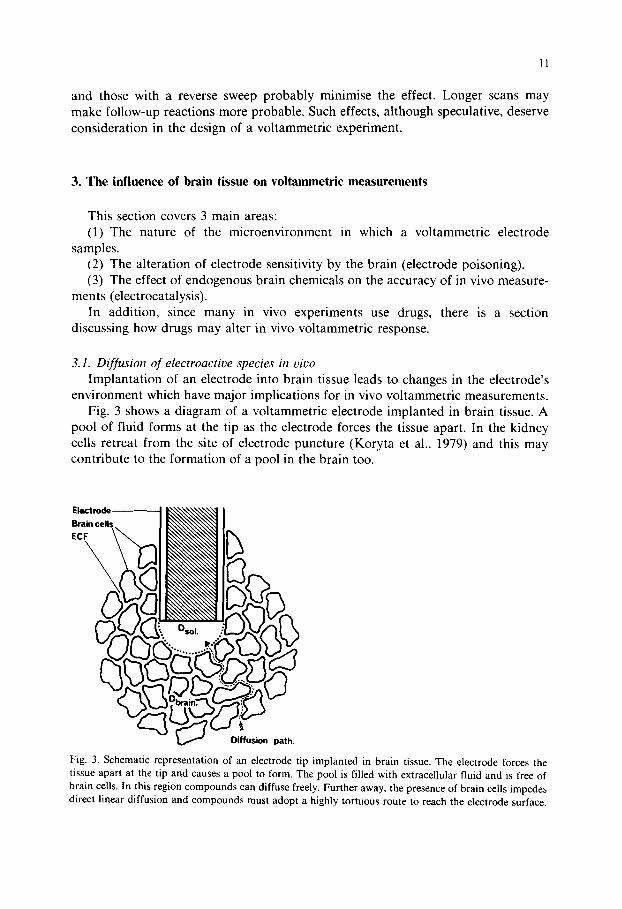
3.1. Diffusion of electroactive species in viuo Implanta t ion of an electrode into brain tissue leads to changes in the electrode's
environment which have major implications for in vivo vol tammetric measurements. Fig. 3 shows a diagram of a voltammetric electrode implanted in brain tissue. A
pool of fluid forms at the tip as the electrode forces the tissue apart. In the kidney cells retreat f rom the site of electrode puncture (Koryta et al., 1979) and this may contr ibute to the formation of a pool in the brain too.
n path
Fig. 3. Schematic representation of an electrode tip implanted in brain tissue. The electrode forces the tissue apart at the tip and causes a pool to form. The pool is filled with extracellular fluid and is free of brain cells. In this region compounds can diffuse freely. Further away, the presence of brain cells impedes direct linear diffusion and compounds must adopt a highly tortuous route to reach the electrode surface.

12
The development of the pool is not instantaneous. A traumatising event, such as the implantation of an electrode, is likely to cause chemical and cellular changes with different time courses. Thus the pool immediately following implantation may be different several weeks later. The pool size in 'acute' experiments is probably due to physical distortion of the tissue by the electrode. With chronically implanted electrodes, gliosis is probably a major determinant (Albery et al., 1983): see below. The astrocytic response to cerebral injury takes several days, for example (Mathew- son and Berry, 1985).
The presence of a pool at the electrode tip affects the access of electroactive species to the electrode surface. The pool is filled with fluid. The composition of the fluid has never been accurately determined, but it is generally considered similar to normal extracellular fluid. This may be qualitatively true, but it is also likely that the relative concentrations of neurochemicals within this pool differ. As a result of tissue damage, normally intracellular constituents would be found in this fluid. Using unperfused push-pull cannulae (which are of similar size to carbon paste electrodes) it has been reported that cell damage occurs in a layer 0.2.0.6 mm thick around the penetration (Yaksh and Yamamura, 1974). When the cannula was then perfused, protein was detected in the initial perfusates. It is likely that the pool at the tip of a voltammetric electrode would also contain more protein than normal extracellular fluid. (The effects of protein on electrode responses are discussed in Section 3.2.)
Other normally intracellular components (GSH for instance) may also influence the voltammetric measurements. GSH is not normally detected in vivo (Adams and Marsden, 1982), but can affect voltammetric measurements of catechols (Tse et al., 1976: Stamford, in press). AA levels too may be higher in the pool. Brazell and Marsden (1982b) reported that the AA oxidation peak in the striatum was 3 times higher immediately after implantation than 90 rain later. (The influence of AA on voltammetric measurements is discussed in Section 3.3.)
Since the pool is essentially devoid of cells, solution diffusion coefficients are considered to apply in this region. Outside the pool, in the brain tissue itself, the diffusional environment is complicated by the presence of cells which provide an impediment to direct diffusion. The extracellular fluid comprises about 20% of the tissue volume (Woodward et al., 1967; Nevis and Collins, 1967), the rest being brain cells. As well as the reduced volume fraction of the extracellular space, diffusion is also limited by tortuosity, i.e. the complexity of the path a compound must follow to diffuse through brain tissue (Nicholson et al., 1979). This is shown by the broken line in Fig. 3).
The tortuosity and volume fraction reduce the measured diffusion coefficient. The extent depends on the compounds investigated. Dayton et al. (1983) reported that the diffusion coefficients of a-methyl DA. AA and DOPAC were reduced in vivo to one third of their solution values. In a more extensive study Adams' group (Rice et al., 1985) showed that anionic compounds (DOPAC, HVA, VMA, AA) behaved in a similar manner. However, cations (DA, 3-MT, NA) had in vivo diffusion coefficients 10 times smaller than their in vitro values (Gerhardt and Adams, 1982). Neutral compounds (DOPEG, MHPG) gave intermediate behaviour.

13
They explained the drastic reduction in diffusion rates for cations as due to electrostatic interaction with anionic glycosaminoglycans. Nicholson and Philips (1981) however, found no difference between anion and cation diffusion in vivo.
What then are the implications of these two different environments for voltam- metric measurements? The effects are best illustrated with reference to the Cottrell equation for chronoamperometric measurements (Adams and Marsden, 1982) with linear diffusion:
nFAD'~C ~2t2
where i = oxidation current, n = number of electrons transferred per oxidised molecule, F = Faraday's constant, A = electrode surface area, D = diffusion coeffi- cient of the electroactive species, C = concentration of electroactive species and t = duration of electrolysis.
The oxidation current measured is determined by the diffusion coefficient of the electroactive species. Use of solution diffusion coefficients (Dsol) to calculate the concentration of a species reaching the electrode through brain tissue will give rise to an erroneously low value. Conversely, the application of a 'b ra in ' diffusion coefficient (Dbrai n) to a species present in the electrode tip pool will give too high a value.
The selection of appropriate diffusion coefficients thus depends on the types of electrochemical measurement used. Consider the two limiting cases: (A) a long oxidative measurement (10-100 s) such as those performed with LSV and DPV and (B) a very short measurement (10-100 ms, perhaps with a subsequent reduction scan).
A very long scan oxidises most of the electroactive species in the pool during a single measurement. Thus, a second sweep measures the sum of the compounds left in the pool and those that have entered it from the brain tissue. Since diffusion through brain tissue is slow, oxidation current on the second measurement is lower than that on the first (Cheng et al., 1979a). This trend continues until an equilibrium is established when the amount of chemical oxidised on each scan is the same as the amount reaching the electrode by diffusion through brain tissue. Under these circumstances, it is the lower 'bra in ' diffusion coefficient which determines the concentration measured. Such measurements are strongly dependent on the inter- vals between scans (Lindsay et al., 1980). Alteration of the timing can result in spurious changes in the size of the electrochemical signal.
Very short electrochemical measurements (10-100 ms) oxidise only a few thou- sand molecules and are almost non-destructive (Dayton et al., 1980a). This is further enhanced when a reducing sweep follows the oxidation scan (Armstrong James et al., 1980). Such measurements causes minimal depletion of the electrode tip pool and replenishment therefore is not rate-limiting. Under these circumstances the oxidation current per measurement is virtually independent of the interval between measurements. Wightman's group (Ewing et al., 1981) showed, using 92 ms duration chronoamps, that striatal oxidation current was the same whether measured at intervals of 4 or 30 s.

14
TABLE IV
INFLUENCE OF INTERSCAN INTERVAL ON THE MEASUREMENT OF DA IN VIVO BY HIGH-SPEED CYCLIC VOLTAMMETRY WITH CARBON FIBRE ELECTRODES
Peak DA release in rat striatum following ipsilateral stimulation of the median forebrain bundle (10 s train, 50 Hz sine waves, 90 p~A r.m.s.). DA release was measured using high speed cyclic voltammetry with interscan intervals of 25, 250, 500, 1000 or 2000 ms. Each animal therefore received 5 stimulations. Data expressed as a percentage of the peak height measured with a 25 ms interscan interval. Means ± S.E.M. (n = 5).
Interscan interval 25 250 500 1 000 2 000 (ins)
DA peak height 100 103 106 99 96 (25 ms value = 100%) _+ 10 + 12 +_ 12 + 10 + 10
Table IV shows that the DA oxidation peak height measured by HSCV is also independent of repetition rate. Measurement intervals as short as 25 ms do not result in any decrease in DA oxidation peak height. Thus very short measurements can be used with solution diffusion coefficients.
In addition to the effects on measured concentration, the time scale of recorded events is also determined by pool size and electrolysis duration. Lindsay el at. (1980) produced a computer simulation to describe these effects. Changes in the extracellu- lar concentration of electroactive species are detected only as they equilibrate with the fluid pool surrounding the electrode tip. A large pool means that equilibration is slower and the electrode responds less accurately to concentration changes within the extracellular fluid (Cheng, 1982). This has been supported experimentally: Following electrical stimulation of the nigrostriatal pathway DA is released into the extracellular fluid of the striatum. When the stimulation is stopped, the DA concentration falls. If carbon paste voltammetric electrodes are used, the decline has a half-life of 81 s (Justice et al., 1981). Studies with much smaller carbon fibre electrodes give half-life values of less than 5 s (Ewing and Wightman, 1984; Stamford et al., 1984c). The electrode tip pool of disc-shaped carbon fibre electrodes has a radius of only 12 ~m (Amatore et al., in press) whereas the value for paste electrodes is presumably much higher. These differences account for the response- times observed in vivo.
Thus far, I have mainly discussed the situation for acutely implanted electrodes. With chronic implantations there is a further variable to be considered - - the presence of glia. It has been reported that the tip of chronically implanted electrodes is surrounded by a border of neuroglia (Albery et al., 1983). At its simplest, this constitutes a further impediment to the access of electroactive species to the electrode surface. The presence of neuroglia in the boundary of the fluid pool may influence diffusion both into and out of the region.
Cultured neuroglia have been shown to accumulate neurotransmitters in vitro. Uptake of DA (Pelton et al., 1981), NA (Kimelberg and Pelton, 1983), 5-HT (Liesi et al., 1981), GABA (Hansson et al., 1984) and glutamate (Hertz and Richardson, 1984) have all been reported. Glia possess monoamine oxidase and catechol-o-meth-

15
yltransferase (Skaper et al., 1976), and it is considered that their major function in vivo may be to 'mop up' extracellular neurotransmitters (Henn and Hamberger, 1971) by uptake and subsequent metabolism. This, of course, prompts the question of whether chronically implanted electrodes measure neuronal or glial amine metabolites.
The extent of the involvement of neuroglial amine metabolism with chronic voltammetric signals is at present unclear. It may be possible to assess any such contribution using the gliotoxin 6-aminonicotinamide (Aikawa and Suzuki, 1985) or by inducing glial hypertrophy with kainate (Coyle, 1982). Such a study would be of value. Direct quantitative comparisons of drug effects in acute and chronic implan- tations might also assist in assessing the contribution (if any) of glial metabolism to recorded signals.
Until now I have assumed, for simplicity, that the size of the pool and of the extracellular space stays more or less constant: This may not be a safe assumption. Bondareff et al. (1970) measured intrastriatal spreading of injected catecholamines and interpreted this in terms of the extracellular space. Mannitol reduced the spread of catecholamines consistent with a decrease in the size of the extracellular space. The spread of catecholamines was also affected by the age of the animals. Bondareff et al. (1971) showed that spreading was reduced in senescent rats and proposed that a decrease in the extracellular space (Bondareff and Narotsky, 1972) accounted for this. However, other workers (Rees et al, 1982) have argued that the space increases with age.
Kent et al. (1985) showed that cerebral noradrenergic mechanisms from the locus coeruleus could alter the extracellular fluid volume. The volume of the extracellular fluid may also change in response to local neuronal depolarisation (Chase and Kopin, 1968). Such changes would be expected to affect the diffusion of compounds through the brain. Substantial swelling of brain cells might reduce the size of the fluid pool at the electrode tip. A quantitative study of the effects of alteration of extracellular space on voltammetric measurements would be welcome to many electrochemists.
3.2. Electrode poisoning When voltammetric electrodes are implanted into brain tissue, there are profound
changes in their behaviour. Three main effects are commonly observed. (1) The sensitivity of the electrodes changes. The extent and direction of such
changes are difficult to predict. Some electrodes lose sensitivity whereas others become more sensitive in vivo.
(2) The resolution between adjacent peaks may change. Compounds which gave well resolved peaks in vitro may be less well separated in vivo. Often resolution is not affected and in some cases it can actually be improved by contact with the brain.
(3) The position of oxidation peaks may be shifted on implantation into the brain.
Table V illustrates some of the changes observed during measurement of DA by HSCV with carbon fibre microelectrodes. The sensitivity was decreased by implan-

16
TABLE V
POISONING OF CARBON FIBRE MICROELECTRODES BY CONTACT WITH BRAIN TISSUE
DA oxidation peak positions (Means+_ S.E.M.: high speed cyclic voltammetry: 300 V/s scan rate) and electrode sensitivity recorded in phosphate-buffered saline (PBS) before and after 30' implantation in vivo. ***P < 0.01 vs preimplantation values.
DA Eox (mV vs Ag/AgCI) Sensitivity
Pre- In vivo Post- post/we implantation implantation implant-
ation
Untreated carbon fibre 491 _+ 29 605 _+ 11 *** 620+ 15 *** 0.43 + 0.08 (n~ (8) (7) (5) (5)
Electrochemically etched fibre 441 +- 5 574_+ 16 *** 0.31 +_0.03 (n) (8) - (8) (8)
tation in vivo and there was also a shift in the DA oxidation potential. As can be seen, the effects differed slightly for the two types of electrode. Interestingly, the resolution between DA and AA was improved. Untreated carbon fibre electrodes are capable of resolving DA and AA in vitro and in vivo (Ewing et al., 1982) whereas DA and AA oxidise at similar potentials with the electrochemically etched electrodes and cannot be separated by HSCV in vitro. Following exposure of etched electrodes to brain tissue there was a shift in DA and AA oxidation peaks. AA now oxidised at a much higher voltage (> 1 V vs Ag/AgCI) and resolution from the DA peak was thus improved.
Deterioration of carbon fibre electrodes in vivo has been observed by a number of workers. Wightman's group observed a 50% loss of sensitivity (Ewing et al., 1981) with concomitant broadening and anodic shift of oxidation peaks. Gonon et al. (1981a) using cylindrical carbon fibre electrodes, observed distortion of AA and DOPAC peaks in vivo during prolonged recording periods (> 3 h), with a gradual loss of separation.
The extent and nature of the electrode alteration by brain tissue depends, to some degree, on the activation of the surface. Lane described iodine-coated platinum electrodes (Lane and Hubbard, 1976) which showed excellent separat ionof AA and catechol oxidation but lost nearly all sensitivity over a few scans in vivo. Brazell and Marsden (1982a) reported that carbon paste electrodes were more sensi~tive in vivo than in vitro prior to implantation. They proposed that priming of the electrode with free amines from CSF might account for this. O'Neill et al. (1982b) observed that 'brain treated' carbon paste electrodes appeared less sensitive to AA and more sensitive to DOPAC than their unexposed counterparts.
The alteration in electrode sensitivity by exposure to brain tissue depends on the electrode type and the species being electrolysed. The point at which these changes occur is less clear. Wightman proposed (Ewing et al., 1981) that poisoning of carbon fibre electrodes occurred during implantation and was not accompanied by further degeneration. Gonon et al. (1981a), on the other hand, showed a gradual deteriora- tion after a few hours in vivo. The two modes are not incompatible. The results in

17
Table V support the work of Wightman and colleagues. The decrease in sensitivity seen here in thirty minutes, if continued over a period of hours would rapidly abolish all responsiveness of the electrode. However, experiments in which stimu- lated DA release was measured showed only a small decline in the DA signal over three hours (Millar et al., 1985). Thus, poisoning appears to occur during the first few minutes of implantation and further deterioration is, at least for DA, less important. Other more highly activated carbon fibre electrodes seem to poison more gradually. The electrically treated electrodes (Gonon et al., 1981b) show excellent separation of AA and DOPAC peaks immediately after implantation. However the resolution is lost after a few hours. It is likely that different mechanisms underlie sudden sensitivity changes and more gradual loss of resolving power.
The causes of poisoning are largely conjectural. Most obvious are protein adsorption and filming by electrogenerated oxidation products. The CSF contains very little protein compared with blood (25 mg/100 ml; Fenstermacher and Rall, 1972). However, during implantation, rupture of cells or blood filled capillaries (Cespuglio et al., 1981) may expose the electrode to much higher concentrations of protein. Protein adsorption onto electrodes (Eddowes and Hill, 1981; Brabec and Schindlerova, 1981) is potential dependent (Mattson and Smith, 1973; Mattson and Jones, 1976) and may occur as a result of electrolysis or electrode surface charge. Films on electrode surfaces can impede access of electroactive species to the electrode (Ikeda et al., 1982) and alter electrode kinetics (Krylov and Fishtik, 1981; Fukui et al., 1982). Proteins containing electroactive amino acids such as tyrosine or tryptophan can generate phenolic products capable of further deteriorating elec- trode response (Kolle and Johnson, 1979). Neuropeptides containing tyrosine or tryptophan (Bennett et al., 1981) may also contribute to this mechanism.
Formation of electrode poisons by electrolysis may contribute to the loss of response seen in vivo. HVA, for instance, present at high levels in the striatum (660 ng/g, Wagner et al., 1982), can impair electrode responses in vitro and in vivo (Ewing et al., 1981) by formation of insoluble oxidation products (Wightman et al., 1978). 5-Hydroxyindoles are also troublesome (Adams and Marsden, 1982), whereas the oxidation products of DA have been reported not to affect electrode responses (Plotsky, 1982).
Film formation or adsorption would be expected to alter electrode sensitivity to all compounds equally. The observation of differential effects (O'Neill et al., 1982b) perhaps indicates a change in surface chemical groupings. Such an effect has been shown to activate electrodes to one species while inhibiting oxidation of others (Engstrom, 1982). This may explain the different effects of 'brain treatment' on other types of carbon electrode too (Brazell and Marsden, 1982a; O'Neill et al., 1982b). Addition of stearic acid to carbon paste retards electron transfer from anionic compounds and accelerates the oxidation of cations. A reverse procedure (e.g. modification of the paste by free CSF amines) in vivo would be expected to increase sensitivity of the electrodes to anionic species (Lane and Hubbard, 1973).
Clearly one cannot assume that calibration of an electrode prior to implantation can be used with any accuracy in vivo. Carbon fibre electrodes, as shown, lose sensitivity, whereas paste electrodes become more sensitive. Since this probably

18
occurs during implantation, postexperiment calibrations seem more valid. To be confident in the accuracy of voltammetric changes it is necessary to establish in which direction the brain alters the sensitivity of the electrode and also whether this change is ' instant' or progressive. A good solution to this problem would be the use of in vivo calibrations, i.e. by local injection or pressure ejection of compounds (at known concentration) at the electrode surface, although geometrical considerations may complicate the interpretation of such data. Postexperiment calibrations are usually considered the most valid feasible solution.
3.3. The influence of electrocatalysis In Section 2.3 some of the potential reactions of oxidised catechols and indoles
were discussed. However, the presence of reducing agents in the brain extracellular fluid alters the pattern of reactivity of these compounds. Of particular interest is the °electrocatalytic' reaction which can have a profound effect on the accuracy of electrochemical measurements in vivo. For simplicity, I shall discuss the specific case for DA, although the rules apply also to other compounds.
The catalytic reaction depends on the presence, near the electrode surface, of oxidised DA (DOQ) and a reducing agent (in its reduced state), e.g. AA. The oxidation of DA is a 2-electron transfer (Eqn. 5.). The current generated by the forward reaction is proportional to the concentration of DA. When a reducing potential is then applied much of the DOQ is reduced back to DA.
In the presence of AA, the stoichiometry is more complex. Consider the situation at an electrode where AA has a higher oxidation potential than DA. The forward reaction for the oxidation of DA occurs as in Eqn. 5. However, DOQ then encounters AA. This gives the reaction shown in Eqn. 6. By oxidising AA to DHA, DA is regenerated from DOQ. This reaction is essentially unidirectional since hydration of DHA renders the AA oxidation irreversible (Perone and Kretlow, 1966; Lechien et al., 1982; Eqn. 7). The DA generated by the catalytic reaction can then be oxidised again (Eqn. 5). Thus, in the presence of AA, DA can be repetitively oxidised. The enhanced oxidation current is proportional to the product of the DA concentration and the square root of the AA concentration (Kovach et al., 1984). At higher potentials (where AA is oxidised directly by the electrode), the oxidation current is the sum of the DA and AA oxidation currents (Dayton et al., 1980b).
The effect of catalytic reactions has been considered mainly in the light of the DA oxidation peak. However, AA also decreases the height of the DOQ reduction peak (J.A. Stamford, in press). This effect is as expected from Eqn. 6.
The interference of AA with DA oxidation is the most frequently discussed. However, other strong reducing agents may also show this response (Ballantine and
H2 O NH2 + 2H+& 2e -
(Eqn,5)

N H° N O H2 HO H2
+ ~ +
O O
O 0
(Eqn.6)
19
O O
÷ H20 (Eqn.7)
Woolfson, 1979). NADH, for example, is oxidisable (rather irreversibly) in vitro (Braun et al., 1975) although it is not detectable in vivo following microinjection (Adams and Marsden, 1982). NADH can however be catalytically oxidised (Ueda et al., 1982) particularly by quinone groups (Tse and Kuwana, 1978). Catalytic oxidation of NADH by DA has been reported in vitro (Degrand and Miller, 1980) and may occur in vivo.
The extent of the catalytic reaction depends on the duration of the electrochem- ical measurement and the size of the electrode. With very short measurements, the extent of the reaction is decreased (Kovach et al., 1984). With DPV at 25 m V / s scan rate the oxidation current from a carbon fibre electrode in 10 - 4 M DA solution was amplified nearly 8-fold by AA (10 3 M). With chronoamperometry (0.125 s, +0.3 V vs Ag/AgC1) the DA oxidation current was additive with the AA oxidation current (Dayton et al., 1980b). With very small electrodes the catalytic effect is also minimised since a much smaller fraction of the regenerated DA returns to the electrode surface as a result of spherical diffusion (Galus et al., 1982). The combination of rapid voltammetric measurements at very small electrodes largely eliminates the catalytic effect. For instance, with 300 V / s cyclic voltammetry no amplification of DA (2 x 10⁻⁵ M) oxidation current was seen in the presence of AA (10 3 M; J.A. Stamford, in press).
For many in vivo electrochemical experiments, very short duration scans are, however, unsuitable and other solutions must be sought. The recent introduction of modified electrodes poses interesting questions. The addition of stearic acid to

20
carbon paste alters the electrode surface such that anions (AA, DOPAC) are repelled and oxidise at much higher potentials than DA (Yamamoto et al., 1982). Other electrodes have been modified with Nafion which is selectively permeable to cations, but not anions.
Since AA oxidises at a higher potential than DA, one would expect such electrodes to exhibit catalysis. Gerhardt et al. (1984) reported that the Nafion electrodes do exhibit minor electrocatalysis. Chronoamperometric current for DA is amplified by 30% in the presence of AA (2 × 10⁻⁴ M).
The case with the stearate-modified electrodes is more controversial. Blaha and Lane (1983) showed that the electrodes were unaffected by AA (up to 600 ktM) using chronoamperometric pulses of +0.25 V. Similarly, no response was seen in vivo following i.v. injection of AA (100 mg/kg) in rats (Blaha and Lane, 1984). When used with DPV, the DA oxidation current did vary with AA concentration (Newell and Colhoun, 1982). LSV with stearate-modified electrodes also gave rise to electrocatalysis: the oxidation current of DA (10 --4 M) was amplified more than 6-fold by the addition of AA (10 3 M) (Knott et al., 1985), although Yamamoto et al. (1982) reported no such effect under very similar voltammetric conditions. Clearly, the 'selectivity' of the anionically modified electrodes is only relative. With long duration measurements they are still susceptible to electrocatalysis.
The same may also apply to the AA 'killer' electrode (Nagy et al., 1982). Used with chronoamperometry, it exhibits little electrocatalysis (ca 5% amplification of catechol oxidation peaks by AA (140 #M). However, these electrodes have not, to my knowledge, been tried with other voltammetric techniques and thus their selectivity is not firmly established.
If the extracellular AA concentration remained constant, the amplification of catechol oxidation signals would also be constant, and any change in peak height would be due to changes in catechol concentration. There is, however, evidence that AA concentrations change in response to drugs (Clemens and Phebus, 1984: O'Neill et al., 1984). There is also a large physiological fluctuation of extracellular striatal AA throughout the light-dark cycle (O'Neill et al., 1982a) and the extracellular levels vary in different brain regions (Brazell and Marsden, 1982b; Stamford et al., 1984b; Louillot et al., 1985).
With voltammetric techniques where AA oxidises earlier than the catechols (Lane et al., 1976; Gonon et al., 1981a; Falat and Cheng, 1982), the catalytic reaction should not occur since any potential adequate to oxidise the catechol should also directly oxidise AA. Thus the compound oxidation current should be a summation rather than amplification (Kovach et al., 1984).
This discussion has covered only AA. Other reducing agents exist in the brain and their significance remains unexplored. Electrocatalysis is a real, but not in- surmountable, problem. Using the guidelines given above (shifting AA oxidation earlier, small electrodes and short measurements) it can be minimised.
3.4. Drug effects on electrochemical measurements Before an in vivo experiment an electrochemist usually checks whether any drugs
he proposes to use are electroactive. Marsden et al. (1984) listed a number of drugs

21
and their oxidation potentials, including the neuroleptics (clozapine, haloperidol and chlorpromazine). To this list can be added many others. Drugs containing aromatic hydroxyl functions particularly tend to be oxidisable (Adams and Mars- den, 1982). Anaesthetics are not electroactive. No peaks could be detected between -1 .0 and + 1.0 V vs Ag/AgCI for urethane, thiopentone, pentobarbitone, y-hy- droxybutyrate, methohexitone, chloralose or chloral hydrate (J.A. Stamford, unpub- lished observations).
Some electroactive drugs have knowingly been detected in vivo. The entry of HA 966 (Broxterman and Mos, 1980), paracetamol (Morgan and Freed, 1981), chlor- amphenicol (Meulemans et al., 1984), a-methyltryptamine (Marsden, 1980) and apomorphine (Marcenac and Gonon, 1985b) into the brain has been reported. Apomorphine has adjacent hydroxyl functions on a fused aromatic ring structure (Cheng et al., 1979b) and oxidises at the same potential as the catechols. This should be considered when measuring its effect on catechols in vivo.
Examining the electroactivity of drugs, however, only partially answers the question of their interference in vivo. Most drugs are metabolised, the neuroleptics particularly so, and the electroactivity of a parent drug says little about its metabolites. Freed and Yamamoto (1981) showed that paracetamol is converted to an electroactive metabolite in the rat brain and the same may apply to other drugs too. On oxidation, some drugs may become compounds which poison the electrode. Phenols are notorious electrode poisons (Kolle and Johnson, 1979) and many drugs contain phenolic structures (tyramine, for instance) and might therefore affect the sensitivity of in vivo electrodes.
Non-electroactive drugs can also alter the response of electrodes. Gonon et al. (1984) reported that caffeine decreased the size of the DOPAC peak in vivo without affecting the AA peak. Caffeine decreased the sensitivity of the electrodes to DOPAC in vitro and they proposed that this might account for the in vivo effect.
| !
1.0 -- 0 + I'0
Eapp V vs AOIAgCl
Fig. 4. Two superimposed, background current-corrected high speed cyclic vohammograms for dopamine (2 x 10 ~ M) in pH 7.4 phosphate-buffered saline (0.1 M) at a carbon fibre microelectrode (voltage scan rate: 300 V/s). The smaller of the two signals was recorded after the addition of pentobarbitone (3 mg/ml) to the buffer.

22
A possibly similar effect was reported by Brazell and Marsden (1982a) who showed that the striatal 5-hydroxyindole peak was 40-50% smaller in pentobarbi- tone-anaesthetised animals compared with those given chloral hydrate. Interestingly, the voltammetric response to local microinjection of 5-HT-or 5-HIAA was also reduced such that the percentage increase (relative to pre-infusion levels) was the same in both groups. An effect of pentobarbitone on electrode sensitivity is a possible explanation.
Fig. 4 shows the effect of pentobarbitone on the in vitro oxidation of DA with HSCV. The oxidation and reduction peak heights are both reduced by the pento- barbitone. This is presumably due to alteration of electrode surface groups. It shows that even non-electroactive drugs can interfere with voltammetric measurements,
In the majority of cases, the interference of drugs with in vivo voltammetric measurements is minimal. Drugs may oxidise at a high potential and thus not affect peaks recorded at lower voltages. The dosage of drug required to exert a pharmaco- logical effect may also be much lower than the level at which it would directly influence the electrochemical measurement. However, it worth considering as a possible explanation for anomalous results.
4. Conclusions and Guidelines
No single voltammetric method is ideal for all applications. In most cases the choice is determined by the nature of the experiment: each experimenter must determine which voltammetric method and which working electrode best suits his specific experimental needs. Over the past few years it has become clear that some combinations of working electrode and voltammetric technique work particularly well together, e.g. DPV with electrically treated carbon fibre electrodes or LSV with carbon paste electrodes. However, each different method brings its own experimen- tal considerations and it is therefore difficult to offer any simple guidelines as to how to avoid pitfalls. Prior to embarking on electrochemical measurements an experimenter should be aware of a number of issues:
(1) The working electrode, for instance, should be miniaturised as much as is feasible. This helps to reduce mechanical trauma. It also reduces current flow and therefore the chance of disrupting local neuronal activity. Indeed the only real disadvantage is that the lower currents may be more susceptible to electrical interference. An appropriate compromise can usually be found.
(2) The duration of the voltammetric scan is usually decided by the experimental design. Where short duration monoamine release is to be measured very fast scan rates are necessary. Where changes in more than one compound are to be recorded the higher resolution of slow-scanning methods is a necessity. Very fast measure- ments generate rather high currents and thus may affect the electrical activity of the brain. They are however virtually insensitive to the interference of electrocatalysis. Slow scans cause a more profound alteration of the chemical environment around the electrode but do not affect local neuronal electrophysiology. Some of the slower scanning methods are vulnerable to electrocatalytic phenomena. Neither approach is wholly problem-free.

23
(3) Calibration of voltammetric electrodes requires an understanding of diffusion in vivo and of electrode poisoning. Short duration measurements cause much less electrolytic depletion than longer scans and can therefore be calibrated using solution diffusion coefficients. Longer scans are dependent on repletion of the electrode tip pool by diffusion through brain tissue which is much slower. It is also important to establish how the brain alters the sensitivity of voltammetric elec- trodes. It is generally accepted that calibration after, rather than before, the experiment is the most valid approach.
These are the main areas of voltammetry where the experimenter can take precautionary measures. Other issues (e.g. the effects of oxidation products on the brain or the influence of drug metabolites on voltammetric signals) may eventually be shown to be only theoretical concerns which do not influence voltammetry in practice. Perhaps by drawing attention to them the questions may be more quickly answered.
5. Summary
In this review, I am aware that I have asked questions and drawn attention to experimental considerations sometimes without providing answers. The article could justifiably be criticised on these grounds. Nevertheless, the fact that we cannot give definite answers should not prevent us from recognising the validity of the ques- tions.
Voltammetry has shown itself to be a successful technique despite many un- answered experimental questions. The purpose, in writing this article, was not to discourage experimentation, but to stimulate interest, in the hope that the meth- odological issues raised may more quickly be resolved.
6. Acknowledgements
I should like to thank the Wellcome Trust for funding and Mary Stock for typing the manuscript.
7. References
Adams, R.N. (1969) Applications of modern electroanalytical techniques to pharmaceutical chemistry, J. Pharm. Sci., 58: 1171-1184.
Adams, R.N. and Marsden, C.A. (1982) Electrochemical detection methods for monoamine measure- ments in vitro and in vivo. In L.L. lversen, S.D. Iversen and S.H. Snyder (Eds.), Handbook of Psychopharmacology, Vol. 15, Plenum, New York, Chapter 1.
Aikawa, H. and Suzuki, K. (1985) Enteric gliopathy in niacin-deficiency induced by CNS glio-toxin, Brain Res., 334: 354-356.
Albery, W.J., Fillenz, M. and O'Neill, R.D. (1983) The compartment model for chronically implanted voltammetric electrodes in the rat brain, Neurosci. Lett., 38: 175-180.

24
Amatore, C., Kelly, R.S., K_ristensen, E.W., Kuhr, W.G. and Wightman, R.M. (1986) Effects of restricted diffusion at ultramicroelectrodes in brain tissue: the pool model: theory and experiment lo~ chronoamperometry, J. Electroanal. Chem., in press.
Armstrong James, M. and Millar, J. (1979) Carbon fibre microelectrodes, J. Neurosci. Meth., 1 : 279 287. Armstrong James, M. and Millar, J. (1984) High speed cyclic voltammetr~ and unit recording with
carbon fibre microelectrodes. In C.A. Marsden (Ed.), Measurement of Neurotransmitter Release In Vivo, Wiley, New York, Chapter 10.
Armstrong James, M., Millar, J. and Kruk, Z.L. (1980) Quantification of noradrenaline ionophoresis. Nature (London), 288: 181-183.
Ballantine, J. and Woolfson, A.D. (1979) Determination of catecholamines in pharmaceutical prepara- tions by differential pulse polarography at the glassy carbon electrode, Int. J. Pharm. 3: 239-246.
Bennett, G.W., Brazell, M.P. and Marsden, C.A. (1981) Electrochemistry of neuropeptides: a possible method for assay and in vivo detection, Life Sci., 29: 1001-1007.
Blaha, C.D. and Lane, R.F. (1983) Chemically modified electrode for in vivo monitoring of brain catecholamines, Brain Res. Bull., 10: 861-864.
Blaha, C.D. and Lane, R.F. (1984) Direct in vivo electrochemical monitoring of dopamine release in response to neuroleptic drugs, Eur. J. Pharmacol.. 98:113-117.
Bondareff, W. and Narotsky, R. (1972) Age changes in the neuronal microenvironment, Science, 176: 1135-1136.
Bondareff, W., Routtenberg, A., Narotzky, R. and Melone, D.G. (1970) Intrastriatal spreading of biogenic amines, Exp. Neurol., 28: 213-229.
Bondareff, W., Narotsky, R. and Routtenberg, A. (1971) Intrastriatal spread of catecholamines in senescent rats, J. Gerontol., 26: 163-167.
Brabec, V. and Schindlerova, I. (1981) Electrochemical behaviour of proteins of graphite electrodes. III: The effect of protein adsorption, Bioelectrochem. Bioenerget., 8: 451-458.
Braun, R.D., Santhanum, K.S.V. and Elving, P.J. (1975) Electrochemical oxidation in aqueous and non-aqueous media of dihydropyridine nucleotides NMNH, NADH and NADPH, J. Am. Chem. Soc., 97: 2591-2598.
Brazell, M.P. and Marsden, C.A. (1982a) Differential pulse voltammetry in the anaesthetised rat: identification of ascorbic acid, catechol and indoleamine oxidation peaks in the striatum and frontal cortex, Br. J. Pharmacol., 75: 539-547.
Brazell, M.P. and Marsden, C.A. (1982b) Intracerebral injection of ascorbate oxidase: effect on in viw~ eleetrochemical recordings, Brain Res., 249: 167-172.
Brazell, M.P., Mason, R. and Marsden, C.A. (1981) In vivo detection of indoleamines in rat striatum and frontal cortex, Neurosci, Lett. Suppl., 7: $57.
Broxterman, H.J. and Mos, J. (1980) Dopamine hypoactivity measured by in vivo voltammetry, Eur..1. Pharmacol., 68: 389-391.
Brun, A. and Rosset, R. (1974) Etude 61ectrochimique de l'oxidation de la dihydroxy-3,4-phenylalanine. M6canisme d'oxidation des catechols en milieu acide, Electroanal. Chem. Interfacial Electrochem., 49: 287-300.
Cespuglio, R. (1982) In vivo measurements by differential pulse voltammetry of 5-hydroxyindole compounds, J. Histochem. Cytochem., 30: 821-823.
Cespuglio, R., Faradji, H., Riou, F., Gonon, F., Pujol, J.-F. and Jouvet, M. (1981) Differential pulse voltammetry in brain tissue I1. Detection of 5-hydroxyindoleacetic acid in the rat striatum, Brain Res., 223: 299-311.
Chase, T.N. and Kopin, l.J. (1968) Stimulus-induced release of substances from olfactory bulb using the push pull cannula, Nature (London), 217: 466-467.
Chavdarian, C.G., Karashima, D., Castagnoli, N. and Hundley, H.K. (1978) Oxidative and cardiovascu- lar studies on natural and synthetic catecholamines, J. Med. Chem., 21: 548-554.
Cheng, H.-Y. (1982) Compartment model for chronoamperometric measurement in vivo, J. Electroanal. Chem., 135: 145-151.
Cheng, H.-Y., Schenk, J., Huff, R. and Adams, R.N. (1979a) In vivo electrochemistry: behaviour of microelectrodes in brain tissue, J. Electroanal. Chem., 100:23-31
Cheng, H.-Y, Strope, E. and Adams, R.N. (1979b) Electrochemical studies of the oxidation pathways of apomorphine, Anal. Chem., 51: 2243-2246.

25
Clark, L.C. and Sachs, G. (1968) Bioelectrodes for tissue metabolism, Ann. N.Y. Acad. Sci., 148: 133-153.
Clemens, J.A. and Phebus, L.A. (1984) Brain dialysis in conscious rats confirms in vivo electrochemical evidence that dopaminergic stimulation releases ascorbate, Life Sci., 35: 671-677.
Conti, J.C., Strope, E., Adams, R.N. and Marsden, C.A. (1978) Voltammetry in brain tissue: chronic recording of stimulated dopamine and 5-hydroxytryptamine release, Life. Sci., 23: 1705-1706.
Coyle, J.T. (1982) Excitatory amino acid neurotoxins. In L.L. Iversen, S.D. Iversen and S.H. Snyder (Eds.), Handbook of Psychopharmacology, 15, Plenum, New York. Chapter 5.
Dayton, M.A., Brown, J.C., Stutts, K.J. and Wightman, R.M. (1980a) Faradaic electrochemistry at microvoltammetric electrodes, Anal. Chem., 52: 946-950.
Dayton, M.A., Ewing, A.G. and Wightman, R.M. (1980b) Response of microvoltammetric electrodes to homogeneous catalytic and slow heterogeneous charge transfer reactions, Anal. Chem., 52: 2392-2396.
Dayton, M.A., Ewing, A.G. and Wightman, R.M. (1981) Evaluation of amphetamine-induced in vivo electrochemical response, Eur. J. Pharmacol., 75: 141-144.
Dayton, M.A., Ewing, A.G. and Wightman, R.M. (1983) Diffusion processes measured at microvoham- metric electrodes in brain tissue, J. Electroanal. Chem., 146: 189-200.
Degrand, C. and Miller, L.L. (1980) An electrode modified with polymer bound dopamine which catalyses NADH oxidation, J. Am. Chem. Soc., 102: 5728-5732.
Eddowes, M.J. and Hill, H.A.O. (1981) Investigation of electron transfer reactions of proteins by electrochemical methods, Biosci. Rep., h 521-532.
Engstrom, R.C. (1982) Electrochemical pretreatment of glassy carbon electrodes, Anal. Chem., 54, 2310-2314.
Ewing, A.G. and Wightman, R.M. (1984) Monitoring the stimulated release of dopamine with in vivo voltammetry. II Clearance of released dopamine from extraceUular fluid, J. Neurochem., 43: 570-577.
Ewing, A.G., Dayton, M.A. and Wightman, R.M. (1981) Pulse voltammetry with microvoltammetric electrodes, Anal. Chem., 53: 1842-1847.
Ewing, A.G., Wightman, R.M. and Dayton, M.A. (1982) In vivo voltammetry with electrodes that discriminate between dopamine and ascorbate, Brain Res., 249: 361-370.
Ewing, A.G., Alloway, K.D., Curtis, S.D., Dayton, M.A., Wightman, R.M. and Rebec, G.V. (1983) Simultaneous electrochemical and unit recording measurements: characterisation of the effects of d-amphetamine and ascorbic acid on neostriatal neurons, Brain Res., 261: 101-108.
Falat, L. and Cheng, H.-Y. (1982) Voltammetric differentiation of ascorbic acid and dopamine at an electrochemically treated graphite/epoxy electrode, Anal. Chem., 54: 2108-2111.
Fenstermacher, J.D. and Rail, D.P. (1972) Physiology and Pharmacology of the CSF. In E. Carpi (Ed.), Pharmacology of the Cerebral Circulation, Vol. 1, Pergamon, Oxford.
Fisher, G., Sayre, G.P. and Bickford, R.G. (1957) Histologic changes in the cats brain after introduction of metallic and plastic coated wire used in electro-encephalography, Proc. Mayo. Clin., 32: 15-22.
Freed, C.R. and Yamamoto, B.K. (1981) Acetaminophen metabolism in cat caudate nucleus measured by in vivo electrochemistry, Soc. Neurosci. Abstr., 7 (252): 13.
Fukui, M., Kitani, A., Degrand, C. and Miller, L.L. (1982) Propagation of a redox reaction through a quinoid polymer film on an electrode, J. Am. Chem. Soc., 104: 28-33.
Gallistel, C.R. (1981) Subcortical stimulation for motivation and reinforcement. In M.M. Patterson and R.P. Kesner (Eds.), Electrical Stimulation Research Techniques, Academic Press, London, pp. 141-179.
Galus, Z., Schenk, J.O. and Adams, R.N. (1982) Electrochemical behaviour of very small electrodes in solution. Double potential step, cyclic voltammetry and chronopotentiometry with current reversal, J. Electroanal. Chem., 135: 1-11.
Gerhardt, G. and Adams, R.N. (1982) Determination of diffusion coefficients by flow injection analysis, Anal. Chem., 54: 2618-2620.
Gerhardt, G.A., Oke, A.F., Nagy, G., Moghaddam, B. and Adams, R.N (1984) Nation-coated electrodes with high selectivity for CNS electrochemistry, Brain Res., 290: 390-395.
Gonon, F.G. and Buda, M. (1985) Regulation of dopamine release by impulse flow and by autoreceptors as studied by in vivo voltammetry in the rat striatum, Neuroscience, 14: 765-774.
Gonon, F., Buda, M., Cespuglio, R., Jouvet, M. and Pujol, J.-F. (1981a). Voltammetry in the striatum of chronic freely moving rats: detection of catechols and ascorbic acid, Brain Res., 223, 69-80.

26
Gonon, F., Fombarlet, C., Buda, M, and Pujol, J.-F. (1981b). Electrochemical treatment of pyrolytic carbon fibre electrodes, Analyt. Chem. 53, 1386-1389.
Gonon, F., Buda, M. and Pujol, J.F. (1984) Treated carbon fibre electrodes for measuring catechols and ascorbic acid. In C.A. Marsden (Ed.), Measurement of Neurotransmitter Release in Vivo, Wiley, Ne,a York, Chapter 7.
Hansson, E., Isaacson, H. and Sellestrom, A. (1984) Characteristics of dopamine and GABA transport in primary cultures of astroglial cells, Acta Physiol. Scand., 121: 333-341.
Harrison, W.H., Whisler, W.W. and Hill, B.J. (1968) Catecholamine oxidation and ionisation properties indicated from the H + release, tritium exchange, and spectral changes which occur during fer- ricyanide oxidation, Biochemistry, 7: 3089-3094.
Hawley, M.D., Tatawawadi, S.V., Piekarski, S. and Adams. R.N. (1967) Electrochemical studies of the oxidation pathways of catecholamines, J. Am. Chem. Soc.. 89: 447-450.
Heacock, R.A. (1959) The chemistry of adrenochrome and related compounds, Chem. Rev.. 59: 181-237. Hefti, F. and Felix, D. (1983) Chronoamperometry in vivo: does it interfere with spontaneous neuronal
activity in the brain? J. Neurosci. Meth., 7: 151-156. Hefti, F. and Melamed, E. (1981) Dopamine release in rat striatum after administration of I-DOPA as
studied with in vivo electrochemistry, Brain Res., 225: 333-346. Henn, F.A. and Hamberger, A. (1971) Glial cell function: uptake of transmitter substances, Proc. Natl.
Acad. Sci. U.S.A., 68: 1685-1690. Hertz, L. and Richardson, J.S. (1984) Is neuropharmacology merely the pharmacology of neurones or are
astrocytes important too, Trends Pharmacol. Sci., 5: 272-276. Hoffer, A. and Osmond, H. (1963) Scurvy and schizophrenia, Dis. Nerv. Syst., 24: 273-285. Huff, R.M. (1980) Drug Induced Release of Catecholamines Detected by In Vivo Electrochemistry,
Ph.D. Thesis, University of Kansas. Hutson, P.H. and Curzon, G. (1983) Monitoring in vivo of transmitter metabolism by electrochemical
methods, Biochem. J., 211: 1-12. Ikeda, T., Schmehl, R., Denisevich, P., Willman, K. and Murray, R.W. (1982) Permeation of electroactive
solutes through ultra thin polymeric films on electrode surfaces, J. Am. Chem. Soc., 104: 2683-2691. Isaacson, R.L. (1981) Brain stimulation effects related to those of lesions. In M.M. Patterson and R.P.
Kesner (Eds.), Electrical Stimulation Research Techniques, Academic Press, London, pp. 205-220. Jackson, W.F. and Duling, B.R. (1983) Toxic effects of silver-silver chloride electrodes on vascular
smooth muscle, Circ. Res., 53: 105-108. Janz, G.J. (1961) Silver-silver halide electrodes. In D.J.G. Ives and G.J. Janz (Eds.), Reference
Electrodes. Theory and Practice, Academic Press. New York, pp. 179-230. Justice, J.B., Neill, D.B., Salamone, J.D. and Garrigues, A. (1981) Extracellular half life of electroactive
compounds in striatum following electrical stimulation of MFB, Neurosci. Lett., Suppl. 7: $63. Kent, T.A., Nagy, G., Oke, A,F., Preskorn, S.H. and Adams, R.N. (1985) Effect of CO 2 on a brain
extracellutar space marker and evidence of its neuronal modulation, Brain Res., 342: 141-144. Kimelberg, H.K. and Pelton, E.W. (1983) High affinity uptake of [3H]norepinephrine by primary
astrocyte cultures and its inhibition by tricyclic antidepressants, J. Neurochem., 40: 1265-1270. Kissinger, P.T., Hart, J.B. and Adams, R.N. (1973) Voltammetry in brain tissue - a new neurophysio-
logical measurement, Brain Res., 55: 209-213. Knott, P.J., Andrews, C.D. and Mueller, K.J. (1985) Voltammetry measurement in vivo of neurotrans-
mitter release in the freely moving rat. In: In Vivo Perfusion and Release of Neuroactive Substances, Academic Press, New York, Chapter 9.
Kolle, R.C. and Johnson, D.C. (1979) Electrochemical removal of phenolic films from a platinum anode. Anal. Chem., 51: 741-744.
Koryta, J., Brezina, M., Pradac, J. and Pradacova, J. (1979) Methods for electroanalysis in vivo. In A.J. Bard (Ed.), Electroanalytical Chemistry, Vol. II, Marcel Dekker, New York, pp. 85-140.
Kovach, P.M., Ewing, A.G., Wilson, R.L. and Wightman, R.M. (1984) In vitro comparison of the selectivity of electrodes for in vivo electrochemistry, J. Neurosci. Meth., 10: 215-227.
Krylov, V.S. and Fishtik, I.F. (1981) Kinetics of electrode processes in the presence of a discrete layer of a specifically adsorbed organic substance, Can. J. Chem., 59: 2026-2031.
Lane, R.F. and Hubbard, A.T. (1973) Electrochemistry of chemisorbed molecules. I1. The influence of

27
charged chemisorbed molecules on the electrode reactions of platinum complexes, J. Phys. Chem., 77: 1411-1421.
Lane, R.F. and Hubbard, A.T. (1976) Differential double pulse voltammetry at chemically modified platinum electrodes for in vivo determination of catecholamines, Analyt. Chem., 48: 1287-1293.
Lane, R.F., Hubbard, A.T., Fukunaga, K. and Blanchard, R.J. (1976) Brain catecholamines: detection in vivo by means of differential pulse voltammetry at surface modified platinum electrodes, Brain Res., 114: 346-352.
Lane, R.F., Hubbard, A.T. and Blaha, C.D. (1978) Brain dopaminergic neurons: in vivo electrochemical information concerning storage, metabolism and release processes, Bioelectrochem. Bioenerget., 5: 504-525.
Lechien, A., Valenta, P., Nurnberg, H.W. and Patriarche, G.J. (1982) Determination of ascorbic acid by differential pulse voltammetry, Fres. Z. Anal. Chem., 311: 105-108.
Liesi, P., Paetau, A., Rechardt, L. and Dahl, D. (1981) Glial uptake of monoamines in primary cultures of rat median raphe nucleus and cerebellum, Histochemistry, 73: 239-250.
Lindsay, W.S., Justice, J.B. and Salamone, J. (1980) Simulation studies of in vivo electrochemistry, Comput. Chem., 4: 19-26.
Louilot, A., Gonon, F., Buda, M., Simon, H., Le Moal, M. and Pujol, J.F. (1985) Effects of d- and l-amphetamine on dopamine metabolism and ascorbic acid levels in nucleus accumbens and olfactory tubercle as studied by in vivo differential pulse voltammetry, Brain Res., 336: 253-263.
Marcenac, F. and Gonon, F.G. (1985a). Fast in vivo monitoring of dopamine release in the rat brain with differential pulse amperometry, Anal. Chem., 57: 1778-1779.
Marcenac, F.M.J. and Gonon, F.G. (1985b) In vivo electrochemical monitoring of apomorphine penetration into the rat brain, Neurosci. Lett., 55: 305-310.
Marsden, C.A. (1980) Involvement of 5-hydroxytryptamine and dopamine neurones in the behavioural effects of alpha-methyltryptamine, Neuropharmacology, 19: 691-698.
Marsden, C.A., Brazell, M.P. and Maidment, N.T. (1984) An introduction to in vivo electrochemistry. In C.A. Marsden (Ed.), Measurement of Neurotransmitter Release In Vivo, Wiley, New York, Chapter 6.
Matthewson, A.J. and Berry, M. (1985) Observations on the astrocyte response to a cerebral stab wound in adult rats, Brain Res., 327: 61-69.
Mattson, J.S. and Jones, T.T. (1976) Infrared spectrophotometric observations of the adsorption of fibrinogen from solution at optically transparent electrode surfaces, Anal. Chem., 48: 2164-2167.
Mattson, J.S. and Smith, C.A. (1973) Enhanced protein adsorption at the solid-solution interface: dependence on surface charge, Science, 181: 1055-1057.
Meulemans, A., Henzel, D., Brun-Pacaud, M., Mohler, J., Vicart, P. and Tran Ba Huy, P. (1984) On-line pharmacokinetics of chloramphenicol in rat cortex by in vivo electrochemical detection~ Chem- otherapy, 30: 353-357.
Millar~ J., Armstrong James, M. and Kxuk, Z.L. (1981) Polarographic assay of ionophoretically applied dopamine and low noise unit recording using a multibarrel carbon fibre microelectrode, Brain Res., 205: 419-424.
Millar, J., Stamford, J.A., Kruk, Z.L. and Wightman, R.M. (1985) Electrochemical, pharmacological and electrophysiological evidence of rapid dopamine release and removal in the rat caudate nucleus following electrical stimulation of the median forebrain bundle, Eur. J. Pharmacol., 109: 341-348.
Morgan, M.E. and Freed, C.R. (1981) Acetaminophen as an internal standard for calibrating in vivo electrochemical electrodes, J. Pharmacol. Exp. Ther., 219: 49-53.
Nagy, G., Rice, M.E. and Adams, R.N. (1982) A new type of enzyme electrode: the ascorbic acid eliminator electrode, Life Sci., 31: 2611-2616.
Neugebauer, R., Helbing, G., Wolter, D., Mohr, W. and Gistinger, G. (1981) The body reaction to carbon fibre particles implanted into the medullary space of rabbits, Biomaterials, 2: 182-184.
Nevis, A.H. and Collins, G.H. (1967) Electrical impedance and volume changes in brain during preparation for electron microscopy, Brain Res., 5: 57-85.
Newell, G.A. and Colhoun, E.H. (1982) Comparison of modified carbon paste and carbon fibre electrodes for the electrochemical measurement of catecholamines in the presence of ascorbic acid, Soc. Neurosci. Abstr, 8 (252): 22.
28
Nicholson, C. and Phillips, J.M. (1981) Ion diffusion modified by tortuosity and volume fraction in the extracellular microenvironment of the rat cerebellum. J. Physiol. (London),321: 225--257.
Nicholson, C., Phillips, J.M. and Gardner-Medwin, A.R. (1979) Diffusion from an iontophoretic point source in the brain: role of tortuosity and volume fraction, Brain Res.. 169: 580-584.
Olds, J. (1958) Self-stimulation in the brain, Science, 127: 315-324. O'Neill, R.D., Fillenz, M. and Albery, W.J. (1982a) Circadian changes in homovanillic acid and
ascorbate levels in the rat striatum using microprocessor controlled voltammetry, Neurosci. Lett., 34: 189-193.
O'Neill, R.D., Grunewald, R.A., Fillenz, M. and Albery, W.J. (1982b) Linear sweep voltammetry with carbon paste electrodes in the rat striatum, Neuroscience, 7: 1945-1954.
O'Neill, R.D., Grunewald, R.A., Fillenz, M. and Albery, W.J. (1983) The effect of unilateral cortical lesions on the circadian changes in rat striatal ascorbate and homovanillic acid levels measured in vivo using voltammetry, Neurosci. Lett. 42:105-110.
O'Neill, R.D., Fillenz, M., Sundstrom, L. and Rawlins, J.N.P. (1984) Voltammetrically monitored brain ascorbate as an index of excitatory amino acid release in the unrestrained rat, Neurosci. Lett., 52: 227-234.
Orrenius, S. and Moldeus, P. (1984) The multiple roles of glutathione in drug metabolism, Trends Pharmacol. Sci., 5: 432-435.
Pelton, E.W., Kimelberg, H.K., Shipherd, S.V. and Bourke, R.S. (1981) Dopamine and norepinephrine uptake and metabolism by astroglial cells in culture, Life Sci., 28: 1655-1663.
Perone, S.P. and Kretlow, W,J. (1966) Application of controlled potential techniques to study of rapid succeeding chemical reaction coupled to electro-oxidation of ascorbic acid, Anal. Chem., 38: 1760-1763.
Perry, T.L., Godin, D.V. and Hansen, S. (1982) Parkinson's disease: a disorder due to nigral glutathione deficiency? Neurosci. Lett., 33: 305-310.
Plotsky, P.M. (1982) Differential voltammetric measurements of catecholamines and ascorbic acid at surface modified carbon filament electrodes, Brain Res., 235: 179-184.
Ponchon, J.-L., Cespuglio, R., Gonon, F., Jouvet, M. and Pujol, J.-F. (1979) Normal pulse polarography with carbon fibre electrodes for in vitro and in vivo determination of catecholamines, Anal. Chem., 51: 1483-1486.
Ranck, J.B. (1975) Which elements are excited in electrical stimulation of mammalian central nervous system: a review. Brain Res. 98, 417-440.
Rees, S., Cragg, B.G. and Everitt, A.V. (1982) Comparison of extracellular space in the mature and ageing brain using a new technique, J. Neurol. Sci., 53: 347-357.
Rice, M.E., Gerhardt, G.A., Hierl, P., Nagy, G. and Adams, R.N. (1985) Diffusion coefficients of neurotransmitters and their metabolites in brain extracellular fluid space, Neuroscience, 15: 891-902.
Schenk, J.O. and Adams, R.N. (1984) Chronoamperometric measurements in the central nervous system. In C.A. Marsden (Ed.), Measurement of Neurotransmitter Release In Vivo. Wiley, New York, Chapter 9.
Senoh, S., Creveling, C.R., Udenfriend, S. and Witkop, B. (1959) Chemical, enzymatic and metabolic studies on the mechanism of oxidation of dopamine, J. Am. Chem. Soc., 81 : 6236-6240.
Shvets-Teneta-Guriy, T.B. (1981) Electrochemical activity on implanted metal electrodes as a brain reaction to a traumatising action. J. Higher Nervous Activity, 31: 148-156.
Silver. I.A. (1965) Some observations on the cerebral cortex with an ultramicro, membrane-covered, oxygen electrode, Med. Electron. Biol. Eng., 3, 377-387.
Skaper, S.D., Adelson, G.L. and Seegmiller, J.E. (1976) Metabolism of biogenic amines in neuroblastoma and glioma cells in culture. J. Neurochem. 27, 1065-1070.
Stamford, J.A. (1985) In vivo voltammetry: promise and perspective, Brain Res. Rev., 10: 119--135. Stamford, J.A. (1986) The effect of electrocatalytic and nucleophilic reactions on fast voltammetric
measurements of dopamine at carbon fibre microelectrodes, Anal. Chem., in press. Stamford, J.A., Kruk, Z.L. and Millar, J. (1984a) A double-cycle high-speed voltammetric technique
allowing direct measurement of irreversibly oxidised species: characterisation and application to the temporal measurement of ascorbate in the rat central nervous system, J. Neurosci. Meth., 10: 107-118.

29
Stamford, J.A., Kruk, Z,L. and Millar, J. (1984b) Regional differences in extracellular ascorbic acid levels in the rat brain determined by high speed cyclic voltammet~, Brain Res., 299: 289-295.
Stamford, J.A., Kruk, Z,L. Millar, J. and Wightman, R~M. (1984c) Striatal dopamine uptake in the rat: in vivo analysis by fast cyclic voltammetry, Neurosci. Lett., 51: 133-138.
Stamford, J.A., Kruk, Z.L. and Millar, J. (1985) Ascorbic acid does not modulate stimulated dopamine release: in vivo voltammetric data in the rat. Neurosci. Lett., 60: 357-362.
Sternson, A.W., McCreery, R., Feinberg, B. and Adams, R.N. (1973) Electrochemical studies of adrenergic neurotransmitters and related compounds. Electroanal. Chem. Interracial Electrochem.. 46: 313-321.
Tayton, K., Phillips, G. and Rails, Z. (1982) Long term effects of carbon fibre on soft tissues, J. Bone Joint Surg., 64-B: 112-114.
Tse, D.C.S. and Kuwana, T. (1978) Electrocatalysis of dihydronicotinamide adenosine diphosphate with quinones and modified quinone electrodes, Anal. Chem., 50:1315 1318.
Tse, D.C.S, McCreery, R.L. and Adams, R.N. (1976) Potential oxidative pathways of brain catechola- mines, J. Med. Chem., 19: 37-39.
Ueda, C., Tse, D.C.S. and Kuwana, T. (1982) Stability of catechol modified carbon electrodes for electrocatalysis of dihydronicotinamide adenine dinucleotide and ascorbic acid, Anal. Chem., 54: 850-856.
Valenstein, E.S. and Beer, B. (1961) Unipolar and bipolar electrodes in self stimulation experiments Am. J, Physiol., 201: 1181-1186.
Wagner, J., Vitali, P., Palfreyman, M.G., Zraika, M. and Huot, S. (1982) Simultaneous determination of 3,4-dihydroxyphenylalanine, 5-hydroxytryptophan, dopamine, 4-hydroxy, 3-methoxyphenylalanine, norepinephrine, 3,4-dihydroxyphenylacetic acid, homovanillic acid, serotonin and 5-hydroxyin- dolacetic acid in rat cerebrospinal fluid and brain by high performance liquid chromatography with electrochemical detection, J. Neurochem., 38: 1241-1254.
Wightman, R.M. (1981) Microvoltammetric electrodes, Anal. Chem., 53: 1125-1130A. Wightman, R.M., Strope, E., Plotsky, P. and Adams, R.N. (1978) In vivo voltammetry: monitoring of
dopamine metabolites in CSF following release by electrical stimulation, Brain Res., 159: 55-68. Wise, R.A. (1972) Spread of current from monopolar stimulation of the lateral hypothalamus, Am. J.
Physiol., 223: 545-548. Woodward, D.L., Reed, D.J. and Woodbury, D,M. (1967) Extracellular space of rat cerebral cortex, Am.
J. Physiol., 212: 367-370. Yaksh, T.L. and Yamamura, H.I. (1974) Factors affecting performance of the push-pull cannula in
brain, J. Appl. Physiol., 37: 428-434. Yamamoto, B.K., Lane, R.F. and Freed, C.R. (1982) Normal rats trained to circle show asymmetric
caudate dopamine release, Life Sci., 30: 2155-2162. Young, T.E., Griswold, J.R. and Hulbert, M.H. (1974) Melanin, I: Kinetics of the oxidative cyclisation of
dopa to dopachrome, J. Org. Chem., 39: 1980-1982. Young, T.E., Babbitt, B.W. and Wolfe, L.A. (1980) Melanin 2: electrochemical study of the oxidation of
alpha methyldopa and 5,6-dihydroxy-2-methyl indole, J. Org. Chem., 45: 2899-2902.