in vivo protection by protein a of hepatic microsomal mixed function oxidase system of...
TRANSCRIPT

Biochemicd Pharmncofogy, Vol. 36, No. 23, pp. 4OSS-UtS8, 1987. Prieted in Great Britain.
OQ%-2952/87 $3.00 + 0.00 @ 1987. Per&%non Jownals Ltd.
IN VWU PROTECTION BY PROTEIN A OF HEPATIC MICROSOMAL MIXED FUNCTION OXIDASE SYSTEM OF
Ccl,-ADMINISTERED RATS
S. P. SRIVASTAVA, K. P. SINGH, A. K. SAXENA, P. K. SETH and P. K. RAY*
Industrial Toxicology Research Centre, Post Box No. 80, M.G. Marg, Lucknow- 001, India
(Received S Jetmary 1987; accepted 18 fune 1987)
Abstract-The in uiuo protection by protein A of hepatic mixed function oxidase system of carbon tetraehioride (Ccl,) administered rats, has been investigated in the present communication. Aryl hydrocarbon hydroxylase activity was decreased by 63% in Ccl, administered rats while in protein A + CC& administered rats the decrease was in the range of 22-25% (group IV-V). The aryl hydrocarbon hydroxylase activity in protein A + CC& administered rats showed significant increase in group IV (P < O.OOS) and group V (P < 0.001) in comparison to CCll alone (group II). Similarly, aniline hydroxylase and aminopyrene N-demethylase were decreased, by 75 and 84% respectively in CC& administered rats and 31% and 54-64%, respectively in protein A + Ccl, administered rats (groups IV and V). The aniline hydroxylase activity was also found enhanced in protein A + CC& administered group IV and V (P < 0.001). In addition the aminopyrene N-demethylase also showed significant increase in its activity in group IV (P < 0.001) and group V (P< 0.001) in comparison to Ccl4 alone. In accordance with these data, serum glutamic oxaloacetic transaminase and glutamic pyruvic transaminase exhibited significantly less increase in their activity in animais receiving protein A and Ccl, than those treated with CCL, alone. Protein A alone was found to have no effect on any of these enzymes. Our results indicate that protein A protects CC& induced injury as judged by the biochemical alterations and suggests that it may be usefui in providing an excellent system for the investigation on the regeneration of the hepatic enzyme activity following toxic insult of CCL,.
Carbon tetrachloride (CClJ, a typical hepatotoxin, causes wide spread damage to liver cells [ 1,2]. The administration of Ccl4 increases lipid peroxidation, destruction of cytochrome P-450, fatty infiltration of liver and necrosis [2-4]. Evidence to date suggests that cytochrome P-450 bioactivation of CC& to reac- tive metabolites is a prerequisite of C&-induced liver damage [S]. It has been postulated that the metabolism of Ccl4 by liver tissue involves a free radical inte~ediate [6,7]. Recently, involvement of a specific form of cytochrome P-450 which has the capacity to generate Ccl3 radicals in reconstituted monooxygenase systems [S] has been suggested fol- lowing exposure to CC& [9].
Protein A, a cell wall glycoprotein of Staphylo- coccus aureus Cowan 1, has a multipotent immuno- modulating property [lO-143. We have reported earlier that it can cause regression of primary rat mammary tumors [ 14,151, and dog tumors 1161 when infused intravenously. In all the protein A-treated animals immunopotentiation was observed.
In this study we have investigated whether or not protein A could minimise or reduce the toxic effects of the hepatotoxin, Ccl*, on the mixed function oxidase (MFO) system.
MATERIALS AND METHODS
Animals and treatment. Female Wistar albino rats (150 * 10 g), obtained from the ITRC animal breed- ing colony raised on a commercial pellet diet and
* To whom all correspondence should be addressed.
40
water provided ad libitum, were used in the present study. The animals were divided into five groups of six to eight rats each and treated as follows: Prior to Ccl4 treatment, the animals of group IV and group V were pretreated for two weeks (twice a week) with intravenous (i.v.) injection of protein A. The treatment of animals with CC& at the doses indicated below is optimal for achieving destruction of cyto- chrome P-450 system. During the entire period of experiment, the animals in group V were treated with intravenous injection of protein A (twice a week). The CC14 treatment was started on the same day in all groups concerned.
Group I: control, injected i.p. with 0.2ml olive oil.
Group II: carbon tetrachloride (OS ml/kg), injected i.p. in 0.2 ml olive oil for 14 days.
Group III: protein A (60pg/kg), i.v. twice a week for two weeks.
Group IV: protein A (60 /&g/kg), was given twice a week for two weeks and finally Ccl4 (OS ml/kg), i.p. in 0.2 ml olive oil for 14 days.
Group V: protein A (60 pg/kg) was given twice a week for four weeks. Ccl4 (0.5 ml/kg), i.p. in 0.2 ml olive oil was started after two weeks of protein A inoculation, and given daily for 14 days.
Preparation of enzyme source from rat liver. The animals were killed by cervical dislocation. Livers were removed, blotted free of blood, and washed with ice-cold saline. Tissues were homogenized in four times its volume of 0.1 M phosphate buffer, pH7.4 containing 0.15 M KCl. The liver homo- genates were centrifuged at 9000g for 20 min at 4”.
55

4056 S. P. SRIVASTAVA et al.
The resulting supernatant was used for the measure- ment of cytochrome P-450 dependent enzymes and glutathione-S-transferase, a phase II enzyme of the liver microsomal detoxification process.
Glutamate oxaloacetate transaminase (GOT) and glutamate pyruvate transaminase (GPT) were measured in the serum of the same animals used for cytochrome P-450 dependent enzymes.
Enzyme assay. Aryl hydrocarbon hydroxylase (AHH) activity was determined according to Dehnen er al. [ 171 with slight modification [ 181. Aminopyrene N-demethylase (ADM) activity was assayed accord- ing to Cochin and Axelrod [19] by measuring the formation of formaldehyde according to Nash [20]. Aniline hydroxylase (AH) activity was assayed by measuring the formation of p-aminophenol [21]. Glutathione-S-transferase (GST) activity was deter- mined according to the method of Habig et al. [22] using 1-chloro 2,4_dinitrobenzene as a substrate. Serum GOT and GPT were measured according to the methods of Bergmeyer and Bernt [23,24] respectively. Protein content was determined accord- ing to Lowry et al. [25], using bovine serum albumin as a reference standard.
Statistical analysis. Statistical analyses were done by using Student’s t-test. Values less than 0.05 were considered significant. The carbon tetrachloride alone group was compared with protein A treated carbon tetrachloride exposed group.
RESULTS
Effect of protein A on aryl hydrocarbon hydroxylase, aniline hydroxylase, aminopyrene demethylase and glutathione-S-transferase activity of hepatic MFO sysem of CCll treated rats
Protection by protein A of CCll-induced depres- sion of aryl hydrocarbon hydroxylase activity is shown in Fig. 1. The rats exposed to Ccl, showed a 63.3% decrease in hepatic AHH-activity. The pro- tein A + CClradministered rats showed a decrease of only 25% (group IV) and 22.6% (group V). The AHH-activity in protein A + Ccl,-administered rats showed significant increase in group IV (P < 0.005) and group V (P < 0.001) in comparison to CC14 alone
Fig. 1. Effect of protein A on carbon tetrachloride inhibited activity of aryl hydrocarbon hydroxylase activity in rat liver. Each value is mean of five animals: Cl, control; n , CC& alone; @, protein A + Ccl,; a, protein A (con-
tinued) + Ccl,; ‘8, protein A alone.
Fig. 2. Effect of protein A on carbon tetrachloride inhibited activity of aniline hydroxylase activity of rat liver. Each value is mean of five animals: q , control; n , Ccl, alone; ffl, protein A + CC&; iSI, protein A(continued) + Ccl.,; 8,
protein A alone.
I r
6 ,” V a
, I
ii
. . . II . . . . . Fig. 3. Effect of protein A on carbon tetrachloride inhibited activity of aminopyrine N-demethylase activity of rat liver. Each value is mean of five animals: q , control; W, Ccl4 alone; K& protein A + Ccl,; q , protein A
(continued) + Ccl.+; q , protein A alone.
I r
Fig. 4. Effect of protein A on carbon tetrachloride inhibited activity of glutathione-S-transferase activity of rat liver. Each value is mean of five animals: 0, control; n , Ccl, alone; kl, protein A + CC14; pJ, protein A
(continued) + CC14; q , protein A alone.
Protein A reverses Ccl4 toxicity 4057
(group II). Protein A alone did not produce any significant effect in comparison to control. A loss of 15% activity was also observed in protein A treated control groups.
The results described in Fig. 2 show a 75% loss of aniline hydroxylase (AH) activity in CC& admin- istered groups while protein A pretreated groups showed 31% (group IV and V) loss of enzyme activity. The AH activity was found to be enhanced in protein A + Ccl,-administered rats in both group IV and group V (P < 0.001) in comparison to Ccl4 (group II). Protein A treated control group showed no change in AH activity. Similarly, an 84.26% decrease in aminopyrene-N-demethylase (ADM) activity was observed in Ccl4 administered rats while in protein A + CCll administered groups, a loss of 64.47% (group IV) and 54.04% (group V) were observed. The ADM-activity was potentiated in pro- tein A + CC14-administered group IV (P < 0.001) and group V (P < 0.001) in comparison to Ccl+ In protein A treated control a decrease of only 8.1% activity was observed which was not significant stat- istically. Figure 4 shows the effect of protein A on GST, a phase II enzyme of detoxification process. The protein A treated control groups showed no significant change in GST-activity. The Ccl, treated rats exhibited a decrease of 46.38% while protein A pretreated animals (group IV), showed a decrease of 26.36%, only. The GST-activity was significantly increased in group IV (P < 0.001) in comparison to CCL,. Protein A alone did not produce any significant change in comparison to control. However, the pro- tein A + CC& treated group (group V) where protein A was continued till the termination of the exper- iment, no improvement in the GST activity was observed.
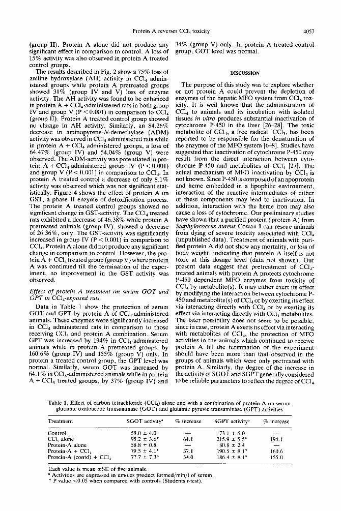
Effect of protein A treatment on serum GOT and GPT in Ccl,-exposed rats
Data in Table 1 show the protection of serum GOT and GPT by protein A of CC&-administered animals. These enzymes were significantly increased in CCll administered rats in comparison to those receiving CC& and protein A combination. Serum GPT was increased by 194% in CCll-administered animals while in protein A pretreated groups, by 160.6% (group IV) and 155% (group V) only. In protein a treated control group, the GPT level was normal. Similarly, serum GOT was increased by 64.1% in CCL,-administered animals while in protein A + CC& treated groups, by 37% (group IV) and
34% (group V) only. In protein A treated control group, GOT level was normal.
DISCUSSION
The purpose of this study was to explore whether or not protein A could prevent the depletion of enzymes of the hepatic MFO system from CC& tox- icity. It is well known that the administration of CC& to animals and its incubation with isolated tissues in vitro produces substantial inactivation of cytochrome P-450 in the liver [2628]. The toxic metabolite of CC14, a free radical ‘CC13, has been reported to be responsible for the denaturation of the enzymes of the MFO system [6-81. Studies have suggested that inactivation of cytochrome P-450 may result from the direct interaction between cyto- chrome P-450 and metabolites of CC& [27]. The actual mechanism of MFO inactivation by CC& is not known. Since P-450 is composed of an apoprotein and heme embedded in a lipophilic environment, interaction of the reactive intermediates of either of these components may lead to inactivation. In addition, interaction with the heme iron may also cause a loss of cytochrome. Our preliminary studies have shown that a purified protein (protein A) from Staphylococcus aureus Cowan I can rescue animals from dying of severe toxicity associated with CC& (unpublished data). Treatment of animals with puri- fied protein A did not show any mortality, or loss of body weight, indicating that protein A itself is not toxic at this dosage level (data not shown). Our present data suggest that pretreatment of CC14- treated animals with protein A protects cytochrome P-450 dependent MFO enzymes from toxicity of Ccl, by metabolite(s). It may either exert its effect by modifying the interaction between cytochrome P- 450 and metabolite(s) of Ccl, or by exerting its effect via interacting directly with Ccl4 or by exerting its effect via interacting directly with Ccl.+ metabolites. The later possibility does not seem to be possible, since in case, protein A exerts its effect via interacting with metabolites of CC14, the protection of MFO activities in the animals which continued to receive protein A till the termination of the experiment should have been more than that observed in the groups of animals which were only pretreated with protein A. Similarly, the degree of the increase in the activity of SGOT and SGPT generally considered to be reliable parameters to reflect the degree of Ccl4
Table 1. Effect of carbon tetrachloride (Ccl,) alone and with a combination of protein-A on serum glutamic oxaloacetic transaminase (GOT) and glutamic pyruvic transaminase (GPT) activities
Treatment SGOT activity” % increase SGPT activity” % increase
Control 58.0 t 4.0 - 73.1 2 6.0 -
CC& alone 95.2 2 3.6* 64.1 215.9 r 5.5* 194.1 Protein-A alone 58.8 ? 0.8 80.8 + 2.4 Protein-A + CCL, 79.5 t 4.1* 37.1 190.5 2 8.1* 160.6 Protein-A (contd) + CCI, 77.7 2 7.3* 34.0 186.4 2 8.1* 155.0
Each value is mean ?SE of five animals. a Activities are expressed in umoles product formed/min/l of serum. * P value co.05 when compared with controls (Students r-test).

4058 S. P. SRIVASTAVA ei al.
hepatocellular toxicity [29,30] were not si~ificantly different whether protein A treatment continued throughout the treatment or was given only prior to CC& administration. Using both the treatment regimens it was observed that protein A caused the protection of hepatic injury as judged by the recovery in the activities of enzymes of MFO system and glutathione-S-transferase and a lesser degree of decreases in the activity of SGOT and SGPT in comparison to respective controls to more or less to the same degree. Though the mechanism of protein A action is unknown, its immunopotentiating ability, particularly its abihty to stimulate the reticu- loendothelial system, could be reiated to this prop- erty [31,32].
In another study we have also found that protein A treatment protects from the carbon tetrachloride- induced lymphoid organotoxicity in rats 1331. Immune function studies are in progress in protein A-treated carbon tetrachloride-exposed rats to know its effect on the immune system. Further, we also observed protection of carbon tetrachloride-induced hepatic injury by protein A in rats [34].
The observations described in this report cor- roborate our earlier findings with cyclophosphamide [35,36]. We discussed earlier that a large dose of cyclophosphamide mediated toxicity could be pre- vented by infusion of protein A [35]. Protein A pretreated animais showed an excellent recovery and accelerated regeneration of the damage and deple- tion of blood cells and enzymes of the hepatic MFO system [36]. In this study also, protein A has been observed to have the ability to show regeneration of the activity of hepatic MFO enzymes following its depletion by CC& metabolites. Thus, the protein A effect appears to be operating through a common mechanism and it is highly unlikely that it is acting directly on either cyclophosphamide or Ccl4 toxic metabolite(s) or interfering in the conversion of cyclophosphamide or CC& to active metabolites.
In summary, because of the fast recovery of MFO levels this system may provide an excellent model for the investigation of liver regeneration. However. more attention is needed to understand the exact mechanism of action of protein A in protecting the MFO system from Ccl,-toxicity reported here and cyclophosphamide toxicity reported earlier [35,36].
Acknowledgement-Authors are thankful to Mr. L. K. Goswami for his secretarial assistance.
REFERENCES
1. F. De Matteis, in Handbook of ~xper~menfu~ Phar- maco~ogy, Voi. 44 (Eds. F. De Matteis and W. N. Aldridge), pp. 95-107. Springer Verlag, New York (1978).
2. R. 0. Recknagel and E. A. Glende Jr. CRC Crif. Rev. Toxic. 2, 263 (1973).
3. E. A. Smuckler, E. Arrhenius and E. Hultin, B&hem. J. 103, SS (1967).
4. 1. A. Castro, H. A. Sasame, H. Sussman and J. R. Gillette, Life Sci. 7, 129 (1968).
5. M. W. Anders, J. Toxic. cfin Toxic. 19, 699 (1983). 6. R. 0. Recknagel, Pharmac. Rev. 19, 145 (1967). 7. T. F. Slater. in Free Radical Mechanisms in Tissue
8. T. Noguchi, K. L. Fong, E. K. Lai, S. S. Alexander, M. M. King, L. Olson, J. L. Poyer and P. B. McCay, Biochem. Pharmac. 31, 615 (1982a).
9. T. Noguchi, K. L. Fong, E. K. Lai, L. Olson and P. B. McCay, B&hem. Pharmac. 31, 609 (1982b).
10. T. Sakane and I. Green, J. Immunol. 120,302 (1978). P. J. Cox and G. Abel. Biochem. Pharmac. 28, 3499 11.
12.
13.
14.
1s.
(1979).
16.
17.
IS.
19.
20. 21.
22.
23.
24.
25.
26. 27.
28. 29.
30.
31.
32.
33.
34.
35.
36.
W. J. Catalona, T. L. Ratliff and R. E. McCool, Nature, Land. 291, 77 (1981). P. K. Ray, S. Raychaudhuri and P. Allen, Cancer Res. 42, 4970 (1982). P. K. Ray and S. Bandhyopadhyay, immunol. Commun. 12, 453 (1983). P. K. Ray, S. Bandyopadhyay, M. Dohadwala, P. Canchanapan and J. Mobini, Cancer Immunol. Immu- nother. 18, 29 (1984). P. K. Ray, D. McLaughlin, P. Allen, S. Bandy- opadhyay, A. Idiculla, L. Clarke, R. Mark, J. E. Rhoads, Jr., J. G. Bassett and D. R. Cooper, J. Riol. Reso. Modif. 3. 293 (1984). W. ‘ Dehnen, R. Tom&as and J. Roos, Analyf. Biuchem. 53,373 (1973). M. Das, P. K. Seth and H. Mukht~, f. Pharmac. exp. Ther. 216, 156 (1981). J. Cochin and J. Axelrod, J. Pharmac. exp. Ther. 125, 105 (1959). T. Nash, Biochem. J. 55, 416 (1953). R. Kato and J. R. Gillette, J. Pharmac. exp. Ther. 150, 279 (196S), W. H. Habig, M. J. Pabst and W. B. Jakoby, J. biol. Chem. 249, 7130 (1974). H. U. Bergmeyer and E. Bernt, in Methoden der enzymatischen Analyse I Chemie (Ed. H. U. Bere- meyer), S 685. Weiuheim, (1970a).’ H. U. Beremever and E. Bernt. in Methoden der enzymatischvera Analyse 1 Chemie’(Ed. H. U. Berg- meyer) S 717. Weinheim, (1970b). 0. H. Lowry, N. J. Rosebrough, A. L. Farr and R. J. Randall, J. biol. Chem. 193, 265 (1951). H. De Groot and W. Hass, FEBf Lett. 115,253 (1980). H. De Groot and W. Haas, Biochem. Pharmac. 30, 2343 (1981). G. Gadeholt, Acta Pharmac. Toxic. 55,216 (1984). H. Frenzel, T. Heidenreich, J. Gellert and R. Teschke, Liver 2, 376 (1982). J. Gellert, L. Golderman and R. Teschke, Intensive Care Med. 9, 333 (1983). K. P. Singh, A. K. Saxena, A. K. Prasad, P. D. Dwivedi, S. I. A. Zaidi and P. K. Ray, J. Immu- nopharmac. in press. A.-K. Prasad, K. P. Singh, A. K. Saxena, N. Mathur and P. K. Ray, J. ~mmunopharmac. submitted. K. P. Singh, A. K. Saxena, S. P. Srivastava, P. K. Seth and P. K. Ray, J. appl. Toxic. submitted. K. P. Singh, A. K. Saxena, S. P. Srivastava, P. K. Seth and P. K. Ray, J. appl. Toxic01 submitted. P. K. Ray, M. Dohadwala and S. Bandyopadhyay, P. Canchanapan and D. McLaughlin, Cancer Chemo. Pharmac. 14, 59 (1985). M. Dohadwala and P. K. Ray, Cancer Chemo. Pharmac. 14, 135 (1985).
Injury (Ed. T. F. Slater), pp. 122. Pion Press, London (1972).