in vivo function of tryptophans in the arabidopsis uv … · in vivo function of tryptophans in the...
TRANSCRIPT
In Vivo Function of Tryptophans in the Arabidopsis UV-BPhotoreceptor UVR8 W
Andrew O’Hara and Gareth I. Jenkins1
Institute of Molecular, Cell and Systems Biology, College of Medical, Veterinary and Life Sciences, Bower Building, University ofGlasgow, Glasgow G12 8QQ, United Kingdom
Arabidopsis thaliana UV RESISTANCE LOCUS8 (UVR8) is a photoreceptor specifically for UV-B light that initiatesphotomorphogenic responses in plants. UV-B exposure causes rapid conversion of UVR8 from dimer to monomer,accumulation in the nucleus, and interaction with CONSTITUTIVELY PHOTOMORPHOGENIC1 (COP1), which functionswith UVR8 in UV-B responses. Studies in yeast and with purified UVR8 implicate several tryptophan amino acids in UV-Bphotoreception. However, their roles in UV-B responses in plants, and the functional significance of all 14 UVR8 tryptophans,are not known. Here we report the functions of the UVR8 tryptophans in vivo. Three tryptophans in the b-propeller core areimportant in maintaining structural stability and function of UVR8. However, mutation of three other core tryptophans and fourat the dimeric interface has no apparent effect on function in vivo. Mutation of three tryptophans implicated in UV-Bphotoreception, W233, W285, and W337, impairs photomorphogenic responses to different extents. W285 is essential forUVR8 function in plants, whereas W233 is important but not essential for function, and W337 has a lesser role. Ala mutants ofthese tryptophans appear monomeric and constitutively bind COP1 in plants, but their responses indicate that monomerformation and COP1 binding are not sufficient for UVR8 function.
INTRODUCTION
Light is a key regulator of plant development, acting throughoutthe lifecycle. Different parameters of the light environment aredetected by specific photoreceptors coupled to networks ofsignal transduction pathways (Jiao et al., 2007). It is well es-tablished that photomorphogenic responses are mediated bythe phytochrome photoreceptors, which detect principally redand far-red light, and the cryptochromes, phototropins, andzeitlupe family proteins, which detect UV-A and blue light (Christie,2007; Li and Yang, 2007; Franklin and Quail, 2010). In addition,relatively low, nondamaging levels of UV-B light (280 to 315 nm)elicit photomorphogenic responses in plants (Frohnmeyer andStaiger, 2003; Ulm and Nagy, 2005; Jenkins, 2009; Heijde andUlm, 2012), including the expression of hundreds of genes and thesuppression of hypocotyl elongation. A subset of genes inducedvia the UV-B photomorphogenic pathway encode proteins thathelp to prevent or repair damage by UV-B and therefore promoteplant growth in sunlight (Ulm et al., 2004; Brown et al., 2005;Favory et al., 2009). Recent research has shown that the Arabi-dopsis thaliana UV RESISTANCE LOCUS8 (UVR8) protein spe-cifically mediates photomorphogenic UV-B responses (Brownet al., 2005; Jenkins, 2009; Heijde and Ulm, 2012) by acting asa UV-B photoreceptor (Rizzini et al., 2011; Christie et al., 2012; Wuet al., 2012). It is therefore important to characterize the UVR8
protein and to understand how it functions to mediate UV-Bresponses.UVR8 was first identified in a screen for Arabidopsis mutants
hypersensitive to UV-B light (Kliebenstein et al., 2002). The uvr8mutant had reduced levels of flavonoid sunscreen pigments andimpaired expression of the CHALCONE SYNTHASE (CHS) genein response to UV-B light. Further uvr8 alleles were isolated intransgene expression screens using the CHS and ELONGATEDHYPOCOTYL5 (HY5) gene promoters to drive luciferase expres-sion (Brown et al., 2005; Favory et al., 2009). Brown et al. (2005)demonstrated that UVR8 acts in a UV-B–specific manner andorchestrates expression of a set of genes that protect againstpotential damage by UV-B exposure, including genes encod-ing flavonoid biosynthesis enzymes, DNA repair enzymes, pro-teins involved in maintaining chloroplast function, and proteinsconcerned with amelioration of oxidative stress. Further genesregulated by UVR8 encode the HY5 and related HY5 HOMO-LOG (HYH) transcription factors (Brown et al., 2005; Brownand Jenkins, 2008; Favory et al., 2009). HY5 has a key role inmediating the UV-B response of many UVR8 regulated genesand in conferring protection against UV-B light (Ulm et al.,2004; Brown et al., 2005; Brown and Jenkins, 2008; Stracke et al.,2010), whereas HYH has a secondary role, acting redundantlywith HY5 in the regulation of some genes (Brown and Jenkins,2008). Some of the genes regulated by UVR8 seem to be involvedin morphogenesis, because the uvr8 mutant is altered in the UV-B–induced suppression of hypocotyl extension (Favory et al., 2009)and regulation of leaf expansion (Wargent et al., 2009). Hence,UVR8 regulates a range of photomorphogenic UV-B responses.UVR8 is constitutively expressed (Kaiserli and Jenkins, 2007)
and conserved among plant species, including in nonvascularplants, such as mosses and algae (Rizzini et al., 2011), suggestingthat the protein appeared early in the evolution of photosynthetic
1 Address correspondence to [email protected] author responsible for distribution of materials integral to the findingspresented in this article in accordance with the policy described in theInstructions for Authors (www.plantcell.org) is: Gareth Jenkins ([email protected]).W Online version contains Web-only data.www.plantcell.org/cgi/doi/10.1105/tpc.112.101451
The Plant Cell, Vol. 24: 3755–3766, September 2012, www.plantcell.org ã 2012 American Society of Plant Biologists. All rights reserved.
plants to help them survive exposure to UV-B in sunlight. An ac-tion spectrum of UVR8 function, derived from dose–responsestudies of the induction of HY5 transcripts indicates that UVR8is most effective at 280 nm with significant action at 290 and300 nm (Brown et al., 2009). Because 280 nm wavelengths donot reach the surface of the earth, the longer wavelength ac-tion of UVR8 is the most physiologically relevant.
UVR8 was found to encode a seven-bladed b-propeller pro-tein (Kliebenstein et al., 2002). Elucidation of the crystal structureof UVR8 (Christie et al., 2012; Wu et al., 2012) shows that itexists as a homodimer that is maintained by salt-bridge inter-actions between charged amino acids at the dimeric interactionsurface. Mutational analysis shows that specific Arg, Asp, andGlu amino acids are key to salt-bridge formation (Christie et al.,2012). Exposure of UVR8 to UV-B light causes dissociation ofthe dimer into monomers; monomerization is observed with thepurified protein (Christie et al., 2012; Wu et al., 2012), in plants,and in heterologous systems (Rizzini et al., 2011). Four trypto-phans of UVR8 (W94, W233, W285, and W337) located adjacentto salt-bridge amino acids at the dimeric interface are sufficientlyclose that their electronic orbitals overlap. These excitonicallycoupled tryptophans are arranged to form two pyramids acrossthe dimeric interface (Christie et al., 2012). Absorption of UV-Blight by one or more pyramid tryptophans results in a loss ofexciton coupling and leads to disruption of the salt-bridges andhence monomerization. W285 and W233 are most strongly im-plicated in UV-B absorption by UVR8 (Rizzini et al., 2011; Christieet al., 2012; Wu et al., 2012).
UV-B exposure stimulates the rapid accumulation of UVR8 inthe nucleus (Kaiserli and Jenkins, 2007). Nuclear localization ofUVR8 is required but is not sufficient for its function in regulatingtarget gene expression. UVR8 binds to chromatin containing theHY5 gene and additional target genes via histones (Brownet al., 2005; Cloix and Jenkins, 2008), suggesting the basis ofa mechanism for UVR8 function in regulating transcription. Itseems likely that UVR8 interacts with other proteins associatedwith chromatin to promote remodeling and the recruitment oractivation of transcription factors that stimulate transcription of itstarget genes.
Oravecz et al. (2006) reported that the CONSTITUTIVELYPHOTOMORPHOGENIC1 (COP1) protein acts as a positive reg-ulator of photomorphogenic UV-B responses. The cop1-4mutantis impaired in the induction of flavonoid biosynthesis and theexpression of numerous genes in response to UV-B, includingHY5. This function is in contrast with the role of COP1 as a re-pressor of photomorphogenesis in dark-grown seedlings, where itacts as an E3 ubiquitin ligase to target positive regulators ofphotomorphogenesis, such as HY5 for proteolytic destruction bythe proteasome (Yi and Deng, 2005). In UV-B responses, COP1 isrequired for regulation of many of the genes regulated by UVR8,suggesting that the two proteins function together. Consistentwith this hypothesis, it was found that UVR8 interacts directly withCOP1 in plants in response to UV-B exposure (Favory et al.,2009). This interaction is reproduced in heterologous systems(Rizzini et al., 2011). UVR8 interacts with two further proteins,REPRESSOR OF UV-B PHOTOMORPHOGENESIS1 (RUP1)and RUP2, which act as negative regulators of UVR8-mediatedresponses in vivo (Gruber et al., 2010).
Experiments with purified UVR8 and with the protein expressedin yeast have implicated a few specific tryptophans in photore-ception. However, UVR8 has 14 conserved tryptophans, and thecontribution of most of these to UVR8 structure and function hasnot been investigated systematically. Furthermore, it is essential toextend the functional analysis of the tryptophans from experi-ments in vitro and in yeast to studies with plants. Thus, in thisarticle we use mutational analysis to study the function of all 14tryptophans of UVR8 in transgenic uvr8-1 mutant plants. Thefindings extend our knowledge of the role of tryptophans in UVR8function, establish the roles of specific tryptophans in UVR8-mediated responses in plants, and show that monomerizationand interaction with COP1 are not sufficient to initiate a UVR8-mediated response in vivo.
RESULTS
UVR8 Structure Identifies Distinct Sets of Tryptophans
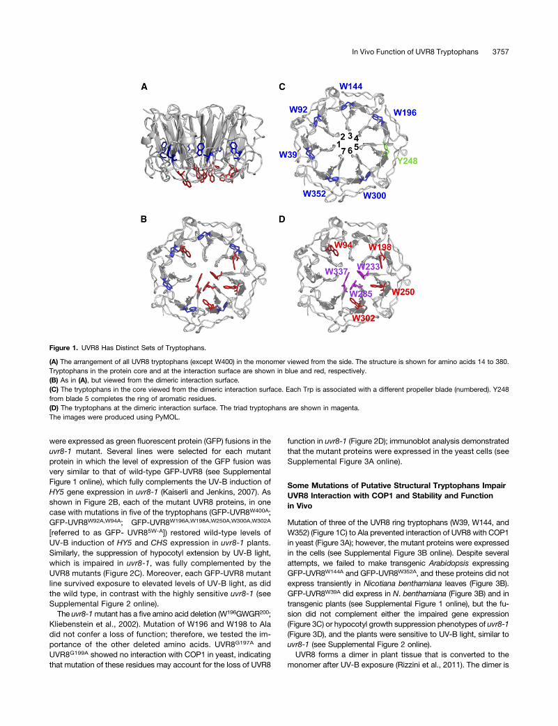
UVR8 has 14 tryptophans that are highly conserved in plantspecies (Rizzini et al., 2011; Wu et al., 2011; Christie et al., 2012).The crystal structure of UVR8 (Figures 1A and 1B) (Christie et al.,2012; Wu et al., 2012) shows the positions of all the tryptophans,except one (W400), which is in the C terminus. The remaining13 tryptophans are organized into two groups. First, within theprotein core is a ring of six tryptophans and one Tyr (Y248),each contributed by a different blade of the b-propeller (Figure1C). Two of these tryptophans (W144 and W352) are conserved inthe structurally related proteins RCC1 (Renault et al., 1998)and HERC2 (Bekker-Jensen et al., 2010; Wu et al., 2011), andthree (W39, W196, and W300) are conserved between UVR8 andHERC2; the remaining Trp is W92. These aromatic residues formhydrogen bonds and hydrophobic interactions between adjacentblades that help to maintain the b-propeller structure. Second,seven tryptophans are located in the dimeric interface (Figure 1D);these include the triad tryptophans W233, W285, and W337 andalso W94, which forms the apex of the excitonically coupled,cross-dimer Trp pyramid implicated in photoreception (Christieet al., 2012). W198, W250, and W302 are at the periphery of thedimeric interaction surface at the opposite side of the triad toW94.
Most of the Conserved Tryptophans of UVR8 Are NotRequired for Function in Plants
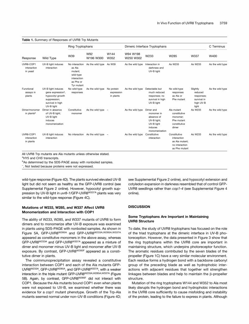
To examine the roles of tryptophans in UVR8 function, we mutatedall of them singly and in several combinations and examined theactivity of the mutant proteins in yeast and transgenic plants.The data obtained for all the tryptophans in this study aresummarized in Table 1. In most cases, Trp was mutated to Ala,because of its nonpolar, nonaromatic nature. In yeast, a verylow fluence rate of narrowband UV-B light stimulates interactionbetween UVR8 and COP1 (Rizzini et al., 2011). Mutation of eightUVR8 tryptophans, in some cases as double mutants (UVR8W250A;UVR8W400A; UVR8W92A,W94A; UVR8W196A,W198A; UVR8W300A,W302A),did not prevent the UV-B–stimulated interaction with COP1(Figure 2A).To examine function in plants, UVR8 proteins with mutations
in one or more of the previously mentioned eight tryptophans
3756 The Plant Cell
were expressed as green fluorescent protein (GFP) fusions in theuvr8-1 mutant. Several lines were selected for each mutantprotein in which the level of expression of the GFP fusion wasvery similar to that of wild-type GFP-UVR8 (see SupplementalFigure 1 online), which fully complements the UV-B induction ofHY5 gene expression in uvr8-1 (Kaiserli and Jenkins, 2007). Asshown in Figure 2B, each of the mutant UVR8 proteins, in onecase with mutations in five of the tryptophans (GFP-UVR8W400A;GFP-UVR8W92A,W94A; GFP-UVR8W196A,W198A,W250A,W300A,W302A
[referred to as GFP- UVR85W-A]) restored wild-type levels ofUV-B induction of HY5 and CHS expression in uvr8-1 plants.Similarly, the suppression of hypocotyl extension by UV-B light,which is impaired in uvr8-1, was fully complemented by theUVR8 mutants (Figure 2C). Moreover, each GFP-UVR8 mutantline survived exposure to elevated levels of UV-B light, as didthe wild type, in contrast with the highly sensitive uvr8-1 (seeSupplemental Figure 2 online).
The uvr8-1mutant has a five amino acid deletion (W196GWGR200;Kliebenstein et al., 2002). Mutation of W196 and W198 to Aladid not confer a loss of function; therefore, we tested the im-portance of the other deleted amino acids. UVR8G197A andUVR8G199A showed no interaction with COP1 in yeast, indicatingthat mutation of these residues may account for the loss of UVR8
function in uvr8-1 (Figure 2D); immunoblot analysis demonstratedthat the mutant proteins were expressed in the yeast cells (seeSupplemental Figure 3A online).
Some Mutations of Putative Structural Tryptophans ImpairUVR8 Interaction with COP1 and Stability and Functionin Vivo
Mutation of three of the UVR8 ring tryptophans (W39, W144, andW352) (Figure 1C) to Ala prevented interaction of UVR8 with COP1in yeast (Figure 3A); however, the mutant proteins were expressedin the cells (see Supplemental Figure 3B online). Despite severalattempts, we failed to make transgenic Arabidopsis expressingGFP-UVR8W144A and GFP-UVR8W352A, and these proteins did notexpress transiently in Nicotiana benthamiana leaves (Figure 3B).GFP-UVR8W39A did express in N. benthamiana (Figure 3B) and intransgenic plants (see Supplemental Figure 1 online), but the fu-sion did not complement either the impaired gene expression(Figure 3C) or hypocotyl growth suppression phenotypes of uvr8-1(Figure 3D), and the plants were sensitive to UV-B light, similar touvr8-1 (see Supplemental Figure 2 online).UVR8 forms a dimer in plant tissue that is converted to the
monomer after UV-B exposure (Rizzini et al., 2011). The dimer is
Figure 1. UVR8 Has Distinct Sets of Tryptophans.
(A) The arrangement of all UVR8 tryptophans (except W400) in the monomer viewed from the side. The structure is shown for amino acids 14 to 380.Tryptophans in the protein core and at the interaction surface are shown in blue and red, respectively.(B) As in (A), but viewed from the dimeric interaction surface.(C) The tryptophans in the core viewed from the dimeric interaction surface. Each Trp is associated with a different propeller blade (numbered). Y248from blade 5 completes the ring of aromatic residues.(D) The tryptophans at the dimeric interaction surface. The triad tryptophans are shown in magenta.The images were produced using PyMOL.
In Vivo Function of UVR8 Tryptophans 3757

observed after SDS-PAGE as long as the protein is not boiled inSDS sample buffer (Rizzini et al., 2011; Christie et al., 2012; Wuet al., 2012). In contrast with GFP-UVR8, the GFP-UVR8W39A
mutant appeared as a monomer in the gel assay both before andafter UV-B treatment (Figure 3E). Interaction of UVR8 with COP1in plants can be monitored using a coimmunoprecipitation assay(Favory et al., 2009). Wild-type GFP-UVR8 interacted with COP1after UV-B exposure, but, by contrast, the GFP-UVR8W39A mu-tant failed to interact with COP1 (Figure 3F).Because Ala mutations of the tryptophans mentioned above
impaired expression and function of UVR8, we tested the effect ofmutations to the aromatic amino acids Phe and Tyr. Each Trp-Pheand Trp-Tyr mutant of W39, W144, and W352 showed UV-B lightstimulated interaction with COP1 in yeast, similar to the wild type(Figure 3G).
W233, W285, and W337 Are Key to UVR8 Function in Yeastand Plants
The triad tryptophans (Figure 1D) were mutated to examine theirrole in UVR8 function in vivo. Mutation of W233, W285, andW337 to Ala, both singly and in combination, caused interactionwith COP1 in yeast in darkness as well as in UV-B light (Figure 4A).By contrast, mutations to Phe or Tyr prevented interaction in bothdarkness and UV-B light (Figure 4B), although the proteins wereexpressed in the cells (see Supplemental Figure 3C online). Asimilar observation was reported previously for W285A and W285F(Rizzini et al., 2011).The function of the triad tryptophans in plants was examined
by expression of GFP fusions in uvr8-1. For each mutation,transgenic lines were selected that had very similar levels ofexpression to GFP-UVR8 (see Supplemental Figure 1 online),which fully complements uvr8-1 (Kaiserli and Jenkins, 2007).We reported recently (Christie et al., 2012) that GFP-UVR8W285A
fails to restore the loss of UV-B–induced HY5 gene expression inuvr8-1, and a similar result was observed for CHS expression(Figure 4C). Furthermore, GFP-UVR8W285A failed to complementthe impaired hypocotyl growth-suppression phenotype (Figure4D), and the transgenic plants were as sensitive to UV-B lightas uvr8-1 (see Supplemental Figure 2 online). Similarly, GFP-UVR8W285F was unable to complement uvr8-1 in each of thesefunctional assays.GFP-UVR8W233A gave partial complementation of UVR8 func-
tion. In each experiment undertaken with uvr8-1/GFP-UVR8W233A
plants, HY5 and CHS transcript levels were greater in UV-B lightthan in its absence, with quantification suggesting that theresponse to UV-B light was ;20 to 25% of that in wild-typeplants. However, the transcript levels in the different experi-ments were quite variable, and the mean values in UV-B lightwere not significantly greater than those in the absence of UV-B light when analyzed by Student’s t test (Figure 4C). Hypo-cotyl growth suppression by UV-B light was partially restored inuvr8-1/GFP-UVR8W233A transgenic plants, and the reduction ofhypocotyl length was statistically significant (Figure 4D), but theplants showed little difference in UV-B sensitivity compared withuvr8-1 (see Supplemental Figure 2 online).GFP-UVR8W337A substantially, but not fully, complemented
uvr8-1 in HY5 and CHS gene expression, giving ;80% of the
Figure 2. Mutation of Eight UVR8 Tryptophans Has Little Effect onFunction in Vivo.
(A) Plasmids containing a GAL4 binding domain (BD) or activation do-main (AD), fused as indicated to UVR8 Trp mutants or COP1, were co-transformed into yeasts, which were then spotted as a dilution series onfull selective media plates. As controls, yeasts were cotransformed withplasmids containing mammalian p53 and antigen T (Ant T; positivecontrol) or no inserts (2; negative control). Yeasts were grown under 0.1mmol m22 s21 of UV-B light (+UV-B) or in darkness (2UV-B) for 72 h.(B) Quantitative assays of HY5 and CHS transcripts, normalized to controlACTIN2 transcript levels, in wild-type Ler, uvr8-1, and uvr8-1 transformed withGFP-UVR8 mutants; UVR85W-A is GFP-UVR8W196A,W198A,W250A,W300A,W302A.Plants were exposed (+UV-B) or not (2UV-B) to 3 mmol m22 s21 of UV-Blight for 3 h. Data are means of three experiments 6SE. The values for+UV-B are significantly greater than those for 2UV-B (P < 0.01) for allgenotypes except uvr8-1, which shows no significant difference (P >0.05).(C) Hypocotyl lengths of wild-type Ler, uvr8-1, and uvr8-1 transformedwith GFP-UVR8 mutants. Seedlings were grown for 4 d in 2 mmol m22 s21
of white light with (+UV-B) or without (2UV-B) 1.5 mmol m22 s21 of UV-Blight. Mean is shown 6SE, n = 30. The values for +UV-B are significantlylower than those for 2UV-B (P < 0.01) for all genotypes except uvr8-1,which shows no significant difference (P > 0.05).(D) Yeast two-hybrid assay, performed as in (A), with UVR8 mutationsfrom within the deleted region of uvr8-1.
3758 The Plant Cell
wild-type response (Figure 4D). The plants survived elevated UV-Blight but did not seem as healthy as the GFP-UVR8 control (seeSupplemental Figure 2 online). However, hypocotyl growth sup-pression by UV-B light in uvr8-1/GFP-UVR8W337A plants was verysimilar to the wild-type response (Figure 4C).
Mutations of W233, W285, and W337 Affect UVR8Monomerization and Interaction with COP1
The ability of W233, W285, and W337 mutants of UVR8 to formdimers and to monomerize after UV-B exposure was examinedin plants using SDS-PAGE with nonboiled samples. As shown inFigure 5A, GFP-UVR8W285A and GFP-UVR8W233A,W285A,W337A
appeared as constitutive monomers in the above assay, whereasGFP-UVR8W233A and GFP-UVR8W337A appeared as a mixture ofdimer and monomer minus UV-B light and monomer after UV-Bexposure. By contrast, GFP-UVR8W285F appeared as a consti-tutive dimer in plants.
The coimmunoprecipitation assay revealed a constitutiveinteraction between COP1 and each of the Ala mutants GFP-UVR8W233A, GFP-UVR8W285A, and GFP-UVR8W337A, with a weakerinteraction in the triple mutant GFP-UVR8W233A,W285A,W337A (Figure5B). Again, by contrast, GFP-UVR8W285F did not interact withCOP1. Because the Ala mutants bound COP1 even when plantswere not exposed to UV-B, we examined whether there wasevidence for a cop1 mutant phenotype. Growth of the Trp-Alamutants seemed normal under non–UV-B conditions (Figure 4D;
see Supplemental Figure 2 online), and hypocotyl extension andcotyledon expansion in darkness resembled that of control GFP-UVR8 seedlings rather than cop1-4 (see Supplemental Figure 4online).
DISCUSSION
Some Tryptophans Are Important in MaintainingUVR8 Structure
To date, the study of UVR8 tryptophans has focused on the roleof the triad tryptophans at the dimeric interface in UV-B pho-toreception. However, the data presented in Figure 3 show thatthe ring tryptophans within the UVR8 core are important inmaintaining structure, which underpins photoreceptor function.The aromatic residues contributed by the seven blades of thepropeller (Figure 1C) have a very similar molecular environment.Each residue forms a hydrogen bond with a backbone carbonylgroup of the preceding blade as well as hydrophobic inter-actions with adjacent residues that together will strengthenlinkages between blades and help to maintain the b-propellerstructure.Mutation of the ring tryptophans W144 and W352 to Ala most
likely disrupts the hydrogen bond and hydrophobic interactionsin the UVR8 core sufficiently to cause misfolding and instabilityof the protein, leading to the failure to express in plants. Although
Table 1. Summary of Responses of UVR8 Trp Mutants
Ring Tryptophans Dimeric Interface Tryptophans C Terminus
Response Wild TypeW39
W92W196 W300
W144W352
W94 W198W250 W302
W233 W285 W337 W400
UVR8-COP1interactionin yeast
UV-B light inducesinteraction
No interactionas Alamutant;wild-typeinteractionas Phe orTyr mutant
As the wild type As W39 As the wild type Interaction indarkness andUV-B light
As W233 As W233 As the wild type
Functionalassays inplants
UV-B light inducesgene expressiona,hypocotyl growthsuppression;survival in highUV-B light
No wild-typeresponses
As the wild type No proteinexpressionin plants
As the wild type Detectable butmuch reducedresponses; nosurvival in highUV-B light
No wild-typeresponsesas Ala orPhe mutant
Slightlyreducedresponses;survival inhigh UV-Blight
As the wild type
Dimer/monomerin plantsb
Dimer in absenceof UV-B light;UV-B lightinducesmonomerization
Constitutivemonomer
As the wild type – As the wild type Dimer andmonomer inabsence ofUV-B light;UV-B lightinducesmonomerization
Ala mutantconstitutivemonomer;Phe mutantconstitutivedimer
As W233 As the wild type
UVR8-COP1interactionin plants
UV-B light inducesinteraction
No interaction As the wild type – As the wild type Constitutiveinteraction
Constitutiveinteractionas Ala mutant;no interactionas Phe mutant
As W233 As the wild type
All UVR8 Trp mutants are Ala mutants unless otherwise stated.aHY5 and CHS transcripts.bAs determined by the SDS-PAGE assay with nonboiled samples.–, Not tested because proteins were not expressed.
In Vivo Function of UVR8 Tryptophans 3759
UVR8W144A and UVR8W352A are expressed in yeast, they fail tointeract with COP1, probably because the protein is unable toadopt the correct conformation for COP1 binding. UVR8W39A
does express in plants but appears as a monomer in the gelassay (Figure 3E), indicating that the dimer is unable to form oris substantially weakened. W39 is involved in linking blades 1and 7 of the protein, which is particularly important in preservingthe overall structure. Moreover, UVR8W39A fails to interact withCOP1 in plants and to functionally complement uvr8-1. It is likelythat impaired interactions in the core of UVR8W39A affect themonomer structure sufficiently to prevent COP1 interaction.The Tyr and Phe mutants of W144, W352, and W39 do interact
with COP1 in a UV-B–dependent manner in yeast, indicatingthat these aromatic residues permit formation of the hydro-phobic interactions that stabilize the UVR8 core. It is in-teresting that mutation of the remaining three ring tryptophans(W92, W196, and W300) does not prevent function in vivo. Infact, two of these tryptophans are mutated to Ala in theUVR85W-A mutant, which complements uvr8-1. A possible expla-nation of the more benign effect of mutations to W92, W196, andW300 is that, in contrast with W39, W144, and W352, these res-idues have adjacent Tyr residues (Y90, Y194, and Y298) that couldmove into the space caused by mutation of the tryptophans to Alaand form a water-mediated hydrogen bond with the backbone
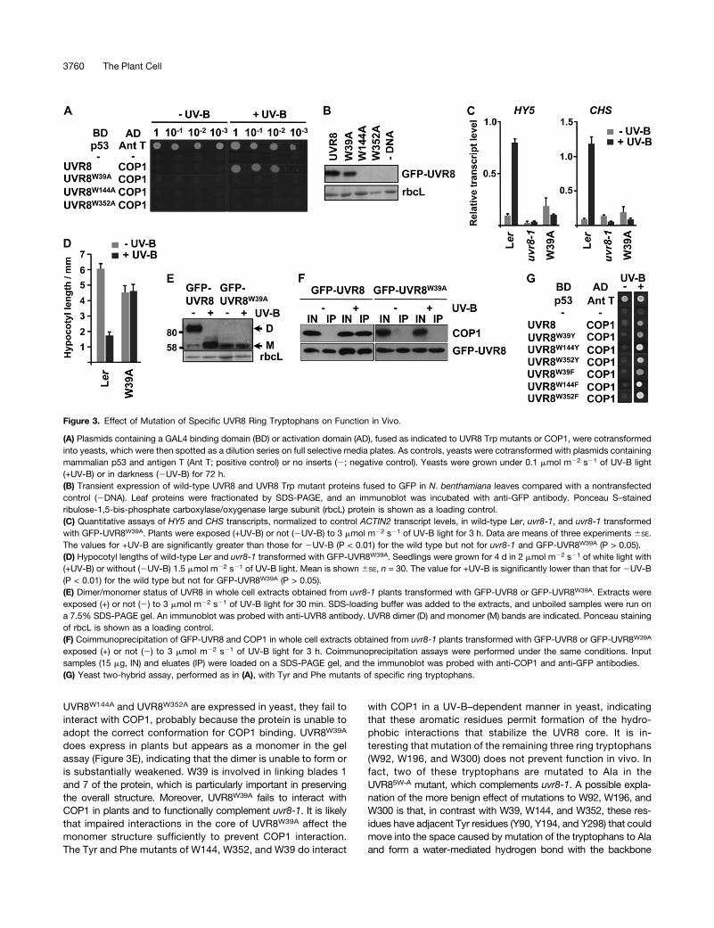
Figure 3. Effect of Mutation of Specific UVR8 Ring Tryptophans on Function in Vivo.
(A) Plasmids containing a GAL4 binding domain (BD) or activation domain (AD), fused as indicated to UVR8 Trp mutants or COP1, were cotransformedinto yeasts, which were then spotted as a dilution series on full selective media plates. As controls, yeasts were cotransformed with plasmids containingmammalian p53 and antigen T (Ant T; positive control) or no inserts (2; negative control). Yeasts were grown under 0.1 mmol m22 s21 of UV-B light(+UV-B) or in darkness (2UV-B) for 72 h.(B) Transient expression of wild-type UVR8 and UVR8 Trp mutant proteins fused to GFP in N. benthamiana leaves compared with a nontransfectedcontrol (2DNA). Leaf proteins were fractionated by SDS-PAGE, and an immunoblot was incubated with anti-GFP antibody. Ponceau S–stainedribulose-1,5-bis-phosphate carboxylase/oxygenase large subunit (rbcL) protein is shown as a loading control.(C) Quantitative assays of HY5 and CHS transcripts, normalized to control ACTIN2 transcript levels, in wild-type Ler, uvr8-1, and uvr8-1 transformedwith GFP-UVR8W39A. Plants were exposed (+UV-B) or not (2UV-B) to 3 mmol m22 s21 of UV-B light for 3 h. Data are means of three experiments 6SE.The values for +UV-B are significantly greater than those for 2UV-B (P < 0.01) for the wild type but not for uvr8-1 and GFP-UVR8W39A (P > 0.05).(D) Hypocotyl lengths of wild-type Ler and uvr8-1 transformed with GFP-UVR8W39A. Seedlings were grown for 4 d in 2 mmol m22 s21 of white light with(+UV-B) or without (2UV-B) 1.5 mmol m22 s21 of UV-B light. Mean is shown 6SE, n = 30. The value for +UV-B is significantly lower than that for 2UV-B(P < 0.01) for the wild type but not for GFP-UVR8W39A (P > 0.05).(E) Dimer/monomer status of UVR8 in whole cell extracts obtained from uvr8-1 plants transformed with GFP-UVR8 or GFP-UVR8W39A. Extracts wereexposed (+) or not (2) to 3 mmol m22 s21 of UV-B light for 30 min. SDS-loading buffer was added to the extracts, and unboiled samples were run ona 7.5% SDS-PAGE gel. An immunoblot was probed with anti-UVR8 antibody. UVR8 dimer (D) and monomer (M) bands are indicated. Ponceau stainingof rbcL is shown as a loading control.(F) Coimmunoprecipitation of GFP-UVR8 and COP1 in whole cell extracts obtained from uvr8-1 plants transformed with GFP-UVR8 or GFP-UVR8W39A
exposed (+) or not (2) to 3 mmol m22 s21 of UV-B light for 3 h. Coimmunoprecipitation assays were performed under the same conditions. Inputsamples (15 mg, IN) and eluates (IP) were loaded on a SDS-PAGE gel, and the immunoblot was probed with anti-COP1 and anti-GFP antibodies.(G) Yeast two-hybrid assay, performed as in (A), with Tyr and Phe mutants of specific ring tryptophans.
3760 The Plant Cell
carbonyl group of the preceding blade as well as hydrophobicinteractions.
Specific Tryptophans at the Dimeric Interface AreConcerned with Photoreception in Vivo
The dimeric interface has a specific spatial arrangement oftryptophans. The triad tryptophans, W233, W285, and W337,are close together and excitonically coupled. W94, which is toone side of the triad in the interacting surface (Figure 1D), isexcitonically coupled with the triad on the opposite monomerand forms the apex of a pyramid with the triad as the base(Christie et al., 2012). The three tryptophans (W198, W250,W302) at the opposite side of the triad to W94 form an outerring of aromatic residues together with Y201, Y253, and F305,which is suggested to shield the triad from solvent (Christieet al., 2012). However, mutation of these three outer trypto-phans to Ala, in the context of the UVR85W-A mutant, does notprevent UVR8 function in plants. The UVR85W-A mutant fullycomplements the impaired gene expression, hypocotyl growthsuppression, and UV-B tolerance phenotypes of uvr8-1. Similarly,
Ala mutations of W198, W250, and W302 do not prevent in-teraction with COP1 in yeast. We conclude that these trypto-phans are not required for UVR8 function in vivo. However, wecannot exclude the possibility that mutations in these aminoacids cause subtle changes in UVR8 function under particularconditions that have not been examined in this study. For in-stance, it will be interesting to examine the dose–response re-lationships of these mutants to see whether there are differencesin response at low, nonsaturating doses.Previous studies in yeast and in vitro have implicated the triad
tryptophans in UV-B photoreception. The UVR8W285F mutantexpressed in yeast does not monomerize in response to UV-Blight (Rizzini et al., 2011), and the purified protein is a consti-tutive dimer that fails to respond to UV-B light based on cir-cular dichroism (CD) spectroscopy (Christie et al., 2012) andfluorescence spectroscopy (Wu et al., 2012). Here we showthat UVR8W285F is a constitutive dimer that is nonfunctional inplants; it fails to restore UV-B induction of gene expression orhypocotyl growth suppression in response to UV-B light in uvr8-1,and the plants are highly sensitive to UV-B light. In contrast withUVR8W285F, UVR8W285A seems to be a constitutive monomer
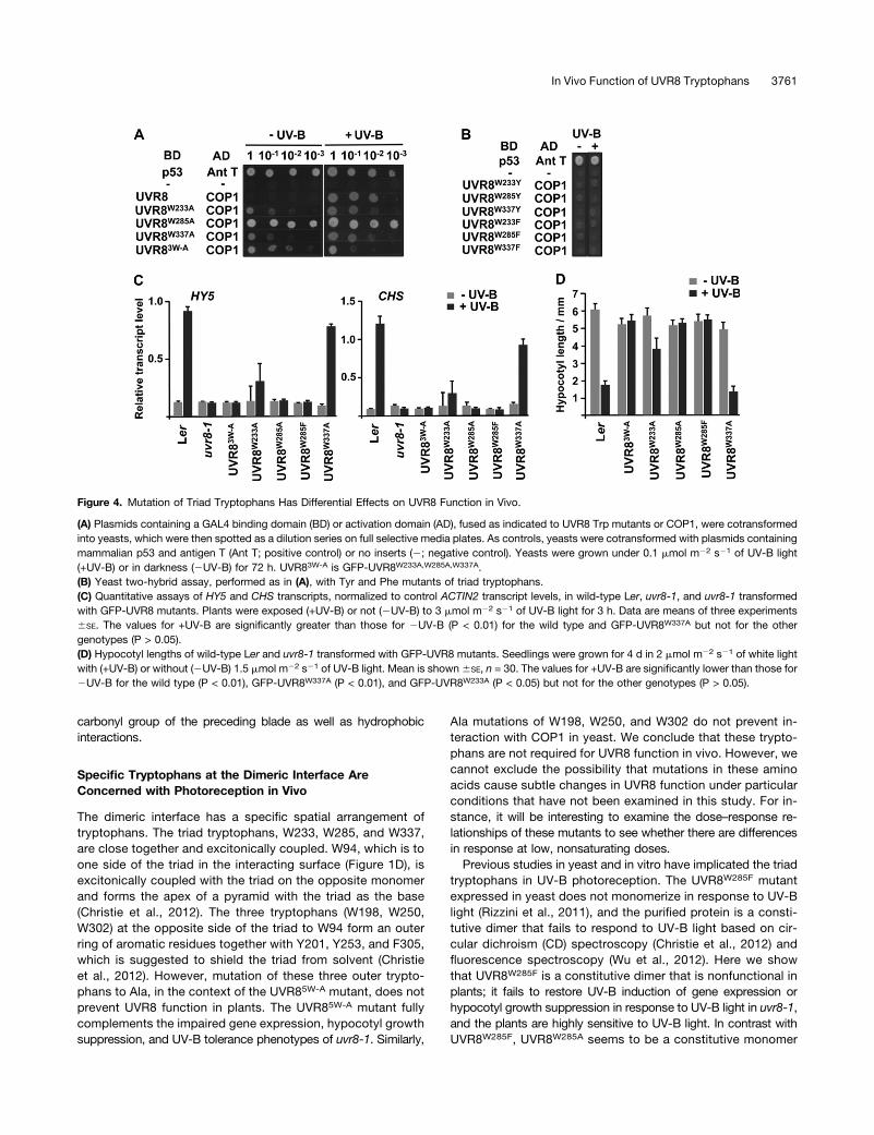
Figure 4. Mutation of Triad Tryptophans Has Differential Effects on UVR8 Function in Vivo.
(A) Plasmids containing a GAL4 binding domain (BD) or activation domain (AD), fused as indicated to UVR8 Trp mutants or COP1, were cotransformedinto yeasts, which were then spotted as a dilution series on full selective media plates. As controls, yeasts were cotransformed with plasmids containingmammalian p53 and antigen T (Ant T; positive control) or no inserts (2; negative control). Yeasts were grown under 0.1 mmol m22 s21 of UV-B light(+UV-B) or in darkness (2UV-B) for 72 h. UVR83W-A is GFP-UVR8W233A,W285A,W337A.(B) Yeast two-hybrid assay, performed as in (A), with Tyr and Phe mutants of triad tryptophans.(C) Quantitative assays of HY5 and CHS transcripts, normalized to control ACTIN2 transcript levels, in wild-type Ler, uvr8-1, and uvr8-1 transformedwith GFP-UVR8 mutants. Plants were exposed (+UV-B) or not (2UV-B) to 3 mmol m22 s21 of UV-B light for 3 h. Data are means of three experiments6SE. The values for +UV-B are significantly greater than those for 2UV-B (P < 0.01) for the wild type and GFP-UVR8W337A but not for the othergenotypes (P > 0.05).(D) Hypocotyl lengths of wild-type Ler and uvr8-1 transformed with GFP-UVR8 mutants. Seedlings were grown for 4 d in 2 mmol m22 s21 of white lightwith (+UV-B) or without (2UV-B) 1.5 mmol m22 s21 of UV-B light. Mean is shown6SE, n = 30. The values for +UV-B are significantly lower than those for2UV-B for the wild type (P < 0.01), GFP-UVR8W337A (P < 0.01), and GFP-UVR8W233A (P < 0.05) but not for the other genotypes (P > 0.05).
In Vivo Function of UVR8 Tryptophans 3761
in yeast (Rizzini et al., 2011) and in plants (Figure 5A) whenassayed by SDS-PAGE with nonboiled samples. Although thiselectrophoretic technique effectively identifies a weaker dimerand is a very convenient assay for cell extracts, it does not rig-orously show whether a protein is monomeric or dimeric. Thus, incontrast with the gel assay, size exclusion chromatographyshows that purified UVR8W285A is a dimer that does notmonomerize in response to UV-B light (Christie et al., 2012).Purified UVR8W233F is also a constitutive dimer when examined bysize exclusion chromatography but appears as a monomer whenexamined by the gel assay in yeast extracts (Rizzini et al., 2011).Nonetheless, it is clear that Ala mutation of W285 weakens thedimer, and it is quite likely that the protein exists as a monomerunder the conditions found within the plant cell. Regardless ofits dimer/monomer status, UVR8W285A, like UVR8W285F, is non-functional in plants, as shown by the lack of induction of HY5(Christie et al., 2012) and CHS transcripts (Figure 4C) in response
to UV-B light, the lack of hypocotyl growth suppression by UV-Blight (Figure 4D), and the high sensitivity of the transgenic uvr8-1/UVR8W285A plants to UV-B light (see Supplemental Figure 2 on-line). The pivotal importance of W285 in UVR8 photoreception isevident, because the other triad tryptophans, including W233, aretotally unable to compensate when it is mutated.Ala and Phe mutants of W233 show very little responsiveness
to UV-B light in biophysical assays of the purified proteins,similar to the equivalent mutants of W285 (Christie et al., 2012;Wu et al., 2012). Moreover, W233 is very important in maintainingexciton coupling in the Trp cluster responsible for photoreception.In plants, UVR8W233A seems to have a weakened dimer but,unlike UVR8W285A, it is not constitutively monomeric and showsa mixture of dimer and monomer under non–UV-B conditions. Inaddition, UVR8W233A shows dimer-to-monomer conversion inuvr8-1 plants and has detectable activity in gene expression andhypocotyl growth suppression in response to UV-B light. Thesedata indicate that W233 is important but not essential for UVR8activity in vivo. They also show that W285 cannot fully com-pensate for mutation of W233, despite its key role in UVR8photoreception.Purified UVR8W337F and UVR8W337A proteins have a partially
reduced response to UV-B light, assayed by CD spectroscopy,but show UV-B–induced dimer-to-monomer conversion (Christieet al., 2012). In plants, UVR8W337A has a weaker dimer than wild-type UVR8, similar to UVR8W233A, and it monomerizes in responseto UV-B light. Expression of UVR8W337A in uvr8-1 substantially,but not completely, restores UV-B–induced gene expression, andthis may explain why the plants are not as completely tolerant ofelevated UV-B light as the wild type. However, the plants ex-pressing UVR8W337A are very similar to the wild type in theirhypocotyl growth suppression response. Thus, W337 hasa significant role in UVR8 function, but it is not as important inphotoreception as W285 and W233.Although W94 is part of the excitonically coupled Trp pyramid,
it is not essential for UVR8 function. In vitro, UVR8W94A showsonly a small reduction in response to UV-B light compared withwild-type UVR8. In plants, mutation of W94, in the context of thedouble UVR8W92A,W94A mutant, has no apparent effect on UVR8function. However, as mentioned above, it is possible that thefunctional significance of some tryptophans, such as W94, maybe revealed under particular conditions, and hence it will bevaluable to examine the role of W94 under varying fluence ratesand wavelengths of UV-B light.Thus, our in vivo analysis of the functions of the pyramid
tryptophans substantially extends the observations reported forpurified UVR8 mutant proteins and shows how each Trp con-tributes to UVR8-mediated photomorphogenic responses inplants. The observed order of importance of the tryptophansin responses in vivo, W285>W233>W337>W94, is essentiallyconsistent with the in vitro assay of photoreception by CDspectroscopy (Christie et al., 2012). The in vivo data support theconclusion from in vitro experiments that W285 is the principalchromophore of UVR8; mutation of W285 to Phe in the purifiedphotoreceptor retunes its spectral sensitivity so that it is able tosense UV-C light (Christie et al., 2012). W233 also has a key rolein UV-B photoreception. Apart from its ability to absorb UV-Blight, W233 is important in maintaining excitonic coupling within
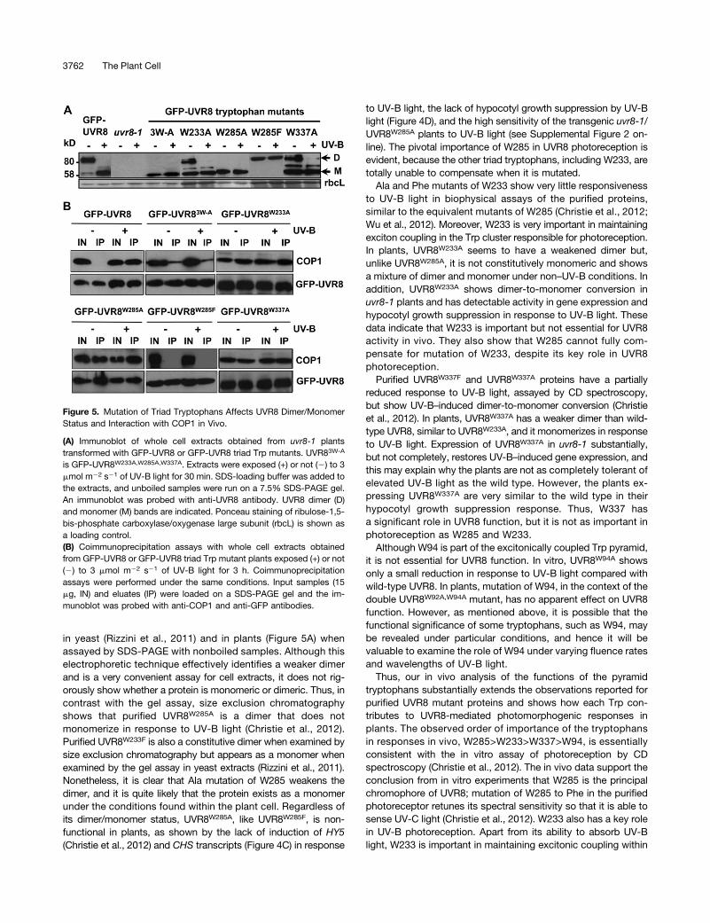
Figure 5. Mutation of Triad Tryptophans Affects UVR8 Dimer/MonomerStatus and Interaction with COP1 in Vivo.
(A) Immunoblot of whole cell extracts obtained from uvr8-1 plantstransformed with GFP-UVR8 or GFP-UVR8 triad Trp mutants. UVR83W-A
is GFP-UVR8W233A,W285A,W337A. Extracts were exposed (+) or not (2) to 3mmol m22 s21 of UV-B light for 30 min. SDS-loading buffer was added tothe extracts, and unboiled samples were run on a 7.5% SDS-PAGE gel.An immunoblot was probed with anti-UVR8 antibody. UVR8 dimer (D)and monomer (M) bands are indicated. Ponceau staining of ribulose-1,5-bis-phosphate carboxylase/oxygenase large subunit (rbcL) is shown asa loading control.(B) Coimmunoprecipitation assays with whole cell extracts obtainedfrom GFP-UVR8 or GFP-UVR8 triad Trp mutant plants exposed (+) or not(2) to 3 mmol m22 s21 of UV-B light for 3 h. Coimmunoprecipitationassays were performed under the same conditions. Input samples (15mg, IN) and eluates (IP) were loaded on a SDS-PAGE gel and the im-munoblot was probed with anti-COP1 and anti-GFP antibodies.
3762 The Plant Cell
the cross-dimer Trp pyramid (Christie et al., 2012). However, it isinteresting that mutation of W233 to Ala does not eliminateUVR8 activity in vivo, whereas very little photochemical activitywas detectable in vitro (Christie et al., 2012; Wu et al., 2012).Evidently other tryptophans, most likely W285, can partly com-pensate for mutation of W233, although this is not seen withmutation of W285. Thus, it is not simply the case that W285 andW233 act as functionally redundant UV-B chromophores forUVR8. Further research is required to determine the precise rolesand functional relationships of W285, W233, and the other tryp-tophans within the pyramid.
Interaction with COP1 Is Not Sufficient for UVR8 Function
Genetic studies indicate that COP1 is required for UVR8-mediatedresponses, and UV-B light stimulates the direct interaction ofUVR8 with COP1. This interaction is believed to initiate sig-naling, although the steps leading to transcriptional regulationare not understood. The constitutively dimeric UVR8W285F mutantdoes not interact with COP1 either in yeast (Rizzini et al., 2011)or in plants (Figure 5B), consistent with its inability to mediateresponses. In fact, the Phe and Tyr mutants of all the triadtryptophans fail to interact with COP1 in yeast (Figure 4B). Bycontrast, UVR8W285A interacts with COP1 in yeast both in dark-ness and in UV-B light, and a similar constitutive interaction isseen in plants. Similarly, UVR8W233A and UVR8W337A interactconstitutively with COP1 in plants and yeast, although in yeast,the interaction in darkness is less strong than for UVR8W285A. Ithas been reported that overexpression of UVR8 leads to a cop1-like phenotype under UV-B light, probably because COP1 is se-questered, restricting its involvement with other light signalingpathways and its availability to act as an E3 ubiquitin ligase(Favory et al., 2009). However, the constitutive binding of COP1 tothe Ala mutants of UVR8 triad tryptophans did not cause a cop1-like phenotype either in light-grown (see Supplemental Figure 2online) or dark-grown (see Supplemental Figure 4 online) plants.This is most likely because transgenic lines were selected in whichthe level of expression of the mutant UVR8 proteins was close tothat of wild-type UVR8, so no overexpression phenotypes wereobserved.
It is not entirely clear why the Ala mutants of the triad tryp-tophans interact constitutively with COP1, whereas the Tyr andPhe mutants do not. Undoubtedly, introduction of the small,nonaromatic Ala residue is more disruptive to the structuralrelationships of the triad tryptophans and adjacent amino acidsthan the aromatic mutations. Wu et al. (2012) reported thatmutation of W285 to Ala substantially alters the orientations ofW337 and W233 and the adjacent Asp D129. However, it is notknown how these changes lead to constitutive interaction withCOP1. It is possible that they affect the conformation of the pro-tein and, in consequence, both weaken the salt-bridges that holdthe dimer together and expose the region that interacts with COP1even under non–UV-B conditions. By contrast, mutation of W285to Phe does not alter its structural relationships with adjacentamino acids (Wu et al., 2012), and hence UVR8W285F does notbind to COP1 in darkness.
The current model of UVR8 function is that UV-B light inducesmonomerization and that consequent binding to COP1 initiates
responses. However, the results presented here for the Alamutants of the triad tryptophans indicate that COP1 binding toUVR8 is not sufficient to induce a response. Each of thesemutants interacts with COP1 constitutively, but there is noresponse with any of them in the absence of UV-B light, andthe extent of response after UV-B exposure is not correlated withCOP1 binding. Moreover, if the monomeric form of these mutantsis present in the absence of UV-B light, which is suggested byboth the gel assay (Figure 5A) and the interaction with COP1(Figure 5B), it can be concluded that monomer formation per se isalso not sufficient for UVR8 function. Rather, the extent ofresponse of the triad mutants to UV-B light in vivo is closelycorrelated with the effect of the mutation on photoreception.Thus, for example, UVR8W285A and UVR8W337A both interactwith COP1 constitutively, but the former is nonresponsive toUV-B light both in vitro and in vivo, whereas the latter has a strongresponse to UV-B light in vitro, as judged by CD spectroscopy(Christie et al., 2012), which is correlated with a near–wild-typelevel of response in vivo. It therefore seems that UV-B photore-ception generates some form of signal that is required for UVR8activity apart from the induction of monomerization and COP1binding. For instance, photoreception may initiate a confor-mational change in the protein that is required for function. Thus,a lack of photochemical activity may impair the production ofa functional UVR8 regardless of whether it is monomeric and canbind COP1. Further research is therefore needed to understandthe mechanism of UVR8 photoreception and how photoreceptionaffects the protein to generate a functional monomer that caninteract with COP1 to initiate photomorphogenic responses.
METHODS
Plant Materials
Seeds of wild-type Arabidopsis thaliana ecotype Landsberg erecta (Ler)were obtained from the Nottingham Arabidopsis Stock Center. The uvr8-1mutant allele in the Ler background (Kliebenstein et al., 2002) was ob-tained from Dan Kliebenstein (University of California, Davis). TheUVR8pro:GFP-UVR8 transgenic line in the uvr8-1 background (line 6-2)was described by Kaiserli and Jenkins (2007). All the GFP-UVR8 mutanttransgenic lines produced in this study are in the uvr8-1 background.
Plasmid Constructs and Transformation
Site-directed mutagenesis was performed using the QuikChange Site-Directed Mutagenesis and Multi QuikChange Site-Directed Mutagenesiskits (Stratagene) following the manufacturer’s instructions. The primersused for site-directed mutagenesis are listed in Supplemental Table 1online. For plant transformation,modifiedUVR8 sequenceswere subclonedinto the pEZR(K)L-C vector downstream of enhanced GFP and the CaMV35S promoter using EcoR1 andSal1 restriction sites, as in the original GFP-UVR8 construct (Brown et al., 2005). DNA sequencing confirmed that thefusions were made correctly. The fusions were introduced into uvr8-1mutant plants byAgrobacterium tumefaciens–mediated transformation, andat least three independent homozygous T3 lines (T2 for GFP-UVR8W337A)were tested for complementation andprotein expression (see SupplementalFigure 1 online).
Data presented for hypocotyl length, UV-B sensitivity, monomer/dimerstatus, and coimmunoprecipitation experiments used the following lines (seeSupplemental Figure 1 online): GFP-UVR8W39A, line 1-2; GFP-UVR8W400A, line9-2; GFP-UVR8W92A,W94A, line 8-1; GFP-UVR8W196A,W198A,W250A,W300A,W302A,
In Vivo Function of UVR8 Tryptophans 3763
line 4-1; GFP-UVR8W233A,W337A,W285A, line 11-5; GFP-UVR8W233A, line 5-2;GFP-UVR8W337A, line 8; GFP-UVR8W285A, line 7-1; GFP-UVR8W285F, line 6-6.For the quantitative transcript measurements, data presented are means of atleast three independent experiments undertaken with all the lines shown inSupplemental Figure 1 online. Similar results were obtained for all linesexpressing a particular fusion.
Yeast Two-Hybrid Assays
Yeast two-hybrid vectors were introduced into the yeast strain AH109.Competent cells were grown on yeast peptone dextrose plates for 2 d.A single colony was resuspended in 300 mL of 40% polyethylene glycol,13 TE, and 0.1 M of LiAc, and 1 mg of each plasmid, pGADT7 andpGBKT7, was added followed by incubation at 42°C for 15 min. Thesamples were centrifuged at 1000g for 5 min, and the pellet was re-suspended in yeast peptone dextrose medium for at least 1 h at roomtemperature. Samples were centrifuged at 1000g for 5min, and the pelletswere resuspended in 0.8% NaCl and left at room temperature for 3 h.The cells were then spread on plates containing synthetic dropout (SD)medium (Clontech) with minus Leu, minus Trp dropout supplement(SD2Leu2Trp) (Clontech) to select for transformation of both plas-mids. Plates were left for 3 d at 30°C in darkness.
A single colony was picked and resuspended in 100 mL of 0.8% NaCl,and 5mL was used for spotting on plates containing SD2Leu2Trp orminusLeu, minus Trp, minus His, minus adenine dropout supplement (SD2Leu2Trp2His2Ade) (Clontech) to select for colonies with interacting proteins.Plates were left for 3 d at 30°C either in darkness or under 0.1mmolm22 s21
of narrowbandUV-B light (Philips TL20W/01RS tube; spectrumpresented inChristie et al., 2012). The data presented are representative of at least threeindependent experiments. Only results with selective medium are shown,but for all transformations presented, colonies grew on nonselective me-dium. In addition, control experiments were performed to show that growthon selective medium was not caused by autoactivation.
To confirm expression of bait and prey proteins in each experiment,protein was extracted using a protocol modified from Grefen et al. (2009).A single colony of yeast containing the plasmids of interest was grownovernight in 10 mL of liquid SD2Leu2Trp at 30°C with constant shaking.When the culture had reached anOD at 550 nm between 1.0 and 2.0, 2 mLwas pelleted, the supernatant was discarded, and the cells were re-suspended in the appropriate volume of LL buffer (50 mM of Tris-HCL, pH6.8, 4% SDS, 8 M of urea, 30% glycerol, 0.1 M of DTT, 0.005% [w/v]bromophenol blue) calculated from the OD. The mixture was then vor-texed for 1 min and incubated at 65°C for 30 min. After centrifugation,proteins were separated on a 10% SDS-PAGE gel, and an immunoblotwas incubated with anti-hemagglutinin (Cell Signaling Technology) oranti-MYC (Cell Signaling Technology) antibodies.
Quantitative Transcript Measurements
Plants were grown for 3 weeks in 20 mmol m22 s21 of constant white light at21°C (warm lightfluorescent tubes;Osram)and thenexposed to3mmolm22 s21
of narrowband UV-B light for 3 h. RNA extraction was performed using theRNeasy Plant Mini Kit (Qiagen). cDNA synthesis was performed as de-scribed by Brown et al. (2005). Quantitative PCR was performed usingthe MX3000 Stratagene real-time PCR system and a Brilliant III SYBRGreen qPCR kit (Stratagene) following the manufacturer’s instructions.The target amplicon was quantified by comparing the amplification incDNA samples with standards of known amount. The PCR conditionswere as follows: 3 min at 95°C, 40 cycles of 10 s at 95°C, 20 s at 60°C,followed by a 60 to 95°C dissociation protocol. The primers used foramplification ofHY5, CHS, and ACTIN2 are shown in Supplemental Table 2online. Stratagene MX software was used to automatically calculate thecycle threshold (Ct) value for each reaction. The Ct values for each standarddilution were plotted against the log of the initial template quantity, and
a standard curve was produced. The initial template quantity in each cDNAsample was determined by comparing the Ct values to the standard curve.Each reaction was performed in duplicate. As a control for variation in RNAquantification, reverse transcription efficiency, and template preparation,the expression of CHS and HY5 transcripts was normalized against theamount of ACTIN2 control transcripts in each sample. The data shown arethe means of at least three independent experiments using differenttransgenic lines. Levels of HY5 and CHS transcripts measured for uvr8-1and the UVR8 mutants in each experiment were determined relative to thelevel for wild-type plants exposed to UV-B light to facilitate comparisonbetween experiments. Statistical differences in mean transcript levels weredetermined using Student’s t test.
Measurements of Hypocotyl Length
Hypocotyl length was measured for seedlings grown on 0.8% agar platescontaining one-half–strength Murashige and Skoog salts. Stratified seedswere exposed to 120 mmol m22 s21 of white light for 4 h in a growthchamber. Seedlings were grown in 2 mmol m22 s21 of white light sup-plemented (or not supplemented in controls) with 1.5 mmol m22 s21 ofnarrowband UV-B light for 4 d. The hypocotyl lengths were measuredusing ImageJ software. The data presented are from three independentexperiments. Statistical differences in hypocotyl lengths were determinedusing Student’s t test.
Sensitivity to UV-B Light
UV-B sensitivity assays were performed as described by Brown et al.(2005) using a broadband UV-B source (UV-B-313 fluorescent tubes[Q-Panel]; spectrum presented in Christie et al., 2012).
UVR8 Dimer/Monomer Status
Plants were grown under 100 mmol m22 s21 of white light for 12 d. Wholecell extracts from leaf tissue were prepared as described by Kaiserli andJenkins (2007). UVR8 dimer/monomer status was examined essentially asdescribed by Rizzini et al. (2011). Whole cell extracts were kept on ice andexposed (or not exposed in controls) to 3 mmol m22 s21 of narrowbandUV-B light for 30 min. Then, 43 loading buffer containing 250 mM of Tris-HCl, pH 6.8, 2% SDS, 20% b-mercaptoethanol, 40% glycerol, and 0.5%bromophenol blue was added to the samples, but they were not boiled.The proteins were loaded on a 7.5% SDS-PAGE gel and an immunoblotincubated with anti-UVR8 antibody (Kaiserli and Jenkins, 2007). The datashown are representative of at least three independent experiments.
Coimmunoprecipitation Assay
Plants were grown under 100 mmol m22 s21 of constant white light at21°C for 12 d and then were put in darkness for 16 h. The plants wereexposed (or not exposed in controls) to 3 mmol m22 s21 of narrowbandUV-B light for 3 h. Whole cell extracts were prepared as described byKaiserli and Jenkins (2007) in the absence or presence of 3 mmol m22 s21
of narrowband UV-B light, and the coimmunoprecipitation assays wereperformed under the same conditions. Extract samples were incubatedfor 30 min on ice with 50 mL of anti-GFP microbeads (mMacs; MiltenyiBiotec). The microcolumn was equilibrated using 200 mL of high salt lysisbuffer (450 mM of NaCl, 1% Triton X-100, 50 mM of Tris-HCl, pH 8, 5 mMof phenylmethylsulfonyl fluoride, protease inhibitors [Complete Mini;Roche]). The lysate containing the microbeads was applied onto thecolumn and non–GFP-tagged proteins left to run through. The columnwas washed five times with 200 mL of high salt lysis buffer and once with300 mM of NaCl, Tris-HCl, pH 7.5. To elute proteins, 20 mL of elutionbuffer (0.1 M triethylamine, pH 11.8, 0.1% Triton X-100) was applied tothe column and left for 5 min at room temperature. A further 50 mL of
3764 The Plant Cell

elution buffer was added, and the eluatewas collected in a tube containing 3mL of 1M of 2-(N-morpholino)-ethane-sulfonic acid, pH3.0, to neutralize thesample. The eluates were analyzed by SDS-PAGE and an immunoblotincubated with anti-GFP (Cell Signaling Technology) and anti-COP1 (Janget al., 2010) antibodies. The data shown are representative of at least threeexperiments.
Transient Expression in Nicotiana benthamiana
Transient expression of GFP-UVR8 fusions was performed in leaves of4-week-old N. benthamiana plants. Agrobacterium with the desiredmutant GFP-UVR8 plasmid DNA was inoculated in 10 mL of Luria-Bertanibroth containing 30 mg mL21 of gentamycin and 50 mg mL21 of kana-mycin. The culture was left at 30°C, constantly shaking (220 rpm) until itreached an OD at 550 nm of at least 1.0. The culture was pelleted bycentrifugation at 2000g for 10 min and washed in 10 mL of sterile 10 mMMgCl2. The cell suspension was diluted to OD550 of 0.2 with 10 mMMgCl2solution to a final volume of 20 mL. A total of 200 mM of acetosyringonewas added, and the solution was left at room temperature for 3 h. Usinga syringe, the Agrobacterium medium was infiltrated into the leaves ofN. benthamiana. TheN. benthamiana plants were put in a growth chamberat 30°C in white light for ;60 h. To prepare a whole cell extract, plantswere ground in liquid N2 with a mortar and pestle, and total protein wasextracted in 500 mL of extraction buffer (25 mM of Tris-HCL, pH 7.5, 1 mMof EDTA, 10% glycerol, 5 mM of DTT, 0.1% Triton X-100, and proteaseinhibitor mix [1 tablet of protease inhibitor mix] [Complete Mini, Roche] per10 mL of extraction buffer). The mixture was centrifuged at 16,000g for10 min at 4°C, and the supernatant, containing total protein extract, wascollected. Proteins were fractionated by SDS-PAGE. An immunoblot wasincubated with anti-GFP antibody.
Light Measurements
Fluence rates of white light were measured using a Skye RS232meter fittedwith a quantum sensor (Skye Instruments). Fluence rates of UV-B light (280to 315 nm) were measured using a RS232 meter with a SKU 430 sensor.
Accession Numbers
The Arabidopsis Genome Initiative locus identifier for UVR8 is At5g63860and for COP1 is At2g32950.
Supplemental Data
The following materials are available in the online version of this article.
Supplemental Figure 1. Levels of GFP-UVR8 Expression in theTransgenic Lines.
Supplemental Figure 2. Sensitivity to UV-B of Transgenic LinesExpressing GFP-UVR8 Mutants.
Supplemental Figure 3. Expression of UVR8 Mutants and COP1 inYeast Two-Hybrid Vectors.
Supplemental Figure 4. Phenotype of Dark-Grown Seedlings Ex-pressing Ala Mutants of Triad Tryptophans.
Supplemental Table 1. Primers Used for Site-Directed Mutagenesisof pSK and pGBK Vectors Containing UVR8.
Supplemental Table 2. Primers Used for Quantitative PCR.
ACKNOWLEDGMENTS
We thank John Christie and all members of the Jenkins and Christielaboratories for discussion of the research and guidance with the methods,
Brian Smith for help in interpreting UVR8 structure, Emanuela Sani forassistance with quantitative PCR, Jason Wargent for advice on statistics,and In-Cheol Jang and Nam-Hai Chua for the COP1 antibody. G.I.J.received research support from the UK Biotechnology and BiologicalSciences Research Council and the Leverhulme Trust. A.O. wassupported by a Biotechnology and Biological Science Research CouncilPhD Studentship.
AUTHOR CONTRIBUTIONS
A.O. performed the research and analyzed data. G.I.J. designed theresearch, analyzed data, and wrote the article.
Received June 12, 2012; revised August 10, 2012; accepted August 23,2012; published September 25, 2012.
REFERENCES
Bekker-Jensen, S., Rendtlew Danielsen, J., Fugger, K., Gromova,I., Nerstedt, A., Lukas, C., Bartek, J., Lukas, J., and Mailand, N.(2010). HERC2 coordinates ubiquitin-dependent assembly of DNArepair factors on damaged chromosomes. Nat. Cell Biol. 12: 80–86.
Brown, B.A., Cloix, C., Jiang, G.H., Kaiserli, E., Herzyk, P.,Kliebenstein, D.J., and Jenkins, G.I. (2005). A UV-B-specific sig-naling component orchestrates plant UV protection. Proc. Natl.Acad. Sci. USA 102: 18225–18230.
Brown, B.A., Headland, L.R., and Jenkins, G.I. (2009). UV-B actionspectrum for UVR8-mediated HY5 transcript accumulation in Arab-idopsis. Photochem. Photobiol. 85: 1147–1155.
Brown, B.A., and Jenkins, G.I. (2008). UV-B signaling pathways withdifferent fluence-rate response profiles are distinguished in matureArabidopsis leaf tissue by requirement for UVR8, HY5, and HYH.Plant Physiol. 146: 576–588.
Christie, J.M. (2007). Phototropin blue-light receptors. Annu. Rev.Plant Biol. 58: 21–45.
Christie, J.M., Arvai, A.S., Baxter, K.J., Heilmann, M., Pratt, A.J.,O’Hara, A., Kelly, S.M., Hothorn, M., Smith, B.O., Hitomi, K.,Jenkins, G.I., and Getzoff, E.D. (2012). Plant UVR8 photoreceptorsenses UV-B by tryptophan-mediated disruption of cross-dimer saltbridges. Science 335: 1492–1496.
Cloix, C., and Jenkins, G.I. (2008). Interaction of the Arabidopsis UV-B-specific signaling component UVR8 with chromatin. Mol Plant 1:118–128.
Favory, J.J., et al. (2009). Interaction of COP1 and UVR8 regulatesUV-B-induced photomorphogenesis and stress acclimation inArabidopsis. EMBO J. 28: 591–601.
Franklin, K.A., and Quail, P.H. (2010). Phytochrome functions inArabidopsis development. J. Exp. Bot. 61: 11–24.
Frohnmeyer, H., and Staiger, D. (2003). Ultraviolet-B radiation-mediatedresponses in plants. Balancing damage and protection. Plant Physiol.133: 1420–1428.
Grefen, C., Obrdlik, P., and Harter, K. (2009). The determination ofprotein-protein interactions by the mating-based split-ubiquitinsystem (mbSUS). Methods Mol. Biol. 479: 217–233.
Gruber, H., Heijde, M., Heller, W., Albert, A., Seidlitz, H.K., andUlm, R. (2010). Negative feedback regulation of UV-B-inducedphotomorphogenesis and stress acclimation in Arabidopsis. Proc.Natl. Acad. Sci. USA 107: 20132–20137.
Heijde, M., and Ulm, R. (2012). UV-B photoreceptor-mediated sig-nalling in plants. Trends Plant Sci. 17: 230–237.
In Vivo Function of UVR8 Tryptophans 3765
Jang, I.-C., Henriques, R., Seo, H.S., Nagatani, A., and Chua, N.-H.(2010). Arabidopsis PHYTOCHROME INTERACTING FACTOR pro-teins promote phytochrome B polyubiquitination by COP1 E3 ligasein the nucleus. Plant Cell 22: 2370–2383.
Jenkins, G.I. (2009). Signal transduction in responses to UV-B radi-ation. Annu. Rev. Plant Biol. 60: 407–431.
Jiao, Y., Lau, O.S., and Deng, X.W. (2007). Light-regulated tran-scriptional networks in higher plants. Nat. Rev. Genet. 8: 217–230.
Kaiserli, E., and Jenkins, G.I. (2007). UV-B promotes rapid nucleartranslocation of the Arabidopsis UV-B specific signaling componentUVR8 and activates its function in the nucleus. Plant Cell 19: 2662–2673.
Kliebenstein, D.J., Lim, J.E., Landry, L.G., and Last, R.L. (2002).Arabidopsis UVR8 regulates ultraviolet-B signal transduction andtolerance and contains sequence similarity to human regulator ofchromatin condensation 1. Plant Physiol. 130: 234–243.
Li, Q.H., and Yang, H.Q. (2007). Cryptochrome signaling in plants.Photochem. Photobiol. 83: 94–101.
Oravecz, A., Baumann, A., Máté, Z., Brzezinska, A., Molinier, J.,Oakeley, E.J., Adám, É., Schäfer, E., Nagy, F., and Ulm, R. (2006).CONSTITUTIVELY PHOTOMORPHOGENIC1 is required for the UV-Bresponse in Arabidopsis. Plant Cell 18: 1975–1990.
Renault, L., Nassar, N., Vetter, I., Becker, J., Klebe, C., Roth, M., andWittinghofer, A. (1998). The 1.7 A crystal structure of the regulator ofchromosome condensation (RCC1) reveals a seven-bladed propeller.Nature 392: 97–101.
Rizzini, L., Favory, J.-J., Cloix, C., Faggionato, D., O’Hara, A.,Kaiserli, E., Baumeister, R., Schäfer, E., Nagy, F., Jenkins, G.I.,
and Ulm, R. (2011). Perception of UV-B by the Arabidopsis UVR8protein. Science 332: 103–106.
Stracke, R., Favory, J.-J., Gruber, H., Bartelniewoehner, L.,Bartels, S., Binkert, M., Funk, M., Weisshaar, B., and Ulm, R.(2010). The Arabidopsis bZIP transcription factor HY5 regulatesexpression of the PFG1/MYB12 gene in response to light and ul-traviolet-B radiation. Plant Cell Environ. 33: 88–103.
Ulm, R., Baumann, A., Oravecz, A., Máté, Z., Adám, E., Oakeley,E.J., Schäfer, E., and Nagy, F. (2004). Genome-wide analysis ofgene expression reveals function of the bZIP transcription factorHY5 in the UV-B response of Arabidopsis. Proc. Natl. Acad. Sci.USA 101: 1397–1402.
Ulm, R., and Nagy, F. (2005). Signalling and gene regulation in re-sponse to ultraviolet light. Curr. Opin. Plant Biol. 8: 477–482.
Wargent, J.J., Gegas, V.C., Jenkins, G.I., Doonan, J.H., and Paul,N.D. (2009). UVR8 in Arabidopsis thaliana regulates multiple as-pects of cellular differentiation during leaf development in responseto ultraviolet B radiation. New Phytol. 183: 315–326.
Wu, D., Hu, Q., Yan, Z., Chen, W., Yan, C., Huang, X., Zhang, J.,Yang, P., Deng, H., Wang, J., Deng, X., and Shi, Y. (2012).Structural basis of ultraviolet-B perception by UVR8. Nature 484:214–219.
Wu, M., Grahn, E., Eriksson, L.A., and Strid, A. (2011). Computa-tional evidence for the role of Arabidopsis thaliana UVR8 as UV-Bphotoreceptor and identification of its chromophore amino acids.J. Chem. Inf. Model. 51: 1287–1295.
Yi, C., and Deng, X.W. (2005). COP1 - from plant photomorphogen-esis to mammalian tumorigenesis. Trends Cell Biol. 15: 618–625.
3766 The Plant Cell
DOI 10.1105/tpc.112.101451; originally published online September 25, 2012; 2012;24;3755-3766Plant Cell
Andrew O'Hara and Gareth I. Jenkins UV-B Photoreceptor UVR8ArabidopsisIn Vivo Function of Tryptophans in the
This information is current as of August 30, 2018
Supplemental Data /content/suppl/2012/09/12/tpc.112.101451.DC2.html /content/suppl/2012/09/11/tpc.112.101451.DC1.html
References /content/24/9/3755.full.html#ref-list-1
This article cites 29 articles, 11 of which can be accessed free at:
Permissions https://www.copyright.com/ccc/openurl.do?sid=pd_hw1532298X&issn=1532298X&WT.mc_id=pd_hw1532298X
eTOCs http://www.plantcell.org/cgi/alerts/ctmain
Sign up for eTOCs at:
CiteTrack Alerts http://www.plantcell.org/cgi/alerts/ctmain
Sign up for CiteTrack Alerts at:
Subscription Information http://www.aspb.org/publications/subscriptions.cfm
is available at:Plant Physiology and The Plant CellSubscription Information for
ADVANCING THE SCIENCE OF PLANT BIOLOGY © American Society of Plant Biologists