in vivo embryogenesis, embryo migration, and … of reproduction 51, 452-464 (1994) in vivo...
TRANSCRIPT
BIOLOGY OF REPRODUCTION 51, 452-464 (1994)
In Vivo Embryogenesis, Embryo Migration, and Embryonic Mortalityin the Domestic Cat 1
WILLIAM F. SWANSON,2 TERRI L. ROTH, and DAVID E. WILDT
National Zoological Park, Smithsonian Institution, Washington, District of Columbia 20008
ABSTRACT
In vivo embryogenesis, embryo migration, and survival were studied in the domestic cat. Queens were naturally mated (three
times daily) on the second and third days of behavioral estrus and, if ovulation occurred, ovariohysterectomized at 64 (n = 8),
76 (n = 11), 100 (n = 8), 124 (n = 7), 148 (n = 6), and 480 h (n = 8) after first copulation. Of 52 cats mated, 48 (92.3%)
ovulated (as evidenced by the presence of ovarian CL), and of these, 38 (79.2%) either produced good-quality embryos or had
implantation sites. From the remaining cats, only unfertilized oocytes (n = 5), degenerating embryos (n = 4), or no oocytes/
embryos (n = 1) were recovered. Embryos at 64, 76, 100, and 124 h after the first copulation typically were 1 to 4 cells (17
of 20; 85.0%), 5 to 8 cells (18 of 28; 64.3% ), 9 to 16 cells (14 of 24; 58.3%), and morulae (15 of 21; 71.4% ), respectively; all
were within the oviducts. At 148 h, embryos primarily were compact morulae or early blastocysts (15 of 18; 83.3%), and all
were within the uterus. For the preimplantation groups, the overall recovery of embryos plus oocytes per CL was 80.6%, and
the mean ( SEM) numbers of CL and embryos were 64 h, 4.8 ± 0.3, 3.1 + 0.8; 76 h, 4.7 ± 0.3, 3.9 + 0.6; 100 h, 5.8 0.5,
3.3 + 0.8; 124 h, 4.4 ± 0.5, 4.0 ± 0.6; and 148 h, 6.5 ± 1.1, 3.7 ± 0.7, respectively. Cats in the 480-h group produced a mean
of 5.6 ± 0.5 CL and 3.9 ± 0.5 implantation sites. In six of eight cats in this group, there was a disparity between CL number
on a given ovary and number of implantation sites in the ipsilateral horn, supporting the concept of transuterine embryo mi-
gration. In summary, results indicated that 1 ) more than 90% of cats ovulated following this multiple mating regimen, but --21%
of these failed to produce any fertilized or viable embryos; 2) embryo developmental rate in vivo was biphasic, with a rapid
cleavage rate to the 5- to 8-cell stage followed by a slower cleavage rate to the morula stage; 3) cat embryos entered the uterus
as compact morulae or early blastocysts -5.5 days after the first copulation; and 4) on the basis of implantation/CL ratio, -30%
of all ovulated cat oocytes underwent either fertilization failure or preimplantation embryonic mortality.
INTRODUCTIONIn vitro fertilization (IVF) and embryo culture have been
the focus of considerable research in the domestic cat (Fellscatus) [1-9] and in several nondomestic felid species in-cluding the leopard cat (Felis bengalensis) [10], tiger(Panthera tigris) [11, 12], puma (Fells concolor) [13], andcheetah (Acinonyx jubatus) [14]. These studies have ex-plored mechanisms associated with sperm capacitation, oo-cyte maturation, gamete interaction, fertilization, and earlyembryonic development in vitro and have contributed sig-nificantly to understanding the reproductive physiology offelids. However, the "normality" of IVF embryos cannot bedetermined without comparative baseline data on in vivoembryogenesis-knowledge that, surprisingly, is unavail-able for all felid species, including the domestic cat.
The chronological events of in vivo embryo develop-ment are known for most common species, including thecow [15], horse [16], pig [17,18], sheep [19], goat [20], rabbit[21, 22], and mouse [23]. Relatively recently, information hasbecome available on preimplantation embryo events in vivofor a few canid species, including the dog [24-26] and sil-
Accepted April 22, 1994.Received January 13, 1994.'This research was supported, in part, by grant HD 23853 from the National
Institute of Child Health and Human Development, the Philip Reed Foundation, Friends
of the National Zoo (FONZ), and the Ralston Purina Company/American Zoo and
Aquarium Association Conservation Endowment Fund.'Correspondence: Dr. William Swanson, Department of Reproductive Physiology,
National Zoological Park/NOAHS Center, Smithsonian Institution, Washington, DC
20008. FAX: (202) 673-4733.
ver and blue fox [26]. However, information on in vivopreimplantation embryo development in the domestic catis scarce and is usually a by-product of studies driven byother objectives [27,28]. Especially problematic has been1) the lack of attention to the time of mating relative toembryo recovery (because the cat is an induced ovulator)[29-32] and 2) the confounding effects of use of exogenousgonadotropins to induce folliculogenesis and ensure ovu-lation after natural breeding [331].
Most of what is known about cat embryology is derivedfrom in vitro studies [1-9] using embryos generated by IVFof in vivo-matured oocytes recovered from queens treatedwith exogenous gonadotropins. Although this approachprovides important information about embryo develop-mental rates and culture requirements in vitro, questionsarise that cannot be addressed without understanding invivo events. For example, in vitro-generated embryos de-velop readily in vitro to morulae under a variety of mediaand culture conditions but then experience a partial de-velopmental arrest at the morula-to-blastocyst transition [34].This developmental "block" has proven resistant to alter-ations in culture conditions [5,6,35], but the early devel-opmental competence of these IVF embryos is difficult todetermine without knowledge of the developmental capac-ity of naturally produced embryos. Similarly, when IVF em-bryos are transferred into gonadotropin-synchronized re-cipients, embryo survival and/or pregnancy rates are low[4, 9, 36]; this is a problem that cannot be addressed ade-quately without comparative information on the survival of
452
BIOLOGY OF REPRODUCTION 51, 452-464 (1994)
In Vivo Embryogenesis, Embryo Migration, and Embryonic Mortality in the Domestic Cat1
WILLIAM F. SWANSON,2 TERRI L. ROTH, and DAVID E. WILDT
National Zoological Park, Smithsonian Institution, Washington, District of Columbia 20008
ABSTRACT
In vivo embryogenesis, embryo migration, and survival were studied in the domestic cat. Queens were naturally mated (three times daily) on the second and third days of behavioral estrus and, if ovulation occurred, ovariohysterectomized at 64 (n = 8), 76 (n = 11), 100 (n = 8), 124 (n = 7), 148 (n = 6), and 480 h (n = 8) after first copulation. Of 52 cats mated, 48 (92.3% ) ovulated (as evidenced by the presence of ovarian CL), and of these, 38 (79.2% ) either produced good-quality embryos or had implantation sites. From the remaining cats, only unfertilized oocytes (n = 5), degenerating embryos (n = 4), or no oocytes/ embryos (n = 1) were recovered. Embryos at 64, 76, 100, and 124 h after the first copulation typically were 1 to 4 cells (17 of 20; 85.0% ), 5 to 8 cells (18 of 28; 64.3% ), 9 to 16 cells (14 of 24; 58.3% ), and morulae (15 of 21; 71.4% ), respectively; all were within the oviducts. At 148 h, embryos primarily were compact morulae or early blastocysts (15 of 18; 83.3%), and all were within the uterus. For the preimplantation groups, the overall recovery of embryos plus oocytes per CL was 80.6%, and the mean (+ SEM) numbers of CL and embryos were 64 h, 4.8 ± 0.3, 3.1 ± 0.8; 76 h, 4.7 + 0.3, 39 ± 0.6; 100 h, 5.8 + 0.5, 3.3 ± 0.8; 124 h, 4.4 ± 0.5, 4.0 ± 0.6; and 148 h, 6.5 ± 1.1, 3.7 ± 0.7, respectively. Cats in the 480-h group produced a mean of 5.6 ± 0.5 CL and 3.9 ± 0.5 implantation sites. In six of eight cats in this group, there was a disparity between CL number on a given ovary and number of implantation sites in the ipsilateral horn, supporting the concept of transuterine embryo mi- gration. In summary, results indicated that 1) more than 90% of cats ovulated following this multiple mating regimen, but ~21% of these failed to produce any fertilized or viable embryos; 2) embryo developmental rate in vivo was biphasic, with a rapid cleavage rate to the 5- to 8-cell stage followed by a slower cleavage rate to the morula stage; 3) cat embryos entered the uterus as compact morulae or early blastocysts —5.5 days after the first copulation; and 4) on the basis of implantation/CL ratio, ~30% of all ovulated cat oocytes underwent either fertilization failure or preimplantation embryonic mortality.
INTRODUCTION
In vitro fertilization (IVF) and embryo culture have been the focus of considerable research in the domestic cat (Felts catus) [1-9] and in several nondomestic felid species in- cluding the leopard cat (Felis bengalensis) [10], tiger (Panthera tigris) [11,12], puma (Felis concolor) [13], and cheetah (Acinonyx jubatus) [14]. These studies have ex- plored mechanisms associated with sperm capacitation, oo- cyte maturation, gamete interaction, fertilization, and early embryonic development in vitro and have contributed sig- nificantly to understanding the reproductive physiology of felids. However, the "normality" of IVF embryos cannot be determined without comparative baseline data on in vivo embryogenesis—knowledge that, surprisingly, is unavail- able for all felid species, including the domestic cat.
The chronological events of in vivo embryo develop- ment are known for most common species, including the cow [15], horse [16], pig [17,18], sheep [19], goat [20], rabbit [21,22], and mouse [23]. Relatively recently, information has become available on preimplantation embryo events in vivo for a few canid species, including the dog [24-26] and sil-
Accepted April 22, 1994. Received January 13, 1994. 'This research was supported, in part, by grant HD 23853 from the National
Institute of Child Health and Human Development, the Philip Reed Foundation, Friends of the National Zoo (FONZ), and the Ralston Purina Company/American Zoo and Aquarium Association Conservation Endowment Fund.
^Correspondence: Dr. William Swanson, Department of Reproductive Physiology, National Zoological Park/NOAHS Center, Smithsonian Institution, Washington, DC
20008. FAX: (202) 673-4733.
ver and blue fox [26]. However, information on in vivo preimplantation embryo development in the domestic cat is scarce and is usually a by-product of studies driven by other objectives [27,28]. Especially problematic has been 1) the lack of attention to the time of mating relative to embryo recovery (because the cat is an induced ovulator) [29-32] and 2) the confounding effects of use of exogenous gonadotropins to induce folliculogenesis and ensure ovu- lation after natural breeding [33].
Most of what is known about cat embryology is derived from in vitro studies [1-9] using embryos generated by IVF of in vivo-matured oocytes recovered from queens treated with exogenous gonadotropins. Although this approach provides important information about embryo develop- mental rates and culture requirements in vitro, questions arise that cannot be addressed without understanding in vivo events. For example, in vitro-generated embryos de- velop readily in vitro to morulae under a variety of media and culture conditions but then experience a partial de- velopmental arrest at the morula-to-blastocyst transition [34]. This developmental "block" has proven resistant to alter- ations in culture conditions [5,6,35], but the early devel- opmental competence of these IVF embryos is difficult to determine without knowledge of the developmental capac- ity of naturally produced embryos. Similarly, when IVF em- bryos are transferred into gonadotropin-synchronized re- cipients, embryo survival and/or pregnancy rates are low [4,9,36]; this is a problem that cannot be addressed ade- quately without comparative information on the survival of
452
EMBRYOGENESIS IN THE DOMESTIC CAT
naturally produced embryos in vivo. Finally, it is likely thata comprehensive examination of in vivo embryo develop-ment, embryo migration, and embryo survival will yield in-formation useful for developing and applying assisted re-productive techniques for the propagation of cats used asmodels for human disease [37] and propagation of endan-gered, nondomestic felid species [38].
This report is the first in a series that will describe invivo embryogenesis in the domestic cat. These studies willsystematically characterize and interrelate developmental,endocrine, and histological traits in the preimplantation andearly postimplantation periods to develop a composite pic-ture of natural reproduction in the domestic cat. Our spe-cific objectives here were to 1) assess the ovulatory re-sponses of cats exposed to a controlled mating regimen; 2)characterize the chronological development of preimplan-tation cat embryos in vivo; 3) examine temporal embryotransport and migration patterns within the oviduct anduterus, respectively, while investigating implantation traits;and 4) evaluate fertility and embryo survival in response tonatural mating.
MATERIALS AND METHODS
Animals
Adult (mean age, 20 2 mo) female domestic cats werehoused in stainless steel cages (1-2 cats per cage) or incommunal pens (2-8 cats per pen) and were provided acommercial feline diet (Purina Cat Chow; Ralston-Purina Co.,St. Louis, MO) and water ad libitum. Two proven breedermales were housed singly in separate pens and fed the samediet. Cats were maintained in a controlled ambient envi-ronment under artificial fluorescent illumination (12L:12Ddaily) during the -1-yr study period.
Estrus Detection and Natural Breeding
Queens were monitored daily for signs of behavioral es-trus that included lordosis posturing, tail deviation, andtreading of the hind feet when stroked on the perineum[39, 40]. The first day of overt estrous behavior was desig-nated as Day 1. If observed behavioral responses were con-sidered ambiguous, queens were allowed to interact brieflywith a male to clarify sexual receptivity; however, this en-counter was always terminated before coitus occurred. Be-ginning on Day 2, queens were allowed to mate three timesdaily at 3-h intervals [41] for two consecutive days (i.e., Day2 and Day 3 of estrus). Males were used in a rotating fash-ion as needed. Each mating encounter was limited to a sin-gle intromission that was confirmed by the queen's dem-onstrating typical after-reaction behavior (i.e, rolling,vocalization, licking of the perineum) [39].
Ovulation Assessment and OvariohysterectomyOccurrence of ovulation in all queens (n = 52) was as-
sessed laparoscopically (n = 60 total evaluations) by ex-
amining all aspects of each ovary for fresh CL [4,42] at oneof six times after first copulation (64 h, n = 8; 76 h, n =14; 100 h, n = 9; 124 h, n = 10; 148 h, n = 9; 480 h, n =10). These times were chosen on the basis of knowledgethat cats exposed to this mating frequency usually ovulate-48-64 h after the first copulation [41]. Although range inovulation onset is variable in this species [41, 43, 44], weanticipated that the 64-h time would coincide with 1- to 2-cell embryo development whereas 480 h would corre-spond to the postimplantation stage. For laparoscopy, anes-thesia was induced with an injection (i.m.) of a ketaminehydrochloride (Vetalar; Parke-Davis, Morris Plains, NJ) plusacepromazine maleate (Ayerst Labs., Rouses Pt., NY) mix-ture (10:1 ratio, 10 mg/kg BW and 0.1 mg/kg BW, respec-tively). A surgical plane of anesthesia was maintained withisoflurane (Aerane; Anaquest, Madison, WI) gas anesthesia(1-2%) delivered by a face mask. At laparoscopy, a Verresneedle was used to manipulate the ovaries to allow com-plete ovarian examination. Ovulating queens (n = 48) weresubjected immediately to a midventral laparotomy andovariohysterectomy. The reproductive organs were excisedcarefully to avoid damaging ovarian structures, embryos, orimplantation sites.
Embryo Recovery/Assessment and ImplantationDetermination
For the preimplantation embryo groups (64-148 h), thereproductive tract was processed immediately by severingthe oviducts from the uterine cornua at the uterotubal junc-tions and the uterine cornua from the uterine body. Eachoviduct and uterine cornu was flushed separately and re-peatedly into plastic petri dishes using 1-5 ml of warm (37°C)tissue culture medium (Ham's F-10; Sigma Chemical Com-pany, St. Louis, MO) supplemented with 5% fetal calf serum(FCS; Irvine Scientific, Santa Ana, CA). Dishes were searchedfor embryos and/or oocytes through use of a stereomicro-scope at 12x magnification, and recovered embryos wereassessed for quality and developmental stage. Criteria forthree quality grades were as follows. Grade 1 embryos hadsymmetrical blastomeres that were dark brown or black incolor with no fragmentation or vacuolation. Grade 2 em-bryos had slightly asymmetrical blastomeres, some withminimal fragmentation or vacuolation. Grade 3 (or degen-erate) embryos had blastomeres of variable size and werevery light or highly variable in color, most having pro-nounced fragmentation or vacuolation. Embryo develop-mental stage was based on number of distinct blastomeresat microscopic examination (i.e., 1 cell, 2 cells, 3 cells, 4cells, 5 to 8 cells, 9 to 16 cells, morula [> 16 cells], compactmorula, or blastocyst). All recovered embryos were cul-tured in vitro for a companion study on embryo develop-ment [45] and so were unavailable for fluorescent stainingof nuclear structures. Because the germinal vesicle, polarbodies, and pronuclei cannot be visualized in cat embryoswithout fluorescent staining [4], cleavage of 1-cell embryos
453EMBRYOGENESIS IN THE DOMESTIC CAT 453
naturally produced embryos in vivo. Finally, it is likely that a comprehensive examination of in vivo embryo develop- ment, embryo migration, and embryo survival will yield in- formation useful for developing and applying assisted re- productive techniques for the propagation of cats used as models for human disease [37] and propagation of endan- gered, nondomestic felid species [38].
This report is the first in a series that will describe in vivo embryogenesis in the domestic cat. These studies will systematically characterize and interrelate developmental, endocrine, and histological traits in the preimplantation and early postimplantation periods to develop a composite pic- ture of natural reproduction in the domestic cat. Our spe- cific objectives here were to 1) assess the ovulatory re- sponses of cats exposed to a controlled mating regimen; 2) characterize the chronological development of preimplan- tation cat embryos in vivo; 3) examine temporal embryo transport and migration patterns within the oviduct and uterus, respectively, while investigating implantation traits; and 4) evaluate fertility and embryo survival in response to natural mating.
MATERIALS AND METHODS
Animals Adult (mean age, 20 + 2 mo) female domestic cats were
housed in stainless steel cages (1-2 cats per cage) or in communal pens (2-8 cats per pen) and were provided a commercial feline diet (Purina Cat Chow; Ralston-Purina Co., St. Louis, MO) and water ad libitum. Two proven breeder males were housed singly in separate pens and fed the same diet. Cats were maintained in a controlled ambient envi- ronment under artificial fluorescent illumination (12L12D daily) during the ~l-yr study period.
Estrus Detection and Natural Breeding Queens were monitored daily for signs of behavioral es-
trus that included lordosis posturing, tail deviation, and treading of the hind feet when stroked on the perineum [39,40]. The first day of overt estrous behavior was desig- nated as Day 1. If observed behavioral responses were con- sidered ambiguous, queens were allowed to interact briefly with a male to clarify sexual receptivity; however, this en- counter was always terminated before coitus occurred. Be- ginning on Day 2, queens were allowed to mate three times daily at 3-h intervals [41] for two consecutive days (i.e., Day 2 and Day 3 of estrus). Males were used in a rotating fash- ion as needed. Each mating encounter was limited to a sin- gle intromission that was confirmed by the queen's dem- onstrating typical after-reaction behavior (i.e, rolling, vocalization, licking of the perineum) [39].
Ovulation Assessment and Ovariohysterectomy Occurrence of ovulation in all queens (n = 52) was as-
sessed laparoscopically (n = 60 total evaluations) by ex-
amining all aspects of each ovary for fresh CL [4,42] at one of six times after first copulation (64 h, n = 8; 76 h, n = 14; 100 h, n = 9; 124 h, n = 10; 148 h, n = 9; 480 h, n = 10). These times were chosen on the basis of knowledge that cats exposed to this mating frequency usually ovulate —48-64 h after the first copulation [41]. Although range in ovulation onset is variable in this species [41,43,44], we anticipated that the 64-h time would coincide with 1- to 2- cell embryo development whereas 480 h would corre- spond to the postimplantation stage. For laparoscopy, anes- thesia was induced with an injection (i.m.) of a ketamine hydrochloride (Vetalar; Parke-Davis, Morris Plains, NJ) plus acepromazine maleate (Ayerst Labs., Rouses Pt., NY) mix- ture (10:1 ratio, 10 mg/kg BW and 0.1 mg/kg BW, respec- tively). A surgical plane of anesthesia was maintained with isoflurane (Aerane; Anaquest, Madison, WI) gas anesthesia (1-2%) delivered by a face mask. At laparoscopy, a Verres needle was used to manipulate the ovaries to allow com- plete ovarian examination. Ovulating queens (n = 48) were subjected immediately to a midventral laparotomy and ovariohysterectomy. The reproductive organs were excised carefully to avoid damaging ovarian structures, embryos, or implantation sites.
Embryo Recovery I Assessment and Implantation Determination
For the preimplantation embryo groups (64-148 h), the reproductive tract was processed immediately by severing the oviducts from the uterine cornua at the uterotubal junc- tions and the uterine cornua from the uterine body. Each oviduct and uterine cornu was flushed separately and re- peatedly into plastic petri dishes using 1-5 ml of warm (37°C) tissue culture medium (Ham's F-10; Sigma Chemical Com- pany, St. Louis, MO) supplemented with 5% fetal calf serum (FCS; Irvine Scientific, Santa Ana, CA). Dishes were searched for embryos and/or oocytes through use of a stereomicro- scope at 12 x magnification, and recovered embryos were assessed for quality and developmental stage. Criteria for three quality grades were as follows. Grade 1 embryos had symmetrical blastomeres that were dark brown or black in color with no fragmentation or vacuolation. Grade 2 em- bryos had slightly asymmetrical blastomeres, some with minimal fragmentation or vacuolation. Grade 3 (or degen- erate) embryos had blastomeres of variable size and were very light or highly variable in color, most having pro- nounced fragmentation or vacuolation. Embryo develop- mental stage was based on number of distinct blastomeres at microscopic examination (i.e., 1 cell, 2 cells, 3 cells, 4 cells, 5 to 8 cells, 9 to 16 cells, morula [> 16 cells], compact morula, or blastocyst). All recovered embryos were cul- tured in vitro for a companion study on embryo develop- ment [45] and so were unavailable for fluorescent staining of nuclear structures. Because the germinal vesicle, polar bodies, and pronuclei cannot be visualized in cat embryos without fluorescent staining [4], cleavage of 1-cell embryos

SWANSON ET AL.
TABLE 1. Ovulatory response and fertility of queens naturally matedusing a controlled breeding regimen.'
Category Number of cats (%)
Total cats naturally mated 52Cats failing to ovulate 8 (15.4%)bCats ovulating 44 (84 .6%/o)b
Total cats ovulating 48C
Ovulating cats with no oocytes or embryos 1 (2.1%)Ovulating cats with only unfertilized oocytes 5 (10.4%)Ovulating cats with embryos or implantations 42 (87.5%)
Cats with embryos or implantations 42Cats with only grade 3 (degenerate) embryos 4 (9.5%)Cats with grade 1 or 2 embryos 30 (71.4%)Cats with implantations 8 (19.1%)
'Queens were naturally mated three times daily at 3-h intervals on Days 2and 3 of behavioral estrus.
bResponse of queens to the initial series of matings.'Includes four previously anovulatory queens that ovulated in response tomating at a subsequent estrus.
within 24 h of culture was used to distinguish these fromnondegenerate, unfertilized oocytes. For the postimplan-tation-interval group (480 h), the number, size (gestationalsac diameter), and distribution of implantation sites withinthe uterine cornua were determined, and gestational sacswere incised to assess presence or absence of a fetus. Ova-ries of all ovariohysterectomized queens were evaluated forCL number (to confirm earlier laparoscopic assessments).
Statistics
Values are presented as means + SEM. Differences inmean number of CL, total embryos, good-quality (grade 1or 2) embryos, degenerate (grade 3) embryos, unfertilizedoocytes, and implantations were evaluated by analysis ofvariance using the Statistical Analysis System [46] and leastsignificant difference (LSD) means comparison. Differencebetween mean number of CL and implantations was ana-lyzed using a Student's t-test. Differences in implantationrate between queens with lower vs. higher ovulation num-ber and differences in embryo/oocyte recovery rates be-tween interval groups were compared using chi-squareanalysis [47].
Ovulatory Response and Embryo Recovery
Of the 52 queens mated three times daily for 2 days be-ginning on Day 2 of estrus, 84.6% ovulated (Table 1). Fourcats failing to ovulate when first subjected to the matingregimen ovulated after a second, identical copulatory seriesduring a subsequent estrus. Three of the nonovulatingqueens were subjected to the same serial breeding regi-men on two or three occasions (two cats, twice; one cat,three times), but none ovulated. Of the 48 ovulating queens,87.5% had embryos that were recovered from the oviductsor uterine horns or had uterine implantation sites (Table1). On the basis of total cats mated, 80.8% ovulated withevidence of fertilization (embryos or implantations). Of the42 cats with embryos or implantation sites, four had onlydegenerate (grade 3) embryos, whereas 30 queens had good-quality embryos and eight (the 480 h group) had uterineimplantation sites. The overall fertility rate (defined as per-centage of mated females with at least one grade 1 or 2embryo or at least one implantation site) was 73.1% (38 of52).
Mean CL number did not differ (p > 0.05) among mostinterval groups (64-480 h); however, CL number for the148-h group was greater (p < 0.05) than that for the 64-,76-, and 124-h groups (Table 2). Mean number of total (grade1, 2, or 3) embryos, good-quality (grade 1 or 2) embryos,degenerate (grade 3) embryos, or unfertilized oocytes didnot differ (p > 0.05) among cats in the preimplantation-interval groups (64-148 h) (Table 2). Mean number of im-plantations in the 480-h group (3.9) did not differ (p >0.05) from the mean number of total embryos (range, 3.1-4.0) or good-quality embryos (range, 2.5-3.0) in the preim-plantation groups (Table 2). Similarly, for the 40 queens inthe preimplantation embryo groups, the disparity betweenCL (n = 206) and embryo (n = 144) number was 30.1%.
For the preimplantation groups, flushing the oviducts anduterine cornua resulted in an overall recovery rate of em-bryos plus oocytes per CL of 80.6% (166 of 206): 64 h, 86.8%(33 of 38); 76 h, 90.4% (47 of 52); 100 h, 76.1% (35 of 46);124 h, 93.5% (29 of 31); 148 h, 56.4% (22 of 39). Recovery
TABLE 2. Mean number (+ SEM) of CL, embryos/unfertilized oocytes, or implantations for ovulating queens atspecific time intervals after first copulation.
Number of Number of embryos and oocytes recoveredInterval group ovulating Implantation(h post-first females CL number Total embryos Grade 1 or 2 Grade 3 Unfertilized numbercopulation) per group per female (Grade 1, 2, or 3) embryos embryos oocytes per female
64 8 4.8+ 0.3' 3.1 + 0.8 2.5+ 0.8 0.6 + 0.6 1.0 +0.7 -76 11 4.7 ± 0.3' 3.9 ±+ 0.6 2.5 + 0.7 1.4 + 0.7 0.4 + 0.4 -
100 8 5.8 ± 0.5lb 3.3 - 0.8 3.0 + 0.8 0.3 ±+ 0.3 1.1 + 0.6
124 7 4.4 ± 0.5' 4.0 ± 0.6 3.0 + 0.5 1.0 0.8 0.1 0.1 -148 6 6.5 + 1.1b 3.7 + 0.7 3.0 + 0.9 0.7 + 0.7 0.0 -480 8 5.6 ±+ 0.5- - - - 3.9 - 0.5
"bWithin columns, different letters indicate that means were significantly different (p < 0.05).
RESULTS
454
:T
454 SWANSON ET AL.
TABLE 1. Ovulatory response and fertility of queens naturally mated using a controlled breeding regimen.9
Category Number of cats (%)
Total cats naturally mated 52 Cats failing to ovulate 8 (15.4%)" Cats ovulating 44 (84.6%)"
Total cats ovulating 48° Ovulating cats with no oocytes or embryos 1 (2.1%) Ovulating cats with only unfertilized oocytes 5 (10.4%) Ovulating cats with embryos or implantations 42 (87.5%)
Cats with embryos or implantations 42 Cats with only grade 3 (degenerate) embryos 4 (9.5%) Cats with grade 1 or 2 embryos 30 (71.4%) Cats with implantations 8 (19.1%)
'Queens were naturally mated three times daily at 3-h intervals on Days 2 and 3 of behavioral estrus.
"Response of queens to the initial series of matings. "Includes four previously anovulatory queens that ovulated in response to mating at a subsequent estrus.
within 24 h of culture was used to distinguish these from nondegenerate, unfertilized oocytes. For the postimplan- tation-interval group (480 h), the number, size (gestational sac diameter), and distribution of implantation sites within the uterine cornua were determined, and gestational sacs were incised to assess presence or absence of a fetus. Ova- ries of all ovariohysterectomized queens were evaluated for CL number (to confirm earlier laparoscopic assessments).
Statistics
Values are presented as means ± SEM. Differences in mean number of CL, total embryos, good-quality (grade 1 or 2) embryos, degenerate (grade 3) embryos, unfertilized oocytes, and implantations were evaluated by analysis of variance using the Statistical Analysis System [46] and least significant difference (LSD) means comparison. Difference between mean number of CL and implantations was ana- lyzed using a Student's f-test. Differences in implantation rate between queens with lower vs. higher ovulation num- ber and differences in embryo/oocyte recovery rates be- tween interval groups were compared using chi-square analysis [47].
RESULTS
Ovulatory Response and Embryo Recovery
Of the 52 queens mated three times daily for 2 days be- ginning on Day 2 of estrus, 84.6% ovulated (Table 1). Four cats failing to ovulate when first subjected to the mating regimen ovulated after a second, identical copulatory series during a subsequent estrus. Three of the nonovulating queens were subjected to the same serial breeding regi- men on two or three occasions (two cats, twice; one cat, three times), but none ovulated. Of the 48 ovulating queens, 87.5% had embryos that were recovered from the oviducts or uterine horns or had uterine implantation sites (Table 1). On the basis of total cats mated, 80.8% ovulated with evidence of fertilization (embryos or implantations). Of the 42 cats with embryos or implantation sites, four had only degenerate (grade 3) embryos, whereas 30 queens had good- quality embryos and eight (the 480 h group) had uterine implantation sites. The overall fertility rate (defined as per- centage of mated females with at least one grade 1 or 2 embryo or at least one implantation site) was 731% (38 of 52).
Mean CL number did not differ (p > 0.05) among most interval groups (64-480 h); however, CL number for the 148-h group was greater (p < 0.05) than that for the 64-, 76-, and 124-h groups (Table 2). Mean number of total (grade 1, 2, or 3) embryos, good-quality (grade 1 or 2) embryos, degenerate (grade 3) embryos, or unfertilized oocytes did not differ (p > 0.05) among cats in the preimplantation- interval groups (64-148 h) (Table 2). Mean number of im- plantations in the 480-h group (39) did not differ (p > 0.05) from the mean number of total embryos (range, 31- 4.0) or good-quality embryos (range, 2.5-3.0) in the preim- plantation groups (Table 2). Similarly, for the 40 queens in the preimplantation embryo groups, the disparity between CL (n = 206) and embryo (n = 144) number was 30.1%.
For the preimplantation groups, flushing the oviducts and uterine cornua resulted in an overall recovery rate of em- bryos plus oocytes per CL of 80.6% (166 of 206): 64 h, 86.8% (33 of 38); 76 h, 90.4% (47 of 52); 100 h, 76.1% (35 of 46); 124 h, 935% (29 of 31); 148 h, 56.4% (22 of 39). Recovery
TABLE 2. Mean number (± SEM) of CL, embryos/unfertilized oocytes, or implantations for ovulating queens at specific time intervals after first copulation.
Number of ovulating
Number of embryos and oocytes recovered
Interval group Implantation
(h post-first females CL number Total embryos Grade 1 or 2 Grade 3 Unfertilized number
copulation) per group per female (Grade 1, 2, or 3) embryos embryos oocytes per female
64 8 4.8 + 0.3" 3.1 + 0.8 2.5 ± 0.8 0.6 + 0.6 1.0 + 0.7 — 76 11 4.7 ± 0.3" 3.9 ± 0.6 2.5 ± 0.7 1.4 + 0.7 0.4 + 0.4 —
100 8 5.8 + 0.5"" 3.3 ± 0.8 3.0 + 0.8 0.3 ± 0.3 1.1 ± 0.6 — 124 7 4.4 + 0.5" 4.0 ± 0.6 3.0 ± 0.5 1.0 ± 0.8 0.1 ± 0.1 — 148 6 6.5 ± 1.1" 3.7 ± 0.7 3.0 + 0.9 0.7 ± 0.7 0.0 — 480 8 5.6 ± 0.5"" — — — — 3.9 ± 0.5
"•"Within columns, different letters indicate that means were significantly different (p < 0.05).

EMBRYOGENESIS IN THE DOMESTIC CAT
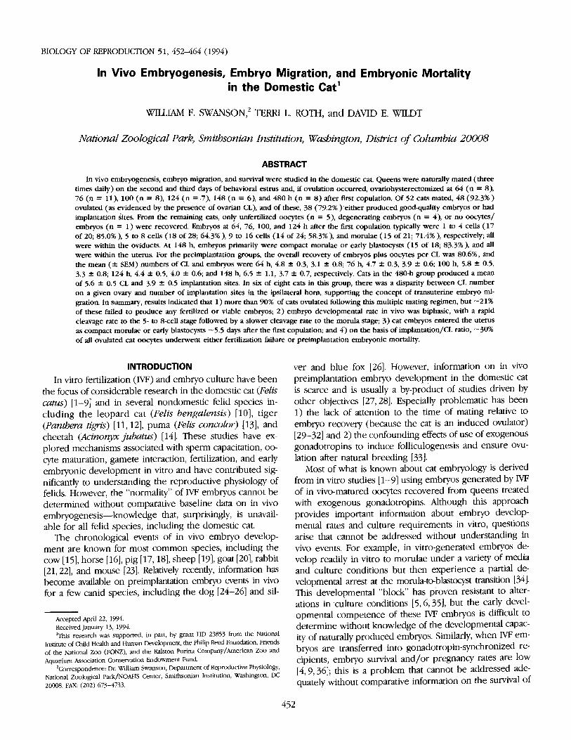
FIG. 1. In vivo cat embryo development at 64 h after the first copulation: (a) percentage of recovered grade 1 or2 embryos at each developmental stage; (b) photomicrograph (x150) of 1-cell and 2-cell embryos typical of this timeinterval. All embryos were recovered from the oviducts.
success did not differ (p > 0.05) among most of the in-terval groups. However, embryo recovery for the 100-h groupwas less (p < 0.05) than that for the 124-h group and sim-ilar (p > 0.05) to that for the 148-h group, which in turn
was less (p < 0.05) than for all other groups. Of the 144embryos recovered, 77.1% were quality grade 1 (n = 83)or 2 (n = 28) and the remaining (n = 33) were classifiedas degenerate (grade 3).
455EMBRYOGENESIS IN THE DOMESTIC CAT 455
V 64 Hours
Stage of Embryo Development
b)
#
FIG. 1. In vivo cat embryo development at 64 h after the first copulation: (a) percentage of recovered grade 1 or 2 embryos at each developmental stage; (b) photomicrograph (x150) of 1-cell and 2-cell embryos typical of this time interval. All embryos were recovered from the oviducts.
success did not differ (p > 0.05) among most of the in- terval groups. However, embryo recovery for the 100-h group was less (p < 0.05) than that for the 124-h group and sim- ilar (/? > 0.05) to that for the 148-h group, which in turn
was less (p < 0.05) than for all other groups. Of the 144 embryos recovered, 77.1% were quality grade 1 (n = 83) or 2 (n = 28) and the remaining (n = 33) were classified as degenerate (grade 3).

456 SWANSON ET AL.
FIG. 2. In vivo cat embryo development at 76 h after first copulation: (a) percentage of recovered grade 1 or 2embryos at each developmental stage; (b) photomicrograph (x200) of 5- to 8-cell embryos typical of this time interval.All embryos were recovered from the oviducts.
456 SWANSON ET AL.
a)
76 Hours
Stage of Embryo Development
b)
*
#
*
FIG. 2. In vivo cat embryo development at 76 h after first copulation: (a) percentage of recovered grade 1 or 2 embryos at each developmental stage; (b) photomicrograph (x200) of 5- to 8-cell embryos typical of this time interval. All embryos were recovered from the oviducts.

EMBRYOGENESIS IN THE DOMESTIC CAT
FIG. 3. In vivo cat embryo development at 100 h after first copulation: (a) percentage of recovered grade 1 or 2embryos at each developmental stage; (b) photomicrograph (x200) of 9- to 16-cell embryos typical of this time in-terval. All embryos were recovered from the oviducts.
457EMBRYOGENESIS IN THE DOMESTIC CAT 457
a) 100 Hours
Stage of Embryo Development
b)
#
#
$
FIG. 3. In vivo cat embryo development at 100 h after first copulation: (a) percentage of recovered grade 1 or 2 embryos at each developmental stage; (b) photomicrograph (x200) of 9- to 16-cell embryos typical of this time in- terval. All embryos were recovered from the oviducts.

SWANSON ET AL.
FIG. 4. In vivo cat embryo development at 124 h after first copulation: (a) percentage of recovered grade 1 or 2embryos at each developmental stage; (b) photomicrograph (x200) of morulae and compacting morulae typical ofthis time interval. All embryos were recovered from the oviducts.
458458 SWANSON ET AL.
124 Hours
0 2-4 cell • 5-8 cell
Q 9-16 cell
• morulae
• compact morulae
Stage of Embryo Development
b)
#
#
#
FIG. 4. In vivo cat embryo development at 124 h after first copulation: (a) percentage of recovered grade 1 or 2 embryos at each developmental stage; (b) photomicrograph (x200) of morulae and compacting morulae typical of this time interval. All embryos were recovered from the oviducts.

EMBRYOGENESIS IN THE DOMESTIC CAT
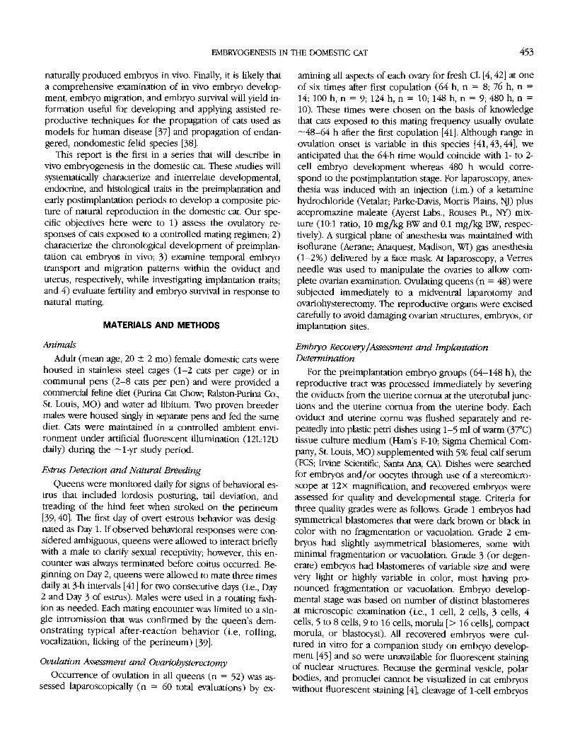
FIG. 5. In vivo cat embryo development at 148 h after first copulation: (a) percentage of recovered grade 1 or 2embryos at each developmental stage; (b) photomicrograph (x200) of compact morulae typical of this time interval.All embryos (except one 9-16-cell embryo within the oviduct) were recovered from the uterine horns.
459EMBRYOGENESIS IN THE DOMESTIC CAT 459
a)
148 Hours
0 9-16 cell U morulae
compact morulae
H blastocysts
Stage of Embryo Development
b)
FIG. 5. In vivo cat embryo development at 148 h after first copulation: (a) percentage of recovered grade 1 or 2 embryos at each developmental stage; <b> photomicrograph (x200) of compact morulae typical of this time interval. All embryos (except one 9-16-cell embryo within the oviduct) were recovered from the uterine horns

SWANSON ET AL.
FIG. 6. CL/implantation disparity in cats at 480 h after the first cop-ulation: (a) mean ( SEM) number of CL and implantation sites. Columnswith different superscripts differ (p < 0.05); (b) number of CL and implan-tations for individual queens within the 480-h-interval group.
Embryo Developmental StagesDevelopmental stages for grade 1 or grade 2 embryos
were characterized for each preimplantation time interval.At 64 h after the first copulation, 65% (13 of 20) of embryoshad completed at least one cleavage division with the re-maining embryos still at the 1-cell stage (Fig. 1). At 76 h,most embryos (64.3%) were 5 to 8 cells (Fig. 2); at 100 h,most (58.3%) were at the 9- to 16-cell developmental stage(Fig. 3). At 124 h, 71.4% (15 of 21) of embryos had devel-oped to the morula or compact morula stage (Fig. 4). All
embryos in the 64-124-h-interval groups were recoveredfrom the oviducts. At 148 h after the first copulation, 61.1%of embryos were compact morulae and 22.2% were blas-tocysts (Fig. 5), and all but one were recovered from theuterine horns. A single 9- to 16-cell, grade 1 embryo wasflushed from the oviduct of one queen; no other embryoswere recovered from this cat.
Implantation and Transuterine MigrationFor queens in the postimplantation group, there was a
disparity of 31.1% (p < 0.05) between number of implan-tation sites (n = 31) and CL (n = 45) (Fig. 6a). Analysis ofresults for individual queens (Fig. 6b) revealed that the im-plantation/CL disparity for queens (n = 4) with 5 CL orfewer was 5.5% (1 of 18) but for cats (n = 4) with morethan 5 CL was 48.1% (13 of 27) (p < 0.01).
In six of the eight cats in the postimplantation group,there were more implantation sites in one uterine horn thanCL on the ipsilateral ovary, indicating transuterine embryomigration from the contralateral horn (Table 3). Implan-tation sites averaged 2.0 + 0.1 cm in diameter and (forqueens with multiple implantation sites in one uterine horn)were spaced an average of 2.5 + 0.3 cm apart. When in-cised, all gestational sacs except a single site in cat #5 con-tained a developing fetus. This vacant gestational sac was1.1 cm in diameter and was positioned immediately adja-cent to the only other implantation site in the left uterinehorn.
The chronological sequence of in vivo embryo devel-opment after natural mating in the domestic cat is sum-marized in Figure 7.
DISCUSSION
Studies of natural reproductive processes in domestic catshistorically have focused on the physiological mechanismsand temporal relationships associated with behavior, ovu-lation induction, and endocrine patterns throughout thevarious phases of the estrous cycle, luteal phase, and preg-nancy (for reviews see [34, 48, 49]). The present datarepresent the first substantive information on in vivoembryogenesis in the domestic cat encompassing embryodevelopmental rate, survival, and intrauterine migration.
TABLE 3. Distribution of CL and implantation sites in naturally mated queens at 480 h after the first copulationand evidence of transuterine embryo migration.
Cat Right ovary Right uterine horn Left ovary Left uterine horn Embryonumber (CL number) (Implantation number) (CL number) (Implantation number) migration?
1 0 1 4 3 yes2 2 2 2 2 unknown3 0 2 5 2 yes4 2 3 3 2 yes5 5 0 1 2 yes6 7 1 0 1 yes7 3 4 4 0 yes8 5 4 2 2 unknown
460460 SWANSON ET AL.
(at
XI 480 Hours
fa 6- 1 lllcrusl a
E
Z 4^ • 1 S 2-
1 MTTTM 1 • corpora lutea
• implantations
lb)
480 Hours
• corpora lutca 0 implantations
12 3 4 5 6 7 Individual Cats
FIG. 6. CL/implantation disparity in cats at 480 h after the first cop- ulation: (a) mean (± SEM) number of CL and implantation sites. Columns with different superscripts differ (p < 0.05); (b) number of CL and implan- tations for individual queens within the 480-h-interval group.
embryos in the 64-124-h-interval groups were recovered from the oviducts. At 148 h after the first copulation, 61.1% of embryos were compact morulae and 22.2% were blas- tocysts (Fig. 5), and all but one were recovered from the uterine horns. A single 9- to 16-cell, grade 1 embryo was flushed from the oviduct of one queen; no other embryos were recovered from this cat.
Implantation and Transuterine Migration For queens in the postimplantation group, there was a
disparity of 31.1% (p < 0.05) between number of implan- tation sites (n = 31) and CL (n = 45) (Fig. 6a). Analysis of results for individual queens (Fig. 6b) revealed that the im- plantation/CL disparity for queens (n = 4) with 5 CL or fewer was 5.5% (1 of 18) but for cats (n = 4) with more than 5 CL was 48.1% (13 of 27) (p < 0.01).
In six of the eight cats in the postimplantation group, there were more implantation sites in one uterine horn than CL on the ipsilateral ovary, indicating transuterine embryo migration from the contralateral horn (Table 3). Implan- tation sites averaged 2.0 ± 0.1 cm in diameter and (for queens with multiple implantation sites in one uterine horn) were spaced an average of 2.5 ± 0.3 cm apart. When in- cised, all gestational sacs except a single site in cat #5 con- tained a developing fetus. This vacant gestational sac was 1.1 cm in diameter and was positioned immediately adja- cent to the only other implantation site in the left uterine horn.
The chronological sequence of in vivo embryo devel- opment after natural mating in the domestic cat is sum- marized in Figure 7.
Embryo Developmental Stages
Developmental stages for grade 1 or grade 2 embryos were characterized for each preimplantation time interval. At 64 h after the first copulation, 65% (13 of 20) of embryos had completed at least one cleavage division with the re- maining embryos still at the 1-cell stage (Fig. 1). At 76 h, most embryos (64.3%) were 5 to 8 cells (Fig. 2); at 100 h, most (58.3%) were at the 9- to 16-cell developmental stage (Fig. 3). At 124 h, 71.4% (15 of 21) of embryos had devel- oped to the morula or compact morula stage (Fig. 4). All
DISCUSSION
Studies of natural reproductive processes in domestic cats historically have focused on the physiological mechanisms and temporal relationships associated with behavior, ovu- lation induction, and endocrine patterns throughout the various phases of the estrous cycle, luteal phase, and preg- nancy (for reviews see [34, 48, 49]). The present data represent the first substantive information on in vivo embryogenesis in the domestic cat encompassing embryo developmental rate, survival, and intrauterine migration.
TABLE 3. Distribution of CL and implantation sites in naturally mated queens at 480 h after the first copulation and evidence of transuterine embryo migration.
Cat Right ovary Right uterine horn Left ovary Left uterine horn Embryo number (CL num ber) (Implantation number) (CL number) (Implantation number) migration?
1 0 1 4 3 yes 2 2 2 2 2 unknown 3 0 2 5 2 yes 4 2 3 3 2 yes 5 5 0 1 2 yes 6 7 1 0 1 yes 7 3 4 4 0 yes 8 5 4 2 2 unknown

EMBRYOGENESIS IN THE DOMESTIC CAT
FIG. 7. Summary diagram of in vivo embryo development in the domestic cat after mating three times per day on Days 2 and 3 of natural estrus.
More than 85% of the cats in the present study ovulatedin response to multiple matings on the second and thirdday of estrus; these results were comparable to those re-ported by others [31,32,41]. Concannon et al. [31] dem-onstrated that ad libitum copulations on Day 3 of estrusresulted in 100% (36/36) of cats ovulating. We have shownearlier that 83% (10 of 12) of cats ovulated when matedthree times on either Day 1, 2, or 3 of estrus, whereas allcats (12 of 12) ovulated when mated three times daily forthe first 3 days of estrus [32, 41]. As induced ovulators, catsdepend on copulatory activity to cause the hypothalamicLHRH secretion that subsequently elicits ovulatory LH surges[31, 32, 41, 44]. Mating frequency has a profound effect onLH surge peak/duration and ovulatory response [31, 32]. Al-though LH was not measured in the present experiments,previous studies have shown that anovulatory queens gen-erally do not produce an LH surge [31, 32, 41, 44]. In thepresent investigation, half of the eight queens failing toovulate in response to the initial series of matings ovulatedduring a later estrus, indirectly suggesting that differentmating experiences within the same female provoked vari-ations in hypothalamo-pituitary response. However, threequeens were consistently anovulatory (despite being matedduring two or three estrous periods) although their age,parity, and sexual behavior were similar to those for therest of the study population. All mated queens exhibitedtypical estrous behavior and postcoital reactions regardlessof whether ovulation resulted, perhaps because sexual be-havior and LHRH release in the cat are uncoupled and arecontrolled by separate hypothalamic centers [50].
Results also indicated that it was unnecessary to matequeens on the first day of estrus to achieve ovulation andto produce viable embryos. Day 2, and not Day 1, was cho-sen for first mating on the basis of recent data suggestingthat follicular oocytes on the first day of estrus are imma-ture and frequently of poorer quality than during later es-trus [8]. In that study, oocytes were laparoscopically aspir-ated from naturally estrual queens receiving hCG (to inducefinal follicular maturation) on either Day 1, 2, or 3 of estrus.
Oocytes recovered from Day 1 follicles were markedlycompromised in fertilizability in vitro compared to oocytesaspirated from Day 2 or 3 follicles. Such results may meanthat matings occurring during the very early stages of estruseither have little functional relevance or serve to promotegonadotropin release that in turn is more important for fol-licle/oocyte maturation than ovulation induction.
A true assessment of embryo developmental rates re-quires relating findings to estimates of time of ovulation, adifficult task in the case of this induced ovulator becauseoocyte release is sensitive to the specifics of the mating reg-imen [34]. Ovulation has been observed to occur from 24to 32 h [43,44] and from 48 to 64 h [41] after first copu-lation, the difference being attributable largely to the num-ber of copulations and the interval between them. Queensmated beginning on Day 3 or 4 of estrus and permittedone to four copulations within one 1/2-h period ovulate24-32 h after the first copulation (44). In contrast, the ear-liest ovulation detected in queens mated beginning on Day1 of estrus (three copulations at 3-h intervals during eachof the first three days of estrus) is 48 h later [41]. Delayingcoitus until the later days of estrus may make the ovarianfollicles more responsive to copulatory stimuli, acceleratingboth the final stage of follicular/oocyte maturation andovulation onset. Our developmental data here supportedthis assertion. With in vivo-matured, in vitro-fertilized oo-cytes, first cleavage division occurs 24-30 h post-insemi-nation [2,4]. In the present study, most (65%) in vivo-gen-erated embryos had cleaved at least once by 64 h after thefirst breeding. Because a parallel study indicated that earlydevelopmental rates of in vivo-generated and IVF embryoswere similar [45], it can be assumed that fertilization in vivooccurred -30 h before recovery of the embryo at 64 h.Within this time frame, ovulation was estimated to have oc-curred -30-36 h after the first mating.
The developmental rate of cat embryos in vivo appearedbiphasic, with an initial fast cleavage period followed by aperiod during which developmental rate within the ovi-ducts was slower. Between the time of ovulation and 64 h
461EMBRYOGENESIS IN THE DOMESTIC CAT 461
Oh 241. ttt ttt
COITUS COITUS
64 h 76h 100 h 124 h 148 h 480 h
FIG. 7. Summary diagram of in vivo embryo development in the domestic cat after mating three times per day on Days 2 and 3 of natural estrus.
More than 85% of the cats in the present study ovulated in response to multiple matings on the second and third day of estrus; these results were comparable to those re- ported by others [31,32,41]. Concannon et al. [31] dem- onstrated that ad libitum copulations on Day 3 of estrus resulted in 100% (36/36) of cats ovulating. We have shown earlier that 83% (10 of 12) of cats ovulated when mated three times on either Day 1, 2, or 3 of estrus, whereas all cats (12 of 12) ovulated when mated three times daily for the first 3 days of estrus [32,41]. As induced ovulators, cats depend on copulatory activity to cause the hypothalamic LHRH secretion that subsequently elicits ovulatory LH surges [31,32,41,44]. Mating frequency has a profound effect on LH surge peak/duration and ovulatory response [31,32]. Al- though LH was not measured in the present experiments, previous studies have shown that anovulatory queens gen- erally do not produce an LH surge [31,32,41,44]. In the present investigation, half of the eight queens failing to ovulate in response to the initial series of matings ovulated during a later estrus, indirecdy suggesting that different mating experiences within the same female provoked vari- ations in hypothalamo-pituitary response. However, three queens were consistently anovulatory (despite being mated during two or three estrous periods) although their age, parity, and sexual behavior were similar to those for the rest of the study population. All mated queens exhibited typical estrous behavior and postcoital reactions regardless of whether ovulation resulted, perhaps because sexual be- havior and LHRH release in the cat are uncoupled and are controlled by separate hypothalamic centers [50].
Results also indicated that it was unnecessary to mate queens on the first day of estrus to achieve ovulation and to produce viable embryos. Day 2, and not Day 1, was cho- sen for first mating on the basis of recent data suggesting that follicular oocytes on the first day of estrus are imma- ture and frequently of poorer quality than during later es- trus [8]. In that study, oocytes were laparoscopically aspir- ated from naturally estrual queens receiving hCG (to induce final follicular maturation) on either Day 1, 2, or 3 of estrus.
Oocytes recovered from Day 1 follicles were markedly compromised in fertilizability in vitro compared to oocytes aspirated from Day 2 or 3 follicles. Such results may mean that matings occurring during the very early stages of estrus either have little functional relevance or serve to promote gonadotropin release that in turn is more important for fol- licle/oocyte maturation than ovulation induction.
A true assessment of embryo developmental rates re- quires relating findings to estimates of time of ovulation, a difficult task in the case of this induced ovulator because oocyte release is sensitive to the specifics of the mating reg- imen [34]. Ovulation has been observed to occur from 24 to 32 h [43,44] and from 48 to 64 h [41] after first copu- lation, the difference being attributable largely to the num- ber of copulations and the interval between them. Queens mated beginning on Day 3 or 4 of estrus and permitted one to four copulations within one 1/2-h period ovulate 24-32 h after the first copulation (44). In contrast, the ear- liest ovulation detected in queens mated beginning on Day 1 of estrus (three copulations at 3-h intervals during each of the first three days of estrus) is 48 h later [41]. Delaying coitus until the later days of estrus may make the ovarian follicles more responsive to copulatory stimuli, accelerating both the final stage of follicular/oocyte maturation and ovulation onset. Our developmental data here supported this assertion. With in vivo-matured, in vitro-fertilized oo- cytes, first cleavage division occurs 24-30 h post-insemi- nation [2,4]. In the present study, most (65%) in vivo-gen- erated embryos had cleaved at least once by 64 h after the first breeding. Because a parallel study indicated that early developmental rates of in vivo-generated and IVF embryos were similar [45], it can be assumed that fertilization in vivo occurred ~30 h before recovery of the embryo at 64 h. Within this time frame, ovulation was estimated to have oc- curred —30-36 h after the first mating.
The developmental rate of cat embryos in vivo appeared biphasic, with an initial fast cleavage period followed by a period during which developmental rate within the ovi- ducts was slower. Between the time of ovulation and 64 h

SWANSON ET AL.
after first copulation, most embryos (65%) had undergoneat least one cleavage division. By 76 h, most embryos (58%)had completed a third cleavage (5 to 8 cells). Given ourestimate of ovulation occurring 30-36 h after the first mat-ing, cat blastomeres cleaved about once every 12 h up tothe 5- to 8-cell stage. Assuming that the first cleavage occurs24-30 h after fertilization (as evidenced by in vitro data)[2,4], the second and third cleavage divisions both oc-curred within the next 12-22 h. Subsequent cleavages thenoccurred about once every 24 h from the 5- to 8-cell to themorula and compact morula stages. Embryo developmentalrates for other species are known to be multiphasic withvariable cleavage intervals, especially during early periodsof development [51]. In cattle, initial cleavage rate to the 4-to 8-cell stage is related to maternal control of protein syn-thesis, and slowed growth is associated with the maternal-zygotic transition [51]. The timing of the maternal-zygotictransition is unknown for the cat, but observed in vivo de-velopmental kinetics indicates that the shift from maternalto embryonic control may well occur at about the 5- to 8-cell stage when the cleavage rate slows substantially.
For 124 h after the first copulation, cat embryos re-mained within the oviducts, growing into morulae. From124 to 148 h, embryos underwent compaction, transversedthe uterotubal junctions, and entered the uterine- cornua ascompact morulae or early blastocysts. Therefore, comparedto embryos of many domestic species, cat embryos weresustained within the oviducts for a longer period, makingthe oviductal-to-uterine transition at a relatively advancedstage. For example, cattle, sheep, rabbit, and pig embryosare typically transported through the oviducts within 2-4days of ovulation, entering the uterus as 4 to 16 cells [15,17-19, 21, 22]. The cat appears most like the horse and the dog,because embryos from the latter two species enter the uterusas late-stage morulae to early-stage blastocysts about 148 hand 216 h post-ovulation, respectively [16, 24-26]. The rel-atively prolonged oviductal transit time in the cat may beassociated with the slow onset of luteal function after cop-ulation, because circulating progesterone does not rise abovebasal concentration until 48-72 h after mating onset[41, 52, 53]. Delayed embryo transport in the cat also maybe affected by circulating estradiol-173, which can remainelevated for 2-5 days after initial matings [41, 53, 54]. Ex-ogenous estrogen retards embryo transport through the cat[27] and rabbit [21] oviduct, whereas progesterone accel-erates transport in the rabbit [21]. Therefore, it is likely thatthe kinetics of embryo location within the cat reproductivetract is hormone mediated and driven.
Cat morulae were similar to horse [16] and dog [26] em-bryos in undergoing compaction at the transition period,before or coincident with entering the uterine cornua.Compaction and the formation of tight intracellular junc-tions appear essential for subsequent blastocoel formation[55]. It is presently unknown whether or not the transitionbetween the oviductal and uterine environments has any
influence on the phenomenon of compaction and cat blas-tocyst formation. However, it was interesting that cat IVFmorulae did not undergo discernible compaction in vitro[45]. We now speculate that perhaps this first visible evi-dence of a difference between in vitro- and in vivo-gen-erated cat embryos holds the clue for addressing the prob-lem of the pronounced in vitro developmental block toblastocyst formation.
As with embryos of the litter-bearing pig [56] and dog[57], cat embryos reportedly undergo transuterine migra-tion before implantation [29, 58]. This was confirmed in thepresent study with six of eight cats manifesting definitiveevidence of transuterine migration so that embryos tendedto become equally spaced throughout the reproductive tract,perhaps to help promote embryo survival. There also wasa tendency for cats having more than 5 CL to have fewerimplantation sites. We have no explanation for this exceptthat perhaps there was a wider range in ovulation time inthe more prolific females, making these queens more sus-ceptible to fertilization failure or embryo loss; alternatively,perhaps some of the oocytes originating from the addi-tional follicles were of poorer fertilizable quality. Nonethe-less, the data clearly suggested that optimal embryo survivalwas associated with the formation of 4 or 5 CL.
The domestic cat as observed in the present study ex-perienced substantial fertilization failure and embryonicmortality from the time of ovulation through early implan-tation. Of 48 ovulating queens, 12.5% experienced a defin-itive infertile estrus (no embryos recovered or implantationsites identified). An additional 8.3% were tentatively clas-sified as experiencing an infertile cycle because only de-generate embryos were recovered. Therefore, the overall"infertility rate" for ovulating queens was -20%. Embryo/oocyte recovery was < 100% in all preimplantation-intervalgroups (mean recovery, 80.6%), possibly as a consequenceof physiological factors (i.e., failure of oocyte retrieval bythe fimbriae or anatomical obstructions) [25] or technicalerror in flushing the reproductive tracts. For example, therelatively low recovery success (56.4%) in the 148-h groupwas potentially due to embryo/oocyte loss associated withsevering the oviducts at the uterotubal junction during thetransition of embryos to the uterus. However, we have noevidence to suggest that recovery rates differed for unfer-tilized oocytes, degenerate embryos, or viable embryos, al-though this is a possibility. Ovulatory queens in the pre-and post-implantation groups had comparable numbers ofovulations and total embryos or implantations, with an overalldisparity of -30% between CL and embryo or implantationnumbers. This disparity was higher than the implantationfailure rate noted by Tsutsui et al. [58] (-16%). Tsutsui etal. examined a larger group (n = 169) of cats from randomsources and with unknown mating and fertility histories. Itmay well be that more of these queens were older, mul-tiparous females in contrast to our predominantly young(mean age, 20 mo), nulliparous (85%) group.
An overall pregnancy failure rate of -30% from ovula-tion through early implantation was comparable to values
462462 SWANSON ET AL.
after first copulation, most embryos (65%) had undergone at least one cleavage division. By 76 h, most embryos (58%) had completed a third cleavage (5 to 8 cells). Given our estimate of ovulation occurring 30-36 h after the first mat- ing, cat blastomeres cleaved about once every 12 h up to the 5- to 8-cell stage. Assuming that the first cleavage occurs 24-30 h after fertilization (as evidenced by in vitro data) [2,4], the second and third cleavage divisions both oc- curred within the next 12-22 h. Subsequent cleavages then occurred about once every 24 h from the 5- to 8-cell to the morula and compact morula stages. Embryo developmental rates for other species are known to be multiphasic with variable cleavage intervals, especially during early periods of development [51]. In cattle, initial cleavage rate to the 4- to 8-cell stage is related to maternal control of protein syn- thesis, and slowed growth is associated with the maternal- zygotic transition [51]. The timing of the maternal-zygotic transition is unknown for the cat, but observed in vivo de- velopmental kinetics indicates that the shift from maternal to embryonic control may well occur at about the 5- to 8- cell stage when the cleavage rate slows substantially.
For 124 h after the first copulation, cat embryos re- mained within the oviducts, growing into morulae. From 124 to 148 h, embryos underwent compaction, transversed the uterotubal junctions, and entered the uterine cornua as compact morulae or early blastocysts. Therefore, compared to embryos of many domestic species, cat embryos were sustained within the oviducts for a longer period, making the oviductal-to-uterine transition at a relatively advanced stage. For example, cattle, sheep, rabbit, and pig embryos are typically transported through the oviducts within 2-4 days of ovulation, entering the uterus as 4 to 16 cells [15,17- 19,21,22]. The cat appears most like the horse and the dog, because embryos from the latter two species enter the uterus as late-stage morulae to early-stage blastocysts about 148 h and 216 h post-ovulation, respectively [16,24-26]. The rel- atively prolonged oviductal transit time in the cat may be associated with the slow onset of luteal function after cop- ulation, because circulating progesterone does not rise above basal concentration until 48-72 h after mating onset [41, 52,53]. Delayed embryo transport in the cat also may be affected by circulating estradiol-17p, which can remain elevated for 2-5 days after initial matings [41,53,54]. Ex- ogenous estrogen retards embryo transport through the cat [27] and rabbit [21] oviduct, whereas progesterone accel- erates transport in the rabbit [21]. Therefore, it is likely that the kinetics of embryo location within the cat reproductive tract is hormone mediated and driven.
Cat morulae were similar to horse [16] and dog [26] em- bryos in undergoing compaction at the transition period, before or coincident with entering the uterine cornua. Compaction and the formation of tight intracellular junc- tions appear essential for subsequent blastocoel formation [55]. It is presently unknown whether or not the transition between the oviductal and uterine environments has any
influence on the phenomenon of compaction and cat blas- tocyst formation. However, it was interesting that cat IVF morulae did not undergo discernible compaction in vitro [45]. We now speculate that perhaps this first visible evi- dence of a difference between in vitro- and in vivo-gen- erated cat embryos holds the clue for addressing the prob- lem of the pronounced in vitro developmental block to blastocyst formation.
As with embryos of the litter-bearing pig [56] and dog [57], cat embryos reportedly undergo transuterine migra- tion before implantation [29,58]. This was confirmed in the present study with six of eight cats manifesting definitive evidence of transuterine migration so that embryos tended to become equally spaced throughout the reproductive tract, perhaps to help promote embryo survival. There also was a tendency for cats having more than 5 CL to have fewer implantation sites. We have no explanation for this except that perhaps there was a wider range in ovulation time in the more prolific females, making these queens more sus- ceptible to fertilization failure or embryo loss; alternatively, perhaps some of the oocytes originating from the addi- tional follicles were of poorer fertilizable quality. Nonethe- less, the data clearly suggested that optimal embryo survival was associated with the formation of 4 or 5 CL.
The domestic cat as observed in the present study ex- perienced substantial fertilization failure and embryonic mortality from the time of ovulation through early implan- tation. Of 48 ovulating queens, 12.5% experienced a defin- itive infertile estrus (no embryos recovered or implantation sites identified). An additional 8.3% were tentatively clas- sified as experiencing an infertile cycle because only de- generate embryos were recovered. Therefore, the overall "infertility rate" for ovulating queens was —20%. Embryo/ oocyte recovery was < 100% in all preimplantation-interval groups (mean recovery, 80.6%), possibly as a consequence of physiological factors (i.e., failure of oocyte retrieval by the fimbriae or anatomical obstructions) [25] or technical error in flushing the reproductive tracts. For example, the relatively low recovery success (56.4%) in the 148-h group was potentially due to embryo/oocyte loss associated with severing the oviducts at the uterotubal junction during the transition of embryos to the uterus. However, we have no evidence to suggest that recovery rates differed for unfer- tilized oocytes, degenerate embryos, or viable embryos, al- though this is a possibility. Ovulatory queens in the pre- and post-implantation groups had comparable numbers of ovulations and total embryos or implantations, with an overall disparity of ~30% between CL and embryo or implantation numbers. This disparity was higher than the implantation failure rate noted by Tsutsui et al. [58] (-16%). Tsutsui et al. examined a larger group (n = 169) of cats from random sources and with unknown mating and fertility histories. It may well be that more of these queens were older, mul- tiparous females in contrast to our predominantly young (mean age, 20 mo), nulliparous (85%) group.
An overall pregnancy failure rate of —30% from ovula- tion through early implantation was comparable to values

EMBRYOGENESIS IN THE DOMESTIC CAT
reported for other domestic species. In pigs, embryonicmortality before Day 25 of pregnancy ranges from 20 to30% [17, 59], whereas 25-30% of cattle pregnancies are lostbefore Day 35 after breeding [60, 61]. There are multiplecauses of early embryonic mortality, including genetic (oo-cyte/embryo chromosomal abnormalities), physiological(age; suboptimal semen quality, endocrine dysfunction), andenvironmental (nutrition, climate) factors [60]. The inci-dence of genetic abnormalities in in vivo-generated cat em-bryos has not yet been studied, but heteroploidy is a sig-nificant cause of early embryonic death in humans [62]. Inour study, most of the physiological and environmental fac-tors (age, health, parity, semen quality, nutrition, climate)were strictly controlled and generally optimized for repro-ductive success. The one uncontrollable variable was theendocrine milieu in mated queens, but previous studies[31, 41, 53, 54] have extensively characterized endocrinepatterns in naturally copulating cats and have demonstrateda general uniformity among ovulating queens. Therefore,although it deserves more systematic attention, the etiologyof embryonic mortality in the cat may well be related pri-marily to the gametes, especially their structural, functionaland genetic competence.
In summary, most queens ovulated in response to mul-tiple matings beginning on the second day of estrus, andthis copulatory regimen was adequate to produce embryosin vivo. After fertilization, the resulting embryos exhibitedan initial rapid phase of cleavage followed by a slower phaseafter three cell cycles. Embryos developed to morulae overa 4-day period within the oviduct, underwent compaction,and entered the uterus 5.5 days after the first copulation.Blastocysts within the uterus migrated between uterine hornsbefore implantation. Lastly, 88% of ovulating females con-ceived (produced fertilized oocytes/embryos); but as manyas 30% of ovulated oocytes failed to fertilize or implant,indicating considerable opportunity for reproductive fail-ure in the naturally estrual and mated domestic cat.
ACKNOWLEDGMENTS
The authors thank Beth Jennette and Jon Anderson for their dedicated technicalassistance.
REFERENCES
1. Hamner CE, Jennings L, Sojka NJ. Cat (Felis catus L.) spermatozoa require ca-pacitation. J Reprod Fertil 1970; 23:477-80.
2. Bowen RA Fertilization in vitro of feline ova by spermatozoa from the ductusdeferens. Biol Reprod 1977; 17:144-147.
3. Niwa K, Ohara K, Hosoi Y, Iritani A. Early events of in vitro fertilization of categgs by epididymal sperm. J Reprod Fertil 1985; 74:657-660.
4. Goodrowe KL, Wall RJ, O'Brien SJ, Schmidt PM, Wildt DE. Developmental com-petence of domestic cat follicular oocytes after fertilization in vitro. Biol Reprod1988; 39:355-372.
5. Johnston LA, Donoghue AM, O'Brien SJ, Wildt DE. Culture medium and proteinsupplementation influence in vitro fertilization and embryo development in thedomestic cat J Exp Zool 1991; 257:350-359.
6. Johnston LA, Donoghue AM, O'Brien SJ, Wildt DE. Influence of temperature andgas atmosphere on in vitro fertilization and embryo development in the do-mestic cat. J Reprod Fertil 1991; 92:377-382.
7. Donoghue AM, Johnston LA, Munson L, Brown JL, Wildt DE. Influence of gonad-otropin treatment interval on follicular maturation, in vitro fertilization, circu-lating steroid concentrations and subsequent luteal function in the domestic cat.Biol Reprod 1992; 46:972-980.
8. Donoghue AM, Johnston LA, Goodrowe KL, O'Brien SJ, Wildt DE. Influence ofday of oestrus on egg viability and comparative efficiency of in vitro fertilizationin domestic cats in natural or gonadotropin-induced oestrus. J Reprod Fertil 1993;98:85-90.
9. Pope CE, Keller GL, Dresser BL. In vitro fertilization in domestic and non-do-mestic cats including sequences of early nuclear events, development in vitro,cryopreservation and successful intra- and interspecies embryo transfer. J ReprodFertil Suppl 1993; 47:189-201.
10. Goodrowe KL, Miller AM, Wildt DE. In vitro fertilization of gonadotropin-stim-ulated leopard cat (Felis bengalensis) follicular oocytes. J Exp Zool 1989; 252:89-95.
11. Donoghue AM, Johnston LA, Seal US, Armstrong DL, Tilson RL, Wolff P, PetriniK, Simmons LG, Gross T, Wildt DE. In vitro fertilization and embryo developmentin vitro and in vivo in the tiger (Panthera tigris). Biol Reprod 1990; 43:733-744.
12. Donoghue AM, Johnston LA, Seal US, Armstrong DL, Simmons LG, Gross T, TilsonRL, Wolff P, Wildt DE. Ability of thawed tiger (Panthera tigris) spermatozoa tofertilize conspecific oocytes and bind and penetrate domestic cat eggs in vitro.J Reprod Fertil 1992; 96:555-564.
13. Miller AM, Roelke ME, Goodrowe KL, Howard JG, Wildt DE. Oocyte recovery,maturation and fertilization in vitro in the puma (Felis concolor). J Reprod Fertil1990; 88:249-258.
14. Donoghue AM, Howard JG, Byers AP, Goodrowe KL, Bush M, Blumer E, LukasJ, Stover J, Snodgrass K, Wildt DE. Correlation of sperm viability with gameteinteraction and fertilization in vitro in the cheetah (Acinomyxjubatus). Biol Re-prod 1992; 46:1047-1056.
15. Betteridge KJ. The anatomy and physiology of pre-attachment bovine embryos.Theriogenology 1988; 29:155-183.
16. Betteridge KJ, Eaglesome MD, Mitchell D, Flood PF, Beriault R. Development ofhorse embryos up to twenty two days after ovulation: observation on fresh spec-imens. J Anat 1982; 135:191-209.
17. Perry JS, Rowlands IW. Early pregnancy in the pig. J Reprod Fertil 1962; 4:175-188.
18. Oxenreider SL, Day BN. Transport and cleavage of ova in swine. J Anim Sci 1965;24:413-417.
19. Chang MC, Rowson LEA. Fertilization and early development of Dorset Hornsheep in the spring and summer. Anat Rec 1965; 152:303-316.
20. Sakkas D, Batt PA, Cameron AWN. Development of preimplantation goat (Capracircus) embryos in vivo and in vitro. J Reprod Fertil 1989; 87:359-365.
21. Greenwald GS. A study of the transport of ova through the rabbit oviduct. FertilSteril 1961; 12:80-95.
22. Alliston CW, Pardee NR. Variability of embryonic development in the rabbit at19 to 168 hours after mating. Lab Anim Sci 1973; 23:665-670.
23. Bowman P, McLaren A. Cleavage rate of mouse embryos in vivo and in vitro. JEmbryol Exp Morphol 1970; 24:203-207.
24. Tsutsui T. Studies on the physiology of reproduction in the dog. V. On cleavageand transport of fertilized ova in the oviduct. Jpn J Anim Reprod 1975; 21:70-75.
25. Renton JP, Boyd JS, Eckersall PD, Ferguson JM, Harvey MJA, Mullaney J, Perry B.Ovulation, fertilization and early embryonic development in the bitch (Canisfamiliaris). J Reprod Fertil 1991; 93:221-231.
26. Valtenen M, Jalkanen L. Species-specific features of oestrous development andblastogenesis in domestic canine species. J Reprod Fertil Suppl 1993; 47:133-137.
27. Herron MA, Sis RF. Ovum transport in the cat and the effect of estrogen admin-istration. Am J Vet Res 1974; 35:1277-1279.
28. Goodrowe KL, Howard JG, Wildt DE. Comparison of embryo recovery, embryoquality, oestradiol-17 and progesterone profiles in domestic cats (Felis catus)at natural or induced oestrus. J Reprod Fertil 1988; 82:553-561.
29. Hill JP, Tribe M. The early development of the cat (Felis domestica). Q J MicroscSci 1924; 68:513-602.
30. Greulich WW. Artificially induced ovulation in the cat (Felis domestica). Anat Rec1934; 58:217-224.
31. Concannon P, Hodgson B, Lein D. Reflex LH release in estrous cats followingsingle and multiple copulations. Biol Reprod 1980; 23:111-117.
32. Wildt DE, Seager SWJ, Chakraborty PK. Effect of copulatory stimuli on incidenceof ovulation and on serum luteinizing hormone in the cat. Endocrinology 1980;107:1212-1217.
33. Denker HW, Eng LA, Mootz U, Hamner CE. Studies on the early developmentand implantation in the cat: I. Cleavage and blastocyst formation. Anat Anz 1978;144:457-468.
463EMBRYOGENESIS IN THE DOMESTIC CAT 463
reported for other domestic species. In pigs, embryonic mortality before Day 25 of pregnancy ranges from 20 to 30% [17, 59], whereas 25-30% of cattle pregnancies are lost before Day 35 after breeding [60,61]. There are multiple causes of early embryonic mortality, including genetic (oo- cyte/embryo chromosomal abnormalities), physiological (age; suboptimal semen quality, endocrine dysfunction), and environmental (nutrition, climate) factors [60]. The inci- dence of genetic abnormalities in in vivo-generated cat em- bryos has not yet been studied, but heteroploidy is a sig- nificant cause of early embryonic death in humans [62]. In our study, most of the physiological and environmental fac- tors (age, health, parity, semen quality, nutrition, climate) were strictly controlled and generally optimized for repro- ductive success. The one uncontrollable variable was the endocrine milieu in mated queens, but previous studies [31,41,53,54] have extensively characterized endocrine patterns in naturally copulating cats and have demonstrated a general uniformity among ovulating queens. Therefore, although it deserves more systematic attention, the etiology of embryonic mortality in the cat may well be related pri- marily to the gametes, especially their structural, functional and genetic competence.
In summary, most queens ovulated in response to mul- tiple matings beginning on the second day of estrus, and this copulatory regimen was adequate to produce embryos in vivo. After fertilization, the resulting embryos exhibited an initial rapid phase of cleavage followed by a slower phase after three cell cycles. Embryos developed to morulae over a 4-day period within the oviduct, underwent compaction, and entered the uterus 5.5 days after the first copulation. Blastocysts within the uterus migrated between uterine horns before implantation. Lastly, 88% of ovulating females con- ceived (produced fertilized oocytes/embryos); but as many as 30% of ovulated oocytes failed to fertilize or implant, indicating considerable opportunity for reproductive fail- ure in the naturally estrual and mated domestic cat.
ACKNOWLEDGMENTS
The authors thank Beth Jennette and Jon Anderson for their dedicated technical assistance.
REFERENCES
1. Hamner CE, Jennings L, Sojka NJ. Cat (Felts catus L.) spermatozoa require ca- pacitation. J Reprod Fertil 1970; 23:477-80.
2. Bowen RA. Fertilization in vitro of feline ova by spermatozoa from the ductus deferens. Biol Reprod 1977; 17:144-147.
3. Niwa K, Ohara K, Hosoi Y, Iritani A. Early events of in vitro fertilization of cat eggs by epididymal sperm. J Reprod Fertil 1985; 74:657-660.
4. Goodrowe KL, Wall RJ, O'Brien SJ, Schmidt PM, Wildt DE. Developmental com- petence of domestic cat follicular oocytes after fertilization in vitro. Biol Reprod 1988; 39:355-372.
5. Johnston LA, Donoghue AM, O'Brien SJ, Wildt DE. Culture medium and protein supplementation influence in vitro fertilization and embryo development in the domestic cat. J Exp Zool 1991; 257:350-359.
6. Johnston LA, Donoghue AM, O'Brien SJ, Wildt DE. Influence of temperature and gas atmosphere on in vitro fertilization and embryo development in the do- mestic cat. J Reprod Fertil 1991; 92:377-382.
7. Donoghue AM, Johnston LA, Munson L, Brown JL, Wildt DE. Influence of gonad- otropin treatment interval on follicular maturation, in vitro fertilization, circu- lating steroid concentrations and subsequent luteal function in the domestic cat. Biol Reprod 1992; 46:972-980.
8. Donoghue AM, Johnston LA, Goodrowe KL, O'Brien SJ, Wildt DE. Influence of day of oestrus on egg viability and comparative efficiency of in vitro fertilization in domestic cats in natural or gonadotropin-induced oestrus. J Reprod Fertil 1993; 98:85-90.
9. Pope CE, Keller GL, Dresser BL. In vitro fertilization in domestic and non-do- mestic cats including sequences of early nuclear events, development in vitro, cryopreservation and successful intra- and Interspecies embryo transfer. J Reprod Fertil Suppl 1993; 47:189-201.
10. Goodrowe KL, Miller AM, Wildt DE. In vitro fertilization of gonadotropin-stim- ulated leopard cat (Felis bengalensis) follicular oocytes. J Exp Zool 1989; 252:89- 95.
11. Donoghue AM, Johnston LA, Seal US, Armstrong DL, Tilson RL, Wolff P, Petrini K, Simmons LG, Gross T, Wildt DE. In vitro fertilization and embryo development in vitro and in vivo in the tiger (Panthera tigris). Biol Reprod 1990; 43:733-744.
12. Donoghue AM, Johnston LA, Seal US, Armstrong DL, Simmons LG, Gross T, Tilson RL, Wolff P, Wildt DE. Ability of thawed tiger (Panthera tigris) spermatozoa to fertilize conspecific oocytes and bind and penetrate domestic cat eggs in vitro. J Reprod Fertil 1992; 96:555-564.
13. Miller AM, Roelke ME, Goodrowe KL, Howard JG, Wildt DE. Oocyte recovery, maturation and fertilization in vitro in the puma (Felis concolor). J Reprod Fertil 1990; 88:249-258.
14. Donoghue AM, Howard JG, Byers AP, Goodrowe KL, Bush M, Blumer E, Lukas J, Stover J, Snodgrass K, Wildt DE. Correlation of sperm viability with gamete interaction and fertilization in vitro in the cheetah (Acinomyx jubatus). Biol Re- prod 1992; 46:1047-1056.
15- Betteridge KJ. The anatomy and physiology of pre-attachment bovine embryos. Theriogenology 1988; 29:155-183.
16. Betteridge KJ, Eaglesome MD, Mitchell D, Flood PF, Beriault R. Development of horse embryos up to twenty two days after ovulation: observation on fresh spec- imens. J Anat 1982; 135:191-209.
17. Perry JS, Rowlands IW. Early pregnancy in the pig. J Reprod Fertil 1962; 4:175- 188.
18. Oxenreider SL, Day BN. Transport and cleavage of ova in swine. J Anim Sci 1965; 24:413-417.
19- Chang MC, Rowson LEA. Fertilization and early development of Dorset Horn sheep in the spring and summer. Anat Rec 1965; 152:303-316.
20. Sakkas D, Batt PA, Cameron AWN. Development of preimplantation goat (Copra bircus) embryos in vivo and in vitro. J Reprod Fertil 1989; 87:359-365.
21. Greenwald GS. A study of the transport of ova through the rabbit oviduct. Fertil Steril 1961; 12:80-95.
22. Alliston CW, Pardee NR Variability of embryonic development in the rabbit at 19 to 168 hours after mating. Lab Anim Sci 1973; 23:665-670.
23. Bowman P, McLaren A. Cleavage rate of mouse embryos in vivo and in vitro. J Embryol Exp Morphol 1970; 24:203-207.
24. Tsutsui T. Studies on the physiology of reproduction in the dog. V. On cleavage and transport of fertilized ova in the oviduct. Jpn J Anim Reprod 1975; 21:70- 75.
25. Renton JP, BoydJS, Eckersall PD, Ferguson JM, Harvey MJA, MullaneyJ, Perry B. Ovulation, fertilization and early embryonic development in the bitch (Cants
familiaris). J Reprod Fertil 1991; 93:221-231. 26. Valtenen M, Jalkanen L. Species-specific features of oestrous development and
blastogenesis in domestic canine species. J Reprod Fertil Suppl 1993; 47:133- 137.
27. Herron MA, Sis RF. Ovum transport in the cat and the effect of estrogen admin- istration. AmJ Vet Res 1974; 35:1277-1279.
28. Goodrowe KL, Howard JG, Wildt DE. Comparison of embryo recovery, embryo quality, oestradiol-178 and progesterone profiles in domestic cats (Felis catus) at natural or induced oestrus. J Reprod Fertil 1988; 82:553-561.
29. Hill JP, Tribe M. The early development of the cat (Felis domestica). QJ Microsc Sci 1924; 68:513-602.
30. Greulich WW. Artificially induced ovulation in the cat (Felis domestica). Anat Rec 1934; 58:217-224.
31. Concannon P, Hodgson B, Lein D. Reflex LH release in estrous cats following single and multiple copulations. Biol Reprod 1980; 23:111-117.
32. Wildt DE, Seager SWJ, Chakraborty PK. Effect of copulatory stimuli on incidence of ovulation and on serum luteinizing hormone in the cat. Endocrinology 1980; 107:1212-1217.
33. Denker HW, Eng LA, Mootz U, Hamner CE. Studies on the early development and implantation in the cat: I. Cleavage and blastocyst formation. Anat Anz 1978; 144:457-468.
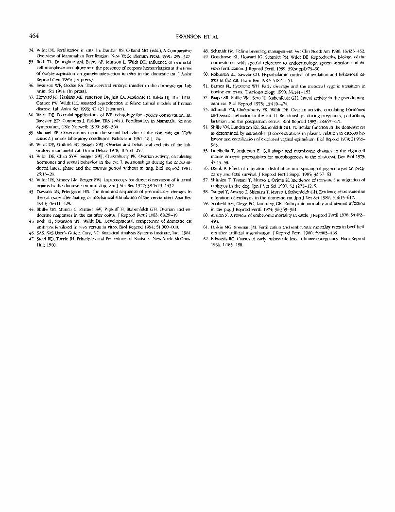
SWANSON ET AL.
34. Wildt DE. Fertilization in cats. In: Dunbar BS, O'Rand MG (eds.), A ComparativeOverview of Mammalian Fertilization. New York: Plenum Press; 1991: 299-327.
35. Roth TL, Donoghue AM, Byers AP, Munson L, Wildt DE. Influence of oviductalcell monolayer co-culture and the presence of corpora hemorrhagica at the timeof oocyte aspiration on gamete interaction in vitro in the domestic cat. J AssistReprod Gen 1994; (in press).
36. Swanson WF, Godke RA. Transcervical embryo transfer in the domestic cat. LabAnim Sci 1994; (in press).
37. HowardJG, Haskins ME, Patterson DF, Just CA, McAloose D, Baker HJ, Thrall MA,Gasper PW, Wildt DE. Assisted reproduction in feline animal models of humandisease. Lab Anim Sci 1993; 42:421 (abstract).
38. Wildt DE. Potential applications of IVF technology for species conservation. In:Bavister BD, Cummins J, Roldan ERS (eds.), Fertilization in Mammals. SeronoSymposium, USA: Norwell; 1990: 349-364.
39. Michael RP. Observations upon the sexual behavior of the domestic cat (Feliscatus L.) under laboratory conditions. Behaviour 1961; 18:1-24.
40. Wildt DE, Guthrie SC, Seager SWJ. Ovarian and behavioral cyclicity of the lab-oratory maintained cat. Horm Behav 1978; 10:251-257.
41. Wildt DE, Chan SYW, Seager SWJ, Chakraborty PK Ovarian activity, circulatinghormones and sexual behavior in the cat. I. Relationships during the coitus-in-duced luteal phase and the estrous period without mating. Biol Reprod 1981;25:15-28.
42. Wildt DE, Kinney GM, Seager SWJ. Laparoscopy for direct observation of internalorgans in the domestic cat and dog. Am J Vet Res 1977; 38:1429-1432.
43. Dawson AB, Friedgood HB. The time and sequence of preovulatory changes inthe cat ovary after mating or mechanical stimulation of the cervix uteri. Anat Rec1940; 76:411-429.
44. Shille VM, Munro C, Farmer SW, Papkoff H, Stabenfeldt GH. Ovarian and en-docrine responses in the cat after coitus. J Reprod Fertil 1983; 68:29-39.
45. Roth TL, Swanson WF, Wildt DE. Developmental competence of domestic catembryos fertilized in vivo versus in vitro. Biol Reprod 1994; 51:000-000.
46. SAS. SAS User's Guide. Cary, NC: Statistical Analysis Systems Institute, Inc.; 1984.47. Steel RD, Torrie JH. Principles and Procedures of Statistics. New York: McGraw-
Hill; 1960.
48. Schmidt PM. Feline breeding management. Vet Clin North Am 1986; 16:435-452.49. Goodrowe KL, Howard JG, Schmidt PM, Wildt DE. Reproductive biology of the
domestic cat with special reference to endocrinology, sperm function and in-vitro fertilization. J Reprod Fertil 1989; 39(suppl):73-90.
50. Robinson BL, Sawyer CH. Hypothalamic control of ovulation and behavioral es-trus in the cat. Brain Res 1987; 418:41-51.
51. Barnes FL, Eyestone WH. Early cleavage and the maternal zygotic transition inbovine embryos. Theriogenology 1990; 33:141-152.
52. Paape SR, Shille VM, Seto H, Stabenfeldt GH. Luteal activity in the pseudopreg-nant cat. Biol Reprod 1975; 13:470-474.
53. Schmidt PM, Chakraborty PK, Wildt DE. Ovarian activity, circulating hormonesand sexual behavior in the cat. II. Relationships during pregnancy, parturition,lactation and the postpartum estrus. Biol Reprod 1983; 28:657-671.
54. Shille VM, Lundstrom KE, Stabenfeldt GH. Follicular function in the domestic catas determined by estradiol-173 concentrations in plasma: relation to estrous be-havior and cornification of exfoliated vaginal epithelium. Biol Reprod 1979; 21:953-963.
55. Ducibella T, Anderson E. Cell shape and membrane changes in the eight-cellmouse embryo: prerequisites for morphogenesis to the blastocyst Dev Biol 1975;47:45-58.
56. Dziuk P. Effect of migration, distribution and spacing of pig embryos on preg-nancy and fetal survival. J Reprod Fertil Suppl 1985; 33:57-63.
57. Shimizu T, Tsutsui T, Murao I, Orima H. Incidence of transuterine migration ofembryos in the dog. Jpn J Vet Sci 1990; 52:1273-1275.
58. Tsutsui T, Amano T, Shimizu T, Murao 1, Stabenfeldt GH. Evidence of transuterinemigration of embryos in the domestic cat. Jpn J Vet Sci 1989; 51:613-617.
59. Scofield AM, Clegg FG, Lamming GE. Embryonic mortality and uterine infectionin the pig. J Reprod Fertil 1974; 36:353-361.
60. Ayalon N. A review of embryonic mortality in cattle. J Reprod Fertil 1978; 54:483-493.
61. Diskin MG, Sreenan JM. Fertilization and embryonic mortality rates in beef heif-ers after artificial insemination. J Reprod Fertil 1980; 59:463-468.
62. Edwards RG. Causes of early embryonic loss in human pregnancy. Hum Reprod1986; 1:185-198.
464464 SWANSON ET AL.
34. Wildt DE. Fertilization in cats. In: Dunbar BS, O'Rand MG (eds.), A Comparative Overview of Mammalian Fertilization. New York: Plenum Press; 1991: 299-327.
35. Roth TL, Donoghue AM, Byers AP, Munson L, Wildt DE. Influence of oviductal cell monolayer co-culture and the presence of corpora hemorrhagica at the time of oocyte aspiration on gamete interaction in vitro in the domestic cat. J Assist Reprod Gen 1994; (in press).
36. Swanson WF, Godke RA Transcervical embryo transfer in the domestic cat. Lab Anim Sci 1994; (in press).
37. Howard JG, Haskins ME, Patterson DF, Just CA, McAloose D, Baker HJ, Thrall MA, Gasper PW, Wildt DE. Assisted reproduction in feline animal models of human disease. Lab Anim Sci 1993; 42:421 (abstract).
38. Wildt DE. Potential applications of IVF technology for species conservation. In: Bavister BD, Cummins J, Roldan ERS (eds.), Fertilization in Mammals. Serono Symposium, USA: Norwell; 1990: 349-364.
39. Michael RP. Observations upon the sexual behavior of the domestic cat (Felis catus L) under laboratory conditions. Behaviour 1961; 18:1-24.
40. Wildt DE, Guthrie SC, Seager SWJ. Ovarian and behavioral cyclicity of the lab- oratory maintained cat. Horm Behav 1978; 10:251-257.
41. Wildt DE, Chan SYW, Seager SWJ, Chakraborty PK. Ovarian activity, circulating hormones and sexual behavior in the cat. I. Relationships during the coitus-in- duced luteal phase and the estrous period without mating. Biol Reprod 1981; 25:15-28.
42. Wildt DE, Kinney GM, Seager SWJ. Laparoscopy for direct observation of internal organs in the domestic cat and dog. Am J Vet Res 1977; 38:1429-1432.
43. Dawson AB, Friedgood HB. The time and sequence of preovulatory changes in the cat ovary after mating or mechanical stimulation of the cervix uteri. Anat Rec 1940; 76:411-429.
44. Shille VM, Munro C, Farmer SW, Papkoff H, Stabenfeldt GH. Ovarian and en- docrine responses in the cat after coitus. J Reprod Fertil 1983; 68:29-39
45. Roth TL, Swanson WF, Wildt DE. Developmental competence of domestic cat embryos fertilized in vivo versus in vitro. Biol Reprod 1994; 51:000-000.
46. SAS. SAS User's Guide. Cary, NC: Statistical Analysis Systems Institute, Inc.; 1984. 47. Steel RD, Torrie JH. Principles and Procedures of Statistics. New York: McGraw-
Hill; 1960.
48. Schmidt PM. Feline breeding management. Vet Clin North Am 1986; 16:435-452. 49. Goodrowe KL, Howard JG, Schmidt PM, Wildt DE. Reproductive biology of the
domestic cat with special reference to endocrinology, sperm function and in- vitro fertilization. J Reprod Fertil 1989; 39(suppl):73-90.
50. Robinson BL, Sawyer CH. Hypothalamic control of ovulation and behavioral es- trus in the cat. Brain Res 1987; 418:41-51.
51. Barnes FL, Eyestone WH. Early cleavage and the maternal zygotic transition in bovine embryos. Theriogenology 1990; 33:141-152.
52. Paape SR, Shille VM, Seto H, Stabenfeldt GH. Luteal activity in the pseudopreg- nant cat. Biol Reprod 1975; 13:470-474.
53. Schmidt PM, Chakraborty PK, Wildt DE. Ovarian activity, circulating hormones and sexual behavior in the cat. II. Relationships during pregnancy, parturition, lactation and the postpartum estrus. Biol Reprod 1983; 28:657-671.
54. Shille VM, Lundstrom KE, Stabenfeldt GH. Follicular function in the domestic cat as determined by estradiol-17B concentrations in plasma: relation to estrous be- havior and comiGcation of exfoliated vaginal epithelium. Biol Reprod 1979; 21:953- 963.
55. Ducibella T, Anderson E. Cell shape and membrane changes in the eight-cell mouse embryo: prerequisites for morphogenesis to the blastocyst Dev Biol 1975; 47:45-58.
56. Dziuk P. Effect of migration, distribution and spacing of pig embryos on preg- nancy and fetal survival. J Reprod Fertil Suppl 1985; 33:57-63.
57. Shimizu T, Tsutsui T, Murao I, Orima H. Incidence of transuterine migration of embryos in the dog. JpnJ Vet Sci 1990; 52:1273-1275.
58. Tsutsui T, Amano T, Shimizu T, Murao I, Stabenfeldt GH. Evidence of transuterine migration of embryos in the domestic cat. Jpn J Vet Sci 1989; 51:613-617.
59. Scofield AM, Clegg FG, Lamming GE. Embryonic mortality and uterine infection in the pig. J Reprod Fertil 1974; 36353-361.
60. Ayalon N. A review of embryonic mortality in cattle. J Reprod Fertil 1978; 54:483-
493. 61. Diskin MG, Sreenan JM. Fertilization and embryonic mortality rates in beef heif-
ers after artificial insemination. J Reprod Fertil 1980; 59:463-468. 62. Edwards RG. Causes of early embryonic loss in human pregnancy. Hum Reprod
1986; 1:185-198.