in vivo action of the hrd ubiquitin ligase complex...
TRANSCRIPT

MOLECULAR AND CELLULAR BIOLOGY,0270-7306/01/$04.0010 DOI: 10.1128/MCB.21.13.4276–4291.2001
July 2001, p. 4276–4291 Vol. 21, No. 13
Copyright © 2001, American Society for Microbiology. All Rights Reserved.
In Vivo Action of the HRD Ubiquitin Ligase Complex:Mechanisms of Endoplasmic Reticulum Quality Control and
Sterol RegulationRICHARD G. GARDNER, ALEXANDER G. SHEARER, AND RANDOLPH Y. HAMPTON*
Section of Cell and Developmental Biology, Division of Biology, University of California, San Diego,La Jolla, California 92093
Received 18 December 2000/Returned for modification 6 February 2001/Accepted 23 March 2001
Ubiquitination is used to target both normal proteins for specific regulated degradation and misfoldedproteins for purposes of quality control destruction. Ubiquitin ligases, or E3 proteins, promote ubiquitinationby effecting the specific transfer of ubiquitin from the correct ubiquitin-conjugating enzyme, or E2 protein, tothe target substrate. Substrate specificity is usually determined by specific sequence determinants, or degrons,in the target substrate that are recognized by the ubiquitin ligase. In quality control, however, a potentially vastcollection of proteins with characteristic hallmarks of misfolding or misassembly are targeted with highspecificity despite the lack of any sequence similarity between substrates. In order to understand the mecha-nisms of quality control ubiquitination, we have focused our attention on the first characterized quality controlubiquitin ligase, the HRD complex, which is responsible for the endoplasmic reticulum (ER)-associateddegradation (ERAD) of numerous ER-resident proteins. Using an in vivo cross-linking assay, we directlyexamined the association of the separate HRD complex components with various ERAD substrates. We havediscovered that the HRD ubiquitin ligase complex associates with both ERAD substrates and stable proteins,but only mediates ubiquitin-conjugating enzyme association with ERAD substrates. Our studies with the sterolpathway-regulated ERAD substrate Hmg2p, an isozyme of the yeast cholesterol biosynthetic enzyme HMG-coenzyme A reductase (HMGR), indicated that the HRD complex discerns between a degradation-competent“misfolded” state and a stable, tightly folded state. Thus, it appears that the physiologically regulated,HRD-dependent degradation of HMGR is effected by a programmed structural transition from a stable proteinto a quality control substrate.
Ubiquitin-mediated, proteasome-dependent degradation isoften employed by the cell to destroy both normal and mis-folded proteins for purposes of regulation and quality control(39, 51). In general, proteins destined for degradation by theproteasome are covalently modified with the small proteinubiquitin through the collective action of a hierarchy of en-zymes (21, 40, 46). A single ubiquitin-activating enzyme, or E1protein, activates ubiquitin for transfer to a small number ofubiquitin-conjugating enzymes, or E2 proteins, which are di-vergent in their targeting function and underlie the first level ofspecificity for substrate selection. The ubiquitin-conjugatingenzymes then covalently attach the ubiquitin moiety to the sidechain of an internal lysine residue within their target substrates(11, 14, 79), in a reaction requiring additional specificity factorsreferred to as ubiquitin ligases, or E3 proteins, which consti-tute the largest class of proteins in the ubiquitination hierar-chy.
Currently known ubiquitin ligases have catalytic subunitsthat fall into two distinct classes: HECT domain proteins, suchas E6-AP and Rsp5p (35, 43, 74), and RING-H2 domain pro-teins, such as Rbx1p and Hrd1p (3, 32, 76, 78). HECT domainubiquitin ligase action involves transient covalent linkage ofubiquitin to the ubiquitin ligase itself, followed by transfer of
the attached ubiquitin moiety to the target substrate (75).RING-H2 domain ubiquitin ligase action appears to involvepromotion of direct ubiquitin transfer between the ubiquitin-conjugating enzyme and the substrate. In addition to catalyzingsubstrate ubiquitination, many ubiquitin ligases also catalyzetheir own ubiquitination (3, 18, 27, 57, 61, 67).
Current models suggest that RING-H2 ubiquitin ligasesfunction by providing specific binding sites for both the targetsubstrate and the relevant ubiquitin-conjugating enzyme toeffect transfer of ubiquitin from the conjugating enzyme to thetarget. In fact, the RING-H2 domain of cCbl mediates directinteraction with UbcH7 (92). The simultaneous binding of thesubstrate and ubiquitin-conjugating enzyme to the RING-H2ubiquitin ligase to stimulate substrate ubiquitination can bereferred to as a mutual binding mechanism. The specific bind-ing of target substrates may be brought about directly by theubiquitin ligase itself or by proteins associated with the ubiq-uitin ligase. For example, in the SCF ubiquitin ligase complex,the RING-H2 protein Hrt1p (also called Rbx1p or Roc1p)employs a scaffold complex containing an effector protein thatspecifically binds to the target substrate (19, 62, 76–78). Dif-ferent substrate-binding proteins are incorporated into thescaffold complex to change the complex’s substrate specificity(19, 23, 63, 77). Similarly, the APC ubiquitin ligase complex,containing the RING-H2 protein Apc11p (53, 62, 76, 90), alsoutilizes a scaffold complex to bind the target substrate (52, 64,89, 90). In contrast, the N-end rule ubiquitin ligase containsmultiple substrate recognition sites within the RING-H2 pro-
* Corresponding author. Mailing address: Section of Cell and De-velopmental Biology, Division of Biology, University of California, SanDiego, La Jolla, CA 92093. Phone: (858) 822-0511. Fax: (858) 534-0555. E-mail: [email protected].
4276

tein itself (1, 2, 30, 70), allowing the same protein to bindmultiple substrates and related allosteric regulators. Otherubiquitin ligases that also directly bind their specific substratetargets include cCbl (47, 54) and Mdm2 (41, 42). In each case,the specificity of the ubiquitin ligase complex is a consequenceof its ability to specifically bind the target protein.
Ubiquitin-mediated degradation is employed in cellularquality control to remove misfolded and misassembled pro-teins (51, 84). For quality control degradation to be effective,the ubiquitination machinery must recognize common struc-tural hallmarks of damage or misfolding in a diverse group ofproteins with little or no primary sequence homology. Re-cently, we and others have identified a RING-H2 ubiquitinligase complex, referred to as the HRD complex below, that isresponsible for degradation of numerous quality control sub-strates (3, 6, 27, 32, 67). Presumably, these and other qualitycontrol ubiquitin ligases also function by promoting associationbetween the substrate and the ubiquitin-conjugating enzyme.However, it is not clear if the mutual binding model of ubiq-uitin ligase function is applicable to the large and varied set ofpossible quality control substrates with no sequence similarity.It may be that the quality control ubiquitin ligases recognizeappropriate substrates by evaluation of the folding state ratherthan specific sequence motifs within the target, although noevidence has yet been presented for such a model.
A significant component of cellular quality control degrada-tion occurs by endoplasmic reticulum (ER)-associated degra-dation (ERAD). Mutant lumenal or integral membrane pro-teins incapable of attaining correct structure or properassembly are destroyed by ERAD (4, 20, 32, 36, 45, 66, 68, 82,85, 87, 88, 93). ERAD is not restricted to aberrant proteins, asa significant fraction of newly made, normal proteins are de-stroyed by ERAD as part of normal ER physiology (55, 80, 83).Furthermore, ERAD is employed in the feedback regulationof HMG-coenzyme (CoA) reductase (HMGR), so that signalsfrom the sterol synthesis pathway control HMGR stability (12,17, 25, 31, 34, 60).
In Saccharomyces cerevisiae, ERAD is mediated in large partby the action of a RING-H2 ubiquitin ligase complex com-posed of the integral ER membrane proteins Hrd1p (Der3p)and Hrd3p (3, 6, 27, 32, 66, 67). Hrd1p contains an N-terminal,multispanning membrane anchor and a C-terminal cytosolicdomain (27), which contains a RING-H2 motif homologous toseveral characterized ubiquitin ligases (18, 47, 49, 57). Consis-tent with this homology, Hrd1p functions both in vitro and invivo as a ubiquitin ligase (3), catalyzing the processive transferof ubiquitin to itself and other proteins. In vitro, Hrd1p showspreference for a misfolded protein as a multiubiquitinationsubstrate (3) and thus may directly participate in some aspectsof quality control recognition. In vivo, Hrd1p is rate limitingfor ubiquitination and degradation of numerous ERAD sub-strates (3, 27, 67) and specifically employs only Ubc7p andUbc1p in its action (3), with Ubc7p being quantitatively moreimportant in the ERAD pathway. Hrd3p is a single-spanningER membrane protein, with the majority of its sequence re-siding in the ER lumen (27, 32, 67, 71). The Hrd3p lumenalregions are both necessary and sufficient for Hrd3p action inERAD (27). One function of the Hrd3p lumenal domain is toregulate the activity and stability of the cytosolic Hrd1pRING-H2 domain through transmembrane communication
mediated by the Hrd1p membrane anchor (27). However,Hrd3p has ERAD functions independent of Hrd1p stabiliza-tion (27).
Because Hrd1p and Hrd3p form an ER membrane-associ-ated ubiquitin ligase complex required for quality controlERAD, it is likely that the complex recognizes common struc-tural features among its diverse substrates. Here, we directlytested the model that in vivo HRD complex promotes associ-ation between ERAD substrates and the ER ubiquitin-conju-gating enzyme Ubc7p. From these studies, it is clear that theHRD complex does indeed mediate fruitful proximity betweenERAD substrates and the appropriate ubiquitin-conjugatingenzyme. Additionally, it appears that the HRD complex scansER proteins for their degradation status and, upon encounter-ing a protein that appears to be misfolded, promotes access ofthe appropriate ubiquitin-conjugating enzyme to the substrate,allowing robust yet selective ubiquitination of the desired tar-get. According to this model, and in contrast to regulatoryubiquitin ligases, the HRD complex does not promote speci-ficity by restricting its interactions only to substrates. Rather,the HRD complex functions in ubiquitination by discerningsubstrate properties subsequent to interaction. This model alsosuggests a mechanism for regulated ERAD of HMGR, whichprovides a significant component of eukaryotic regulation ofthe sterol pathway. Regulated HMGR ERAD appears to oc-cur through a controlled structural transition of the HMGRtransmembrane domain that allows it to be recognized as aquality control substrate by the HRD machinery. These studieshave mechanistic implications for understanding both the spec-ificity of quality control degradation and the clinically relevantaxis of sterol regulation that employs ERAD to control thecellular synthesis of cholesterol (29).
MATERIALS AND METHODS
Materials and reagents. All enzymes were obtained from New England Bio-labs (Beverly, Mass.). Chemical reagents were obtained from Sigma Chemical(St. Louis, Mo.). Dithiosuccinimidylproprionate (DSP) was obtained from Pierce(Rockford, Ill.). Protein A-Sepharose CL-4B was obtained from AmershamPharmacia (Piscataway, N.J.). Lovastatin, L-659,699, and zaragozic acid weregenerously donated by Merck (Rahway, N.J.). ECL enhanced chemilumines-cence immunodetection reagents were from Amersham Corp. (ArlingtonHeights, Ill.). Anti-Myc 9E10 antibody was used as a cell culture supernatantobtained by growing the 9E10 hybridoma (ATCC CRL 1729) in RPMI 1640culture medium (Life Technologies, Grand Island, N.Y.) with 10% fetal calfserum. Antihemagglutinin (HA) antibody was an ascites fluid obtained fromBabco (Berkeley, Calif.). Affinity-purified goat anti-mouse immunoglobulin-horseradish peroxidase (HRP) conjugate was obtained from Sigma Chemical.
Recombinant DNA and molecular cloning. The splicing-by-overlap-extensionmethod was use to make epitope-tagged versions of each gene (38). PCR wasperformed as previously described (24). A list of primers used is available uponrequest. All epitope-tagged plasmids were verified for their ability to completelycomplement a null mutation in their respective genomic copy.
The plasmid that expressed a single, myc epitope-tagged version of Hmg1p(1myc-Hmg1p) was constructed as follows. Partial complementary primers thatencoded a single myc epitope sequence (EQKLISEEDL) were used to amplify afragment of HMG1 from pRH144-2 (31), which resulted in a single myc epitopecoding sequence inserted between codons 621 and 622 of HMG1. The PCRfragment was cloned between the BsrGI and PflMI sites in pRH144-2. Theresulting plasmid was named pRH945.
The plasmid that expressed a double, HA epitope-tagged version of Ubc7p(2HA-Ubc7p) was constructed as follows. The UBC7 gene was amplified fromRHY623 genomic DNA (25). A DNA fragment that encoded two tandem HAepitope tags was amplified from pGTEP (obtained from B. Futcher). The twoPCR fragments were spliced together by PCR, and the resulting DNA fragmentwas cloned between the PstI and SalI sites in pRH423 (34), which resulted in
VOL. 21, 2001 IN VIVO ACTION OF HRD UBIQUITIN LIGASE COMPLEX 4277

pRH685. The 1.3-kb PvuII-SphI fragment from pRH685, which contained the2HA-UBC7 gene, was cloned between the Ecl136II and SphI sites in pRH507.The resulting TRP1 2HA-UBC7-containing plasmid was called pRH373.
The plasmid that expressed a triple HA epitope-tagged version of Ubc6p(3HA-Ubc6p) was made as follows. The UBC6 gene was amplified from RHY623genomic DNA and cloned between the PstI and NheI sites in pRH442 (25). Theresulting plasmid was called pRH1149. A DNA fragment encoding a triple HAepitope tag was amplified from pGTEP and cloned between the NheI and SalIsites in pRH1149, which resulted in pRH1151. A 1.6-kb fragment which con-tained the 3HA-UBC6 gene was cloned between the PstI and KpnI sites inpRH507 (85). The resulting TRP1 3HA-UBC6-containing plasmid was calledpRH1217.
The plasmid used to make hrd3D::LEU2 was constructed as follows. A 3.1-kbXhoI-SpeI fragment from pRH508 which contained the HRD3 gene (32) wasinserted between the BamHI and EcoRI sites in pBluescript KSII, resulting inpRH1175. The LEU2 gene was PCR amplified from pRS405, digested with XbaI,and inserted between the BsaBI and NheI sites in pRH1175. The resultinghrd3D::LEU2 plasmid was named pRH1185.
The plasmid used to make ubc7D::LEU2 was constructed as follows. A 650-bpfragment containing the coding region for the UBC7 gene was PCR amplifiedfrom pRH685 and inserted between the PstI and SalI sites in pBluescript KSII,which resulted in pRH1176. pRH1176 contained an HpaI and a BsrGI site withthe UBC7 sequence. A 1.5-kb HpaI-BsrGI fragment from pRS405 containing theLEU2 gene was inserted between the HpaI and BsrGI sites in pRH1176. Theresulting ubc7D::LEU2 plasmid was named pRH1186.
Strains and media. Escherichia coli DH5a strains were grown at 37°C inLuria-Bertani medium with ampicillin (100 mg/ml). Yeast strains were grown at30°C in minimal medium supplemented with glucose and the appropriate aminoacids, as described (24). The lithium acetate method was used to transform yeastcells with plasmid DNA (44).
Yeast strains RHY1914 (MATa his3D200 lys2-801 ade2-101 leu2D ura3-52::1MYC-HMG2::URA3 trp1::HISG met2 hmg1::LYS2 HMG2) and RHY1915(MATa his3D200 lys2-801 ade2-101 leu2D ura3-52::1MYC-HMG1::URA3trp1::HISG met2 hmg1::LYS2 HMG2) were used as parent strains. The plasmidcontaining 3HA-Hrd1p, pRH1196, was digested with BglII and integrated at theHRD1 genomic locus. The plasmid containing 3HA-Hrd3p, pRH1263, was di-gested with BsrGI and integrated at the HRD3 genomic locus. The plasmidcontaining 2HA-Ubc7p, pRH373, was digested with BsgI and integrated at theTRP1 genomic locus. The plasmid containing 3HA-Ubc6p, pRH1217, was di-gested with BsrGI and integrated at the UBC6 genomic locus. Transformantswere selected for TRP1 prototrophy. The plasmid containing hrd1D::LEU2,pRH1184 (85), was digested with XhoI and BamHI and integrated at the HRD1genomic locus. The plasmid containing hrd3D::LEU2, pRH1185, was digestedwith XhoI and NdeI and integrated at the HRD3 genomic locus. The plasmidcontaining ubc7D::LEU2, pRH1186, was digested with PstI and SalI and inte-grated at the UBC7 genomic locus. The plasmid containing cue1::LEU2, pTX118(5), was digested with ApaI and SacI and integrated at the CUE1 genomic locus.Transformants were selected for LEU1 prototrophy.
Ubiquitination assays. Ubiquitination assays were performed as previouslydescribed (3).
Degradation assay. Cycloheximide-chase assays were performed as previouslydescribed (24).
Cross-linking assays. Cross-linking assays were done as previously described(27), except that 30 ml of anti-HMGR antiserum was used for the immunopre-cipitation. In a given set of experiments, samples were always run on a single gelso that transfer, immunoreactivity, and exposure times were kept constant be-tween samples that were to be compared.
Flow cytometry. Flow cytometry was performed as previously described (24),using a FACSscan (Beckton Dickinson, Palo Alto, Calif.) analytical flow mi-crofluorimeter with settings for fluorescein-labeled antibody analysis. To exam-ine the effects of lovastatin and zaragozic acid on Hmg2p-green fluorescentprotein (GFP) steady-state levels, the drugs were added to early-log-phase cul-tures (optical density at 600 nm [OD600] of ,0.2) to the desired final concen-trations, and the cells were grown for an additional 4 h (two doublings). In somecases, cells were grown to log phase, with 10% glycerol initially added to themedium or 10% glycerol added at the time of drug addition.
TLC. Thin-layer chromatography (TLC) was performed as previously de-scribed (28).
Protease protection assays. Microsomes were isolated as previously described(27). Protease protection assays were performed as previously described (27)except that anti-Hmg2p antiserum was used to detect Hmg2p.
RESULTS
ERAD of numerous ER lumenal and membrane proteinsrequires the Hrd1p/Hrd3p ubiquitin ligase complex (4, 6, 66,67), including the regulated ERAD of the yeast HMGRisozyme Hmg2p (3, 27, 32). In addition to Hrd1p and Hrd3p,ERAD requires the ubiquitin-conjugating enzyme Ubc7p (4,34, 37, 66) and its membrane association factor Cue1p (5).From this, it seems likely that some or all of the HRD complexproteins and their associated factors could directly interactwith substrates to target them for ubiquitination.
To test this hypothesis, we required an assay that wouldallow detection of the transient interactions that might existbetween the ER membrane-bound HRD ubiquitin ligase com-plex components and the targeted degradation substrates. Fur-thermore, the substrates and HRD complex under study are,for the most part, integral membrane proteins, and the assayhad to be amenable to the physical constraints inherent withsuch molecules. Finally, to study the participation of the HRDcomplex in regulation of Hmg2p, the assay had to allow for thepresence and action of small signaling molecules from themevalonate pathway that our studies show are at low cellularabundance and transiently produced (25, 28, 31). Assays nor-mally used for interaction studies are the yeast two-hybridassay, coimmunoprecipitation, and chemical cross-linking. Tomeasure transient interactions, however, the two-hybrid assaywas not a viable option because by nature, the transient inter-actions would likely be of low affinity, nor is there a transparentadaptation of this technique for studying interactions betweenmembrane proteins. Similarly, because the HRD complex com-ponents and some of its substrates are integral membraneproteins (27, 31, 32), a cross-linking approach was required inlieu of the more usual coimmunoprecipitation studies of ubiq-uitin ligase activity and substrate association (47, 49, 78, 86).Coimmunoprecipitation assays were not applicable to our sys-tem, since the solubilization methods required to extract theproteins from the ER membrane would likely disrupt the in-teractions between substrates and membrane-bound HRD pro-teins and would most certainly dilute or remove the transientsignaling molecules that control ERAD of Hmg2p. In contrast,chemical cross-linking has been used with great success todetermine transient interactions between components of theER membrane-bound Sec61p translocon and its translocationsubstrates (48, 58, 59, 65, 72). Additionally, we developed an invivo modification of the cross-linking assay (27) which allowsthe signaling molecules that control Hmg2p ERAD to remainand function at normal levels and location. Although cross-linking between two proteins indicates their close proximityand is therefore informative of potential protein-protein inter-actions, the absence of cross-linking does not necessarily pre-clude an interaction. With this caveat, we proceeded with thecross-linking studies to test interactions between the HRDcomplex and its substrates.
To analyze substrate-HRD complex interactions, we used thein vivo chemical cross-linking assay previously developed toexamine interactions between the subunits of the HRD com-plex itself (27). For ease of detection, we used functional, HAepitope-tagged versions of each protein in the HRD complex,which have been described previously (3, 27, 71). In addition,we employed a uniquely available set of ERAD substrates
4278 GARDNER ET AL. MOL. CELL. BIOL.

from our studies of regulated degradation of yeast HMGR (26,32), each with an added myc epitope tag. These included themevalonate pathway-regulated Hmg2p, the homologous butconstitutively stable Hmg1p, and the constitutively degraded6myc-Hmg2p.
Specific cross-linking of Ubc7p with ERAD substrates. Thecurrent model for RING-H2 ubiquitin ligase function is thatthe ligase promotes proximity between the ubiquitin-conjugat-ing enzyme and the target substrate through their mutual bind-ing, which leads to direct ubiquitination of the substrate.However, ubiquitin ligase-mediated proximity of ubiquitin-conjugating enzyme and its substrate has never been demon-strated in vivo. We first used the cross-linking assay to examineproximity between the regulated ERAD substrate Hmg2p andUbc7p, the principal ubiquitin-conjugating enzyme employedin its degradation (3, 34). When Hmg2p was immunoprecipi-tated from cells incubated with increasing concentrations ofthe cross-linker DSP (56), Ubc7p coimmunoprecipitated withHmg2p in a cross-linker concentration-dependent fashion (Fig.1a). Another ER-associated ubiquitin-conjugating enzyme,Ubc6p, which does not significantly participate in the ubiquiti-nation and degradation of Hmg2p (3, 34), did not cross-link toHmg2p (Fig. 1a). In contrast to Hmg2p, Ubc7p did not dem-onstrate any significant cross-linking to Hmg1p (Fig. 1a), theexceedingly stable isozyme of HMGR (26, 31). Thus, only aubiquitin-conjugating enzyme required for Hmg2p degrada-tion cross-linked to Hmg2p, and this cross-linking was in largepart restricted to degradation substrate and not a similar, sta-ble protein.
In order for two proteins to be cross-linked by DSP, free andaccessible lysine residues within a target protein must be withina short distance of lysine residues within the bait protein. Aslysine residues are generally the sites for attachment of ubiq-uitin (21, 46), it may be that the stable Hmg1p has limitedaccess to its lysine residues for Ubc7p. Accordingly, perhapsUbc7p did not cross-link with Hmg1p because there were noaccessible lysine residues within Hmg1p in close proximity toUbc7p.
To test if Ubc7p could be cross-linked to Hmg1p, we per-formed the cross-linking assay on mutant and degraded ver-sions of Hmg1p. From our previous mutagenic analyses thatdissected the in cis determinants for Hmg2p degradation (26),we constructed a number of Hmg1p mutants that were nolonger stable but were subject to Ubc7p-dependent degrada-tion (unpublished observations). One of these degradation-competent Hmg1p mutants, termed hydrophilic-Hmg1p, wasthe result of a limited number of substitutions that convertedthe hydrophobic residues in the first 26 residues to hydrophilicones but did not change the number of lysine residues withinHmg1p, similar to hydrophilic-Hmg2p previously described
FIG. 1. Ubc7p cross-linked to degraded Hmg2p in a mevalonatepathway-regulated manner but not to stable Hmg1p. Interaction of2HA-Ubc7p with 1myc-HMGR was assessed by the in vivo cross-linking assay. Cells were grown to mid-log phase and removed toamine-free medium. DSP was added at the indicated concentrations toseparate aliquots of cells, and cross-linking proceeded at 30°C for 30min. Lysates were prepared, and 1myc-Hmg2p was immunoprecipi-tated with antiserum raised against its catalytic domain. Immunopre-cipitates were denatured under reducing conditions and analyzed byimmunoblotting with either the 9E10 antibody (a-myc) to detect 1myc-Hmg2p or the 12CA5 antibody (a-HA) to detect 2HA-Ubc7p. (a)Ubc7p cross-linked to Hmg2p but did not cross-link to Hmg1p. Cross-linking assay was done with strains expressing either 1myc-Hmg2p or1myc-Hmg1p and coexpressing either 2HA-Ubc7p or 3HA-Ubc6p.The reduced immunoreactivity of 1myc-HMGR with DSP at 400 mg/mlis due to modification of the lysine residue in the myc epitope se-quence, not reduced immunoprecipitation of HMGR (unpublishedobservations). The small amount of 2HA-Ubc7p that is present withDSP at 0 mg/ml in this and the following figures is similar to thatobserved when preimmune serum is used instead of HMGR-specificantiserum (unpublished observations), indicating that it is nonspecificimmunoprecipitation. (b) Ubc7p cross-linked to a degraded version ofHmg1p; cross-linking assay with strains expressing either 1myc-Hmg2p, 1myc-Hmg1p, or hydrophilic-1myc-Hmg1p and coexpressing2HA-Ubc7p. (c) Addition of either lovastatin or L-659,699 decreased
Ubc7p cross-linking. Regulated Ubc7p cross-linking to Hmg2p wasobserved by preincubating the cells with either no or 50 mg of lova-statin (Lov) or 10 mg of L-659,599 (L659) per ml for 2 h prior toaddition of DSP. Cross-linking assay was performed as in Fig. 1. (d)Cross-linking of Ubc7p to 6myc-Hmg2p was not regulated by themevalonate pathway. Ubc7p cross-linking to 6myc-Hmg2p was ob-served by preincubating the cells with either 0 or 50 mg lovastatin or 10mg of L-659,599 per ml for 2 h prior to addition of DSP.
VOL. 21, 2001 IN VIVO ACTION OF HRD UBIQUITIN LIGASE COMPLEX 4279

FIG. 2. HRD gene-encoded proteins required for Ubc7p-Hmg2p cross-linking. (a) The appropriate null alleles of each gene required forHmg2p degradation were introduced into the strain coexpressing 1myc-Hmg2p and 2HA-Ubc7p. Correct introduction of each null allele wasdetermined by observation of 1myc-Hmg2p stabilization in a cycloheximide-chase assay, performed as in Fig. 5 (left panels). Lysates from each
4280 GARDNER ET AL. MOL. CELL. BIOL.

(26). When hydrophilic-Hmg1p was immunoprecipitated fromcells treated with DSP, Ubc7p now increasingly coimmunopre-cipitated with the mutant Hmg1p in a cross-linker concentra-tion-dependent manner (Fig. 1b, lanes hydrophilic versus wt).Thus, it appeared that the difference in Ubc7p cross-linking tonormal Hmg1p was a physiological result of its stability ratherthan an intrinsic inability of Hmg1p to cross-link to Ubc7p.
Hmg2p is unique as an ERAD substrate in that its ubiquiti-nation and degradation rates are feedback regulated by down-stream signals from the mevalonate pathway (25, 31). Reducedproduction of downstream products of the mevalonate path-way by inhibition or downregulation of pathway enzymes re-sults in the stabilization of Hmg2p (25, 31). We determined ifUbc7p cross-linking to Hmg2p was altered in accord with themevalonate pathway regulation of the Hmg2p degradationrate. To examine if cross-linking between Ubc7p and Hmg2pwas affected by this axis of regulation, we tested the effects ofdrugs that result in stabilization of Hmg2p. Incubation of cellswith inhibitors of early pathway enzymes, such as the HMGRinhibitor lovastatin and the upstream HMG-CoA synthase in-hibitor L-659,699, blocks Hmg2p ubiquitination and degrada-tion (25, 31, 34). Incubation of cells with either of these drugsdrastically reduced Ubc7p cross-linking to Hmg2p (Fig. 1c).The same drug treatments had no effect on Ubc7p cross-link-ing to 6myc-Hmg2pD63–219 (Fig. 1d), a grossly misfolded,epitope-tagged mutant of Hmg2p missing 157 residues of thenative Hmg2p sequence, including a putative transmembranespan that undergoes constitutive, unregulated Ubc7p-depen-dent degradation (25, 32).
In vivo action of the HRD ubiquitin ligase complex. Thus,Ubc7p cross-linking to ER proteins was directly correlatedwith their degradation status. Proteins that are naturally stableor are stabilized by physiological conditions within the cell didnot cross-link to Ubc7p. Ubc7p-dependent degradation sub-strates did cross-link to Ubc7p, suggesting that Ubc7p directlyassociated with ERAD substrates in a highly specific and se-lective manner. In the case of regulated Hmg2p, the cross-linking between Hmg2p and Ubc7p was physiologically regu-lated in a manner entirely consistent with the regulation ofHmg2p ubiquitination and degradation by mevalonate path-way production.
HRD complex mediated Ubc7p cross-linking to substrates.We next investigated the role of the Hrd1p/Hrd3p ubiquitinligase complex in promoting the proximity of Ubc7p to Hmg2p.To do so, we tested the effects of null alleles of the genesencoding these proteins on Hmg2p-Ubc7p cross-linking. Asexpected, when the individual null alleles were introduced into
the strain that coexpressed the epitope-tagged versions ofHmg2p and Ubc7p, the presence of either the hrd1D or thehrd3D allele resulted in complete stabilization of Hmg2pthrough reduced ubiquitination (Fig. 2a and b, respectively).Consistent with their effect on Hmg2p ubiquitination and deg-radation, the presence of either the hrd1D or the hrd3D alleleresulted in almost complete loss of Ubc7p cross-linking toHmg2p (Fig. 2c). Importantly, the effects of the hrd1D andhrd3D alleles were not explained by altered steady-state levelsor cellular distribution of Ubc7p. In both of these mutants,Ubc7p stability, steady-state levels, and microsomal associationwere all similar to those in wild-type cells (Fig. 2d and e).Similar to the hrd1D and hrd3D alleles, introduction of thecue1D allele, which results in loss of the Ubc7p ER membraneanchor Cue1p (5), also stabilized Hmg2p through reducedubiquitination (Fig. 2a and b). The presence of the cue1D allelesimilarly reduced Ubc7p cross-linking to Hmg2p (Fig. 2c).However, loss of Cue1p prevented Ubc7p from associatingwith the ER membrane and resulted in rapid degradation ofthe subsequently soluble Ubc7p protein (Fig. 2d and e). Thus,reduced Ubc7p cross-linking to Hmg2p in cue1D cells waslikely due to the mislocalization and reduced steady-state lev-els of Ubc7p. In contrast to the effects of the hrd1D and hrd3Dalleles, introduction of the hrd2-1 allele, which stabilizesHmg2p by altering proteasome activity but not Hmg2p ubiq-uitination (32) (Fig. 2a and b), had no effect on Ubc7p cross-linking to Hmg2p (Fig. 2c). From these combined results, theHrd1p/Hrd3p complex was required to promote a functionalproximity between Ubc7p and the target ERAD substrateHmg2p, consistent with its proposed function as a RING-H2ubiquitin ligase complex.
Cross-linking of HRD ubiquitin ligase complex to sub-strates. The Hrd1p/Hrd3p ubiquitin ligase complex is requiredfor the specific ubiquitination of ERAD substrates by Ubc7pand appeared to promote a functional association betweenUbc7p and ERAD substrates. This suggested that the HRDcomplex itself associated with target substrates. Accordingly,we evaluated cross-linking of the HRD complex componentsHrd1p and Hrd3p with ERAD substrates. Because Hmg2pdegradation is regulated by the mevalonate pathway (25, 31),we were particularly interested in the role that potential HRDcomplex-substrate interactions played in substrate selectionand physiological regulation.
To examine such interactions, we used strains that coex-pressed either 3HA-Hrd1p or 3HA-Hrd3p and a relevant1myc-HMGR protein. Using the same in vivo cross-linkingassay, we observed that both Hrd1p and Hrd3p cross-linked to
indicated time point after addition of cycloheximide were prepared and immunoblotted to determine the level of 1myc-Hmg2p. (b) The presenceof cue1D, hrd1D, and hrd3D blocked Hmg2p-regulated ubiquitination. Ubiquitination assays of cells carrying the indicated null allele wereperformed in the presence of no drug or zaragozic acid (ZA, 10 mg/ml). Upper panels are the result of antiubiquitin (a-Ub) immunoblotting forcovalently linked Ub-Hmg2p conjugates. Lower panels are the result of parallel immunoblotting of an aliquot (1/8 total volume) of the sameimmunoprecipitates with the 9E10 anti-myc antibody (a-myc) to assess total immunoprecipitated Hmg2p. (c) The appropriate null alleles of eachgene required for Hmg2p degradation were introduced into the strain coexpressing 1myc-Hmg2p and 2HA-Ubc7p. Cross-linking assay wasperformed as in Fig. 1. (d) 2HA-Ubc7p levels and degradation were unaffected in hrd1D and hrd3D cells, but 2HA-Ubc7p was rapidly degradedin cue1D cells. Cells expressing 2HA-Ubc7p and the indicated hrd allele were grown to log phase. Lysates from each indicated time point afteraddition of cycloheximide were prepared and immunoblotted to determine the level of 2HA-Ubc7p. (e) Membrane fractionation of the cells frompanel d was performed by osmotically lysing cells and preparing a crude microsomal fraction. The ability of 2HA-Ubc7p to remain membranebound was assayed under conditions of buffer, 2.5 M NaCl, 2.5 M urea, 0.8 M potassium acetate (KOAc, pH 11.6), or 1% Triton X-100. LanesS, supernatant fractions. Lanes P, pellet fractions.
VOL. 21, 2001 IN VIVO ACTION OF HRD UBIQUITIN LIGASE COMPLEX 4281

Hmg2p (Fig. 3a and b, respectively). Surprisingly, and unlikeUbc7p cross-linking, both Hrd1p and Hrd3p also cross-linkedto the stable HMGR isozyme Hmg1p (Fig. 3a and b, respec-tively). Furthermore, and consistent with this observation,cross-linking of either Hrd1p or Hrd3p to Hmg2p was com-pletely unaffected by incubation of cells with lovastatin orL-659,699 (Fig. 3d and e, respectively), which stabilizedHmg2p (31, 34) and blocked Ubc7p cross-linking to Hmg2p(Fig. 1c). Thus, the Hrd1p/Hrd3p complex appeared to asso-ciate with both stable and degraded proteins alike and associ-ation with Hmg2p was not regulated. The cross-linking ofHrd1p and Hrd3p to both Hmg1p and Hmg2p did not appearto be due simply to nonspecific cross-linking of all ER proteinsto Hmg1p or Hmg2p, as other ER proteins not involved inHRD-dependent degradation did not cross-link to eitherHmg1p or Hmg2p, such as Ubc6p (Fig. 1a) and Sec12p (Fig.3c).
We next examined cross-linking of each HRD protein toHmg2p and Hmg1p in a variety of null mutants to evaluatefunctional relationships. Hrd3p cross-linking to Hmg2p orHmg1p was equivalent in wild-type cells and cells carryingeither the hrd1D, ubc7D, or cue1D allele (Fig. 4a and unpub-lished observations), indicating that Hrd3p association withER proteins was independent of Hrd1p, Ubc7p, or Cue1pexpression. Hrd1p cross-linking to Hmg2p or Hmg1p wasequivalent in wild-type cells and cells carrying the ubc7D orcue1D allele, but was strongly diminished by the presence ofthe hrd3D allele (Fig. 4b and unpublished observations). How-ever, Hrd1p steady-state levels are eightfold lower in hrd3Dcells due to its rapid degradation (27) (Fig. 4d).
To determine if Hrd3p was necessary for Hrd1p cross-linking to ER proteins, we required a mutant of Hrd3p thatallowed normal Hrd1p stabilization but was deficient forERAD. Previously, we demonstrated that deletion of thefirst 356 residues of Hrd3p, resulting in the truncation mu-tant Hrd3p357–833, allowed normal Hrd1p stabilization butdid not allow ERAD (27). Use of this mutant allowed us toassess the role of Hrd3p in HRD complex cross-linking to ERproteins independent of its function in Hrd1p stabilization. Infact, Hrd1p cross-linking to Hmg2p was reduced in cells thatexpressed the truncation mutant Hrd3p357–833 as the onlysource of Hrd3p (Fig. 4c), despite the normal steady-statelevels of wild-type Hrd1p (27) (Fig. 4d). We also tested theeffect of this ERAD-deficient hrd3 truncation allele on Ubc7pcross-linking with substrate. Despite the presence of normalUbc7p and Hrd1p levels (Fig. 4d), Ubc7p cross-linking toHmg2p was reduced in cells expressing only the truncationmutant Hrd3p357–833 as the only source of Hrd3p (Fig. 4e), andthe effect was identical to that of the hrd3D allele. Further-more, loss of the N-terminal region also resulted in greatlyreduced Hrd3p cross-linking with substrate (Fig. 4f). Thus, theHrd3p N-terminal region was required for Hrd3p-substrateassociation and appeared to be critical in controlling Hrd1p-Ubc7p cross-linking with substrate under normal Hrd1psteady-state levels. Accordingly, Hrd3p functions both in themaintenance of Hrd1p levels and in a separate ERAD functionrequired to target Ubc7p and Hrd1p to the substrate. Impor-tantly, functional disruption of Hrd1p and Hrd3p cross-linkingto ERAD substrates indicated that both Hrd1p and Hrd3pcross-linking was physiologically relevant.
Hmg2p is recognized for degradation by a regulated struc-tural change. Both Hrd1p and Hrd3p in the HRD ubiquitinligase complex appeared to associate indiscriminately withboth stable proteins and ERAD substrates. However, the com-plex specifically mediated functional Ubc7p cross-linking withproteins slated for either constitutive or regulated ERAD.Thus, the Hrd1p/Hrd3p ubiquitin ligase complex discriminatedERAD substrates from stable proteins at the level of Ubc7precruitment through some feature of the targeted protein thatdistinguishes it as an ERAD substrate. Considering the qualitycontrol function of the HRD complex, a reasonable criterionfor ERAD substrates would be molecular hallmarks consistentwith misfolded or incompletely folded proteins.
According to this model, constitutively degraded substrateswould always cross-link with Ubc7p, but physiologically regu-lated substrates would only cross-link with Ubc7p when deg-radation signals were high. In fact, both the Hmg2p degrada-tion rate and HRD-dependent Ubc7p cross-linking to Hmg2pare altered accordingly by a feedback regulatory signal gener-ated from the conditions of cellular mevalonate pathway pro-duction (25, 31) (Fig. 1c). Because the HRD complex targetsmisfolded proteins for degradation, we investigated if a meva-lonate pathway-regulated alteration in the Hmg2p folding stateprogrammed its controlled ERAD by the quality control HRDcomplex.
We used a class of compounds known as chemical chaper-ones to examine the role of the Hmg2p folding state in HRDcomplex recognition, Ubc7p interaction, and subsequent deg-radation of Hmg2p. Such agents have been demonstrated toenhance the folding of various mutant and normal proteins incell and whole-animal studies (8, 9, 73). Importantly, additionof such chemical chaperones to cells has been shown to blocksubstrate-specific ERAD by enhancing the folding of a numberof mutant, misfolded proteins. For example, addition of thechemical chaperone glycerol to mammalian cells enhances thefolding of the cystic fibrosis transmembrane conductance reg-ulator (CFTR) DF508 mutant, which is normally retained inthe ER and destroyed by ERAD (82), thereby allowing passageof the mutant CFTR through the secretory pathway, restoringnormal CFTR-mediated chloride ion transport into the cell (8,73). Similarly, addition of the chemical chaperone 4-phenylbu-tyric acid to mammalian cells enhances folding and secretion ofthe PiZ isoform of a1-antitrypsin (10), which is also normallyretained in the ER and subject to ERAD (13).
We first determined if addition of the chemical chaperoneglycerol to yeast cells could stabilize the normally regulatedfluorescence reporter protein Hmg2p-GFP (15, 25, 33). Whencells expressing Hmg2p-GFP were grown in 10% glycerol, theHmg2p-GFP steady-state level was significantly elevated,which was observed as a rightward shift in the fluorescencehistogram of glycerol-treated cells compared to untreated cells(Fig. 5a). Such an increase in Hmg2p-GFP steady-state level isnormally indicative of Hmg2p-GFP stabilization (15, 25, 26,33). In contrast, identical treatment of a strain expressing theextremely stable Hmg1p-GFP resulted in only a minor alter-ation in cellular fluorescence in either case (Fig. 6a). Theincrease in Hmg2p-GFP steady-state levels in glycerol-treatedcells was identical to that in cells treated with lovastatin (Fig.5a). Importantly, glycerol did not alter production of down-stream mevalonate pathway products (Fig. 5b). Furthermore,
4282 GARDNER ET AL. MOL. CELL. BIOL.

FIG. 3. Hrd1p/Hrd3p ubiquitin ligase complex cross-linked with both ERAD substrates and stable proteins. (a) Hrd1p cross-linked to both thedegraded Hmg2p and the stable Hmg1p. Strains expressing either 1myc-Hmg2p or 1myc-Hmg1p and coexpressing 3HA-Hrd1p were subjected tothe cross-linking assay as described for Fig. 1. 1myc-Hmg2p and 1myc-Hmg1p were detected using the 9E10 antibody (a-myc), and 3HA-Hrd1pwas detected using the 12CA5 antibody (a-HA). The lower immunoreactivity of 1myc-HMGR with DSP at 400 mg/ml lane is due to modificationof the lysine residue in the myc epitope sequence, not reduced immunoprecipitation of HMGR (data not shown). Preimmune serum substitutedfor anti-Hmg2p antiserum in the indicated sample is indicated as Pre. (b) Hrd3p cross-linked to both the degraded Hmg2p and the stable Hmg1p.Strains expressing either 1myc-Hmg2p or 1myc-Hmg1p and coexpressing 3HA-Hrd3p were subjected to the cross-linking assay as described forpanel a. (c) Sec12p did not cross-link to Hmg2p. Strains expressing either 1myc-Hmg2p or 1myc-Hmg1p and coexpressing 3HA-Sec12p weresubjected to the cross-linking assay. (d) Hrd1p cross-linking to Hmg2p was unregulated by the mevalonate pathway. Lack of regulated Hrd1pcross-linking to Hmg2p was observed by preincubating the cells with either no drug, lovastatin (Lov) (50 mg/ml), or L-659,599 (L659) (10 mg/ml)for 2 h prior to addition of DSP. (e) Hrd3p cross-linking to Hmg2p was unregulated by the mevalonate pathway. Lack of regulated Hrd3pcross-linking to Hmg2p was observed by preincubating the cells as in panel d.
VOL. 21, 2001 IN VIVO ACTION OF HRD UBIQUITIN LIGASE COMPLEX 4283

addition of lovastatin to glycerol-treated cells had no addedeffect on the glycerol-induced increase in Hmg2p-GFP steady-state levels (unpublished observations). Not only did incuba-tion of cells with glycerol increase Hmg2p-GFP steady-statelevels, but the normal, zaragozic acid-induced reduction inHmg2p-GFP steady-state levels was also completely blockedby addition of glycerol at the same time as zaragozic acid (Fig.5c). Zaragozic acid is an inhibitor of the downstream enzymesqualene synthase and results in enhanced Hmg2p ubiquitina-tion and degradation through build-up of the pathway mole-cule farnesyl diphosphate (25, 34).
Glycerol is most effective at stabilizing proteins with pointmutations that result in misfolding of the mutant protein.Therefore, we did not expect glycerol to have much effect on6myc-Hmg2pD63–219, a grossly misfolded mutant of Hmg2pthat is missing 157 residues of the native Hmg2p sequence(32), including a putative transmembrane span. This large de-letion of Hmg2p sequence would prevent 6myc-Hmg2pD63–219
from ever achieving a normally folded sequence, and consis-tent with this, addition of glycerol did not significantly affectthe steady-state level of 6myc-Hmg2pD63–219 (Fig. 5a, rightpanel).
FIG. 4. Hrd1p/Hrd3p cross-linking to Hmg2p did not require the presence of Ubc7p or Cue1p. (a) Hrd3p cross-linking to Hmg2p in thepresence of either the hrd1D, ubc7D, or cue1D allele. The appropriate null alleles of each gene required for Hmg2p degradation were introducedinto the strain coexpressing 1myc-Hmg2p and 3HA-Hrd3p. Cross-linking assay was performed as in Fig. 1. (b) Hrd1p cross-linking to Hmg2p inthe presence of either the hrd1D, ubc7D, or cue1D allele. The appropriate null alleles of each gene required for Hmg2p degradation wereintroduced into the strain coexpressing 1myc-Hmg2p and 3HA-Hrd1p, and the cross-linking assay was performed. (c) Hrd3p function was requiredfor Hrd1p cross-linking to Hmg2p under normal Hrd1p expression levels. Cells expressing 1myc-Hmg2p and 3HA-Hrd1p from its native promoterand coexpressing either the wild-type HRD3 allele (wt), the hrd3D allele, or the truncated hrd3 allele (hrd3357–833) were subjected to thecross-linking assay. (d) Expression of Hrd3p357–833 as the only source of Hrd3p allowed stabilization of Hrd1p, but did not allow ERAD.Cycloheximide-chase assays of strains expressing 1myc-Hmg2p and either 3HA-Hrd1p or 2HA-Ubc7p and containing either the wild-type HRD3allele, the hrd3D allele, or the truncated hrd3 allele. Cells were grown to mid-log phase (OD600 of 0.5), and cycloheximide was added to a finalconcentration of 50 mg/ml. Lysates were prepared at the indicated time points. The 9E10 anti-myc antibody was used to detect 1myc-Hmg2p. Ananti-HA ascites fluid was used to detect 3HA-Hrd1p and 2HA-Ubc7p. (e) Hrd3p function was required for Ubc7p cross-linking to Hmg2p undernormal Hrd1p expression levels. Cells expressing 1myc-Hmg2p, 2HA-Ubc7p, and Hrd1p from its native promoter and coexpressing either thewild-type HRD3 allele, the hrd3D allele, or the truncated hrd3 allele, were subjected to the cross-linking assay as in Fig. 1. (f) The N-terminal regionof Hrd3p was required for Hrd3p cross-linking to Hmg2p. Cells expressing 1myc-Hmg2p and carrying the wild-type HRD3 allele, the hrd3D allele,or the hrd3357–833 allele were subjected to the cross-linking assay as in Fig. 3.
4284 GARDNER ET AL. MOL. CELL. BIOL.

It was likely that glycerol’s effect occurred through stabili-zation of Hmg2p, possibly by enhancing Hmg2p folding.Therefore, we directly determined the effect of glycerol on thedegradation of regulated 1myc-Hmg2p and grossly misfolded6myc-Hmg2pD63–219 by the cycloheximide-chase assay. Incuba-tion of cells in glycerol resulted in stabilization of 1myc-Hmg2pto a similar extent as incubation of cells with lovastatin (Fig.6a). In a similar experiment, the constitutive degradation of6myc-Hmg2pD63–219 was unaltered by incubation of cells withglycerol (Fig. 6a), consistent with its severely perturbed struc-ture (26, 32).
Finally, we examined the effect of glycerol in the cross-linking between Ubc7p and Hmg2p. When cells were incu-bated with glycerol, Ubc7p cross-linking to Hmg2p was re-duced, and the effect was similar to incubation of cells withlovastatin (Fig. 6b). This was not the result of decreased cross-linking efficiency for Ubc7p, as Ubc7p cross-linked similarly to6myc-Hmg2pD63–219 in both the presence and absence of glyc-erol (Fig. 6c). This was also not a result of decreased Ubc7pstability or membrane association (unpublished observations),nor was it a result of decreased Hrd1p cross-linking to sub-strate, as Hrd1p cross-linked to Hmg2p identically in the pres-
ence and absence of glycerol (Fig. 6d). Thus, enhanced foldingof Hmg2p specifically blocked functional HRD-mediated prox-imity between Ubc7p and Hmg2p and was without effect onHRD complex association with substrate.
These results supported a model in which the HRD complexdetects proteins with appropriate structural features and pro-motes functional proximity between Ubc7p and the substrateto initiate ERAD. The glycerol experiments implied that theHmg2p structure was different between the stable state and thedegradation-competent state, with the stable state having amore completely folded structure. To determine if Hmg2p didhave a dynamic structure, we examined the sensitivity of theHmg2p structure to exogenously added proteases under dif-ferent mevalonate pathway conditions. A similar assay hasbeen employed successfully to evaluate the structural differ-ences between the wild-type form of CFTR and the rapidlydegraded CFTR DF508 mutant (91). We probed the proteasesensitivity of Hmg2p in isolated microsomes from yeast cellstreated with either no drug, stabilizing lovastatin, or the deg-radation-enhancing zaragozic acid.
A noticeable and reproducible difference was observed inthe proteolytic sensitivity of Hmg2p from the zaragozic acid-
FIG. 5. Chemical chaperone glycerol increased the steady-state level of Hmg2p and blocked the degradation-enhancing effect of zaragozic acid.(a) Incubation of cells with glycerol increased the steady-state levels of Hmg2p-GFP. Cells expressing the indicated HMGR-GFP variant weregrown in minimal medium to mid-log phase in the presence or absence of glycerol (final concentration, 10%). Lovastatin (final concentration, 25mg/ml) was added to the indicated sample, and all samples were incubated at 30°C for an additional 4 h. Steady-state GFP fluorescence wasanalyzed by flow cytometry. (b) Glycerol did not affect mevalonate pathway production. TLC analysis of the nonsaponifiable lipid fraction fromyeast cells treated with no drug (nd), 10% glycerol (G), or 50 mg of lovastatin (L) per ml. Cells were incubated with [14C]acetate in the presenceof the indicated drug for 6 h. Lipids were extracted and gently saponified, and the nonsaponifiable lipid fraction was extracted and applied to aTLC plate. Migration of standards is marked on the right. (c) Glycerol blunted the degradation-enhancing effect of zaragozic acid. Cells expressingHmg2p-GFP were grown in minimal medium to mid-log phase. Zaragozic acid (ZA; final concentration, 10 mg/ml) and/or glycerol (finalconcentration, 10%) was added to the indicated samples, which were incubated at 30°C for an additional 4 h. Steady-state GFP fluorescence wasanalyzed by flow cytometry.
VOL. 21, 2001 IN VIVO ACTION OF HRD UBIQUITIN LIGASE COMPLEX 4285

treated cells compared to the no-drug cells and the lovastatin-treated cells (Fig. 7, right panels, lanes Z versus nd and L). Thecomplete absence of lower-molecular-weight fragments in thezaragozic acid-treated cells suggested that Hmg2p from zara-gozic acid-treated cells had a different structure that altered itssensitivity to exogenously added proteases. Furthermore, thereduced presence of these fragments in the untreated samplecorrelated with its intermediate rate of degradation. Thechange in Hmg2p proteolytic sensitivity seen with the zaragozicacid-treated cells was blocked by incubation of these cells inlovastatin prior to addition of zaragozic acid (Fig. 7, rightpanels, lane ZL), indicating that the effect was regulated byupstream blockade of the mevalonate pathway. Thus, in amanner consistent with the mevalonate pathway’s regulation ofHmg2p ubiquitination and degradation, Hmg2p had a differ-ential sensitivity to exogenously added proteases that de-pended on production of downstream molecules of the meva-lonate pathway. These results, and those with the chemicalchaperone, strongly suggest that Hmg2p undergoes a meval-onate pathway-regulated structural transition that allows it tobe recognized by the HRD complex as a degradation substrate.
DISCUSSION
The ER-associated proteins Hrd1p and Hrd3p function as aRING-H2 ubiquitin ligase complex required for the targetingand ubiquitination of ER-associated, proteasome-dependentdegradation substrates (3, 27), such as the ER lumenal protein
CPY* (6, 67) and the ER membrane protein Hmg2p (27, 32).A current model for RING-H2 ubiquitin ligase action is thatthe ubiquitin ligase mediates direct association between thesubstrate and the appropriate ubiquitin-conjugating enzyme,allowing the specific transfer of ubiquitin to the target protein.From our studies, we demonstrate that the HRD complex doesmediate functional association between the ER-associatedubiquitin-conjugating enzyme Ubc7p and ERAD substratesthrough a unique substrate-targeting mechanism, which mayact in an inspection process to target ERAD substrates forubiquitination and subsequent proteasome-dependent degra-dation.
Mechanism of substrate selection: a scanning mechanismfor the HRD complex. Ubiquitin ligases must interact withsubstrates as part of the targeting mechanism. In the cases ofother characterized ubiquitin ligases, specific sequence motifsin the substrate serve as key specificity determinants for ubiq-uitin ligase binding and subsequent ubiquitination (1, 2, 19, 23,41, 42, 47, 53, 54, 57, 62, 63, 76, 77, 90). Unlike other ubiquitinligase proteins and associated components, however, bothHrd1p and Hrd3p associated with stable proteins in addition todegradation substrates (Fig. 8). Thus, we suggest that theHrd1p/Hrd3p complex “scans” the ER for candidate sub-strates, querying all accessible proteins and associating withstable proteins and degradation substrates alike. When abound protein meets the criteria for ERAD, the HRD complexmediates a functional association between the substrate andthe ER-associated ubiquitin conjugating enzyme Ubc7p,
FIG. 6. Chemical chaperone glycerol stabilized Hmg2p and reduced Ubc7p cross-linking to Hmg2p. (a) Glycerol stabilized 1myc-Hmg2p butnot 6myc-Hmg2p. Cells expressing either 1myc-Hmg2p or 6myc-Hmg2p were grown in minimal medium to mid-log phase. Glycerol (finalconcentration, 15%) or lovastatin (lovastatin; final concentration, 25 mg/ml) was added to the indicated samples, and the cells were incubated at30°C for 4 h. Cells were then subjected to a cycloheximide-chase assay. Lysates were prepared at the indicated time points after cycloheximideaddition and immunoblotted with the 9E10 antibody to assess the levels of myc-HMGR. (b and c) Glycerol blocked Ubc7p cross-linking to Hmg2pbut not 6myc-Hmg2p. Cells coexpressing either 1myc-Hmg2p or 6myc-Hmg2p and 2HA-Ubc7p were grown in minimal medium to mid-log phase.Cells were treated with either no drug, 15% glycerol, or lovastatin (25 mg/ml) for 2 h at 30°C. A similar cross-linking assay was performed as inFig. 1. (d) Glycerol did not block Hrd1p cross-linking to Hmg2p. Cells coexpressing 1myc-Hmg2p and 3HA-Hrd1p were grown in minimal mediato mid-log phase. Cells were treated with either no drug or 15% glycerol for 2 h at 30°C. A similar cross-linking assay was performed as in Fig.4.
4286 GARDNER ET AL. MOL. CELL. BIOL.

thereby allowing direct transfer of ubiquitin from Ubc7p to thesubstrate (Fig. 8). Substrate specificity lies not in substrate-HRD complex association, but in the subsequent HRD-depen-dent functional association of the ubiquitin-conjugating en-zyme with the substrate. A reasonable criterion for the HRDcomplex to proceed with functional association of Ubc7p witha substrate to allow substrate ubiquitination would be recog-nition of hallmarks of misfolding. By this model, the differencebetween a degradation substrate and a stable protein would bethe exposure of such hallmarks by the degradation substratebut not the stable protein. In this regard, the soluble Hrd1pC-terminal domain, which contains the RING-H2 motif, doespreferentially program the ubiquitination of a misfolded pro-tein in an in vitro ubiquitination assay (3), indicating thatHrd1p might participate directly in some aspect of qualitycontrol recognition.
Our model of HRD ubiquitin ligase action also allowed for asimple mechanism for the sterol pathway regulation of HMGRdegradation by the apparent quality control HRD complex.Whereas misfolded degraded substrates always expose qualitycontrol hallmarks and are constitutively recognized by theHRD complex, HMGR would only expose similar hallmarkswhen degradation signals from the sterol pathway were high.Thus, regulated degradation of HMGR would occur through a
controlled structural transition from a stable protein to a qual-ity control substrate (Fig. 8). Consistent with this, the chemicalchaperone glycerol, which stabilizes some ER-associated mis-folded proteins such as CFTR (73), blocked the degradation ofHmg2p but not the degradation of the grossly misfolded 6myc-Hmg2p. Furthermore, glycerol blocked Ubc7p cross-linking toHmg2p but not 6myc-Hmg2p. Glycerol addition had no effecton HRD complex association with Hmg2p, nor did any of themevalonate pathway inhibitors that stabilized Hmg2p. In fact,Hmg2p did appear to undergo a mevalonate pathway struc-tural transition, as measured by protease sensitivity. Thoughthis structural transition could be the prelude to exposure ofspecific degrons, we do not think that is likely based on the factthat exhaustive mutational analysis of the entire Hmg2p trans-membrane sequence did not reveal any signature sequencesindicative of a degron, but did demonstrate that structuralinformation was required for Hmg2p degradation (26). Takentogether, these observations imply that regulation of Hmg2pdegradation was not due to altered association of Hmg2p withthe HRD complex. Rather, HRD-dependent Hmg2p degrada-tion occurred through a regulated structural transition of theHmg2p transmembrane domain from a stable state to one thatresembled a quality control substrate, allowing the HRD com-plex to proceed with functional Ubc7p-Hmg2p association andsubsequent Hmg2p ubiquitination.
It is quite possible that this substrate-scanning mechanismfor the HRD ubiquitin ligase complex is restricted to ER mem-brane proteins and a different mechanism is employed forlumenal ERAD substrates, as our experiments were limited tomembrane-associated ERAD substrates. Consistent with this,the protein Der1p is required for ERAD of the lumenal CPY*(50), but is not utilized for ERAD of the membrane proteinHmg2p (unpublished observations). Thus, a different targetingmechanism for lumenal substrates by the HRD complex may beemployed, and interactions between the various HRD complexcomponents and lumenal ERAD substrates should be exam-ined. However, a unique set of lumenal substrates similar tothat which we have employed in this study—a regulated ERADsubstrate, a constitutive ERAD substrate, and a stable proteinnot involved in ER quality control—is not available for lume-nal proteins. A well-characterized, constitutively degraded, lu-menal ERAD substrate does exist in CPY* (6, 20, 37, 67), andwe are initiating similar studies to determine CPY*-HRD com-plex interactions. Without a regulated lumenal ERAD sub-strate and a stable lumenal protein not involved in ER qualitycontrol, it will be difficult to extend the studies that we havereported here.
Mechanism(s) of Ubc7p recruitment. The fact that Ubc7pcross-linked only to degradation substrates and not to stableproteins leads to a few possible models for Ubc7p recruitmentto the substrate by the Hrd1p/Hrd3p complex. First, Ubc7pmay not be constitutively associated with the Hrd1p/Hrd3pcore complex and may only be recruited to join the complexwhen a degradation substrate is bound by Hrd1p and Hrd3p,possibly as the result of an allosteric alteration in the Hrd1p/Hrd3p complex induced only by degradation substrate binding.Alternatively, Ubc7p may be constitutively associated with thecore Hrd1p/Hrd3p complex, but is not in the correct positionto effect ubiquitination of a bound protein. Again, only uponthe binding of a degradation substrate by the Hrd1p and Hrd3p
FIG. 7. Hmg2p transmembrane domain underwent structural tran-sition from stable to degradation-competent state. Limited proteolyticanalysis of intact microsomes containing 1myc-Hmg2p revealed analtered sensitivity to trypsin when cells were incubated with zaragozicacid. Cells expressing 1myc-Hmg2p were grown to mid-log phase inminimal medium. The cells were treated with either no drug (nd),lovastatin (L; 100 mg/ml, final concentration) for 30 min, zaragozic acid(Z; 10 mg/ml, final concentration) for 15 min, or both lovastatin andzaragozic acid (ZL) for 30 min at 30°C. These treatments did notappreciably alter the steady-state level of Hmg2p during the period oftreatment (unpublished observations). Intact, isolated microsomeswere prepared from these cells. Separate aliquots of isolated micro-somes were treated with trypsin (0.5 mg/ml) for 30 min at 0°C in theabsence of detergent. The degree of protease digestion was observedby immunoblotting with a polyclonal antiserum specific for Hmg2psequences located in both the lumen and cytosol. All samples hadsimilar amounts of undigested Hmg2p in the microsomes prior totreatment with trypsin (no-trypsin lanes, left panel). All total-proteinloads were identical in each lane, as assessed from India ink staining ofthe Western blot (data not shown). Full-length Hmg2p is indicatedwith an arrow. Lower-molecular-weight bands are indicated with thebrackets. The asterisk indicates a nonspecific band that cross-reactswith the anti-Hmg2p antiserum.
VOL. 21, 2001 IN VIVO ACTION OF HRD UBIQUITIN LIGASE COMPLEX 4287
would the complex undergo an allosteric alteration that sub-sequently brings Ubc7p into a correct orientation with thesubstrate to effect substrate ubiquitination. Lastly, Ubc7p maybe constitutively associated with the Hrd1p/Hrd3p core com-plex in a fixed position conducive to effect substrate ubiquiti-nation so that the complex need not undergo any structuralalteration. Instead, the complex distinguishes degradation sub-strates from stable proteins by the misfolded structure of thedegradation substrate that allows Ubc7p direct access to thebound protein’s lysine residues, which are used as the sites forthe covalent attachment of ubiquitin (21, 46). Only those pro-teins to be ubiquitinated and degraded would have lysine res-idues accessible to Ubc7p.
In this regard, our studies delineating the sequence deter-minants required for Hmg2p-regulated degradation may beinstructive. HRD-dependent ubiquitination of Hmg2p requirestwo distinct lysine residues, Lys6 and Lys357, which appear tobe specifically involved in regulation of Hmg2p stability andmay serve as the ubiquitination sites within Hmg2p (26). Per-haps the regulated Hmg2p structural transition brings aboutrepositioning of Lys6 and Lys357, allowing for their close prox-imity to Ubc7p when Hmg2p is engaged by the HRD complex,which would result in their subsequent ubiquitination. Alter-natively, Lys6 and Lys357 may act directly to promote globalstructural transitions of Hmg2p in response to signals from the
mevalonate pathway. In this case, Lys6 and Lys357 would notbe the recipients of Ubc7p-dependent ubiquitination, but to-gether would be intimately involved in the structural changesthat bring other lysine residues in close proximity to Ubc7pwhen Hmg2p is engaged by the HRD complex. Resolving thedetails of the regulated Hmg2p structural transition and thegeneral ERAD scanning mechanism is a central concern of ourongoing work and may provide the key to understanding themode of HRD complex-dependent Ubc7p recruitment to thesubstrate.
In addition to Ubc7p, Ubc1p has recently been shown to bedirected by Hrd1p in vivo to program the ubiquitination ofboth CPY* and Hmg2p (3, 22). This suggests that the HRDcomplex also mediates an interaction between ERAD sub-strates and Ubc1p, although this has yet to be tested.
Role of each component of the complex in substrate binding.Consistent with its role as a ubiquitin ligase (3), Hrd1p wasrequired for Ubc7p cross-linking to ERAD substrates. Thecytosolic RING-H2 domain of Hrd1p mediates direct recruit-ment of Ubc7p, as this domain is required for direct, in vivoand in vitro interaction between Hrd1p and Ubc7p (3, 16). TheRING-H2 motif similarly binds ubiquitin-conjugating enzymesin other homologous ubiquitin ligases (47, 57), and the crystalstructure between cCbl and UbcH7 demonstrated that thecCbl RING-H2 motif mediates direct interaction between the
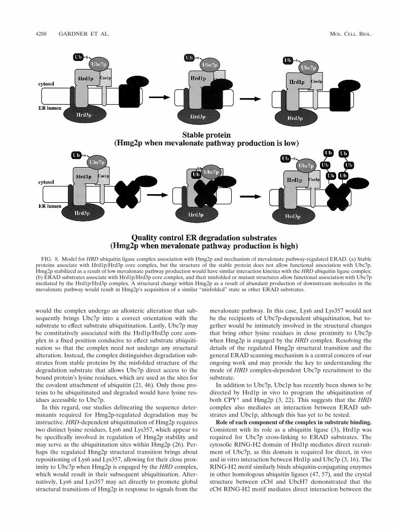
FIG. 8. Model for HRD ubiquitin ligase complex association with Hmg2p and mechanism of mevalonate pathway-regulated ERAD. (a) Stableproteins associate with Hrd1p/Hrd3p core complex, but the structure of the stable protein does not allow functional association with Ubc7p.Hmg2p stabilized as a result of low mevalonate pathway production would have similar interaction kinetics with the HRD ubiquitin ligase complex.(b) ERAD substrates associate with Hrd1p/Hrd3p core complex, and their misfolded or mutant structures allow functional association with Ubc7pmediated by the Hrd1p/Hrd3p complex. A structural change within Hmg2p as a result of abundant production of downstream molecules in themevalonate pathway would result in Hmg2p’s acquisition of a similar “misfolded” state as other ERAD substrates.
4288 GARDNER ET AL. MOL. CELL. BIOL.

ubiquitin ligase and the ubiquitin-conjugating enzyme (92).Hrd1p directly binds Ubc7p (3, 16) and directs Ubc7p prox-imity to substrates, so it is likely that Hrd1p contains somesubstrate-binding activity. Because Hrd1p directs ubiquitina-tion of only ERAD substrates, it must have a way of distin-guishing between the degradation substrates and the stableproteins that it interacts with. It is possible that Hrd1p mayengage all proteins with its transmembrane domain but onlybinds ERAD substrates with its RING-H2 domain, thus bring-ing Ubc7p in proximity to the degradation substrate. In fact,the cytosolic domain of Hrd1p has a preference for misfoldedproteins in an in vitro ubiquitination assay (3), possibly indi-cating that the Hrd1p cytosolic domain possesses a componentof substrate recognition. Hrd1p functions in in vivo ERAD inthe absence of Hrd3p when expressed at sufficiently high levels(27, 67), further supporting the idea of a substrate recognitioncomponent within Hrd1p.
To interact with such a diverse array of proteins, the com-plex must have a general binding mechanism. We cannot ruleout that the complex recognizes specific sequences in eachtarget degradation substrate, such as degrons (81), but noconsensus sequence has been revealed through our mutagenicor computer analyses (26; unpublished observations). In fact,our recent analysis of the Hmg2p sequence (26) and our stud-ies here indicate that the complex recognizes structural cueswithin Hmg2p rather than specific sequences. To allow recog-nition of common structural features among a variety of pro-teins, perhaps a protein in the complex has a substrate-bindingsite with binding affinities similar to those of a chaperone. It isquite possible that the initial substrate-scanning site is in thelumen of the ER, where both lumenal proteins and ER mem-brane proteins would exist. Hrd3p is the only protein of theestablished ER ubiquitin ligase complex that is almost entirelylumenal (27, 67, 71) and, from our cross-linking data, the onlyprotein that interacted with all tested substrates in vivo in theabsence of the other complex components when present atnormal steady-state levels. These observations suggest thatHrd3p may serve a primary role in initial substrate recognition.
Hrd3p may also function directly to control Hrd1pRING-H2 ubiquitin ligase activity (27), which promotes prox-imity between Ubc7p and the substrate, thereby indirectly con-trolling Ubc7p ubiquitin-conjugating activity. In support ofthis, a completely lumenal version of Hrd3p functions normallyin ERAD (27), indicating that the action of Hrd3p to programthe cytosolic ubiquitination of substrates occurs through a lu-men-to-cytosol signaling process. Because Hrd3p interacts di-rectly with the Hrd1p transmembrane domain (27), Hrd3paction to allow cytosolic ERAD events is likely transducedthrough the Hrd1p transmembrane domain. Thus, a defunctsignaling process, through either elimination of Hrd1p or lossof Hrd3p function, may prevent Ubc7p association with sub-strates. In fact, elimination of Hrd1p did prevent Ubc7p cross-linking to substrates. Loss of Hrd3p function, by expression ofonly the Hrd3p N-terminal truncation mutant, also preventedUbc7p cross-linking to substrates despite maintenance of nor-mal Hrd1p levels (27). Additionally, although Hrd1p was main-tained at normal levels by the Hrd3p truncation mutant, theHrd3p truncation mutant did not allow Hrd1p cross-linking tosubstrates. This was most likely the result of an inability of theHrd3p truncation mutant itself to associate with substrates and
is consistent with the Hrd3p N-terminal region’s functioning inthe initial substrate recognition of the complex. Perhaps theinitial binding of substrate with Hrd3p induces allostericchanges in the complex that result in the activation of Hrd1pubiquitin ligase function. These allosteric changes would initi-ate correct temporal and spatial activation of Hrd1p RING-H2ubiquitin ligase function and would promote physical associa-tion between ERAD substrates and Hrd1p and Ubc7p, leadingto ubiquitination of the bound substrate. It may be that thetruncated Hrd3p maintains Hrd1p in an inactive conformation,preventing any functional association of Hrd1p and Ubc7pwith substrate. However, the deletion of such a large portion ofHrd3p may result in an aberrant structure of the remainingpart of Hrd3p, possibly confounding this interpretation.
The action of the HRD ubiquitin ligase complex involvescoordination of processes and information on both sides of theER membrane (27). One likely model for this coordination isthat degradation proceeds by interaction of the Hrd1p/Hrd3pcomplex with a substrate engaged by the Sec61p retrotranslo-cation complex, which is required for ER degradation (94).The combined interaction of both Hrd3p and Hrd1p with theSec61p-engaged substrate would allow the Hrd1p RING-H2domain to promote Ubc7p association with substrate, resultingin subsequent ubiquitination of the substrate. Further explo-ration of the mechanism by which the HRD ubiquitin ligasecomplex operates on both sides of a membrane will be impor-tant in understanding specific aspects of ERAD and processesthat employ ubiquitination as a mode of transmembrane signaltransduction.
Implications of Hmg2p interaction with the HRD complex.The structural transition mechanism for Hmg2p-regulateddegradation has several important implications. It has recentlybeen demonstrated that regulated degradation of mammalianHMGR is mediated by ubiquitination (69), in a manner verysimilar to Hmg2p (34). Although some differences exist in themolecular details of the two systems, it is very likely that thesame mechanism of signal-induced transition to a quality con-trol substrate underlies this important axis of sterol synthesisregulation. At present, it is not clear if the ability to undergothis structural transition is an autonomous feature of theHMGR molecule or if it is mediated by ancillary machinerythat brings the transition about. In either case, however, it ispossible that small molecules could be designed or discoveredto drive the transition in a specific and clinically useful manner.More generally, as quality control degradation systems likelyexist in the cytosol, and quite possibly the nucleus or mitochon-dria, it may be that entry of substrates into these systems occursby similar mechanisms and could be harnessed in much thesame fashion as the ER quality control degradation apparatus.The observation of a regulated transition to a quality controlsubstrate allows the framing of both basic and clinical lines ofinquiry to examine the utility and biological generality of thismode of protein regulation.
ACKNOWLEDGMENTS
We thank Merck for the generous gifts of zaragozic acid, L-659,699,and lovastatin. R.G.G. thanks C. Melissa Morelli for essential techni-cal support required to achieve strong signal intensity despite limitedapplication of reagents. R.Y.H. thanks J. Theriot for opening unex-pected conceptual doors.
VOL. 21, 2001 IN VIVO ACTION OF HRD UBIQUITIN LIGASE COMPLEX 4289

This work was supported by NIH grant DK5199601 (R.Y.H.) and aSearle Scholarship (R.Y.H.).
REFERENCES
1. Baker, R. T., and A. Varshavsky. 1991. Inhibition of the N-end rule pathwayin living cells. Proc. Natl. Acad. Sci. USA 88:1090–1094.
2. Bartel, B., I. Wunning, and A. Varshavsky. 1990. The recognition componentof the N-end rule pathway. EMBO J. 9:3179–3189.
3. Bays, N. W., R. G. Gardner, L. P. Seelig, C. A. Joazeiro, and R. Y. Hampton.2000. Hrd1p is a membrane-anchored ubiquitin ligase required for endo-plasmic reticulum-associated degradation. Nat. Cell Biol. 3:24–29.
4. Biederer, T., C. Volkwein, and T. Sommer. 1996. Degradation of subunits ofthe Sec61p complex, an integral component of the ER membrane, by theubiquitin-proteasome pathway. EMBO J. 15:2069–2076.
5. Biederer, T., C. Volkwein, and T. Sommer. 1997. Role of Cue1p in ubiquiti-nation and degradation at the ER surface. Science 278:1806–1809.
6. Bordallo, J., R. K. Plemper, A. Finger, and D. H. Wolf. 1998. Der3p-Hrd1pis required for endoplasmic reticulum-associated degradation of misfoldedlumenal and integral membrane proteins. Mol. Biol. Cell 9:209–222.
7. Bordallo, J., and D. H. Wolf. 1999. A RING-H2 finger motif is essential forthe function of Der3/Hrd1 in endoplasmic reticulum associated protein deg-radation in the yeast Saccharomyces cerevisiae. FEBS Lett. 448:244–248.
8. Brown, C. R., L. Q. Hong-Brown, J. Biwersi, A. S. Verkman, and W. J. Welch.1996. Chemical chaperones correct the mutant phenotype of the delta F508cystic fibrosis transmembrane conductance regulator protein. Cell StressChaperones 1:117–125.
9. Brown, C. R., L. Q. Hong-Brown, and W. J. Welch. 1997. Correcting tem-perature-sensitive protein folding defects. J. Clin. Investig. 99:1432–1444.
10. Burrows, J. A., L. K. Willis, and D. H. Perlmutter. 2000. Chemical chaper-ones mediate increased secretion of mutant alpha 1-antitrypsin (alpha 1-AT)Z: a potential pharmacological strategy for prevention of liver injury andemphysema in alpha 1-AT deficiency. Proc. Natl. Acad. Sci. USA 97:1796–1801.
11. Chau, V., J. W. Tobias, A. Bachmair, D. Marriott, D. J. Ecker, D. K. Gonda,and A. Varshavsky. 1989. A multiubiquitin chain is confined to specific lysinein a targeted short-lived protein. Science 243:1576–1583.
12. Chun, K. T., S. Bar-Nun, and R. D. Simoni. 1990. The regulated degradationof 3-hydroxy-3-methylglutaryl-CoA reductase requires a short-lived proteinand occurs in the endoplasmic reticulum. J. Biol. Chem. 265:22004–22010.
13. Ciccarelli, E., M. A. Alonso, D. Cresteil, A. Bollen, P. Jacobs, and F. Alvarez.1993. Intracellular retention and degradation of human mutant variant of Aalpha-1 antitrypsin in stably transfected Chinese hamster ovary cell lines.Eur. J. Biochem. 213:271–276.
14. Cook, W. J., L. C. Jeffrey, E. Kasperek, and C. M. Pickart. 1994. Structureof tetraubiquitin shows how multiubiquitin chains can be formed. J. Mol.Biol. 236:601–609.
15. Cronin, S. R., A. Khoury, D. K. Ferry, and R. Y. Hampton. 2000. Regulationof HMG-CoA reductase degradation requires the P-type ATPase Cod1p/Spf1p. J. Cell Biol. 148:915–924.
16. Deak, P. M., and D. H. Wolf. 2001. Membrane topology and function ofder3/hrd1p as a ubiquitin-protein ligase (e3) involved in endoplasmic retic-ulum degradation. J. Biol. Chem. 276:10663–10669.
17. Edwards, P. A., S. F. Lan, R. D. Tanaka, and A. M. Fogelman. 1983. Me-valonolactone inhibits the rate of synthesis and enhances the rate of degra-dation of 3-hydroxy-3-methylglutaryl coenzyme A reductase in rat hepato-cytes. J. Biol. Chem. 258:7272–7275.
18. Fang, S., J. P. Jensen, R. L. Ludwig, K. H. Vousden, and A. M. Weissman.2000. Mdm2 is a RING finger-dependent ubiquitin protein ligase for itselfand p53. J. Biol. Chem. 275:8945–8951.
19. Feldman, R. M. R., C. C. Correll, K. B. Kaplan, and R. J. Deshaies. 1997. Acomplex of Cdc4p, Skp1p, and Cdc53p-Cullin catalyzes ubiquitination of thephosphorylated CDK inhibitor Sic1p. Cell 91:221–230.
20. Finger, A., M. Knop, and D. H. Wolf. 1993. Analysis of two mutated vacuolarproteins reveals a degradation pathway in the endoplasmic reticulum or arelated compartment of yeast. Eur. J. Biochem. 218:565–574.
21. Finley, D., and V. Chau. 1991. Ubiquitination. Annu. Rev. Cell Biol. 7:25–69.22. Friedlander, R., E. Jarosch, J. Urban, C. Volkwein, and T. Sommer. 2000. A
regulatory link between ER-associated protein degradation and the unfold-ed-protein response. Nat. Cell Biol. 2:379–384.
23. Galan, J. M., and M. Peter. 1999. Ubiquitin-dependent degradation of mul-tiple F-box proteins by an autocatalytic mechanism. Proc. Natl. Acad. Sci.USA 96:9124–9129.
24. Gardner, R., S. Cronin, B. Leader, J. Rine, and R. Hampton. 1998. Sequencedeterminants for regulated degradation of yeast 3-hydroxy-3-methylglutaryl-CoA reductase, an integral endoplasmic reticulum membrane protein. Mol.Biol. Cell 9:2611–2626.
25. Gardner, R. G., and R. Y. Hampton. 1999. A highly conserved signal controlsdegradation of 3-hydroxy-3-methylglutaryl-coenzyme A (HMG-CoA) reduc-tase in eukaryotes. J. Biol. Chem. 274:31671–31678.
26. Gardner, R. G., and R. Y. Hampton. 1999. A ‘distributed degron’ allowsregulated entry into the ER degradation pathway. EMBO J. 18:5994–6004.
27. Gardner, R. G., G. M. Swarbrick, N. W. Bays, S. Cronin, S. Wilhovsky, L.
Seelig, C. Kim, and R. Y. Hampton. 2000. Endoplasmic reticulum degrada-tion requires lumen to cytosol signaling: transmembrane control of Hrd1p byHrd3p. J. Cell Biol. 151:69–82.
28. Gardner, R. G., and R. Y. Hampton. 2000. An oxysterol-derived signal for3-hydroxy-3-methylglutaryl CoA reductase degradation in yeast. J. Biol.Chem. 276:8681–8694.
29. Goldstein, J. L., and M. S. Brown. 1990. Regulation of the mevalonatepathway. Nature 343:425–430.
30. Gonda, D. K., A. Bachmair, I. Wunning, J. W. Tobias, W. S. Lane, and A.Varshavsky. 1989. Universality and structure of the N-end rule. J. Biol.Chem. 264:16700–16712.
31. Hampton, R. Y., and J. Rine. 1994. Regulated degradation of HMG-CoAreductase, an integral membrane protein of the endoplasmic reticulum, inyeast. J. Cell Biol. 125:299–312.
32. Hampton, R. Y., R. G. Gardner, and J. Rine. 1996. Role of 26S proteasomeand HRD genes in the degradation of 3-hydroxy-3-methylglutaryl-CoA re-ductase, an integral endoplasmic reticulum membrane protein. Mol. Biol.Cell 7:2029–2044.
33. Hampton, R. Y., A. Koning, R. Wright, and J. Rine. 1996. In vivo examina-tion of membrane protein localization and degradation with green fluores-cent protein. Proc. Natl. Acad. Sci. USA 93:828–833.
34. Hampton, R. Y., and H. Bhakta. 1997. Ubiquitin-mediated regulation of3-hydroxy-3-methylglutaryl-CoA reductase. Proc. Natl. Acad. Sci. USA 94:12944–12948.
35. Hein, C., J. Y. Springael, C. Volland, R. Haguenauer-Tsapis, and B. Andre.1995. NP11, an essential yeast gene involved in induced degradation of Gap1and Fur4 permeases, encodes the Rsp5 ubiquitin-protein ligase. Mol. Mi-crobiol. 18:77–87.
36. Hill, K., and A. A. Cooper. 2000. Degradation of unassembled Vph1p revealsnovel aspects of the yeast ER quality control system. EMBO J. 19:550–561.
37. Hiller, M. M., A. Finger, M. Schweiger, and D. H. Wolf. 1996. ER degrada-tion of a misfolded luminal protein by the cytosolic ubiquitin-proteasomepathway. Science 273:1725–1728.
38. Ho, S. N., H. D. Hunt, R. M. Horton, J. K. Pullen, and L. R. Pease. 1989.Site-directed mutagenesis by overlap extension using the polymerase chainreaction. Gene 77:51–59.
39. Hochstrasser, M. 1995. Ubiquitin, proteasomes, and the regulation of intra-cellular protein degradation. Curr. Opin. Cell Biol. 7:215–223.
40. Hochstrasser, M. 1996. Ubiquitin-dependent protein degradation. Annu.Rev. Genet. 30:405–439.
41. Honda, R., and H. Yasuda. 1999. Association of p19ARF with Mdm2 inhibitsubiquitin ligase activity of Mdm2 for tumor suppressor p53. EMBO J. 18:22–27.
42. Honda, R., and H. Yasuda. 2000. Activity of MDM2, a ubiquitin ligase,towards p53 or itself is dependent on the RING finger domain of the ligase.Oncogene 19:1473–1476.
43. Huibregtse, J. M., M. Scheffner, and P. M. Howley. 1991. A cellular proteinmediates association of p53 with the E6 oncoprotein of human papilloma-virus types 16 or 18. EMBO J. 10:4129–4135.
44. Ito, H., Y. Fukuda, K. Murata, and A. Kimura. 1983. Transformation ofintact yeast cells treated with alkali cations. J. Bacteriol. 153:163–168.
45. Jensen, T. J., M. A. Loo, S. Pind, D. B. Williams, A. L. Goldberg, and J. R.Riordan. 1995. Multiple proteolytic systems, including the proteasome, con-tribute to CFTR processing. Cell 83:129–135.
46. Jentsch, S. 1992. The ubiquitin-conjugation system. Annu. Rev. Genet. 26:179–207.
47. Joazeiro, C. A. P., S. S. Wing, H. Huang, J. D. Leverson, T. Hunter, and Y.Liu. 1999. The tyrosine kinase negative regulator c-CBL as a RING-typeE2-dependent ubiquitin-protein ligase. Science 286:309–312.
48. Kalies, K. U., T. A. Rapoport, and E. Hartmann. 1998. The beta subunit ofthe Sec61 complex facilitates cotranslational protein transport and interactswith the signal peptidase during translocation. J. Cell Biol. 141:887–894.
49. Kamura, T., D. M. Koepp, M. N. Conrad, D. Skowyra, R. J. Moreland, O.Iliopoulos, W. S. Lane, W. G. Kaelin, Jr., S. J. Elledge, R. C. Conaway, J. W.Harper, and J. W. Conaway. 1999. Rbx1, a component of the VHL tumorsuppressor complex and SCF ubiquitin ligase. Science 284:657–661.
50. Knop, M., A. Finger, T. Braun, K. Hellmuth, and D. H. Wolf. 1996. Der1, anovel protein specifically required for endoplasmic reticulum degradation inyeast. EMBO J. 15:753–763.
51. Kopito, R. R. 1997. ER quality control: the cytoplasmic connection. Cell88:427–430.
52. Kramer, K. M., D. Fesquet, A. L. Johnson, and L. H. Johnston. 1998.Budding yeast RSI1-APC2, a novel gene necessary for initiation of anaphase,encodes an APC subunit. EMBO J. 17:498–506.
53. Leverson, J. D., C. A. P. Joazeiro, A. M. Page, H. Huang, P. Hieter, and T.Hunter. 2000. The APC11 RING-H2 finger mediates E2-dependent ubiq-uitination. Mol. Biol. Cell 11:2315–2325.
54. Levkowitz, G., H. Waterman, S. A. Ettenberg, M. Katz, A. Y. Tsygankov, I.Alroy, S. Lavi, K. Iwai, Y. Reiss, A. Ciechanover, S. Lipkowitz, and Y.Yarden. 1999. Ubiquitin ligase activity and tyrosine phosphorylation underliesuppression of growth factor signaling by c-Cbl/Sli-1. Mol. Cell 4:1029–1040.
55. Liao, W., S.-C. Yeung, and L. Chan. 1998. Proteasome-mediated degradation
4290 GARDNER ET AL. MOL. CELL. BIOL.

of apolipoprotein B targets both nascent peptides cotranslationally beforetranslocation and full-length apolipoprotein B after translocation into theendoplasmic reticulum. J. Biol. Chem. 273:27225–27230.
56. Lomant, A. J., and G. Fairbanks. 1976. Chemical probes of extended bio-logical structures: synthesis and properties of the cleavable protein crosslink-ing reagent [35S]dithiobis(succinimidyl proprionate). J. Mol. Biol. 104:243–261.
57. Lorick, K. L., J. P. Jensen, S. Fang, A. M. Ong, S. Hatakeyama, and A. M.Weissman. 1999. RING fingers mediate ubiquitin-conjugating enzyme (E2)-dependent ubiquitination. Proc. Natl. Acad. Sci. USA 96:11364–11369.
58. Mothes, W., S. Prehn, and T. A. Rapoport. 1994. Systematic probing of theenvironment of a translocating secretory protein during translocationthrough the ER membrane. EMBO J. 13:3973–3982.
59. Musch, A., M. Wiedmann, and T. A. Rapoport. 1992. Yeast Sec proteinsinteract with polypeptides traversing the endoplasmic reticulum membrane.Cell 69:343–352.
60. Nakanishi, M., J. L. Goldstein, and M. S. Brown. 1988. Multivalent controlof 3-hydroxy-3-methylglutaryl coenzyme A reductase: mevalonate-derivedproduct inhibits translation of mRNA and accelerates degradation of en-zyme. J. Biol. Chem. 263:8929–8937.
61. Nuber, U., S. E. Schwarz, and M. Scheffner. 1998. The ubiquitin-proteinligase E6-associated protein (E6-AP) serves as its own substrate. Eur. J. Bio-chem. 254:643–649.
62. Ohta, T., J. J. Michel, A. J. Schottelius, and Y. Xiong. 1999. ROC1, ahomolog of APC11, represents a family of cullin partners with an associatedubiquitin ligase activity. Mol. Cell 3:535–541.
63. Patton, E. E., A. R. Willems, D. Sa, L. Kuras, D. Thomas, K. L. Craig, andM. Tyers. 1998. Cdc53 is a scaffold protein for multiple Cdc34/Skp1/F-boxprotein complexes that regulate cell division and methionine biosynthesis inyeast. Genes Dev. 12:692–705.
64. Peters, J. M. 1999. Subunits and substrates of the anaphase-promoting com-plex. Exp. Cell Res. 248:339–349.
65. Pilon, M., K. Romisch, D. Quach, and R. Schekman. 1998. Sec61p servesmultiple roles in secretory precursor binding and translocation into theendoplasmic reticulum membrane. Mol. Biol. Cell 9:3455–3473.
66. Plemper, R. K., R. Egner, K. Kuchler, and D. H. Wolf. 1998. Endoplasmicreticulum degradation of a mutated ATP-binding cassette transporter Pdr5proceeds in a concerted action of Sec61 and the proteasome. J. Biol. Chem.273:32848–32856.
67. Plemper, R. K., J. Bordallo, P. M. Deak, C. Taxis, R. Hitt, and D. H. Wolf.1999. Genetic interactions of Hrd3p and Der3p/Hrd1p with Sec61p suggesta retro-translocation complex mediating protein transport for ER degrada-tion. J. Cell Sci. 112:4123–4134.
68. Qu, D., J. H. Teckman, S. Omura, and D. H. Perlmutter. 1996. Degradationof a mutant secretory protein, alpha-1-antitrypsin Z, in the endoplasmicreticulum requires proteasome activity. J. Biol. Chem. 271:22791–22795.
69. Ravid, T., R. Doolman, R. Avner, D. Harats, and J. Roitelman. 2000. Theubiquitin-proteasome pathway mediates the regulated degradation of mam-malian HMG-CoA reductase. J. Biol. Chem. 275:35840–35847.
70. Reiss, Y., D. Kaim, and A. Hershko. 1988. Specificity of binding of NH2-terminal residue of proteins to ubiquitin-protein ligase. Use of amino acidderivatives to characterize specific binding sites. J. Biol. Chem. 263:2693–2698.
71. Saito, Y., T. Yamanushi, T. Oka, and A. Nakano. 1999. Identification ofSEC12, SED4, truncated SEC16, and EKS1/HRD3 as multicopy suppressorsof ts mutants of Sar1 GTPase. J. Biochem. 125:130–137.
72. Sanders, S. L., K. M. Whitfield, J. P. Vogel, M. D. Rose, and R. W. Schek-man. 1992. Sec61p and BiP directly facilitate polypeptide translocation intothe ER. Cell 69:353–365.
73. Sato, S., C. L. Ward, M. E. Krouse, J. J. Wine, and R. R. Kopito. 1996.Glycerol reverses the misfolding phenotype of the most common cysticfibrosis mutation. J. Biol. Chem. 271:635–638.
74. Scheffner, M., J. M. Huibregtse, R. D. Vierstra, and P. M. Howley. 1993. TheHPV-16 E6 and E6-AP complex functions as a ubiquitin-protein ligase in the
ubiquitination of p53. Cell 75:495–505.75. Scheffner, M., U. Nuber, and J. M. Huibregtse. 1995. Protein ubiquitination
involving an E1–E2-E3 enzyme ubiquitin thioester cascade. Nature 373:81–83.
76. Seol, J. H., R. M. Feldman, W. Zachariae, A. Shevchenko, C. C. Correll, S.Lyapina, Y. Chi, M. Galova, J. Claypool, S. Sandmeyer, K. Nasmyth, andR. J. Deshaies. 1999. Cdc53/cullin and the essential Hrt1 RING-H2 subunitof SCF define a ubiquitin ligase module that activates the E2 enzyme Cdc34.Genes Dev. 13:1614–1626.
77. Skowyra, D., K. L. Craig, M. Tyers, S. J. Elledge, and J. W. Harper. 1997.F-box proteins are receptors that recruit phosphorylated substrates to theSCF ubiquitin-ligase complex. Cell 91:209–219.
78. Skowyra, D., D. M. Koepp, T. Kamura, M. N. Conrad, R. C. Conaway, J. W.Conaway, S. J. Elledge, and J. W. Harper. 1999. Reconstitution of G1 cyclinubiquitination with complexes containing SCFGrr1 and Rbx. Science 284:662–665.
79. Thrower, J. S., L. Hoffman, M. Rechsteiner, and C. M. Pickart. 2000. Rec-ognition of the polyubiquitin proteolytic signal. EMBO J. 19:94–102.
80. Travers, K. J., C. K. Patil, L. Wodicka, D. J. Lockhart, J. S. Weissman, andP. Walter. 2000. Functional and genomic analyses reveal an essential coor-dination between the unfolded protein response and ER-associated degra-dation. Cell 101:249–258.
81. Varshavsky, A. 1991. Naming a targeting signal. Cell 64:13–15.82. Ward, C. L., S. Omura, and R. R. Kopito. 1995. Degradation of CFTR by the
ubiquitin-proteasome pathway. Cell 83:121–127.83. Wei, X., R. Eisman, J. Xu, A. D. Harsch, A. E. Mulberg, C. L. Bevins, M. C.
Glick, and T. F. Scanlin. 1996. Turnover of the cystic fibrosis transmembraneconductance regulator (CFTR): slow degradation of wild-type and deltaF508 CFTR in surface membrane preparations of immortalized airway ep-ithelial cells. J. Cell. Physiol. 168:373–384.
84. Wickner, S., M. R. Maurizi, and S. Gottesman. 1999. Posttranslational qual-ity control: folding, refolding, and degrading proteins. Science 286:1888–1893.
85. Wilhovsky, S., R. Gardner, and R. Hampton. 2000. HRD gene dependence ofER-associated degradation. Mol. Biol. Cell 11:1697–1708.
86. Xie, Y., and A. Varshavsky. 1999. The E2–E3 interaction in the N-end rulepathway: the RING-H2 finger of E3 is required for the synthesis of multiu-biquitin chain. EMBO J. 18:6832–6844.
87. Yang, M., S. Omura, J. S. Bonifacino, and A. M. Weissman. 1998. Novelaspects of degradation of T cell receptor subunits from the endoplasmicreticulum (ER) in T cells: importance of oligosaccharide processing, ubiq-uitination, and proteasome-dependent removal from ER membranes. J. Exp.Med. 187:835–846.
88. Yu, H., G. Kaung, S. Kobayashi, and R. R. Kopito. 1997. Cytosolic degra-dation of T-cell receptor alpha chains by the proteasome. J. Biol. Chem.272:20800–20804.
89. Yu, H., J. M. Peters, R. W. King, A. M. Page, P. Hieter, and M. W. Kirschner.1998. Identification of a cullin homology region in a subunit of the anaphase-promoting complex. Science 279:1219–1222.
90. Zachariae, W., A. Shevchenko, P. D. Andrews, R. Ciosk, M. Galova, M. J.Stark, and K. Nasmyth. 1998. Mass spectrometric analysis of the anaphase-promoting complex from yeast: identification of a subunit related to cullins.Science 279:1216–1219.
91. Zheng, F., N. Kartner, and G. L. Lukacs. 1998. Limited proteolysis as aprobe for arrested conformational maturation of DELTA-F508 CFTR. Nat.Struct. Biol. 5:180–183.
92. Zheng, N., P. Wang, P. D. Jeffrey, and N. P. Pavletich. 2000. Structure of ac-Cbl-UbcH7 complex: RING domain function in ubiquitin-protein ligases.Cell 102:533–539.
93. Zhou, M., E. A. Fisher, and H. N. Ginsberg. 1998. Regulated co-translationalubiquitination of apolipoprotein B100. A new paradigm for proteasomaldegradation of a secretory protein. J. Biol. Chem. 273:24649–24653.
94. Zhou, M., and R. Schekman. 1999. The engagement of Sec61p in the ERdislocation process. Mol. Cell 4:925–934.
VOL. 21, 2001 IN VIVO ACTION OF HRD UBIQUITIN LIGASE COMPLEX 4291