in vitro study of bacterial growth inhibition in ...aac.asm.org/content/23/5/766.full.pdf · in...
TRANSCRIPT

Vol. 23, No. 5ANTIMICROBIAL AGENTS AND CHEMOTHERAPY, May 1983, p. 766-7730066-4804/83/050766-08$02.00/0Copyright © 1983, American Society for Microbiology
In Vitro Study of Bacterial Growth Inhibition in ConcentratedSugar Solutions: Microbiological Basis for the Use of Sugar in
Treating Infected WoundsJORGE CHIRIFE,' LE6N HERSZAGE,2 ARABELLA JOSEPH,3 AND ELISA S. KOHN3
Departamento de Industrias, Facultad de Ciencias Exactas y Naturales, Universidad de Buenos Aires,Ciudad Universitaria, 1428 Buenos Aires1; Viamonte 1620, 20 C, 1055 Buenos Aires2; and Instituto Nacional
de Microbiologia, "Dr. Carlos G. Malbrdn," Buenos Aires,3 Argentina
Received 2 December 1982/Accepted 11 February 1983
The use of sugar for the treatment of infected wounds was investigated in invitro experiments with bacteria pathogenic to humans, such as Escherichia coli,Pseudomonas aeruginosa, Klebsiella pneumoniae, and Staphylococcus aureus.
Studies showed that solutions of appropriate sugar concentration incubated at pH7.0 and 35°C were lethal to the bacterial species studied. On the basis of theseresults, it is proposed that an important function of sugar in the treatment ofinfected wounds is to create an environment of low water activity (a,), whichinhibits or stresses bacterial growth.
Various naturally occurring substances havebeen used in wound treatment throughout his-tory. Among them, sugar (sucrose), honey (mainconstituents are glucose, fructose, and maltose),and molasses (main constituents are sucrose andglucose) are the most common. However, it isstill uncertain how sugar acts on wounds, asrecently reviewed by Forrest (9, 10).
In 1976, Herszage and Montenegro of BuenosAires began treating wounds with ordinary sugarbecause of the complicated evolution (despiteconventional therapy) and critical condition oftwo in-patients with postsurgical necrotic celluli-tis. Sugar was used simply on the basis ofexisting folk therapy. In view of the successobtained, they began using sugar systematicallyfor the treatment of infected wounds. Ordinarygranulated sugar (purchased in the supermarket)was used since it was found not to contain anyforeign substance with antibacterial properties;the sugar was not mixed with any antiseptic orany other substance with proven or supposedantibacterial action; and antibiotics were notused concurrently. The procedure consisted of(i) wide opening of the wound; (ii) drying oftissues with gauze; and (iii) filling the woundwith as much sugar as possible, taking care to fillevery cavity, and adding more sugar periodical-ly. Herszage et al. (12) reported 120 cases withinfected wounds and other superficial lesionswhich were treated with sugar with a cure rate of99.2%. The time for cure varied between 9 daysand 17 weeks; however, it was usual for odorand secretion to diminish within 24 h and todisappear after 72 to 96 h of treatment. Thisreport, however, was only a short summary of
their findings and did not include hundreds ofphotographs showing the evolution of casestreated. The patients varied between 3 monthsand 94 years in age and included 50 females and70 males, of whom 6 were diabetics.
In most cases treated with sugar, it was ob-served that wounds were healed without de-bridement of necrotic tissues or any other surgi-cal procedure except for a complete opening ofthe wound. After 5 or 7 days, it was possible toremove the necrotic tissue with forceps as if itwere a piece of dressing; this process occurredeven in wounds contaminated with fecal materi-al. Diabetic patients showed the same responseas others, even in the presence of hyperglyce-mia. Glossy protecting covering formed in thewounds, and the resulting cicatrices were unusu-ally resistant. The pH values of all woundstreated with sugar ranged from 6.8 to 7.4. Photo-graphs showing the evolution of wound healingin a representative case treated with sugar areshown in Fig. 1 and 2. For the sake of brevity,only one case is shown here. The work ofHerszage et al. (12) was not a controlled studysince the dramatic improvement resulting fromthis treatment initially prevented them, for ethi-cal reasons, from undertaking a randomizedcontrolled study. In spite of this, the accumulat-ed evidence strongly suggests that sugar playeda role in the treatment of infected wounds.We are proposing that an important function
of sugar in the treatment of infected wounds is tocreate an environment of low water activity (aw)which inhibits or stresses bacterial growth. Alow a, also means high osmotic pressure (7i)since both are thermodynamically related ac-
766
on July 6, 2018 by guesthttp://aac.asm
.org/D
ownloaded from

SUGAR AND BACTERIAL GROWTH IN INFECTED WOUNDS 767
,
_ -; :+gS 0 f - .. . -.-_ f . . .. D -F; . . t. . ._: .;. j - S_ v
V D
cording to the equation mr = (RT/V} x log (1Ia,),where V is the partial molal volume of water. Inthis way, a solution of low a, has high osmoticpressure.
Bacteria, like all other forms of life, requirewater for growth, and these water requirementsare best defined in terms of water activity (aw) ofthe substrate (6, 18). When the aqueous solu-tions in the environment of the microorganismare concentrated by the addition of a solute suchas sugar (sucrose), the consequences for micro-bial growth result mainly (although not only)from the change in aw. At present, numerousdata are available on the relationship betweenaw and the ability of microorganisms to grow,and it has been reported that every microorgan-ism has a limiting a, below which it will notgrow (5). Table 1 shows the limiting a, values
FIG. 1. Evolution of sugar-treated wound in a 16-year-old male, Alvear Hospital, admitted 15 October1979 on account of severe traumatisms of left lowerlimb; femur fractured; wound in left limb with sectionof left popliteal artery and vein. Fracture nail used.Anastomosis with saphenous vein for the arterial andvenous injury. After 4 days, blisters all over the limb(Meleney gangrenous cellulitis). (A) Surgical wound,19 October 1979; blisters on the tense wounded skin.(B) Sutures removed; the wound is opened widely;muscular masses of necrotic aspect. (C) Sugar treat-ment begins. (D) First week of treatment; necrotictissue becomes circumscribed. (E) Sixth week;marked improvement of the wound.
(and calculated equivalent sugar concentrations)for various bacteria pathogenic to humans, suchas Klebsiella spp., Salmonella spp., Pseudomo-nas spp., Escherichia coli, Clostridium perfrin-gens, and Staphylococcus aureus. Of the wholerange of bacteria that infect human skin, subcu-taneous tissues, and mucous membranes, thelowest a, is tolerated by S. aureus, which canproliferate with an a, as low as 0.86. We haverecently reported some preliminary in vitro ex-periments on inhibiting S. aureus ATCC 6538Pin sucrose solutions (3). S. aureus was chosen asa test organism since, as mentioned, it is thepathogen most resistant to low aw.
This study has two objectives: (i) to extendthe studies of bacterial growth inhibition inconcentrated sucrose solutions to other patho-genic bacteria relevant to infected wounds and
VOL. 23, 1983
on July 6, 2018 by guesthttp://aac.asm
.org/D
ownloaded from
ANTIMICROB. AGENTS CHEMOTHER.
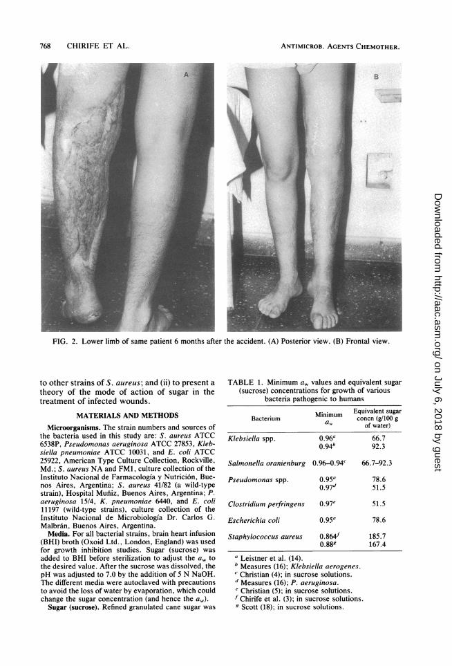
FIG. 2. Lower limb of same patient 6 months after the accident. (A) Posterior view. (B) Frontal view.
to other strains of S. aureus; and (ii) to present a
theory of the mode of action of sugar in thetreatment of infected wounds.
MATERIALS AND METHODS
Microorganisms. The strain numbers and sources ofthe bacteria used in this study are: S. aureus ATCC6538P, Pseudomonas aeruginosa ATCC 27853, Kleb-siella pneumoniae ATCC 10031, and E. coli ATCC25922, American Type Culture Collection, Rockville,Md.; S. aureus NA and FM1, culture collection of theInstituto Nacional de Farmacologia y Nutrici6n, Bue-nos Aires, Argentina; S. aureus 41/82 (a wild-typestrain), Hospital Mufiiz, Buenos Aires, Argentina; P.aeruginosa 15/4, K. pneumoniae 6440, and E. coli11197 (wild-type strains), culture collection of theInstituto Nacional de Microbiologfa Dr. Carlos G.Malbran, Buenos Aires, Argentina.Media. For all bacterial strains, brain heart infusion
(BHI) broth (Oxoid Ltd., London, England) was usedfor growth inhibition studies. Sugar (sucrose) was
added to BHI before sterilization to adjust the aw tothe desired value. After the sucrose was dissolved, thepH was adjusted to 7.0 by the addition of 5 N NaOH.The different media were autoclaved with precautionsto avoid the loss of water by evaporation, which couldchange the sugar concentration (and hence the aw).Sugar (sucrose). Refined granulated cane sugar was
TABLE 1. Minimum a, values and equivalent sugar(sucrose) concentrations for growth of various
bacteria pathogenic to humans
Minimum Equivalent sugarBacterium
awconcn (g/100 g
of water)
Klebsiella spp. 0.96a 66.70.94b 92.3
Salmonella oranienburg 0.96-0.94c 66.7-92.3
Pseudomonas spp. 0.95a 78.60.97d 51.5
Clostridium perfringens 0.97e 51.5
Escherichia coli 0.95a 78.6
Staphylococcus aureus 0.864f 185.70.889 167.4
a Leistner et al. (14).b Measures (16); Klebsiella aerogenes.c Christian (4); in sucrose solutions.d Measures (16); P. aeruginosa.e Christian (5); in sucrose solutions.f Chirife et al. (3); in sucrose solutions.g Scott (18); in sucrose solutions.
768 CHIRIFE ET AL.
on July 6, 2018 by guesthttp://aac.asm
.org/D
ownloaded from

SUGAR AND BACTERIAL GROWTH IN INFECTED WOUNDS
E\5
m4 S~*- -* ' -aw=0843aw=0.848
0 1 2 3 4 5 6 7 8 9 10INCUBATION, TIME days
FIG. 3. Inhibition of growth of S. aureus ATCC6538P (A), FM1 (A), and NA (0) in BHI with addedsugar at pH 7.0, incubated at 35°C.
purchased in 1-kg packages from a local supermarketand used as it was.Growth inhibition studies. Growth studies were
made in 250-ml screw-top glass bottles containingabout 18 g each of inoculated sugar-supplementedmedia, incubated at 35°C in a constant temperaturecabinet.Enumeration procedure. Counts were determined by
the use of plate count agar (Difco Laboratories, De-troit, Mich.) for S. aureus, K. pneumoniae, and E.coli; CLED medium with Andrade indicator (OxoidLtd.) was used for P. aeruginosa. The samples wereserially diluted with 0.1% peptone (Oxoid Ltd.) beforeplating. The plates were incubated at 35°C for 24 to 48h, and the colonies were counted.
Determination of aw. The a, of sugar-supplementedmedia and the a, in sugar-treated wound cavities weredetermined at 25.0 ± 0.1°C with an electronic hygrom-eter Humicap HMI 14 A manufactured by Vaisala,Helsinki, Finland, as described by Favetto et al. (G.Favetto, S. Resnik, J. Chirife, and C. Ferro Fontan, J.Food Sci., in press).
RESULTSThe relationship between aw and sucrose con-
centration may be obtained from the equation aw= Xl exp (-6.47 X22), where Xl and X2 aremolar fractions of water and sucrose, respec-tively, defined as Xl = (moles of water)/(molesof water + moles of sucrose); 1 = XI + X2.
Figure 3 shows the behavior of S. aureusATCC 6538P, NA, and FM1 in BHI media withthe aw adjusted with sugar to the range of 0.84 to0.86, incubated at 35°C; initial inoculum levelswere in the range of 103 to 106 CFU/ml. Asexpected, sugar produced complete growth inhi-bition of all three strains. Control experiments(BHI without sugar) were also performed (datanot shown) and showed that the three S. aureusstrains reached 109 (or above) CFU/ml after 24to 36 h of incubation.
Figure 4 shows the inhibitory effect of a sugar-saturated broth (aw, 0.828) on S. aureus at highinitial inoculum levels (_108 CFU/ml). The num-ber of CFU of strains ATCC 6538P and 41/82 permilliliter declined throughout the incubation pe-
INCUBATION TIME,days
FIG. 4. Growth inhibition and survival of S. aureus41/82 (0), and ATCC 6538P (0) in BHI saturated withsugar at pH 7.0, incubated at 35°C.
riod to the point of almost complete loss ofviability by the end of the period studied (60 to70 days).
Figure 5 shows the results of experiments ofgrowth inhibition and survival of P. aeruginosaATCC 27853, E. coli ATCC 25922, K. pneumo-niae ATCC 10031, and S. aureus ATCC 6538P insugar-saturated broth (aw, 0.828) incubated at35°C, pH 7.0. Control experiments in brothwithout sugar were also performed (data notshown). It is noteworthy that viable cells of P.aeruginosa, E. coli, and K. pneumoniae de-clined rapidly, leading to almost total loss ofviable population in 2 to 3 days of incubation. S.aureus, however, was far more resistant (al-though its viable population also declined con-tinuously).
Figures 6 and 7 show similar growth inhibition
4
3cn 0 5
10 is 20 25 30 35 40INCUBATION TIME,days
FIG. 5. Growth inhibition and survival of E. coliATCC 25922 (0), P. aeruginosa ATCC 27853 (O), K.pneumoniae ATCC 10031 (A), and S. aureus ATCC6538P +) in BHI saturated with sugar at pH 7.0,incubated at 35°C.
769VOL. 23, 1983
on July 6, 2018 by guesthttp://aac.asm
.org/D
ownloaded from

ANTIMICROB. AGENTS CHEMOTHER.
FIG. 6. Growth inhibition and surmoniae 6440 with sugar (@) and coIaeruginosa 15/4 with sugar (0) and ccat pH 7.0, incubated at 35°C.
and survival curves for other s11197, K. pneumoniae 6440, and15/4, in sugar-saturated broth (a,bated at 35°C, pH 7.0.
DISCUSSIONThe explanation of the role c
treatment of infected wounds isperhaps impossible to reduce to a
nism, such as its antibacterial actless, we are proposing here thafunction of sugar is to create an elow a, which inhibits or stregrowth. Infected wounds in mosons heal by drainage and debrider, it has to be stressed that an iresult of the interrelationshipfactors: the medium, the bacterifenses of the host (15). Thereforreasonable to assume that in patered defense mechanisms, sugar iin the control of infection by dimiial virulence.Herszage et al. (12) followed t]
bacterial flora present in va:wounds being treated with sugatially present included, among (
coccus spp., Klebsiella spp., E. Xgens, and S. aureus, of which aldisappeared during the first daysment. These in vivo results hsimilarity to our in vitro resultsuggest that an aw-based expl;sugar action is likely.The aw of a bacterial cultur4
reduced by exposing the medspheres of appropriate relative
equilibrium, the a, of the medium is equal to therelative humidity divided by 100. Turner andSalmonsen (19) exposed very small volumes ofcultures of three different serotypes of Klebsiel-la spp. to a relative humidity of 85% at 25°C andfound that no bacteria survived after 24 h. Thisfinding is in good agreement with our results insucrose solution (Fig. 5) since in the experi-ments of Turner and Salmonsen (19), the a, ofthe growth medium should have been close to0.85, which resulted in the rapid death of Klebsi-ella spp.
In a recent preliminary report (3) on the effectof sucrose on the in vitro growth of S. aureus,we showed that the sucrose concentration need-ed to achieve complete growth inhibition of this
5 6 7 microorganism was 183 g/100 g water. With this
vival of K. pneu- experiment, we tried to illustrate the conve-ntrol (A) and p. nience of maintaining a high sugar concentra-)ntrol (A) in BHI tion, which can be obtained by initially filling the
wound with as much sugar as possible (see Fig.1C) and then adding periodically more sugar.We recommended this procedure bearing in
,trains, E. coli mind the uptake of tissue water, which dilutesP. aeruginosa the sugar placed in the wound and which resultsW9 0.828) incu- in a low sugar concentration that may aid rather
than inhibit bacterial growth. It is noteworthy,however, that the sucrose concentration neededto achieve growth inhibition of most human
)f sugar in the pathogens other than S. aureus is much lowers complex and than 183 g/100 g water (see Table 1). For in-i single mecha- stance, postoperative wounds are frequentlyLion. Neverthe- contaminated with E. coli or PseudomonasI an important strains which may be inhibited by sugar concen-environment of trations as low as 52 and 79 g, respectively per-sses bacterial 100 g of water.ist normal per-Lement; howev-infection is thebetween threeia, and the de-re, it would beients with low-may play a rolenishing bacteri-
he evolution ofrious infectedr. Bacteria ini-others, Stepto-coli, C. perfrin-1l but S. aureusof sugar treat-
iave a strikingLs (Fig. 5) andanation of the
e may also beiium to atmo-humidities; at
C-,
TIME, days
FIG. 7. Growth inhibition and survival of E. coliATCC 25922 with sugar (A) and control (A) and E. coli11197 with sugar (0, 0) and control (O) in BHI at pH7.0, incubated at 350C.
770 CHIRIFE ET AL.
-L0 1 2 3 4T M E , d a y s
6
-g5-nU-
C-)4mO
3
2
on July 6, 2018 by guesthttp://aac.asm
.org/D
ownloaded from

SUGAR AND BACTERIAL GROWTH IN INFECTED WOUNDS 771
The actual sugar concentration (or a,) in awound cavity is not constant but is a function ofthe frequency of the addition of fresh sugar andthe amount of liquid released by the tissues,which is time dependent. At the beginning ofevery treatment, the sugar concentration shouldbe very high, approaching saturation, which forbody temperature is about 225 g/100 g water andcorresponds to a, about 0.83. Then, water activ-ity is progressively raised owing to the uptake ofwater from the surrounding tissues until moresugar is again placed in the wound and the a,drops to a very low value. In this way, bacteriain the wound cavity are subjected to a series ofosmotic shocks owing to the continuous changeof a, of the medium; this implies that bacteriashould be adapted to the differences in a, insideand outside the protoplasm. It is known (1) thatabrupt changes in the water activity of bacterialcultures cause injury and death of the cells, evenwithout going beyond the conditions which aresuitable for growth. Bayer (1) showed that 90%of logarithmically growing cells of E. coli sur-vived exposure for 10 min to 50% sucrose fol-lowed by a slow dilution. However, when thecultures were subjected to an osmotic shockproduced by a sudden reduction of the a, of themedium, the viability of the cells decreaseddramatically. Thus, although conditions in thesugar-treated wound are such that the limitingaw for growth is temporarily surpassed, bacterialcells will be stressed, and it is reasonable toassume that under this condition it would beeasier for the immune system to play its role.We have shown that the limiting aw with
sucrose for in vitro growth of S. aureus at 35°Cand pH 7.0 is around 0.864. At higher aw valuesgrowth occurs, but the initiation of growth isdelayed (lag period). Scott (18) reported that thelag period for S. aureus growth in various staticliquid media at 30°C was 1 to 3 days at an aw of0.90. We have found that lag periods of S.aureus at 35°C in sucrose-supplemented liquidmedia were 1 to 2 days at an aw of around 0.89(3). These lag periods may be important inbacterial inhibition in sugar-treated wounds. Thegrowth rate of bacteria is also very much affect-ed by reduction in aw. Scott (18) showed that thegrowth rate of various strains of S. aureus couldbe strongly diminished by decreasing the awfrom its optimal value for growth (0.993). At aw0.90 and 0.94 (both values well above the limit-ing aw), the growth rates were only 12.3 and51.5%, respectively, of the maximum. Christian(4) has shown that the limiting aw for growth ofSalmonella oranienberg at 30°C in sucrose-add-ed medium was 0.96, which corresponded toabout 67 g of sucrose per 100 g of water.However, a sucrose concentration of only 35g/100 g of water was enough to reduce the
growth rate to 59% of its maximum value.It is well known that cells of E. coli and
Pseudomonas spp. suspended in a hypertonic(low a,) solution of a nonpenetrant solute (suchas sucrose) plasmolyze rapidly as they adjustthermodynamically by losing water (13). Deplas-molysis is then essential for resumption ofgrowth in a medium of lowered a, (7, 8). Deplas-molysis consists of the rehydration of the cell,which occurs because of the entry of osmoticallyactive solutes to balance intracellular and extra-cellular a,. This process, however, is slow inthe presence of sucrose. Kroll and Anagnosto-poulos (13) have shown that deplasmolysis of P.aeruginosa in a sucrose solution having only22.7 g of sucrose per 100 g water (aw, 0.987)takes almost 3 h. The following experiment wasperformed on a 60-year-old male patient with apostsurgical abdominal wound who was receiv-ing daily sugar treatment. The wound cavity wasfilled with about 265 g of ordinary granulatedsugar and covered with gauze. After 2.5 h, thesugar was transformed into a syrup that still hadundissolved sugar granules; a homogenized sam-ple was taken, and the aw was determined withan electronic hygrometer (as described above).The a, was found to correspond to that of asaturated sucrose solution (aw, 0.848 at 25°C).Additional samples were taken at 4 and 10 h(from the beginning of treatment), and the corre-sponding a, values were 0.897 and 0.951, re-spectively. The last value is still low enough toinhibit or severely restrict growth of most hu-man pathogens. The evolution of a, in sugar-treated infected wounds is the subject of addi-tional research.
All of the above facts may help to explain howsugar may reduce infection in a wound, even ifthere is a dilution owing to water uptake fromthe surroundings. It may be noted that this liquidflow contributes to cleaning the wound.One may be concerned about the effect of the
low water activity on the tissue cells with whichthe sugar is in contact. The internal water activi-ty of the tissue cells (a, cell) may be assumed tobe >0.990 (11), whereas the water activity in thesugar side (or wound cavity) (a, cav) is consider-ably lower; thus, a flux of water exists becauseof a water activity driving force. Thus, it may beargued that dehydration of the tissue surround-ing the wound may eventually kill these cells.However, in living organisms it does not happenbecause a flux of water from the inner regionsreplaces water as fast as it is transferred fromthe surrounding cells to the wound cavity. Inthis way external tissue cells remain wet, andthe healing process is not adversely affected bysugar (see Fig. 1 and 2). This does not occur,however, in a dead organism, in which sugarproduces dehydration of the surrounding cells.
VOL. 23, 1983
on July 6, 2018 by guesthttp://aac.asm
.org/D
ownloaded from

ANTIMICROB. AGENTS CHEMOTHER.
FIG. 8. Effect of saturated sugar solution on beef tissue after 24 h at room temperature. Dark areas caused byso-called osmotic dehydration may be observed in the tissue with which the sugar is in contact.
This was shown in the following experiment. Acavity was opened in the center of a large pieceof beef (about 3 kg), filled with sugar, andallowed to remain for 24 h at room temperature.The sugar produced dehydration of the externaltissue cells, as evidenced by the dark areassurrounding the cavity filled with sugar (Fig. 8).
Recently, Forrest (9, 10) reviewed the effec-tive wound treatments that had been discoveredby prehistoric and primitive peoples. He indicat-ed that honey was among the agents most usedby ancient civilizations for the treatment ofwounds. Wounds were usually washed withwater (or milk) and dressed with honey. Forrest(10) indicated that honey has antibacterial prop-erties dependent on the production of hydrogenperoxide from glucose. However, it is also pos-sible that honey may have acted on wounds bycreating an environment of low water activity,which inhibits bacterial activity. Rueg and Blanc(17) have measured the water activity of severalcommercially available natural liquid and crys-tallized honeys. They found that the mean a, ofliquid honeys was 0.562, and that of crystallizedhoneys was 0.589. These values are quite lowand certainly would be inhibitory for bacterialgrowth.We may conclude that it is likely that sugar
reduces infection through lowering the a, andthat the experimental evidence indicates thatsugar treatment does not negatively influencewound healing. There are, however, additionalfacts which must be studied, among them theeffects of sugar on macrophages and the inci-dence on wound healing.
ACKNOWLEDGMENTS
The authors acknowledge the financial support from Subse-cretarfa de Ciencia y Tecnologia de la Republica Argentina(Programa Nacional de Tecnologia de Alimentos).
LITERATURE CITED
1. Bayer, M. E. 1967. Response of cell walls of Escherichiacoli to a sudden reduction of the environmental osmoticpressure. J. Bacteriol. 93:1104-1112.
2. Chirife, J., C. Ferro Fontin, and E. A. Benmergui. 1980.The prediction of water activity in aqueous solutions inconnection with intermediate moisture foods. IV. awPrediction in aqueous non-electrolyte solutions. J. FoodTechnol. 15:59-70.
3. Chirife, J., G. Scarmatto, and L. Herszage. 1982. Scien-tific basis for the use of granulated sugar in the treatmentof infected wounds. Lancet i:560.
4. Christian, J. H. B. 1955. The water relations of growthand respiration of Salmonella oranienburg at 30'C. Aust.J. Biol. Sci. 8:490-497.
5. Christian, J. H. B. 1981. Specific solute effects on micro-bial water relations, p. 825-854. In L. B. Rockland andG. F. Stewart (ed.), Water activity: influences on foodquality. Academic Press, Inc., New York.
772 CHIRIFE ET AL.
on July 6, 2018 by guesthttp://aac.asm
.org/D
ownloaded from
SUGAR AND BACTERIAL GROWTH IN INFECTED WOUNDS 773
6. Christian, J. H. B., and J. A. Waltho. 1962. The waterrelations of staphylococci and micrococci. J. Appl. Bact.25:369-377.
7. Dhavises, G., and G. D. Anagnostopoulos. 1979. Influenceof amino acids and water activity on the growth ofEscherichia coli B/rll. Microbios Lett. 7:105-115.
8. Dhavises, G., and G. D. Anagnostopoulos. 1979. Influenceof amino acids and water activity on the growth ofEscherichia coli B/r/1. Microbios Lett. 7:149-159.
9. Forrest, R. D. 1982. Early history of wound treatment. J.R. Soc. Med. 75:198-205.
10. Forrest, R. D. 1982. Development of wound therapy fromthe Dark Ages to the present. J. R. Soc. Med. 75:268-273.
11. Gimeno, A. L., and M. A. F. de Gimeno. 1965. Nocionesde fisiologia celular. EUDEBA, Editorial Universitaria deBuenos Aires, Buenos Aires.
12. Herszage, L., J. R. Montenegro, and A. L. Joseph. 1980.Tratamiento de las heridas supuradas con azficar granu-
lado comercial. Bol. Trab. Soc. Argent. Cir. 41:315-330.13. Kroll, R. G., and G. D. Anagnostopoulos. 1981. Potassium
fluxes on hyperosmotic shock and the effect of phenol and
bronopol (2-bromo-2-nitropropan-1,3-diol) on deplasmo-lysis of Pseudomonas aeruginosa. J. Appi. Bacteriol.51:313-323.
14. Leistner, L., W. Rodel, and K. Krispien. 1981. Microbiolo-gy of meat and meat products in high and intermediate-moisture ranges, p. 855-916. In L. B. Rockland and G. F.Stewart (ed.), Water activity: influences on food quality.Academic Press, Inc., New York.
15. Meakins, J. L. 1976. Pathophysiologic determinants andprediction of sepsis. Surg. Clin. N. Am. 56:847-857.
16. Measures, J. C. 1975. Role of amino acids in osmoregula-tion in non-halophilic bacteria. Nature (London) 257:398-400.
17. Rueg, M., and B. Blanc. 1981. The water activity of honeyand related sugar solutions. Lebensm.-Wiss. Technol.14:1-6.
18. Scott, W. J. 1953. Water relations of Staphylococcusaureus at 30°C. Aust. J. Biol. Sci. 6:549-564.
19. Turner, A. G., and P. A. Salmonsen. 1973. The effect ofrelative humidity on the survival of three serotypes ofKlebsiella. J. Appl. Bacteriol. 36:497-499.
VOL. 23, 1983
on July 6, 2018 by guesthttp://aac.asm
.org/D
ownloaded from