in vitro propagation and dynamics of t cells from skin
TRANSCRIPT

Acta Derm Venereol 90
INVESTIGATIVE REPORT
Acta Derm Venereol 2010; 90: 468–473
© 2010 The Authors. doi: 10.2340/00015555-0927Journal Compilation © 2010 Acta Dermato-Venereologica. ISSN 0001-5555
In order to explore the mechanisms of inflammatory skin disorders, we established two methods of expan-ding skin-derived lymphocytes, one using high levels of interleukin (IL)-2 and IL-4 (method A) and the other using low levels of cytokines and anti-CD3/CD28 microbeads (method B). Both methods provide advan-tages for functional studies. With either of these two, we could obtain more than 107 cells/ from a 3 mm skin biopsy in 21 days from 23 out of 26 biopsies of various skin diseases. The relevance of these cells was confir-med by shifted T-cell receptor β chain variable region (TCR-Vβ) repertoire and antigen-dependent prolifera-tion in antigen-driven skin disorders. The propagation of skin-resident lymphocytes, seen especially in method A, seems to be mediated by a functional defect of regu-latory T cells residing in skin sequentially expanding under the conditions of our methods. Key words: skin-derived lymphocytes; T-cell receptor repertoire; regula-tory T cells.
(Accepted March 22, 2010.)
Acta Derm Venereol 2010; 90: 468–473.
Hideo Hashizume, Department of Dermatology, Hama-matsu University School of Medicine, 1-20-1 Handa-yama Higashi-ku, Hamamatsu 431-3192, Japan. E-mail: [email protected]
Skin has an exquisite immune system that protects against invasion by pathogens. Dysregulation of the skin im-mune system may influence chronic inflammatory skin diseases, including atopic dermatitis (AD) and psoriasis (1). An ideal way of exploring the mechanisms of such conditions would be to obtain large numbers of inflam-matory cells resident in skin. However, it is difficult to expand a low number of cells from skin explants to obtain sufficient numbers of cells to investigate, and special techniques and devices are required (2, 3). We describe here the establishment of two simple methods of expanding skin-derived lymphocytes, which provide advantages for functional studies.
MATERIALS AND METHODS
PatientsTwenty-three patients with AD (6 men, 3 women; mean age 44 years (range 36–61 years)), psoriasis (6 men, 1 woman; mean age 47.5 years (range 26–70 years)), acute/chronic eczema, which could not be further defined (2 men, 2 women; mean age 64 years (range 55–87 years)), pityriasis lichenoides (1 man, age 68 years) or drug eruption (2 men, age 56 and 64 years) were included in the study. Two healthy individuals (age 48 and 64 years) donated normal skin and a subject with an erythematous lesion following a tuberculin skin reaction (1 man, 64 years) also participated in this study. A total of 26 skin specimens were obtained from these 25 patients/persons. All of the patients were informed about the purpose of this study and agreed to participate. All studies were conducted in accordance with the Declaration of Helsinki Principles for Human Tissue Research and were approved by the Institutional Review Board of Aarhus University and Hamamatsu University School of Medicine.
Reagents, monoclonal antibodies and culture mediaFluorescein isothiocyanate (FITC)-labelled anti-Vαβ common frame and anti-cutaneous lymphocyte antigens (CLA) mono-clonal antibodies (MoAbs) were obtained from PharMingen, Sorrent Valley, CA, USA. Twenty-four T-cell receptor β chain variable region (TCR-Vβ) gene products were analysed with IOtest Beta Mark, TCR-Vβ repertoire kit (Beckman Coulter Co., Marseille, France). FITC- and phycoerythrin (PE)-labelled anti-CD3 (SK7), anti-CD4 (SK3), and anti-CD8 (SK1) were obtained from Beckton Dickinson, San Jose, CA, USA. MoAbs against chemokine receptors were obtained from R & D System Inc. Minneapolis, MN, USA.
To detect the expression of Foxp3, a PE anti-human Foxp3 staining set (e-Bioscience, San Diego, CA, USA) was used according to the manufacturer’s protocol.
Cells were cultured in RPMI-1640 (Gibco, Paisley, UK) supplemented with L-glutamine (2 mM), sodium pyruvate (1 mM), 5 × 10⁻⁵ M 2-mercaptoethanol, 1% of a 100 × mixture of non-essential amino acids (Gibco), antibiotics and pooled human AB serum or 10% heat-inactivated foetal calf serum. For culture of these cells, the medium was supplemented with recombinant human interleukin 2 (rIL-2, Takeda Pharmaceutical Co., Tokyo, Japan and R & D System Inc.) and/or recombinant human IL-4 (R & D System Inc.).
Establishment of skin-derived lymphocyte cell linesSkin-derived lymphocyte cell lines were established by two methods (A and B), as follows.
In vitro Propagation and Dynamics of T cells from Skin Biopsies by Methods Using Interleukins-2 and -4 or Anti-CD3/CD28 Antibody-coated MicrobeadsHideo HASHIzUME1–4, Anker HANSEN2, Lars K. POULSEN2, Allan Randrup THOMSEN3, Masahiro TAKIGAWA4 and Kristian THESTRUP-PEDERSEN1
1Department of Dermatology, Aarhus University Hospital, Aarhus, 2Laboratory of Allergology, Rigshospitalet, Copenhagen University, 3Department of Virology, Copenhagen University, Copenhagen, Denmark, and 4Department of Dermatology, Hamamatsu University School of Medicine, Hamamatsu, Japan

469Propagation of skin-resident T cells
Method A. A 3-mm skin biopsy was cut into 1 mm square pieces with a sharp blade and immersed in 10 ml RPMI-1640 containing IL-2 (1,000 U/ml) and IL-4 (250 U/ml) in a 25 ml culture bottle (Corning) for 14–21 days. When the cell density yielded more than 106 cells/ml, half of the culture medium was exchanged with RPMI-1640, as above, containing IL-2 and IL-4 at the same concentrations. Method B. A 3-mm skin biopsy was cut as in method A and cultured in 10 ml RPMI-1640 containing IL-2 (50 U/ml) and 10 μl anti-CD3/CD28 Ab-coated microbeads (CD3/CD28 T-cell ex-pander, Dynal Biotech, Oslo, Norway) in a 25 ml culture bottle for 7 days. On day 8, the cells were harvested and the bound microbeads were detached with a magnetic device (Dynal), and transferred into a new flask containing RPMI-1640 with IL-2 and new microbeads at the same concentration (re-stimulation). At 6–8-day intervals re-stimulation was performed in order to further expand the cells for 2 months.
Cell preparations for antigen-presenting cellsFreshly isolated peripheral blood mononuclear cells (PBMC) were incubated in a 6-well-plate for 45 min, and the adhe-rent cells were harvested and cultured in RPMI with serum containing GM-CSF (10 ng/ml, R & D) and IL-4 (10 ng/ml) for 14–21 days to obtain monocyte-derived dendritic cells as antigen-presenting cells (APCs).
Lymphocyte proliferation assay with 5- or 6-(N-succinimidyl oxy-carbonyl)-fluorescein 3’,6’-diacetate labelling methodAfter labelling with 5- or 6-(N-succinimidyloxycarbonyl)-fluorescein 3’,6’-diacetate (CFSE), as described previously (4), cells were cultured in 96-well flat-bottomed plates (Corning Glass Works, Corning, NY, USA) in the presence or absence of antigens (tetanus toxoid (TT) 3 fpu/ml, purified protein derivatives (PPD) 1–10 μg/ml or salazosulfapyridine10 μg/ml and APCs in selected cell lines, and were analysed with flow cytometry after 3 days culture.
Flow cytometric analysisAliquots of 105 cells were stained with FITC-, PE- and PerCP-conjugated MoAbs, and analysed as described previously (4).
Statistical analysisMann-Whitney U test was used for non-parametric analysis and Student’s t-test for parametric analysis. p < 0.05 was considered statistically significant.
RESULTS
Propagation of skin-derived lymphocytes from a skin biopsy
The cell number increased progressively to more than 107 cells/specimen from all the skin pieces in method A (filled symbols) and B (open symbols), in 23 out of 26 biopsies (Fig. 1a and b). In method A, the proliferation activity peaked between 14 and 20 days in culture and decreased over the next 30 days (Fig. 1b). The optimal proliferation response was observed at a cell density of approximately 5 × 10⁵ cells/ml (Fig. 1b). In method B, although the proliferation activity was inferior to
that in method A (Fig. 1a), periodic stimulations with anti-CD3/CD28 Ab-coated microbeads could main-tain cell viability for at least 3 months in 12 out of 14 biopsies.
Analysis of phenotype and TCR-Vβ usage of skin-derived cell lines
The phenotypes of cells expanded by methods A and B from skin lesions were examined for their TCR-Vβ repertoire of lymphocytes. In both methods, the ex-panded cells from all the specimens were CD3+CD14-
Fig. 1. Proliferation of cells derived from skin specimens. (a) Absolute number of cells expanded from skin lesions by methods A (filled symbols) and B (open symbols). Total cell number per skin piece was indicated. The x-axis indicates culture period. Circle, AD; square, drug eruption; triangle, psoriasis. (b) Cell division activity depending on cell density and culture days. After labelling with 5- or 6-(N-succinimidyloxycarbonyl)-fluorescein 3’,6’-diacetate, cells harvested at day 15 and day 30 were cultured for 72 h at different cell densities (2 × 10⁶/ml and 5 × 10⁵/ml), followed by flow cytometry (FCM) analysis. Numbers indicate the percentage of divided cells in the total cells, as shown in a box.
Table I. Phenotype of expanded cells during culture
D12* D20* D28*
Method A PPD reaction CD4 77.5 14.0 n.d.CD8 20.3 85.9 n.d.
Psoriasis CD4 59.6 93.1 99.5CD8 24.6 4.7 0.5
Drug eruption CD4 81.1 49.0 15.9CD8 17.6 47.9 77.5
Normal skin CD4 97.6 88.0 25.5CD8 0.15 11.7 74.2
Method B Drug eruption CD4 35.6 29.8 11.4CD8 64.4 67.6 85.2
Normal skin CD4 60.5 55.5 40.0CD8 35.2 45.0 58.0
The percentage of cells positive for CD4/CD8 among total lymphocytes is indicated. *Indicates culture days. n.d.: not done.PPD: purified protein derivatives.
Acta Derm Venereol 90
470 H. Hashizume et al.
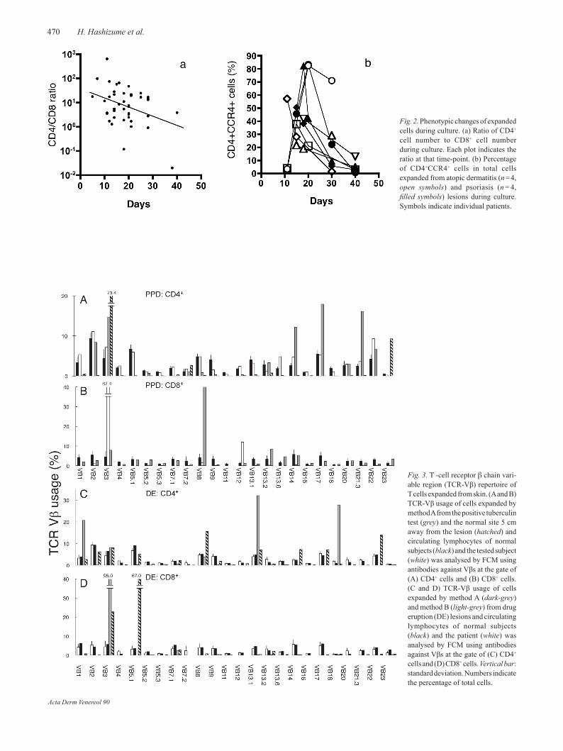
Fig. 2. Phenotypic changes of expanded cells during culture. (a) Ratio of CD4+ cell number to CD8+ cell number during culture. Each plot indicates the ratio at that time-point. (b) Percentage of CD4+CCR4+ cells in total cells expanded from atopic dermatitis (n = 4, open symbols) and psoriasis (n = 4, filled symbols) lesions during culture. Symbols indicate individual patients.
Fig. 3. T -cell receptor β chain vari-able region (TCR-Vβ) repertoire of T cells expanded from skin. (A and B) TCR-Vβ usage of cells expanded by method A from the positive tuberculin test (grey) and the normal site 5 cm away from the lesion (hatched) and circulating lymphocytes of normal subjects (black) and the tested subject (white) was analysed by FCM using antibodies against Vβs at the gate of (A) CD4+ cells and (B) CD8+ cells. (C and D) TCR-Vβ usage of cells expanded by method A (dark-grey) and method B (light-grey) from drug eruption (DE) lesions and circulating lymphocytes of normal subjects (black) and the patient (white) was analysed by FCM using antibodies against Vβs at the gate of (C) CD4+ cells and (D) CD8+ cells. Vertical bar: standard deviation. Numbers indicate the percentage of total cells.
Acta Derm Venereol 90

471Propagation of skin-resident T cells
CD19-CD20- cells, indicating T lymphocytes. A decrease in CD4/CD8 ratio was noted (Table I and Fig. 2a) with a transient increase in CD4+CCR4+ cells (Fig. 2b) during the culture in method A, but not in method B.
We established two cell lines by method A from the skin samples of a positive tuberculin lesion and clinically normal skin 5 cm away from the tuberculin lesion in the same individual (Fig. 3A and B). CD4+ cells from the tuberculin reaction showed preferential usage of Vβ3, Vβ14, Vβ17 and Vβ21.3, while those from normal skin predominantly expressed Vβ3 and Vβ23 (Fig. 3A). Among CD8+ cells, the expanded cells from the lesion highly expressed Vβ8 (Fig. 3B), whereas all the cells from the normal skin site were negative for CD8. This suggests that CD8+ T cells ac-cumulating in inflamed skin expand easily compared with normal skin.
We next compared the Vβ usage of cells expanded from drug eruptions using methods A and B at 21 days (Fig. 3C and D). We observed divergence in the T-cell receptor repertoire of expanded cells between the two methods. While CD4+ cells bearing Vβ1, Vβ13.1 and Vβ18 proliferated preferentially by method A, those positive for Vβ8, Vβ14 and Vβ22 expanded significantly by method B (Fig. 3C). Additionally, we found that 95% of CD8+ cells were positive for Vβ3 in method A, and 65% were Vβ5.1 in method B (Fig. 3D). The highly skewed TCR-Vβ repertoire of the skin-derived cells and their remarkable difference from the circulating lymphocytes suggest special compartmentalization of skin-specific T cells.
Relevance of lymphocytes derived from inflammatory skin lesions
To investigate whether the lymphocytes expanded from skin lesions by these two methods were pathogenic, we investigated the immune responses of expanded lymp-hocytes to causative antigens in the tuberculin reaction and drug eruption. Despite an absence of proliferative response in the culture without PPD or APCs, irrespec-tive of their CD4 or CD8 expression, CFSE-labelled cells expanded from the tuberculin reaction by method A were proliferating in response to PPD antigens, but not to tetanus toxoid (TT) in the presence of autolo-gous APCs (Fig 4a). The cells expanded from normal skin in the tested individual did not respond to PPD. In the same way, we found drug-specific proliferation of skin-resident cells from salazosulfapyridine-induced eruption lesion by method B (Fig 4b), although CD4+ cells predominantly expanded rather than CD8+ cells (data not shown). These observations indicated that the cells expanded from skin lesions by our methods contained antigen-specific T cells in the antigen-driven skin diseases.
Transient expansion of CD4+CD25+Foxp3+ cells by high level of IL-2 and anti-CD3/CD28 microbeads
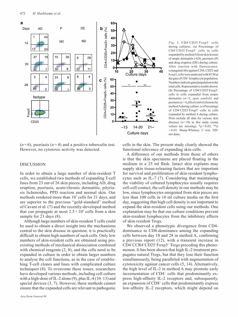
A high level of IL-2 promotes vigorous expansion of natural regulatory T cells (Tregs), being parallel to augmentation of an anti-tumour effect in patients with cancer (5, 6). Because of the high IL-2 level in method A, we investigated the number of Tregs among CD4+ cells expanded from various disease skins including AD (n = 5) and psoriasis (n = 4) by enumeration of cells co-expressing CD25 and Foxp3 (6). In method A, CD4+CD25+Foxp3+ cell population was 5.0% ± 4.7% (mean ± SD) at day 10–13 (Fig 5A–C) and significantly increased to 23.2% ± 17.4% from day 14 to day 20, while they decreased to 7.9% ± 6.7% after day 21 and disappeared after day 35. In cultures from AD lesions, comparable increments of CD4+CD25+Foxp3+ cells were found between methods A (40.1%) and B (32%) at day 16.
Cytotoxic activity of blood lymphocytes against expanded skin-resident cells
We investigated the cytotoxic activity of blood lymp-hocytes against expanded cells from skin lesions of AD
Fig. 4. Antigen response of the expanded cells by (a) method A and (b) method B in inflammatory conditions. After labelling with 5-or 6-(N-succinimidyloxycarbonyl)-fluorescein 3’,6’-diacetate (CFSE), the expanded cells were cultured for 72 h with or without antigen-presenting cells (APCs) and in the presence of antigens, followed by FCM analysis. (a) Proliferation of the cells expanded by method A from positive tuberculin test site (TS) in response to PPD antigens. +: added; –: not added; TT: tetanus toxioid; N: cells expanded from normal site near the positive test site in the same individual. Number indicates percentage of total cells. (b) Proliferation of the cells expanded by method B from the drug eruption lesion in response to the culprit drug, salazosulfapyridine (drug, 0, 1 and 10 μg/ml). +: added; –: not added. Numbers indicate percentage of total cells.
Acta Derm Venereol 90
472 H. Hashizume et al.
(n = 6), psoriasis (n = 4) and a positive tuberculin test. However, no cytotoxic activity was detected.
DISCUSSION
In order to obtain a large number of skin-resident T cells, we established two methods of expanding T-cell lines from 23 out of 26 skin pieces, including AD, drug eruption, psoriasis, acute/chronic dermatitis, pityria-sis lichenoides, PPD reaction and normal skin. Our methods rendered more than 107 cells for 21 days, and are superior to the previous “gold-standard” method of Cavani et al. (7) and the recently-developed method that can propagate at most 2.5 × 10⁵ cells from a skin sample for 21 days (8).
Although large numbers of skin-resident T cells could be used to obtain a direct insight into the mechanisms central to the skin disease in question, it is practically difficult to obtain high numbers of such cells. Only low numbers of skin-resident cells are obtained using pre-existing methods of mechanical dissociation combined with chemical reagents (2, 8), and the cells need to be expanded in culture in order to obtain larger numbers to analyse the cell functions, as in the case of establis-hing T-cell clones and lines with complicated culture techniques (8). To overcome these issues, researchers have developed various methods, including cell culture with a high-dose of IL-2 alone (9), plus IL-4 (10–13) and special devices (3, 7). However, these methods cannot ensure that the expanded cells are relevant to pathogenic
cells in the skin. The present study clearly showed the functional relevance of expanding skin cells.
A difference of our methods from those of others is that the skin specimens are placed floating in the medium in a 25 ml flask. Intact skin explants may supply skin tissue-releasing factors that are important for survival and proliferation of skin-resident lympho-cytes such as IL-7 (7). Considering that maintaining the viability of cultured lymphocytes usually requires cell-cell contact, the cell density in our methods may be low, since lymphocytes emigrated from skin pieces are less than 100 cells in 10 ml culture media on the first day, suggesting that high cell density is not important to expand the skin-resident cells using our methods. One explanation may be that our culture conditions prevent skin-resident lymphocytes from the inhibitory effects of skin-resident Tregs.
We observed a phenotypic divergence from CD4-dominance to CD8-dominance among the expanding cells between day 18 and 28 in method A, confirming a previous report (12), with a transient increase in CD4+CCR4+CD25+Foxp3+ Tregs preceding this pheno-menon. It has been shown that high IL-2 treatment pro-pagates natural Tregs, but that they lose their function simultaneously, being paralleled with augmentation of cytotoxicity against cancer cells (5, 14). Alternatively, the high level of IL-2 in method A may promote early incrementation of CD4+ cells that predominantly ex-press high-affinity IL-2 receptors and, subsequently, an expansion of CD8+ cells that predominantly express low-affinity IL-2 receptors, which might depend on
Fig. 5. CD4+CD25+Foxp3+ cells during cultures. (a) Percentage of CD4+CD25+Foxp3+ cells in cells expanded by method A from skin lesions of atopic dermatitis (AD), psoriasis (P) and drug eruption (DE) during culture. After reaction with fluorescence-conjugated Abs against CD4, CD25 and Foxp3, cells were analysed with FCM at the gate of CD4+ lymphocyte population. Numbers indicate gated population in the total cells. Representative results shown. (b) Percentage of CD4+CD25+Foxp3+ cells in cells expanded from atopic dermatitis (n = 5, open symbols) and psoriasis (n = 4, filled symbols) lesions by method A during culture. (c) Percentage of CD4+CD25+Foxp3+ cells in cells expanded by method A during culture. Plots include all data for various skin diseases (n = 10) in this study (some values are missing). *p < 0.02, **p < 0.03. Mann-Whitney U test. ND: not done.
Acta Derm Venereol 90

473Propagation of skin-resident T cells
their profile of IL-2 receptor expression (15). Although method A is a very simple way to obtain a large number of skin-resident cells, including Tregs, it must be noted that these cells may be phenotypically and functionally biased under Th2 cytokine condition. On the other hand, method B is useful for functional investigation of skin-resident lymphocytes, although repeated stimulations with anti-CD3/CD28 microbeads are necessary for propagation.
Easy isolation and expansion of skin-resident cells will enable further investigation of how T cells contri-bute to cutaneous pathology in skin diseases; the two methods described here will be a beneficial strategy for use in investigating this issue.
ACKNOWLEDGEMENTThis study was partially supported by funding from the Danish Dermatological Association.
The authors declare no conflict of interest.
REFERENCES
Wittmann M, Werfel T. Interaction of keratinocytes with in-1. filtrating lymphocytes in allergic eczematous skin diseases. Curr Opin Allergy Clin Immunol 2006; 6: 329–334.Schaerli P, Britschgi M, Keller M, Steiner UC, Steinmann 2. LS, Moser B, et al. Characterization of human T cells that regulate neutrophilic skin inflammation. J Immunol 2004; 173: 2151–2158.Clark RA, Chong BF, Mirchandani N, Yamanaka K, Murphy 3. GF, Dowgiert RK, et al. A novel method for the isolation of skin resident T cells from normal and diseased human skin. J Invest Dermatol 2006; 126: 1059–1070.Hashizume H, Takigawa M, Tokura Y. Characterization of 4. drug-specific T cells in phenobarbital-induced eruption. J Immunol 2002; 168: 5359–5368.Wei S, Kryczek I, Edwards RP, zou L, Szeliga W, 5. Banerjee M, et al. Interleukin-2 administration alters the CD4+FOXP3+ T-cell pool and tumor trafficking in
patients with ovarian carcinoma. Cancer Res 2007; 67: 7487–7494.Wu Y, Borde M, Heissmeyer V, Feuerer M, Lapan AD, 6. Stroud JC, et al. FOXP3 controls regulatory T cell fun-ction through cooperation with NFAT. Cell 2006; 126: 375–387.Cavani A, Nasorri F, Prezzi C, Sebastiani F, Albanesi C, 7. Girolomani G. Human CD4+ T lymphocytes with remar-kable regulatory functions on dendritic cells and nickel-specific Th1 immune responses. J Invest Dermatol 2000; 114: 295–302.Yamanaka K, Clark R, Rich B, Dowgiert R, Hirahara K, 8. Hurwitz D, et al. Skin-derived interleukin-7 contributes to the proliferation of lymphocytes in cutaneous T-cell lym-phoma. Blood 2006; 107: 2440–2445.Campbell JJ, Murphy KE, Kunkel EJ, Brightling CE, Soler 9. D, Shen z, et al. CCR7 expression and memory T-cell di-versity in humans. J Immunol 2001; 166: 877–884.Aiba S, Tagami H. In vitro propagation of tissue-infiltrating 10. lymphoid cells from lesional skin by culture in IL 2-contain-ing medium. Acta Derm Venereol 1986; 66: 391–397.Reinhold U, Kukel S, Goeden B, Neumann U, Wehrmann 11. W, Kreysel HW. Interleukin-4 promotes the expansion of skin-infiltrating lymphocytes from atopic dermatitis in vitro. J Invest Dermatol 1991; 96: 370–375.Kaltoft K, Bisballe S, Dyrberg T, Boel E, Rasmussen PB, 12. Thestrup-Pedersen K. Establishment of two continuous T-cell strains from a single plaque of a patient with mycosis fungoides. In Vitro Cell Dev Biol 1992; 28A: 161–167.Bang K, Lund M, Mogensen SC, Thestrup-Pedersen K. In 13. vitro culture of skin-homing T lymphocytes from inflam-matory skin diseases. Exp Dermatol 2005; 14: 391–397.Thestrup-Pedersen K, Parhar R, Wu K, Bertilsson PA, Meyer 14. B, Abu-Amero S, et al. Skin-homing CD8+ T lymphocytes show preferential growth in vitro and suppress CD4+ T-cell proliferation in patients with early stages of cutaneous T-cell lymphoma. Acta Derm Venereol 2007; 87: 118–126.Elkord E. Role of regulatory T cells in allergy: implications 15. for therapeutic strategy. Inflamm Allergy Drug Targets 2006; 5: 211–217.Ohashi Y, Takeshita T, Nagata K, Mori S, Sugamura K. 16. Differential expression of the IL-2 receptor subunits, p55 and p75 on various populations of primary periphe-ral blood mononuclear cells. J Immunol 1989; 143: 3548–3555.
Acta Derm Venereol 90