in vitro micronucleus induction by polymethyl methacrylate bone cement in cultured human lymphocytes
TRANSCRIPT
Genetic Toxicology E L S E V I E R Mutation Research 32l (1994) 133-137
In vitro micronucleus induction by polymethyl methacrylate bone cement in cultured human lymphocytes
M . P a o l a B i g a t t i a , , L a u r a L a m b e r t i a, F r a n c e s c o P a o l o R i z z i b, M a r i o C a n n a s c,
G i a m p i e t r o A l l a s i a d
a Dipartimento di Biologia Animale, Laboratori di Antropologia, Via Accademia Albertina 17, 10123 Turin, Italy, b U.S.S.L. 1-24, Via S. Secondo 29, Turin, Italy, c Dipartimento di Scienze Mediche, Facolt?t di Medicina di Novara,
a Dipartimento di Matematica, Unil~ersith di Torino, Turin, Italy
(Received 13 May 1993) (Revision received 29 October 1993)
(Accepted 29 October 1993)
Abstract
Human lymphocytes cultured in vitro were used to assess the ability of polymethyl methacrylate (PMMA), currently used in orthopaedic surgery as bone cement, to induce micronuclei in binucleated cells. The results of the study show a significant increase in the micronucleus frequency in treated cultures and therefore the genotoxic effect of PMMA bone cement or its ingredients (methyl methacrylate, dimethyl para-toluidinc and hydroquinone) usually present in self-curing methacrylate bone cement and released in small quantities after polymerisation. This effect is evident during the stage immediately after the polymerisation process, and after a certain period of time (5 days in our experimental model).
Key words." PMMA bone cement; Micronuclei; Lymphocyte culture; In vitro test
I. Introduct ion
Polymethyl methacrylate (PMMA, CAS No. 9011-17-7) is an acrylic resin which is widely used as a biomaterial due to its excellent biocompati- bility and haemocompatibility. As well as in den- tistry and ophthalmology, it is primarily used in the biomedical field together with other polymers in the production of hollow fibres used for the dialysis process. It is also widely used in or-
* Corresponding author. Tel. 011/8122374; Fax 011/8124561.
thopaedic practice as a cement for arthroprosthe- ses.
Whereas there is an extensive literature on the biocompatibility and haemocompatibil i ty of the material, reports concerning its possible genotoxi- city are (to our knowledge) both scanty and in- conclusive. The appearance of local sarcomas in mice or rats following the implant of PMMA films or discs (including the orthopaedic formula- tion, Surgical Simplex P) has been highlighted by some authors (Stinson, 1964; Tomatis, 1966; La- vorgna et al., 1972). A more comprehensive re- view of this subject can be found in the 1979
0165-1218/94/$07.00 © 1994 Elsevier Science B.V. All rights reserved SSDI 0165-1218(93)E0131-7
134 M.P. Bigatti et al. /Mutat ion Research 321 (1994) 133-137
IARC monographs. Epidemiological studies (De- Wijn and Van Mullem, 1981) have confirmed the absence of tumours correlated with the implant of cemented prostheses, even after a number of years have elapsed. Since widespread use is still made of cemented prostheses despite the intro- duction onto the market of "non-cemented" prostheses using biological fixtures, the aim of the present study was to evaluate the genotoxic potential of the cement by reproducing in vitro an analogous situation to that observed in vivo, namely by adding a recently polymerised cement (PMMA bone cement) to lymphocyte cultures.
Our earlier studies (Cannas et al., 1987; Bigatti et al., 1989) on human lymphocytes cultured in vitro showed the inability of polymethyl methac- rylate to induce sister-chromatid exchange (SCE) in the orthopaedic formulation. The aim of the present study was to evaluate the possible geno- toxic effect of PMMA bone cement by using the micronucleus test as a complementary cytogenic end-point.
2. Materials and methods
2.1. PMMA bone cement
PMMA bone cement was prepared using Sur- gical Simplex P (Howmedica International Ltd., London, UK) marketed as a powder to be mixed with a liquid just before use. The liquid mainly consists of methyl methacrylate (MMA) added to N,N-dimethyl-para-toluidine (DMPT), which serves to trigger the reaction, and hydroquinone, in the following proportions: 97.4% w/w, 2.6%
w / w and 75 + 15 ppm. The powder is sterile and consists of polymethyl methacrylate (15% w/w) and 75% w / w methyl methacrylate-styrene copolymer with the addition of barium sulphate U.S.P. (10% w/w) to make the final product radio-opaque. Spherical samples of PMMA bone cement, with a mean weight of 0.14 g (range 0.09-0.27 g), were obtained by mixing the cement and the liquid mechanically in sterile conditions. Heavier samples (0.7-1.0 g) caused insufficient cell growth or the death of cells. The autocat- alytic reaction is triggered off and completed within the space of a few minutes and the final product has the appearance of a solid mass.
2.2. Micronuclei (MN)
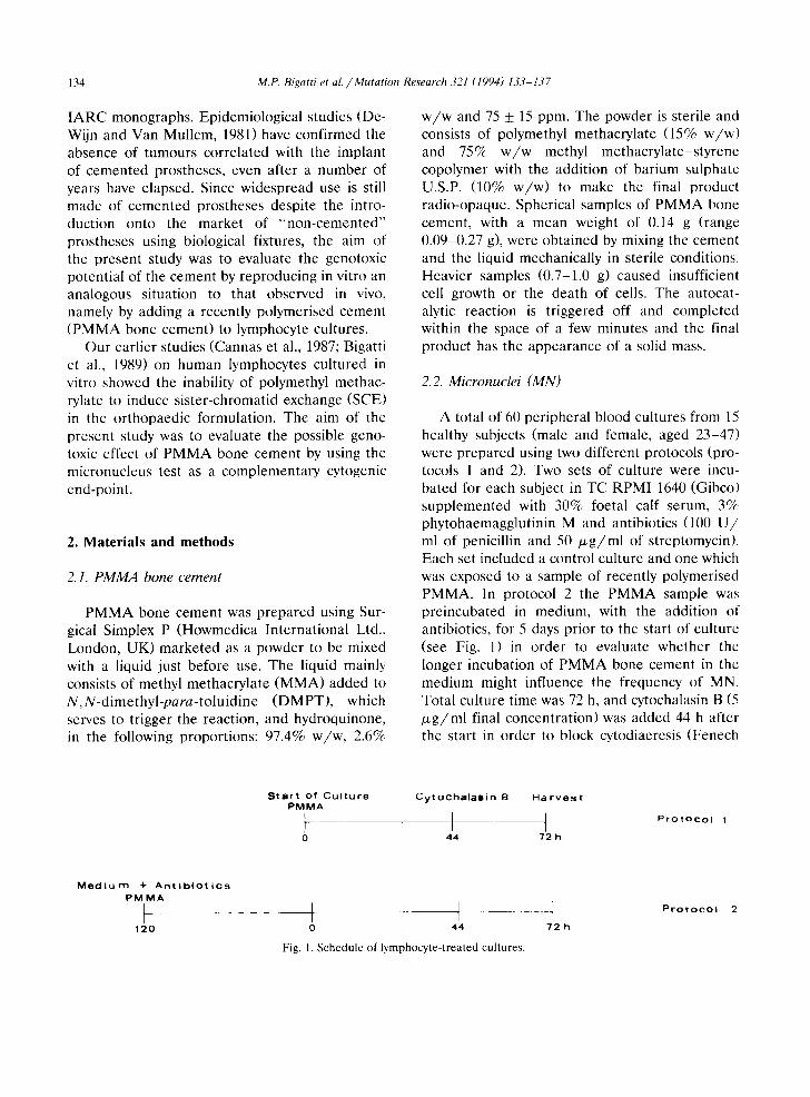
A total of 60 peripheral blood cultures from 15 healthy subjects (male and female, aged 23-47) were prepared using two different protocols (pro- tocols 1 and 2). Two sets of culture were incu- bated for each subject in TC RPMI 1640 (Gibco) supplemented with 30% foetal calf serum, 3% phytohaemagglutinin M and antibiotics (100 U / mI of penicillin and 50 # g / m l of streptomycin). Each set included a control culture and one which was exposed to a sample of recently polymerised PMMA. In protocol 2 the PMMA sample was preincubated in medium, with the addition of antibiotics, for 5 days prior to the start of culturc (see Fig. 1) in order to evaluate whether the longer incubation of PMMA bone cement in the medium might influence the frequency of MN. Total culture time was 72 h, and cytochalasin B (5 ~ g / m l final concentration) was added 44 h after the start in order to block cytodiaeresis (Fenech
S t a r t o f C u l t u r e C y t o c h a l a l D i n B H a r v e s t P M M A
) i I 0 44 72 h
P r o t o c o l 1
M e d i u m + A n t i b i O t i C s P M M A
I . . . . .
12(1 t I I 0 44 72 h
Fig. I. Schedule of lymphocyte-treated cultures.
P r o t o c o l
M.P. Bigatti et al. /Mutation Research 321 (1994) 133-137 135
and Morley , 1985). A f t e r harves t the cu l tu res were cen t r i fuged , the pe l l e t ed cells were resus- p e n d e d in a hypo ton ic so lu t ion consis t ing of 1 pa r t R P M I 1640 med ium, 4 par t s d is t i l led wa te r and 2% foeta l cal f serum, and kep t at 25°C for 10 min. A f t e r cen t r i fuga t ion the s u p e r n a t a n t was fully r emoved and smear s made . The sl ides were left to dry be fo re be ing fixed with m e t h a n o l and acet ic acid ( 3 : 1 ) and s ta ined with G i e m s a 5% (using the m e t h o d of Van H u m m e l e n and Ki r sch -Volde r s (1990), sl ightly modif ied) . Sl ides were scored b l ind and 1000 b inuc l ea t ed cells were scored p e r cu l tu re to d e t e r m i n e the mic ronuc leus f requency. The c r i te r ia used to score micronuc le i were those normal ly r e p o r t e d in the l i t e ra tu re (Fenech , 1993). Posi t ive cont ro l cu l tu res were p r e p a r e d by add ing mi tomycin C (0.3 / x g / m l ) 48 h af te r the s tar t of cul ture .
2.3. Stat is t ical analysis
Firs t the Wi lcoxon s igned ranks test ( S Y S T A T sta t is t ical packages ) was used to c o m p a r e mi- c ronuc leus f r equenc ies in cont ro l and t r e a t ed cul- tures using the two d i f fe ren t protocols . Then in o r d e r to achieve a more eff ic ient use of data , a t - test was used to eva lua te the effects of t rea t - men t s and p ro toco l s using the fol lowing con t ras t s (see Tab le 1):
col l + c o l 2 - c o l 3 - c o l 4
p ro toco l 1 vs p ro toco l 2
col 1 - col 2 + col 3 - col 4
cont ro l vs P M M A
c o l l - c o l 2 - c o l 3 + c o l 4
test for the addi t iv i ty of effects
Since it can be seen tha t the s t a n d a r d devia- t ion of each co lumn increases app rox ima te ly in l ine with the square roo t of the mean , we first p e r f o r m e d a square root t r ans fo rma t ion to ho- mogen i se the co lumn var iances .
3. Results
The m e a n f requency of s p o n t a n e o u s micronu- clei f rom the 15 donors was 4.0 _+ 3.49 (p ro toco l
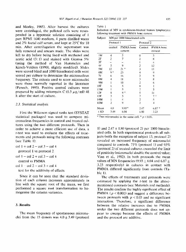
Table 1 Induction of MN in cytokinesis-blocked human lymphocytes following treatment with PMMA bone cement
Subject MN per 1000 binucleated cells
Protocol 1 Protocol 2
control PMMAbone Control PMMAbone cement cement
1M 4 5 0 3 2F 2 7 2 8 3F - - ! 4 4F 2 9 2 12 5M 12 21 ~ 3 11 6M 9 ~ 23 4 9 7F 6 16 2 9 8M 4 11 3 8 9M 1 10 2 6
10F 1 5 0 1 l lM 1 5 2 5 12F 8 9 ~' 5 11 13F 3 5 7 7 ~t 14M 1 4 2 5 15M 2 9 2 4
Mean 4.0 9.93 * 2.47 6.87 * + SD 3.49 6.04 1.80 3.23
a Two micronuclei in the same cell. * p < 0.01.
1) and 2.47 _+ 1.80 (p ro toco l 2) pe r 1000 b inucle- a ted cells. In both expe r imen ta l p ro toco ls all sub- jects (with the except ion of subject 13, p ro toco l 2) revea led an inc reased f requency of micronuc le i c o m p a r e d to controls . 71% (pro toco l 1) and 93% (pro tocol 2) of t r e a t e d cul tures exceeded the limit of posit ivi ty (micronucle i doub le the cont ro l value; Vian et al., 1992). In bo th p ro toco ls the mean values of M N f requenc ies (9.93 _+ 6.04 and 6.87 _+ 3.23 respect ively) in cu l tu res in contac t with P M M A di f fe red signif icant ly f rom cont ro ls (Ta- ble 1).
The effects of t r e a tme n t s and pro toco ls were e s t ima ted by apply ing the t - test to the above- m e n t i o n e d con t ras t s (see Mate r i a l s and methods) . The resul ts conf i rm the highly signif icant effect of P M M A ( p < 0.001) and suggest a d i f fe rence be- tween pro toco ls with p < 0.05 and no signif icant in terac t ion . The re fo re , a s ignif icant d i f fe rence be tween the re la t ive increases due to P M M A u n d e r the two d i f ferent p ro toco ls does not ap- p e a r to e m e r g e because the effects of P M M A and the p ro toco l are addi t ive.
136 M.P. Bigatti et a l . / Mutation Research 321 (1994) 133-137
It is worth noting that in both treated and control cultures using protocol 2 (prepared in medium kept with antibiotics at 37°C for 120 h) there was a decrease in the mean frequency of MN. The probability given by the t-test of less than p < 0.05 of chance occurrence points to some evidence of a real effect caused by the protocol. We arc not yet able to explain the reason for this decrease but if thc effect of the protocol is additive, as it would appear, both the control and treated groups should be equally affected.
No correlation was observed between the number of MN and the weight of samples.
In positive control cultures the mean fre- quency of MN per 1000 binucleated cells was 32.57 + 4.99.
4. Di scuss ion
After the polymerisation process small quanti- ties of ingredients usually present in self-curing methacrylate bone cements arc released and their rate of diffusion depends on storage conditions. A study by Brauer et al. (1977) revealed that, in air, the monomer content of the cured resins decreased from 3.3% after 1 h to 2.4% after 215 days. In water, at 37°C, over 65% of the ex- tractable monomer is leached out within 10 rain. After 6 h, the diffusion rate of monomer into aqueous phase was low. Residual peroxide de- creases from 0.8% to 0.14% after 15 months. The hydroquinone content within 24 h after curing amounted to 9 ppm. DMPT (0.6% as much in cured cement) does not leach out. The monomer, which is responsible for the main toxic effects observed in the use of cemented prosthesis im- plants, is the ingredient which is released in the largest amount, and in fact 2 - 5 % of monomer is not polymerised. Individually these substances may be genotoxic. The genetic activity of MMA has been investigated using in vitro and in vivo systems (Hacmiya et al., 1982; P o s s e t al., 1984; Wagemakers et al., 1984; National Toxicology Program, 1986; Marez et al., 1991). However, the results remain in part contradictory. With regard to the genotoxicity of hydroquinone and benzene
metabolites in general, numerous data have been reported in the literature. A genotoxic effect is certainly shown both on germinal (Ciranni and Adler, 1991) and somatic (Tunek et al., 1982; Ciranni et al., 1988; Barale et al., 199(I) cells. The use of anti-kinetochore antibody also appears to reveal that the mechanism of action occurs at the level of the mitotic spindle (Robertson ct al., 1991).
The present study shows a highly significant increase in MN frequency in human lymphocytes treated with PMMA (Surgical Simplex P) and consequently a genotoxic effect of this substance or of the aphorised residual ingredients which continue to be released in small amounts from the polymer. A lack of SCE induction, but a significant decline in the proliferation rate index was observed in our earlier analogous experi- ments in which human lymphocytes cultured in vitro were treated with PMMA bone cement (Bi- gatti et al., 1989).
The different results obtained in the SCE and MN tests could be due to differences in thc sensitivity of the two assay systems. Nevertheless, the MN incidence, a clastogenic and aneugenic end-point, has a specificity which is distinct from SCE. It is therefore possible that PMMA, under the conditions tested, broke chromosomes a n d / o r impaired the spindle apparatus which develop into MN. At all events, these experimental data do not allow us to establish whether MN are a consequence of aneuploidy or breakage.
Acknowledgements
This work was supported by Regione Piemonte (Progetto finalizzato No. 197, 1989). The authors wish to thank the referees for their valuable comments and suggestions, in particular with re- gard to the statistical analyses.
References
Baralc, R., A. Marazzini, C. Betti, V. Vangelisti, N. Loprieno and 1. Barrai (1990) Genotoxicity of two metabolites of benzene: phenol and hydroquinone show strong synergistic effects in vivo, Mutation Res., 244, 15 2(1.
M.P. Bigatti et aL /Mutation Research 321 (1994) 133-137 137
Bigatti, M.P., L. Lamberti, M. Cannas and E. Rossi (1989) Lack of sister-chromatid exchange induction by polymethyl methacrylate bone cement in human lymphocytes cultured in vitro, Mutation Res., 227, 21-24.
Brauer, G.M., D.J. Termini and G. Dickson (1977) Analysis of the ingredients and determination of the residual compo- nents of acrylic bone cements, J. Biomed. Mater. Res., 11, 577-607.
Cannas, M., P. Bigatti, E. Rossi and P. Rossi (1987) In vitro research on the possibility of chromosomal damage caused by polymethyl methacrylate in orthopaedics, It. J. Orthop. Traumat., XIII, 387-391.
Ciranni, R., and I.-D. Adler (1991) Clastogenic effects of hydroquinone: induction of chromosomal aberrations in mouse germ cells, Mutation Res., 263, 223-229.
Ciranni, R., R. Barale, G. Ghelardini and N. Loprieno (1988) Benzene and the genotoxicity of its metabolites. II. The effect of the route of administration on the micronuclei and bone marrow depression in mouse bone marrow cells, Mutation Res., 209, 23-28.
DeWijn, J.R., and P.J. Van Mullem (1981) Biocompatibility of acrylic implants, in: D.F. William (Ed.), Biocompatibility of Clinical Implant Materials, Vol. 2, CRC Press, Boca Raton, FL, pp. 99-125.
Fenech, M. (1993) The cytokinesis-block micronucleus tech- nique: a detailed description of the method and its appli- cation to genotoxicity studies in human populations, Muta- tion Res., 285, 35-44.
Fenech, M., and A.A. Morley (1985) Measurement of mi- cronuclei in lymphocytes, Mutation Res., 147, 29-36.
Hacmiya, N., A. Taketami and Y. Takizawa (1982) Mutagenic- ity of environmental substances, Nippon Koshu Eisei Zasshi, 29, 236-239.
IARC (1979) Monographs on the Evaluation of the Carcino- genic Risk of Chemicals to Humans, Vol. 19, Methyl Methacrylate and Polymethyl Methacrylate, International Agency for Research on Cancer, Lyon, pp. 187-211.
Lavorgna, J.J., N.A. Burstein, A.L. Schiller and W.H. Harris (1972) The carcinogenesis of plastics used in orthopedic
surgery. An assessment of the incidence in rats and the possible relevance to man, Clin. Orthop. Rel. Res., 88, 223-227.
Marez, T., P. Shirali, H.F. Hildebrand and J.M. Haguenoer (1991) Increased frequency of sister chromatid exchange in workers exposed to high doses of methylmethacrylate, Mutagenesis, 6, 127-129.
National Toxicology Program (1986) Toxicology and carcino- genesis studies of methylmethacrylate in F344/n rats and B6C3F mice (inhalation studies), National Toxicology Pro- gram Technical Report Series.
Poss, R., W.G. Thilly and D.A. Kaden (1979) Methyl- methacrylate is a mutagen for Salmonella typhimurium, J. Bone Joint Surg., 61, 1203-1207.
Robertson, M.L., D.A. Eastmond and M.T. Smith (1991) Two benzene metabolites, catechol and hydroquinone, produce a synergistic induction of micronuclei and toxicity in cul- tured human lymphocytes, Mutation Res., 249, 201-209.
Stinson, N.E. (1964) The tissue reaction induced in rats and guinea-pigs by polymethylmethacrylate (acrylic) and stain- less steel (18/8/Mo), Br. J. Exp. Pathol., 45, 21-29.
Tomatis, L. (1966) Subcutaneous carcinogenesis by C and H labelled polymethylmethacrylate films, Tumori, 52, 165- 172.
Tunek, A., B. Hogstedt and T. Olofsson (1982) Mechanism of benzene toxicity. Effects of benzene and benzene metabo- lites on bone marrow cellularity, number of granulopoietic stem cells and frequency of micronuclei in mice, Chem.- Biol. Interact., 39, 128-138.
Van Hummelen, P., and M. Kirsch-Volders (1990) An im- proved method for the in vitro micronucleus test using human lymphocytes, Mutagenesis, 5, 203-204.
Vian, L., N. Bichet and D. Gouy (1993) The in vitro micronu- cleus test on isolated human lymphocytes, Mutation Res., 291, 93-102.
Waegemakers, T.H.J.M., and M.P.M. Bensik (1984) Non- mutagenicity of 27 aliphatic acrylate esters in the Salmonella microsome test, Mutation Res., 137, 95 102.