in vitro differentiation of retinal cells from human...
TRANSCRIPT
3169Research Article
IntroductionEmbryonic stem (ES) cells are pluripotent cells derived from theinner cell mass of blastocyst stage embryos that can maintain anundifferentiated state indefinitely and differentiate into derivativesof all three germ layers: the ectoderm, mesoderm and endoderm(Evans and Kaufman, 1981). The pluripotency of ES cells has raisedthe possibility that they might be used to treat various degenerativediseases. However, the clinical application of human ES celltherapy faces ethical difficulties concerning the use of humanembryos, as well as tissue rejection following implantation. Oneway to circumvent these issues is to generate pluripotent cellsdirectly from somatic cells. Development normally proceedsirreversibly from embryo to adult as cells progressively differentiateinto their final, specialized cell types. Remarkably, adult somaticcells can be reprogrammed and returned to the naive state ofpluripotency found in the early embryo simply by forcing expressionof a defined set of transcription factors. Forced expression of justthree transcription factors, Oct4, Sox2 and Klf4, can reprogramsomatic cells into induced pluripotent stem (iPS) cells (Nakagawaet al., 2008; Takahashi et al., 2007; Takahashi and Yamanaka, 2006;Yu et al., 2007). iPS cells have been shown to be functionallyequivalent to ES cells, as they express ES cell markers, have similargene expression profiles, form teratomas, and contribute to all celltypes in chimeric animals, including the germ line (Maherali et al.,2007; Okita et al., 2007; Wernig et al., 2007). These propertiesmakes ES cells and iPS cells an attractive potential donor source
for cell replacement therapies in tissues damaged by disease or injury(Hanna et al., 2007; Lindvall and Kokaia, 2006; Osakada andTakahashi, 2009; Wernig et al., 2008).
The adult mammalian central nervous system (CNS) containsendogenous neural stem cells that are capable of proliferation anddifferentiation, with newly generated neurons in the hippocampusand olfactory bulbs integrating into pre-existing neural circuits(Lledo et al., 2006; Zhao et al., 2008). In the adult retina, Müllerglia act as endogenous progenitors in response to injury, but aretoo few in number to restore function after damage has occurred(Ooto et al., 2004; Osakada et al., 2007). Thus, transplantation ofdonor cells to replace damaged or lost cells is a promising approachfor regeneration therapy. Retinas with photoreceptor degenerationcan be repaired by transplantation of photoreceptor precursors orES cell-derived progenitors, which are able to form synapticconnections to the host retina and improve visual function (Lambaet al., 2009; MacLaren et al., 2006). In addition, transplantation ofES cell-derived retinal pigment epithelia (RPE) has been reportedto improve visual function in RPE degeneration diseases, such asage-related macular degeneration (Haruta et al., 2004; Lund et al.,2006). Unlike somatic stem cells, ES cells and iPS cells are ableto propagate indefinitely. If photoreceptor and/or RPE cells couldbe differentiated from human ES cells or iPS cells under definedconditions, and the safety of their transplantation could be ensured,this approach would represent enormous potential for therapeutictreatment of retinal degeneration.
The use of stem-cell therapy to treat retinal degeneration holdsgreat promise. However, definitive methods of retinaldifferentiation that do not depend on recombinant proteinsproduced in animal or Escherichia coli cells have not beendevised. Here, we report a defined culture method using low-molecular-mass compounds that induce differentiation ofhuman embryonic stem (ES) cells and induced pluripotent stem(iPS) cells into retinal progenitors, retinal pigment epitheliumcells and photoreceptors. The casein kinase I inhibitor CKI-7,the ALK4 inhibitor SB-431542 and the Rho-associated kinaseinhibitor Y-27632 in serum-free and feeder-free floatingaggregate culture induce retinal progenitors positive for RX,MITF, PAX6 and CHX10. The treatment induces hexagonalpigmented cells that express RPE65 and CRALBP, form ZO1-positive tight junctions and exhibit phagocytic functions.Subsequent treatment with retinoic acid and taurine induces
photoreceptors that express recoverin, rhodopsin and genesinvolved in phototransduction. Both three-factor (OCT3/4,SOX2 and KLF4) and four-factor (OCT3/4, SOX2, KLF4 andMYC) human iPS cells could be successfully differentiated intoretinal cells by small-molecule induction. This method providesa solution to the problem of cross-species antigeniccontamination in cell-replacement therapy, and is also usefulfor in vitro modeling of development, disease and drugscreening.
Supplementary material available online athttp://jcs.biologists.org/cgi/content/full/122/17/3169/DC1
Key words: ES cell, Differentiation, iPS cell, Regeneration, Retina,Transplantation
Summary
In vitro differentiation of retinal cells from humanpluripotent stem cells by small-molecule inductionFumitaka Osakada1,2,3,*, Zi-Bing Jin1, Yasuhiko Hirami1, Hanako Ikeda1,2, Teruko Danjyo2, Kiichi Watanabe2,Yoshiki Sasai2 and Masayo Takahashi11Laboratory for Retinal Regeneration, Center for Developmental Biology, RIKEN, 2-2-3 Minatojima-minamimachi, Chuo-ku, Kobe 650-0047, Japan2Organogenesis and Neurogenesis Group, Center for Developmental Biology, RIKEN, 2-2-3 Minatojima-minamimachi, Chuo-ku, Kobe 650-0047,Japan3Systems Neurobiology Laboratory, The Salk Institute for Biological Studies, 10010 North Torrey Pines Road, La Jolla, CA 92037, USA*Author for correspondence ([email protected])
Accepted 19 June 2009Journal of Cell Science 122, 3169-3179 Published by The Company of Biologists 2009doi:10.1242/jcs.050393
Jour
nal o
f Cel
l Sci
ence
3170
However, the use of human pluripotent stem cells as a donor cellsource for transplantation therapy requires defined and controlleddifferentiation conditions. Although much progress has been madein the differentiation and propagation of human ES cells, definitivemethods of retinal differentiation have not been devised (Hirano etal., 2003; Ikeda et al., 2005; Lamba et al., 2006; Osakada et al.,2008; Osakada et al., 2009; Zhao et al., 2002). Previously, weshowed that retinal progenitors, photoreceptors and RPE cells canbe generated from ES cells by mimicking developmental processesin a stepwise fashion in vitro (Ikeda et al., 2005; Osakada et al.,2008; Osakada et al., 2009). However, this culture method requiresthe addition of recombinant Dkk1 and Lefty-A (also known asLefty2) proteins, which are produced in animal cells or E. coli,raising the possibility of infection or immune rejection due to cross-species contamination.
By contrast, using chemical compounds to induce differentiationoffers several advantages compared with using recombinantproteins. Not only are they non-biological products, but they showstable activity, have small differences between production lots, andare low-cost. Thus, establishment of chemical compound-basedculture systems will be necessary for human pluripotent cell-basedtransplantation therapies (Ding and Schultz, 2004). Here we showthat chemical compounds can induce retinal progenitors,photoreceptors, and RPE cells from human ES and iPS cells.
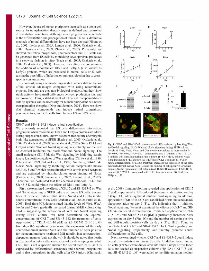
ResultsCKI-7 and SB-431542 induce retinal specificationWe previously reported that ES cells differentiate into retinalprogenitors when recombinant Dkk1 and Lefty-A proteins are addedduring suspension culture, known as serum-free culture of embryoidbody-like aggregates, or SFEB (Ikeda et al., 2005; Osakada et al.,2008; Osakada et al., 2009; Watanabe et al., 2005). Since Dkk1 andLefty-A inhibit Wnt and Nodal signaling, respectively, we focusedon chemical inhibitors that block Wnt and Nodal signaling. Thesmall molecule CKI-7 blocks Wnt signaling by inhibiting caseinkinase I, a positive regulator of Wnt signaling (Chijiwa et al., 1989;Peters et al., 1999; Sakanaka et al., 1999). Similarly, SB-431542blocks Nodal signaling by inhibiting activin receptor-like kinase(ALK)4, 5 and 7, which heterodimerize with activin type I receptorsand are activated by phosphorylation upon binding of Nodal(Eiraku et al., 2008; Inman et al., 2002; Laping et al., 2002).Therefore, we postulated that the chemical inhibitors CKI-7 andSB-431542 could mimic the effects of Dkk1 and Lefty-A.
First, we examined the effects of CKI-7 and SB-431542 on Wntand Nodal signaling in SEFB culture of mouse ES cells. Severallines of evidence indicate that Wnts, Nodal and Cripto inhibitneural commitment in ES cells (Aubert et al., 2002; Parisi et al.,2003). Real-time PCR demonstrated that the levels of Wnt1, Wnt3,Nodal and Cripto gradually increased during SFEB culture (Fig.1A-D), suggesting the activation of Wnt and Nodal signalingduring SFEB culture. We next determined the optimalconcentrations of CKI-7 and SB-431542 for treatment of cells.Application of CKI-7 (0.1-10 μM) or SB-431542 (0.1-10 μM)from days 0-5 significantly increased the expression of the earlyneuroectodermal marker Sox1 and the number of cells positivefor the neural markers nestin and βIII-tubulin, in a concentration-dependent manner (data not shown). It should be noted that nestinis expressed in mitotically active areas of the developing and adultCNS, but is not a specific marker for neural stem cells, as it isexpressed by differentiated astrocytes and neuronal progenitorsand is also upregulated in glial cells after CNS injury (Chojnacki
et al., 2009). Immunoblotting revealed that application of CKI-7(5 μM) suppressed SFEB-induced β-catenin stabilization on day5 (Fig. 1E), indicating that it inhibited Wnt signaling. In addition,application of SB-431542 (5 μM) abolished SFEB-induced Smad2phosphorylation on day 5 (Fig. 1F), indicating that it inhibitedNodal signaling. We next examined the effects of CKI-7 and SB-431542 on neural differentiation. Combined application of CKI-7 (5 μM) and SB-431542 (5 μM) significantly increased Sox1expression on day 5 (Fig. 1G) and the number of nestin-positiveand βIII-tubulin-positive cells on day 8 (Fig. 1H,I). Thus, weconclude that CKI-7 and SB-431542 block Wnt signaling andNodal signaling, respectively, and thereby promote neuraldifferentiation of ES cells.
Next, we examined whether CKI-7 and SB-431542 could induceneural differentiation in human ES cells. Undifferentiated humanES cells (khES-1) were dissociated into small clumps of five to tencells and seeded as suspension cultures (Fig. 2A). CKI-7 (5 μM)and SB-431542 (5 μM) were added to the differentiation medium
Journal of Cell Science 122 (17)
Fig. 1. CKI-7 and SB-431542 promote neural differentiation by blocking Wntand Nodal signaling. (A-D) Wnt and Nodal signaling during SFEB culture.Levels of Wnt1, Wnt3, Nodal and Cripto were normalized to those on day 0.*P<0.05, **P<0.01, ***P<0.001, compared to day 0 (Dunnett’s test). (E) CKI-7 inhibits Wnt signaling during SFEB culture. (F) SB-431542 inhibits Nodalsignaling during SFEB culture. (G-I) Effects of CKI-7 and SB-431542 onneural differentiation. SFEB/CS treatment increases the expression of the earlyneuroectodermal marker Sox1 (G) and the number of cells positive for neuralmarkers Nestin (green) and βIII-tubulin (red; H, SFEB treatment; I, SFEB/CStreatment). **P<0.01, compared with SFEB (unpaired t-test, G). Scale bar,100 μm (H,I).
Jour
nal o
f Cel
l Sci
ence

3171Retinal specification by small molecules
of the suspension culture for 21 days (SEFB/CS culture). Inaddition, to improve cell survival during differentiation, the Rho-associated kinase inhibitor Y-27632 (10 μM), which preventsdissociation-induced cell death in human ES cells (Watanabe et al.,2007), was added 1 hour before dissociation and was maintained
in the differentiation medium during the first 15 days of floatingculture. Under these conditions, ES cells formed embryoid body-like aggregates. Expression of the undifferentiated ES cell markersNANOG and OCT3/4 decreased during the suspension culture.SEFB/CS treatment significantly reduced the levels of NANOG andOCT3/4 on day 21 (Fig. 2B,C). Subsequently, these aggregates wereplated onto poly-D-lysine-laminin-fibronectin-coated slides on day21, and cultured until day 40 (Fig. 2D). Immunostaining revealedthat the neural progenitor marker nestin (NES) was stronglyexpressed by day 30, and 94.8±3.3% of colonies were positive forNES on day 40 (Fig. 2E). The neuronal marker βIII-tubulin wasrarely detected on or before day 30, and substantially increasedduring days 35-40. By day 40, 92.7±3.3% of colonies were positivefor βIII-tubulin (Fig. 2F,G). By contrast, expression of NANOG andOCT3/4 had disappeared by day 40.
To characterize SFEB/CS-induced neural tissues, we nextperformed quantitative RT-PCR for regional gene markers alongthe rostral-caudal axis. SFEB/CS treatment increased the expressionlevels of the rostral CNS markers BF1 (telencephalon), RX (retinaand diencephalon), and SIX3 (rostral diencephalon and brain tissuerostral to it), and decreased the caudal markers IRX3 (caudaldiencephalon and brain tissue caudal to it), GBX2 (rostral hindbrain),and HOXB4 (hindbrain and spinal cord), compared with SFEBtreatment (Fig. 2H-M). These results indicate that SFEB/CStreatment preferentially induces the rostral-most CNS in human EScells.
Following neural tube formation in vertebrates, progenitors inthe optic vesicle and the optic cup express Mitf in the outer layerthat will give rise to the RPE, Rx in the inner layer that will giverise to the neural retina, and Pax6 in both layers (Baumer et al.,2003; Furukawa et al., 1997; Ikeda et al., 2005; Mathers et al.,1997) (Fig. 2N). To determine whether CKI-7 and SB-431542promote retinal specification of ES cells, we examined theexpression of these markers in SFEB/CS-treated ES cells byimmunocytochemistry. After 30 days in SFEB culture, colonieswere rarely positive for MITF and RX. However, SFEB/CStreatment significantly increased the number of MITF-positive(MITF+) colonies (Fig. 2O,P; 32.5±3.0% of total colonies,22.8±3.1% of total cells). SEFB/CS treatment was as efficient astreatment with recombinant proteins Dkk1 (100 ng/ml) and Lefty-A (500 ng/ml; SFEB/DL; Fig. 2P). On day 35, 25.4±2.9% ofcolonies were RX+ in SFEB/CS culture (Fig. 2O). The inducedRX+ cells were frequently found in close proximity to MITF+ cells.
Fig. 2. Retinal specification of human ES cells by CKI-7 and SB-431542.(A) Schematic diagram of the culture procedure for retinal differentiation.(B,C) CKI-7 and SB-431542 decrease the expression of NANOG and OCT3/4,markers of the undifferentiated state. *P<0.05, **P<0.01, compared withSFEB (unpaired t-test). (D) Phase-contrast image of human ES cells treatedwith CKI-7 and SB-431542. (E-G) Human ES cells treated with CKI-7 andSB-431542 express the neural markers NES (red) and βIII-tubulin (green) onday 40. (H-M) Regional characterization of SFEB/CS-treated neural tissuesderived from human ES cells. Fold expression is the ratio of expression indifferentiated versus undifferentiated ES cells. (N) Multi-step commitment inthe development of retinal cells. Pluripotent stem cells derived from the innercell mass (blastocyst) differentiate into retinal progenitors corresponding tothose in the eye primordium (optic vesicle/optic cup) that give rise to RPE andphotoreceptors (adult retina). (O) RX+ and MITF+ retinal progenitor cellsdevelop from human ES cells under SFEB/CS culture conditions. (P) Effect ofCKI-7 and SB-431542 on the percentage of MITF+ colonies. **P<0.01,***P<0.001, compared with SFEB. NS, not significant (Tukey’s test).(Q) Formation of rosette-like clusters positive for PAX6. Scale bars: 300 μm(D), 100 μm (G,Q), 30 μm (O).
Jour
nal o
f Cel
l Sci
ence

3172
In addition, 79.2±5.1% of colonies were positive for PAX6, withsome forming rosette-like clusters in SFEB/CS cultures (Fig. 2Q).Thus, SFEB/CS treatment is able to induce retinal progenitors fromhuman ES cells.
Small molecule induced, ES-cell-derived retinal progenitorsare competent to differentiate into RPE and photoreceptorsNext, we examined whether SFEB/CS-induced retinal progenitorscould differentiate into RPE cells. On day 35, most MITF+ cellscoexpressed PAX6, consistent with the in vivo marker profile ofthe embryonic RPE (Fig. 3A). On day 40, pigmented cells appeared.On day 60, pigmented cells with the squamous and hexagonalmorphology characteristic of RPE cells were observed in SFEB/CScultures (26.0±4.0% of colonies, 18.1±1.9% of total cells; Fig. 3B).No significant difference in the frequency of pigment cell inductionwas found between SFEB/CS treatment and SFEB/DL treatment(Fig. 3C). Immunostaining with an anti-ZO1 antibody showed thatpigment cells derived from human ES cells formed tight junctionswith a polygonal morphology by day 100 (Fig. 3D). We alsoexamined the expression of genes related to the cellular functionsof RPE cells. SFEB/CS-treated human ES cells expressed bothRPE65 and CRALBP on day 120 (Fig. 3E,F). Retinal pigmentepithelium-specific protein 65 kDa (RPE65) is strongly expressedin RPE cells and is involved in the conversion of all-trans retinolto 11-cis retinal and in visual pigment regeneration, whereascellular retinaldehyde-binding protein (CRALBP; also known asRLBP1) is involved in vitamin A metabolism. Furthermore, in the
adult retina, RPE cells phagocytose the outer segment ofphotoreceptors to maintain photoreceptor function. We conducteda latex bead phagocytosis assay with our SFEB/CS-inducedpigmented cells, and showed that Phalloidin-stained polygonal cellsincorporated the latex beads (Fig. 3G). Thus, we conclude thatSFEB/CS-treated cells are competent to differentiate into pigmentcells with typical RPE characteristics.
Journal of Cell Science 122 (17)
Fig. 3. Differentiation of RPE from SFEB/CS-treated human ES cells. (A) SB-431542 and CKI-7 treatment induced MITF+ (green)/PAX6+ (red) RPEprogenitors from human ES cells. (B) Generation of polygonal pigment cells.(C) Effect of SB-431542 and CKI-7 on the percentage of pigment cells.***P<0.001, compared with SFEB. NS, not significant (Tukey’s test).(D) Tight junction formation of SFEB/CS-treated cells, as shown by anti-ZO1antibody staining (red). (E) Maturity of human ES-cell-derived pigment cells.RPE65, retinal pigment epithelium-specific protein 65 kDa; CRALBP, cellularretinaldehyde-binding protein. (F) Quantitative RT-PCR analysis of RPE65 inSFEB-CS-treated human ES cells. (G) Induced pigment cells have phagocyticfunction. Phalloidin-stained polygonal cells (green) incorporated latex beads(red). Scale bars: 30 μm (A,B,D), and 10 μm (G).
Fig. 4. Differentiation of photoreceptors from SFEB/CS-treated human EScells. (A,B) Quantitative PCR analysis for the photoreceptor precursor markerCRX (A) and the mature photoreceptor marker RCVRN (B). Fold expression isthe ratio of expression in differentiated versus undifferentiated human EScells. SFEB/CS-cultured human ES cells were treated with retinoic acid andtaurine (RA/T). (C,D) Immunostaining for the photoreceptor marker RHO. Anouter process (arrows) and inner process (arrowheads) are present inSFEB/CS+RA/T-treated cells (D). Scale bars: 30 μm (C,D). (E) Effect of CKI-7 and SB-431542 on the percentage of RHO+ cells. Treatment with retinoicacid and taurine (RA+T). **P<0.01, ***P<0.001, compared withSFEB+RA/T. NS, not significant (Tukey’s test). (F) RT-PCR analysis ofhuman ES cells treated with SFEB/CS+RA/T. Expression of photoreceptormarkers and phototransduction genes on days 100 and 140.
Jour
nal o
f Cel
l Sci
ence

3173Retinal specification by small molecules
We then asked whether SFEB/CS-induced retinal progenitorscould differentiate into retinal photoreceptors. We previouslyreported that the chemical compounds retinoic acid and taurine, bothof which are critical for photoreceptor development, promotephotoreceptor differentiation in an ES cell culture system (Osakadaet al., 2008). When we treated SFEB/CS-induced ES cells withretinoic acid (100 nM) and taurine (100 μM) beginning at day 90(SFEB/CS + RA/T treatment), the photoreceptor precursor markerCRX and the early photoreceptor markers NRL and recoverin
(RCVRN) were detected on day 100 (Fig. 4A,B). By day 140, thedifferentiated cells expressed the mature photoreceptor markerrhodopsin (RHO; Fig. 4C). Cultured photoreceptors do not formouter segments, but putative outer and inner processes wereobserved in human ES cell-derived RHO+ cells (Fig. 4D), andSFEB/CS treatment significantly increased the number of RHO+cells (20.1±3.9% of total colonies, 6.5±1.2% of total cells). Thedifferentiation efficiencies of SFEB/CS and SFEB/DL treatment didnot significantly differ (Fig. 4E). To determine the maturity ofinduced photoreceptors, we examined the expression of genesresponsible for phototransduction. Human ES cells treated withSFEB/CS + RA/T expressed RCVRN (rods and cones), phosducin(PDC, rods and cones), phosphodiesterases (PDE6b, rods; PDE6c,cones), RHO (rods), rhodopsin kinase (GRK1, rods), and arrestinS-antigen (SAG, rods) by day 140 (Fig. 4F). These results indicatethat the SFEB/CS + RA/T-treated ES cells are competent to respondto light. In addition, other types of retinal neurons were observedunder these culture conditions at low efficiency, includingHPC1+/PAX6+ amacrine cells, PKCα+ bipolar cells, andPAX6+/Islet1/2+ ganglion cells (<1%), as observed with SFEB/DLculture (Osakada et al., 2008). Thus, we conclude that SFEB/CS-induced retinal progenitors are competent to differentiate intophotoreceptors in response to retinoic acid and taurine.
Retinal differentiation of human induced pluripotent stem (iPS)cellsFinally, we determined whether CKI-7 and SB-431542 couldinduce retinal differentiation in human iPS cells. iPS cells (clone253G1) generated from human dermal fibroblasts by retroviral genetransfer of OCT3, SOX2, and KIF4 expressed pluripotent stem cellmarkers NANOG, OCT3/4, TRA-1-60, and TRA-1-81, but not pan-neural markers NES and βIII-tubulin (Fig. 5A,B; and data notshown). iPS cells were seeded as suspension cultures in thepresence of Y-27632 (10 μM, days 0-14), CKI-7 (5 μM, days 0-20), and SB-431542 (5 μM, days 0-20). Under these conditions,human iPS cells grew as floating aggregates, in a manner similarto human ES cells treated with Y-27632 (10 μM, days 0-14), CKI-7 (5 μM, days 0-20), and SB-431542 (5 μM, days 0-20). On day21, these aggregates were plated onto poly-D-lysine-laminin-fibronectin-coated slides. Immunostaining revealed that most(>80%) of the colonies are positive for the neural progenitor markersNES, βIII-tubulin, and NCAM on day 40 (Fig. 5C; and data notshown).
To characterize SFEB/CS-induced cells, we next performedquantitative RT-PCR for rostral-caudal CNS markers. SFEB/CS
Fig. 5. Retinal specification of human iPS cells by CKI-7 and SB-431542.(A,B) Expression of pluripotent cell markers NANOG and TRA-1-60 inhuman iPS cells. (C) Neural induction of human iPS cells by SFEB/CStreatment. (D-I) Regional characterization of SFEB/CS-treated iPS cells. Foldexpression is ratio of expression in differentiated versus undifferentiated iPScells. *P<0.05, compared with SFEB (unpaired t-test). (J-O) Time-courseanalysis of the expression of markers of the undifferentiated state, NANOG (J)and OCT3/4 (K) and retinal progenitor markers PAX6 (L), RX (M), MITF (N)and CHX10 (O) during SFEB/CS culture. Fold expression is ratio ofexpression on each day compared to day 0. *P<0.05, **P<0.01, ***P<0.001,compared to day 0 (Dunnett’s test). (P) Differentiation of RX+/PAX6+ neuralretina progenitors from SFEB/CS-treated human iPS cells. (Q) Differentiationof MITF+/PAX6+ RPE progenitors from SFEB/CS-treated iPS cells.(R) Effect of CKI-7 and SB-431542 on the percentage of MITF+ colonies.***P<0.001, compared with SFEB alone (unpaired t-test). (S) Formation ofrosette-like clusters positive for PAX6. Scale bar, 100 μm (A-C), 30 μm (P,Q),and 300 μm (S).
Jour
nal o
f Cel
l Sci
ence

3174
treatment promoted expression of the rostral CNS markers BF1(telencephalon), RX (retina and diencephalon) and SIX3 (rostraldiencephalon and more rostral brain tissue) and suppressedexpression of the caudal markers IRX3 (caudal diencephalon andmore caudal brain tissue), GBX2 (rostral hindbrain) and HOXB4(hindbrain and spinal cord; Fig. 5D-I). Time-course analysisdemonstrated that expression levels of the undifferentiated-statemarkers NANOG and OCT3/4 decreased by day 10 (Fig. 4J,K). Thelevels of the retinal progenitor markers PAX6, RX, MITF and CHX10peaked on days 30-40 and gradually declined thereafter (Fig. 5L-O). Immunostaining demonstrated that SFEB/CS treatment inducedretinal progenitors positive for both RX/PAX6 and MITF/PAX6 onday 35 (29.0±3.3% of colonies; Fig. 5P,Q). Cells positive for eitherRX (16.9±2.5% of total cells) or MITF (22.1±4.1% of total cells)were also generated. SFEB/CS treatment significantly increased thenumber of MITF+ colonies compared with SFEB treatment (Fig.5R; supplementary material Fig. S1B). PAX6+ rosette-like clusterswere also observed in SFEB/CS cultures (76.8±3.9% of colonies;Fig. 5S).
We then determined whether SFEB/CS-treated iPS cells coulddifferentiate into retinal cells. On day 40, pigment cells appearedin SFEB/CS cultures (29.1±4.0% of colonies). These cellsaccumulated more pigment and had adopted a polygonalmorphology with a squamous appearance by day 60 (27.2±4.4%of total cells; Fig. 6A; supplementary material Fig. S1C). Thesepigment cells formed polygonal actin bundles (Fig. 6B) and ZO1+tight junctions by day 90, and expressed RPE65 and CRALBP (Fig.6C), consistent with characteristics of the RPE. We also examinedphotoreceptor differentiation from SFEB/CS-treated iPS cells.Human iPS cells were treated with Y-27632 (10 μM, days 0-14),CKI-7 (5 μM; days 0-20), and SB-431542 (5 μM; days 0-20), andsubsequently with retinoic acid (100 nM; days 90-140) and taurine(100 μM; days 90-140). On day 120, 26.5±8.3% of total colonieswere immunopositive for the photoreceptor marker RCVRN inSFEB/CS + RA/T culture (Fig. 6D). On day 140, 5.4±1.9% of totalcells expressed RHO, a rod photoreceptor marker (Fig. 6E). Weperformed RT-PCR to test for expression of genes responsible forphototransduction. SFEB/DL- and RA/T-treated iPS cells expressedPDC, PDE6b, PDE6c, RHO, GRK1 and SAG, indicating thathuman iPS cell-derived photoreceptor cells possess the functionalcomponents required for light response. Taken together, theseresults indicate that small molecule induction of human iPS cellscan cause differentiation into retinal cells.
In addition, we compared the differentiation potential of four linesof human iPS cells (253G1, 253G4, 201B6, and 201B7) and oneline of human ES cells (khES-1). 253G1 and 253G4 were generatedby retroviral transduction of three factors, OCT3, SOX2, and KIF4(3F hiPSC), and 201B6 and 201B7 were generated by transductionof four factors, OCT3, SOX2, KIF4 and MYC (4F hiPSC). Thesepluripotent stem cells were treated with Y-27632 (10 μM, days 0-14), CKI-7 (5 μM; days 0-20) and SB-431542 (5 μM; days 0-20),and plated onto poly-D-lysine-laminin-fibronectin-coated slides onday 21. All lines of human iPS cells tested differentiated into neuralcells positive for NES and βIII-tubulin on day 40 (supplementarymaterial Fig. S1A). The khES-1 (hESC), 253G1, 253G4 (3FhiPSC) and 201B7 (4F hiPSC) lines generated pigment cells thatexpressed RPE65 and CRALBP (Fig. 6C; supplementary materialFig. 1B,C). However, 201B6 (4F hiPSC) did not differentiate intopigment cells. The efficiencies of MITF+ cell, ZO1+ cell, andpigment cell differentiation did not differ significantly betweenkhES-1, 253G1(3F hiPSC) and 201B7 (4F hiPSC) cells in SFEB/CS
culture (Fig. 6E). These results suggest that 201B6 is a pseudo iPScell colony or does not maintain pluripotency under our conditions,despite the expression of markers of the undifferentiated state. Thus,we conclude that the selection of iPS cell colonies rather than thepresence or absence of MYC affects the differentiation capacity ofiPS cells.
Taken together, our data show that the SFEB/CS method caninduce retinal specification in human iPS and ES cells.
Journal of Cell Science 122 (17)
Fig. 6. Generation of RPE and photoreceptors from both three- and four-factorhuman iPS cells. (A) Generation of pigment cells from human iPS cells inSFEB/CS cultures. (B) Phalloidin staining shows the polygonal shape of thepigment cells. (C) RT-PCR analysis for markers of the mature RPE, RPE65and CRALBP in two lines of human iPS cells. (D,E) Generation of RCVRN+(D) and RHO+ (E) photoreceptors from human iPS cells in SFEB/CS + RA/Tculture. (F) RT-PCR analysis of phototransduction genes in two lines of humaniPS cells. (G) Comparison of the differentiation of one human ES cell line(khES-1) and two human iPS cell lines (253G1 and 201B7). The 253G1 linewas generated by three-factor induction (OCT3/4, SOX2, and KLF4) and the201B7 line by four-factor induction (OCT3/4, SOX2, KLF4 and MYC). Scalebars: 30 μm (A,D,E) and 10 μm (B).
Jour
nal o
f Cel
l Sci
ence

3175Retinal specification by small molecules
DiscussionPhotoreceptor loss in retinal degeneration is the major cause ofblindness (Hartong et al., 2006; Rattner and Nathans, 2006). Celltransplantation of photoreceptors and/or RPE cells is one of themost promising therapeutic strategies for incurable retinaldegenerative diseases (MacLaren and Pearson, 2007; Osakada andTakahashi, 2009). However, the clinical application of celltransplantation is hampered by the fact that cells are often culturedwith materials from other animals, such as feeder cells, serum andrecombinant proteins, and this poses a risk in terms of adverseimmune responses and potential exposure to xenopathogens (Martinet al., 2005). In the present study, we have established a method ofinducing retinal differentiation of human ES cells and iPS cells usingthe chemical compounds CKI-7, SB-431542 and Y-27632. Thesechemical compounds are non-biological, do not trigger immuneresponses, have stable activity, show little difference betweenproduction lots, and are inexpensive. Therefore, retinaldifferentiation methods using chemical compounds are ideal forclinical applications. Our small molecule-based differentiationmethod provides a solution to the problem of cross-species antigeniccontamination in cell replacement therapy, and also contributes toin vitro modeling of development, disease and drug screening (Dingand Schultz, 2004; Pouton and Haynes, 2007).
In the present study, we conclude that inhibition of β-catenin(Wnt signaling) and pSmad (Nodal signaling) is important for retinalcell differentiation. To determine whether other signaling pathwaysmight be involved in retinal specification, we also tested the effectsof Shh, Wnt, BMP4, Nodal (without Lefty-A), IGF, FGF1, FGF2,and FGF antagonists (data not shown). However, addition of theseproteins showed only marginal effects, if any, on retinal celldifferentiation in our ES cell system. The patterning signals thatinduce the retinal primordia in the embryo have not yet beenelucidated. How Wnt and Nodal signaling pathways control theexpression of eye field transcription factors such as Six3, Pax6, Rx,Chx10 and Mitf deserve further investigation. In vitro methods ofstudying mouse and human pluripotent stem cells will pave the wayto understanding the molecular and cellular mechanisms of retinalspecification during development, and also bridge the gap betweenmouse embryology and human development.
We have used potent and specific inhibitors to block Wnt andNodal signaling. CKI-7 is a specific inhibitor of casein kinase I,and does not inhibit other kinases such as protein kinase A, proteinkinase C, Ca2+/CaM kinase II, and myosin light chain kinase atconcentrations as high as 100 μM (Chijiwa et al., 1989). SB-431542is a specific inhibitor of ALK4, 5 and 7, and has no effect on moredivergent ALK family members that recognize bone morphogeneticproteins (BMPs) (Inman et al., 2002). SB-431542 also exhibits noeffect on components of the ERK, JNK or p38 MAP kinasepathways (Laping et al., 2002). Accumulating evidence indicatesthat CKI-7 and SB-431542 suppress Wnt and Nodal signaling,respectively. However, we found that the differentiation efficiencyof CKI-7 and SB-431542 was lower than that of Dkk1 and Lefty-A, although the difference was not statistically significant. We alsoobserved that the aggregates formed by cells treated with CKI-7and SB-431542 were smaller than those made by cells treated withDkk1 and Lefty-A (data not shown). As far as we have examined,SB-431542 increased the expression levels of Hes5 and Hesr2,downstream components of Notch signaling (Louvi and Artavanis-Tsakonas, 2006), but not Hes1, Hesr1 or Hesr3 (supplementarymaterial Fig. S2). These observations raise the possibility that non-specific effects of SB-431542 might affect ES cell differentiation.
Identification of specific inhibitors should help our understandingof signaling pathways in retinal specification, and also contributeto establishment of efficient and selective differentiation methods(Ding and Schultz, 2004).
From the therapeutic point of view, direct reprogramming ofsomatic cells to generate iPS cells provides an invaluable resourcefor regenerative medicine, enabling the generation of patient-specific cells of any lineage without the use of embryonic material.We found that the efficiency of retinal differentiation of human iPScells was comparable to that of human ES cells. In addition, retinaldifferentiation of three-factor human iPS cells (OCT3/4, SOX2 andKLF4) was similar to that of four-factor human iPS cells (OCT3/4,SOX2, KLF4 and MYC), indicating that the differentiation capacityof human iPS cells does not depend on the specific combination ofreprogramming factors. One line of human iPS cells never generatedpigmented cells, suggesting that partial or aberrant reprogrammingresults in impaired ability to differentiate into the required cell type.Indeed, abnormal expression of a single gene, such as Nat1, Grb2,Apc or Nanog, renders ES cells refractory to differentiation(Yamanaka et al., 2000). Thus, we conclude that the selection andvalidation of iPS cells rather than the sets of reprogramming factorsused are critical for generation of iPS cells.
Another concern for transplantation therapy is the possibility oftumor formation as a result of contamination with undifferentiatedES cells or iPS cells (Choo et al., 2008; Fukuda et al., 2006). Thus,purification to remove undifferentiated cells is required for donorcell preparation. Moreover, although transgenes are largely silencedin iPS cells, reactivation of transgenes, in particular Myc, can leadto tumorigenesis (Okita et al., 2007). We have been able to generatemouse and human iPS cells using recombinant Oct4, Sox2, Klf4and Myc proteins without transfection of viral vectors or plasmids(Kim et al., 2009; Zhou et al., 2009). However, if human iPS cellscan be generated with only small molecules, feeder-cell-free,animal-product-free, and gene-insertion-free retinal cells could beobtained from patients’ cells using our small molecule inductionsystem. Thus, identification of small molecules that inducereprogramming is important for clinical grade preparation of iPScells.
In addition, choosing the proper cell type and stage for donorsis critical for successful transplantation. MacLaren et al. havedemonstrated that integration of donor rod photoreceptors in thehost retina requires rod photoreceptors of a corresponding stage topostnatal days 3-6 (MacLaren et al., 2006). These studies suggestthat the ontogenic stage of transplanted photoreceptors determinesthe ability of these cells to integrate into the diseased retina, furtherunderscoring the importance of cell type- and stage-specificpurification of differentiated ES and/or iPS cells. Selection ofspecific types of ES-cell-derived progenitors for transplantation intohost mice can be easily achieved using mouse ES cells withknocked-in fluorescence or antibiotic-resistance genes at specificmarker loci. However, knock-in technology is not suitable for humanES cells or iPS cells. Thus, identification of surface antigens markingpostnatal days 3-6 rod photoreceptors and purification of ES andiPS cell-derived rod photoreceptors corresponding postnatal days3-6 stage are crucial (Osakada and Takahashi, 2009).
Finally, the host environment is also crucial for photoreceptortransplantation (Fisher et al., 2005). Retinal degeneration ischaracterized by microglial activation and glial scar formation,which may impede integration and survival of transplanted cells.Robust integration of transplanted retinal cells into the retinas ofhost mice deficient in both vimentin and glial fibrillary acidic protein
Jour
nal o
f Cel
l Sci
ence

3176
has been reported (Kinouchi et al., 2003). Moreover, matrixmetalloproteases and chondroitinases that degrade the extracellularmatrix in the diseased retina aid in the integration of transplantedphotoreceptors (Suzuki et al., 2007; Suzuki et al., 2006). Disruptionof the outer limiting membrane also increases photoreceptorintegration following transplantation (West et al., 2008). Thesestudies indicate that the glial barrier in the host retina preventsintegration of donor photoreceptors. Therefore, in addition toimmunosuppression, the host retinal environment must bemodulated for successful transplantation.
In conclusion, this small molecule-based method provides asolution to the problem of cross-species antigenic contamination incell replacement therapy, which represents a significant step towardclinical application of human ES cell or iPS cell-basedtransplantation therapy for retinal diseases (Ding and Schultz, 2004).Additionally, patient-specific iPS cell-derived retinal cells willfacilitate the development of transplantation therapies withoutimmune rejection, and promote an improved understanding ofdisease pathogenesis (Dimos et al., 2008; Ebert et al., 2009; Parket al., 2008). For successful retinal regeneration, methods ofpurifying donor retinal cells and optimizing host conditions, as wellas use of animal models of human diseases to determine the efficacy(functional recovery) and safety (immune response and tumorformation) of treatments will be crucial.
Materials and MethodsMouse ES cell cultureMouse ES cells were maintained as described previously (Ikeda et al., 2005; Osakadaet al., 2008; Osakada et al., 2009; Ueno et al., 2006; Watanabe et al., 2005). For theSFEB (serum-free culture of embryoid body-like aggregates) method, ES cells wereincubated at 5�104 cells/ml in a bacterial-grade dish with differentiation medium[Glasgow minimal essential medium (GMEM), 5% KnockOut Serum Replacement(KSR), 0.1 mM non-essential amino acids, 1 mM pyruvate, and 0.1 mM 2-mercaptoethanol]. CKI-7 (0.1-10 μM; Sigma) or SB-431542 (0.1-10 μM; Sigma)was applied to the medium for 5 days while cells were in suspension culture.
Human ES cell cultureHuman ES cells were used in accordance with the human ES cell research guidelinesof the Japanese government. Human ES cells (khES-1) were maintained as previouslydescribed (Osakada et al., 2008; Osakada et al., 2009; Ueno et al., 2006). Briefly,undifferentiated human ES cells were maintained on a feeder layer of mitomycin-C-treated mouse embryonic fibroblasts in a humidified atmosphere of 2% CO2 and 98%air at 37°C. ES cells were passaged every 3-4 days.
For differentiation into retinal cells, ES colonies were treated with Y-27632 (10μM) for 1 hour, and dissociated into clumps (5-10 cells per clump) with 0.25% trypsinand 0.1 mg/ml collagenase IV in PBS containing 1 mM CaCl2 and 20% KSR. Feederswere removed by incubation of the ES cell suspension on a gelatin-coated dish. EScell clumps, at a density of 8.8�102 clumps/ml, were incubated in a non-adhesive2-methacryloyloxyethyl phosphorylcholine (MPC)-treated dish (Nunc) in DMEM/F-12 supplemented with 0.1 mM 2-mercaptoethanol, 0.1 mM non-essential amino acids,2 mM L-glutamine, and 20% KSR for 3 days, in 20% KSR-containing ESdifferentiation medium (GMEM, 0.1 mM non-essential amino acids, 1 mM pyruvate,and 0.1 mM 2-mercaptoethanol) for 3 days, then in 15% KSR-containing ESdifferentiation medium for 9 days, and finally in 10% KSR-containing ESdifferentiation medium for 6 days. Y-27632 (10 μM) was added for the first 15 daysof suspension culture. CKI-7 (5 μM) and SB-431542 (5 μM) were applied to themedium for 21 days during suspension culture. The medium was changed every 3days. ES cell aggregates were then re-plated en bloc on poly-D-lysine-laminin-fibronectin-coated eight-well culture slides (BD Biocoat) at a density of 15-20aggregates/cm2. In adherent cultures, cells were incubated in 10% KSR-containingES differentiation medium. For photoreceptor differentiation, SFEB/DL- or SFEB/CS-treated differentiated cells were further incubated in photoreceptor differentiationmedium [GMEM, 5% KSR, 0.1 mM non-essential amino acids, 1 mM pyruvate, 0.1mM 2-mercaptoethanol, N2 supplement, 100 nM retinoic acid (Sigma), 100 μMtaurine (Sigma), and 50 units/ml penicillin, 50 μg/ml streptomycin] for 50 days. Themedium was changed daily.
Human iPS cell cultureRetroviral transduction of Oct3/4, Sox2, Klf4, and Myc into human cells and theculture conditions for these cells were previously described (Hirami et al., 2009;Nakagawa et al., 2008; Takahashi et al., 2007). Briefly, the human iPS cell lines
253G1and 253G4 were established by retroviral transduction of OCT3/4, SOX2 andKLF4. The human iPS cell lines 201B6 and 201B7 were established by retroviraltransduction of OCT3/4, SOX2, KLF4 and MYC. Undifferentiated human iPS cellswere maintained on a feeder layer of mitomycin-C-treated SNL cells (a mousefibroblast STO cell line expressing the neomycin-resistance gene cassette and LIF)in DMEM-F-12 supplemented with 0.1 mM 2-mercaptoethanol, 0.1 mM non-essentialamino acids, 2 mM L-glutamine, 20% KSR, and 4 ng/ml basic fibroblast growthfactor (Upstate Biotechnology) in a humidified atmosphere of 5% CO2 and 95% airat 37°C. These iPS cells were passaged with 0.25% trypsin and 0.1 mg/ml collagenaseIV (Gibco) in PBS containing 1 mM CaCl2 and 20% KSR every 3-4 days.
For retinal differentiation, iPS colonies were treated with Y-27632 (10 μM) for 1hour, and dissociated into clumps (5-10 cells per clump) with 0.25% trypsin and 0.1mg/ml collagenase IV in PBS containing 1 mM CaCl2 and 20% KSR. Feeders wereremoved by incubation of the iPS cell suspension on a gelatin-coated dish for 1 hour.iPS cell clumps, at a density of 8.8�102 clumps/ml, were incubated in a non-adhesiveMPC-treated dish (Nunc) in DMEM-F-12 supplemented with 0.1 mM 2-mercaptoethanol, 0.1 mM non-essential amino acids, 2 mM L-glutamine, and 20%KSR for 3 days, in 20% KSR-containing ES differentiation medium (GMEM, 0.1mM non-essential amino acids, 1 mM pyruvate, and 0.1 mM 2-mercaptoethanol) for3 days, then in 15% KSR-containing ES differentiation medium for 9 days, and finallyin 10% KSR-containing ES differentiation medium for 6 days. Y-27632 (10 μM)was added for the first 15 days of suspension culture. CKI-7 (5 μM) and SB-431542(5 μM) were added to the medium for 21 days during suspension culture. The mediumwas changed every 3 days. Formed cell aggregates were then re-plated en bloc onpoly-D-lysine-laminin-fibronectin-coated eight-well culture slides (BD Biocoat) at adensity of 15-20 aggregates/cm2. In adherent cultures, cells were incubated in 10%KSR-containing ES differentiation medium. For photoreceptor differentiation,SFEB/CS-treated differentiated cells were further incubated in the photoreceptordifferentiation medium [GMEM, 5% KSR, 0.1 mM non-essential amino acids, 1 mMpyruvate, 0.1 mM 2-mercaptoethanol, N2 supplement, 100 nM retinoic acid (Sigma),100 μM taurine (Sigma), and 50 units/ml penicillin, 50 μg/ml streptomycin] for 50days. The medium was changed daily.
ImmunocytochemistryCells were immunolabeled as described previously (Ikeda et al., 2005; Mizuseki etal., 2003; Osakada et al., 2008; Osakada et al., 2009; Osakada et al., 2007; Ueno etal., 2006). The primary antibodies used were as follows: mouse anti-βIII-tubulin(1:500, Sigma), mouse anti-CD133 (1:100, Miltenyi Biotec), rat anti-Crx (1:200), ratanti-E-cadherin (1:50, Takara), mouse anti-microtubule-associated protein-2(a+b)(1:500, Sigma), mouse anti-Mitf (1:30, Abcam), goat anti-Nanog (1:20, R&D), mouseanti-N-cadherin (1:500, BD pharmingen), rabbit anti-NCAM (1:200, Chemicon),rabbit anti-nestin (1:1000, Covance), mouse anti-Oct3/4 (1:200, BD pharmingen),mouse anti-Pax6 (1:500, R&D), rabbit anti-Pax6 (1:600, Covance), mouse anti-rhodopsin (RET-P1, 1:2000, Sigma), rabbit anti-Rx (1: 200), mouse anti-TRA-1-60(1:200, Chemicon), anti-TRA-1-81 (1:200, Chemicon), and rabbit anti-ZO1 (1:100,Zymed). Antibodies against Crx and Rx were obtained as previously described (Ikedaet al., 2005). The secondary antibodies used were as follows: anti-mouse IgG, anti-rabbit IgG, anti-rat IgG, anti-goat IgG, and anti-mouse IgM antibodies conjugatedwith Cy3 or Cy2 (1:300, Jackson). For enhancement of immunoreactive signal,specimens were incubated with biotinylated secondary antibodies (1: 200, Vector),and then with Texas Red-Avidin or FITC-Avidin (1:1000, Vector). Cell nuclei werecounterstained with 4�,6-diamidino-2-phenylindole (DAPI; 1 μg/ml, MolecularProbes). Labeled cells were imaged with a laser-scanning confocal microscope (Zeiss).
Real-time PCRTotal RNA was extracted with the RNeasy kit (Qiagen), treated with RNase-freeDNase I, and reverse-transcribed with a first-strand cDNA synthesis kit (Amersham)as previously described (Osakada et al., 2008; Osakada et al., 2007). QuantitativePCR was performed with the StepOnePlus Real-Time PCR system (AppliedBiosystems). Specific primers and their corresponding probes were designed withthe Universal ProbeLibrary system (Roche). The expression levels were normalizedto those of β-actin. The primers used for quantitative PCR are listed in Table 1.
RT-PCR analysisTotal RNA was extracted with the RNeasy kit (Qiagen), treated with RNase-freeDNase I, and reverse-transcribed with a first-strand cDNA synthesis kit (Amersham)as previously described (Osakada et al., 2008; Osakada et al., 2007). The cDNA wasused as a template for PCR with ExTaq (Takara). Human adult retinal cDNA(Clontech) was used as a positive control. The PCR products were separated byelectrophoresis on an agarose gel and detected under UV illumination. The primersused for RT-PCR are listed in Table 2.
Western blot analysisCells were harvested and homogenized in ice-cold lysis buffer. After normalizationof protein concentrations and denaturation, samples were subjected to 4-12% sodiumdodecyl sulphate polyacrylamide gel electrophoresis (SDS-PAGE; Wako), followedby transfer to polyvinylidene difluoride membranes (GE). The membranes wereprobed with rabbit anti-Smad2/3 (1:1000; Cell Signaling), rabbit anti-phospho
Journal of Cell Science 122 (17)
Jour
nal o
f Cel
l Sci
ence

3177Retinal specification by small molecules
Smad2/3 (1:1000; Cell Signaling), or rabbit anti-β-catenin (1:1000; Upstate)antibodies, and then with horseradish peroxidase-conjugated goat anti-rabbit IgG(1:1000; Dako). The bound antibodies were detected with an enhancedchemiluminescence detection system (Amersham).
Phagocytosis assayCells were incubated in medium containing Cy3-conjugated 1 μm polystyrenemicrospheres at a concentration of 1.0�108 beads/ml for 6 hours at 37°C as describedpreviously (Osakada et al., 2008). For visualization of F-actin, the cells were stainedwith Alexa-Fluor-488-conjugated Phalloidin (Molecular Probes). The fluorescencesignal was observed with a laser-scanning confocal microscope.
Statistical analysisValues are expressed as means ± s.e.m. 100-200 colonies were examined in eachexperiment. All statistical analyses were performed using GraphPad PRISM version5.0 (GraphPad Software Inc.). The statistical significance of differences wasdetermined by one-way analysis of variance followed by Dunnett’s test or Tukey’stest, or with an unpaired t-test. Probability values less than 5% were consideredsignificant.
We thank H. Suemori and N. Nakatsuji (Kyoto University, Kyoto,Japan) for the human ES cell line, K. Takahashi and S. Yamanaka(Kyoto University) for the human iPS cell line, M. Kikkawa, A. Nomoriand K. Iseki for technical assistance, and members of the Takahashilaboratory and the Sasai laboratory for helpful discussions. This workwas supported by Grants-in-Aid from the Ministry of Education,Culture, Sports, Science and Technology, and the Leading Project (M.T).This study was also supported by Grants-in-Aid for Scientific Researchfrom the Japan Society for the Promotion of Science, the MochidaMemorial Foundation for Medical and Pharmaceutical Research, theKanae Foundation for the Promotion of Medical Science, the UeharaMemorial Foundation, and the Naito Foundation (F.O.).
ReferencesAubert, J., Dunstan, H., Chambers, I. and Smith, A. (2002). Functional gene screening
in embryonic stem cells implicates Wnt antagonism in neural differentiation. Nat.Biotechnol. 20, 1240-1245.
Table 1. Primers used for real-time PCR
Primer sequence (5′-3′)Gene Forward Reverse
MouseCripto GCCTATGGGATTCCCTTCC ACAGCGGGATACAGGGACTHes1 ACACCGGACAAACCAAAGAC CGCCTCTTCTCCATGATAGGHes5 GATGCTCAGTCCCAAGGAGA AGCTTCAGCTGCTCTATGCTGHesr1 CATGAAGAGAGCTCACCCAGA CGCCGAACTCAAGTTTCCHesr2 GTGGGGAGCGAGAACAATTA GTTGTCGGTGAATTGGACCTHesr3 CTGAATTGCGACGATTGGT GCAAGACCTCAGCTTTCTCCNodal CCAACCATGCCTACATCCA CACAGCACGTGGAAGGAACSox1 GTGACATCTGCCCCCATC GAGGCCAGTCTGGTGTCAGWnt1 TACTGGCACTGACCGCTCT CTTGGAATCCGTCAACAGGTWnt3 CTCGCTGGCTACCCAATTT GAGGCCAGAGATGTGTACTGCβ-actin CTAAGGCCAACCGTGAAAAG ACCAGAGGCATACAGGGACA
HumanBF1 TACTACCGCGAGAACAAGCA TCACGAAGCACTTGTTGAGGCHX10 GCTGGACACCAGCCAGAC GCAGATTTGGACATTTTTCGATCRALBP AGATCTCAGGAAGATGGTGGAC GAAGTGGATGGCTTTGAACCCRX CACCAGGCTGTGCCCTAC CTTCCAGCTCCTCCAGTTGGBX2 AAAGAGGGCTCGCTGCTC ATCGCTCTCCAGCGAGAAHOXB4 TGGATGCGCAAAGTTCAC GCTGGACACCAGCCAGACIRX3 AAAAGTTACTCAAGACAGCTTTCCA GAAATTCCTTCTCCAGCTCCAMITF AGAGTCTGAAGCAAGAGCACTG TGCGGTCATTTATGTTAAATCTTCNANOG ATGCCTCACACGGAGACTGT AGGGCTGTCCTGAATAAGCAOCT3/4 GCAAAACCCGGAGGAGGAGTC CCACATCGGCCTGTGTATATCPAX6 TCACCATGGCAAATAACCTG CAGCATGCAGGAGTATGAGGRCVRN TAACGGGACCATCAGCAAG CCTCGGGAGTGATCATTTTGRPE65 CAATGGGTTTCTGATTGTGGA CCAGTTCTCACGTAAATTGGCTARX GGCAAGGTCAACCTACCAGA CTTCATGGAGGACACTTCCAGSIX3 CCGGAAGAGTTGTCCATGTT CTCCTCCAGCGTCTCACAGβ-actin ATTGGCAATGAGCGGTTC GGATGCCACAGGACTCCA
Table 2. Primers used for RT-PCR
Primer sequence (5′-3′)Gene Forward Reverse
CRALBP AGATCTCAGGAAGATGGTGGAC GAAGTGGATGGCTTTGAACCCRX GCCCCACTATTCTGTCAACG CTTCCAGCTCCTCCAGTTGNRL GAGCCCAGAGGAGACAGGA TTTAGCTCCCGCACAGACATPDC TCAAAGGAACGAGTCAGCAG CTGCTGCAAGGCATGTTAAAPDE6b CAGTGATGAACACCGACACC ATTTGACCAGGTCCAGTTCGPDE6c CTGAGGTGGCCTCTAGGTTG GCTGGTGTGATGAAGCCTTAGRCVRN TAACGGGACCATCAGCAAG CCTCGGGAGTGATCATTTTGRHO CACCAGGCTGTGCCCTAC GCCTCATCGTCACCCAGTGRK1 GGACTGGTTCCTGGACTTCA AAGCCAGGGTTCTCCTCATTRPE65 CAATGGGTTTCTGATTGTGGA CCAGTTCTCACGTAAATTGGCTASAG CTGATCCGCAAAGTACAGCA TCAGCGTCTTGGTCAAAGTGGAPDH ACCACAGTCCATGCCATCAC TCCACCACCCTGTTGCTGTA
Jour
nal o
f Cel
l Sci
ence

3178
Baumer, N., Marquardt, T., Stoykova, A., Spieler, D., Treichel, D., Ashery-Padan, R.and Gruss, P. (2003). Retinal pigmented epithelium determination requires the redundantactivities of Pax2 and Pax6. Development 130, 2903-2915.
Chijiwa, T., Hagiwara, M. and Hidaka, H. (1989). A newly synthesized selectivecasein kinase I inhibitor, N-(2-aminoethyl)-5-chloroisoquinoline-8-sulfonamide, andaffinity purification of casein kinase I from bovine testis. J. Biol. Chem. 264, 4924-4927.
Chojnacki, A. K., Mak, G. K. and Weiss, S. (2009). Identity crisis for adult periventricularneural stem cells: subventricular zone astrocytes, ependymal cells or both? Nat. Rev.Neurosci. 10, 153-163.
Choo, A. B., Tan, H. L., Ang, S. N., Fong, W. J., Chin, A., Lo, J., Zheng, L., Hentze,H., Philp, R. J., Oh, S. K. et al. (2008). Selection against undifferentiated humanembryonic stem cells by a cytotoxic antibody recognizing podocalyxin-like protein-1.Stem Cells 26, 1454-1463.
Dimos, J. T., Rodolfa, K. T., Niakan, K. K., Weisenthal, L. M., Mitsumoto, H., Chung,W., Croft, G. F., Saphier, G., Leibel, R., Goland, R. et al. (2008). Induced pluripotentstem cells generated from patients with ALS can be differentiated into motor neurons.Science 321, 1218-1221.
Ding, S. and Schultz, P. G. (2004). A role for chemistry in stem cell biology. Nat. Biotechnol.22, 833-840.
Ebert, A. D., Yu, J., Rose, F. F., Jr, Mattis, V. B., Lorson, C. L., Thomson, J. A. andSvendsen, C. N. (2009). Induced pluripotent stem cells from a spinal muscular atrophypatient. Nature 457, 277-280.
Eiraku, M., Watanabe, K., Matsuo-Takasaki, M., Kawada, M., Yonemura, S.,Matsumura, M., Wataya, T., Nishiyama, A., Muguruma, K. and Sasai, Y. (2008).Self-organized formation of polarized cortical tissues from ESCs and its activemanipulation by extrinsic signals. Cell Stem Cell 3, 519-532.
Evans, M. J. and Kaufman, M. H. (1981). Establishment in culture of pluripotential cellsfrom mouse embryos. Nature 292, 154-156.
Fisher, S. K., Lewis, G. P., Linberg, K. A. and Verardo, M. R. (2005). Cellular remodelingin mammalian retina: results from studies of experimental retinal detachment. Prog.Retin. Eye Res. 24, 395-431.
Fukuda, H., Takahashi, J., Watanabe, K., Hayashi, H., Morizane, A., Koyanagi, M.,Sasai, Y. and Hashimoto, N. (2006). Fluorescence-activated cell sorting-basedpurification of embryonic stem cell-derived neural precursors averts tumor formationafter transplantation. Stem Cells 24, 763-771.
Furukawa, T., Kozak, C. A. and Cepko, C. L. (1997). rax, a novel paired-type homeoboxgene, shows expression in the anterior neural fold and developing retina. Proc. Natl.Acad. Sci. USA 94, 3088-3093.
Hanna, J., Wernig, M., Markoulaki, S., Sun, C. W., Meissner, A., Cassady, J. P., Beard,C., Brambrink, T., Wu, L. C., Townes, T. M. et al. (2007). Treatment of sickle cellanemia mouse model with iPS cells generated from autologous skin. Science 318, 1920-1923.
Hartong, D. T., Berson, E. L. and Dryja, T. P. (2006). Retinitis pigmentosa. Lancet 368,1795-1809.
Haruta, M., Sasai, Y., Kawasaki, H., Amemiya, K., Ooto, S., Kitada, M., Suemori, H.,Nakatsuji, N., Ide, C., Honda, Y. et al. (2004). In vitro and in vivo characterizationof pigment epithelial cells differentiated from primate embryonic stem cells. Invest.Ophthalmol. Vis. Sci. 45, 1020-1025.
Hirami, Y., Osakada, F., Takahashi, K., Okita, K., Yamanaka, S., Ikeda, H., Yoshimura,N. and Takahashi, M. (2009). Generation of retinal cells from mouse and human inducedpluripotent stem cells. Neurosci. Lett. 458, 126-131.
Hirano, M., Yamamoto, A., Yoshimura, N., Tokunaga, T., Motohashi, T., Ishizaki, K.,Yoshida, H., Okazaki, K., Yamazaki, H., Hayashi, S. et al. (2003). Generation ofstructures formed by lens and retinal cells differentiating from embryonic stem cells.Dev. Dyn. 228, 664-671.
Ikeda, H., Osakada, F., Watanabe, K., Mizuseki, K., Haraguchi, T., Miyoshi, H.,Kamiya, D., Honda, Y., Sasai, N., Yoshimura, N. et al. (2005). Generation ofRx+/Pax6+ neural retinal precursors from embryonic stem cells. Proc. Natl. Acad. Sci.USA 102, 11331-11336.
Inman, G. J., Nicolas, F. J., Callahan, J. F., Harling, J. D., Gaster, L. M., Reith, A.D., Laping, N. J. and Hill, C. S. (2002). SB-431542 is a potent and specific inhibitorof transforming growth factor-beta superfamily type I activin receptor-like kinase (ALK)receptors ALK4, ALK5, and ALK7. Mol. Pharmacol. 62, 65-74.
Kim, D., Kim, C. H., Moon, J. I., Chung, Y. G., Chang, M. Y., Han, B. S., Ko, S.,Yang, E., Cha, K. Y., Lanza, R. et al. (2009). Generation of human inducedpluripotent stem cells by direct delivery of reprogramming proteins. Cell Stem Cell4, 472-476.
Kinouchi, R., Takeda, M., Yang, L., Wilhelmsson, U., Lundkvist, A., Pekny, M. andChen, D. F. (2003). Robust neural integration from retinal transplants in mice deficientin GFAP and vimentin. Nat. Neurosci. 6, 863-868.
Lamba, D. A., Karl, M. O., Ware, C. B. and Reh, T. A. (2006). Efficient generation ofretinal progenitor cells from human embryonic stem cells. Proc. Natl. Acad. Sci. USA103, 12769-12774.
Lamba, D. A., Gust, J. and Reh, T. A. (2009). Transplantation of human embryonic stemcell-derived photoreceptors restores some visual function in crx-deficient mice. Cell StemCell 4, 73-79.
Laping, N. J., Grygielko, E., Mathur, A., Butter, S., Bomberger, J., Tweed, C., Martin,W., Fornwald, J., Lehr, R., Harling, J. et al. (2002). Inhibition of transforming growthfactor (TGF)-beta1-induced extracellular matrix with a novel inhibitor of the TGF-betatype I receptor kinase activity: SB-431542. Mol. Pharmacol. 62, 58-64.
Lindvall, O. and Kokaia, Z. (2006). Stem cells for the treatment of neurological disorders.Nature 441, 1094-1096.
Lledo, P. M., Alonso, M. and Grubb, M. S. (2006). Adult neurogenesis and functionalplasticity in neuronal circuits. Nat. Rev. Neurosci. 7, 179-193.
Louvi, A. and Artavanis-Tsakonas, S. (2006). Notch signalling in vertebrate neuraldevelopment. Nat. Rev. Neurosci. 7, 93-102.
Lund, R. D., Wang, S., Klimanskaya, I., Holmes, T., Ramos-Kelsey, R., Lu, B.,Girman, S., Bischoff, N., Sauve, Y. and Lanza, R. (2006). Human embryonic stemcell-derived cells rescue visual function in dystrophic RCS rats. Cloning Stem Cells8, 189-199.
MacLaren, R. E. and Pearson, R. A. (2007). Stem cell therapy and the retina. Eye 21,1352-1359.
MacLaren, R. E., Pearson, R. A., MacNeil, A., Douglas, R. H., Salt, T. E., Akimoto,M., Swaroop, A., Sowden, J. C. and Ali, R. R. (2006). Retinal repair by transplantationof photoreceptor precursors. Nature 444, 203-207.
Maherali, N., Sridharan, R., Xie, W., Utikal, J., Eminli, S., Arnold, K., Stadtfeld, M.,Yachechko, R., Tchieu, J., Jaenisch, R. et al. (2007). Directly reprogrammed fibroblastsshow global epigenetic remodeling and widespread tissue contribution. Cell Stem Cell1, 55-70.
Martin, M. J., Muotri, A., Gage, F. and Varki, A. (2005). Human embryonic stem cellsexpress an immunogenic nonhuman sialic acid. Nat. Med. 11, 228-232.
Mathers, P. H., Grinberg, A., Mahon, K. A. and Jamrich, M. (1997). The Rx homeoboxgene is essential for vertebrate eye development. Nature 387, 603-607.
Mizuseki, K., Sakamoto, T., Watanabe, K., Muguruma, K., Ikeya, M., Nishiyama, A.,Arakawa, A., Suemori, H., Nakatsuji, N., Kawasaki, H. et al. (2003). Generation ofneural crest-derived peripheral neurons and floor plate cells from mouse and primateembryonic stem cells. Proc. Natl. Acad. Sci. USA 100, 5828-5833.
Nakagawa, M., Koyanagi, M., Tanabe, K., Takahashi, K., Ichisaka, T., Aoi, T., Okita,K., Mochiduki, Y., Takizawa, N. and Yamanaka, S. (2008). Generation of inducedpluripotent stem cells without Myc from mouse and human fibroblasts. Nat. Biotechnol.26, 101-106.
Okita, K., Ichisaka, T. and Yamanaka, S. (2007). Generation of germline-competentinduced pluripotent stem cells. Nature 448, 313-317.
Ooto, S., Akagi, T., Kageyama, R., Akita, J., Mandai, M., Honda, Y. and Takahashi,M. (2004). Potential for neural regeneration after neurotoxic injury in the adultmammalian retina. Proc. Natl. Acad. Sci. USA 101, 13654-13659.
Osakada, F. and Takahashi, M. (2009). Drug development targeting the glycogen synthasekinase-3beta (GSK-3beta)-mediated signal transduction pathway: targeting the Wntpathway and transplantation therapy as strategies for retinal repair. J. Pharmacol. Sci.109, 168-173.
Osakada, F., Ooto, S., Akagi, T., Mandai, M., Akaike, A. and Takahashi, M. (2007).Wnt signaling promotes regeneration in the retina of adult mammals. J. Neurosci. 27,4210-4219.
Osakada, F., Ikeda, H., Mandai, M., Wataya, T., Watanabe, K., Yoshimura, N., Akaike,A., Sasai, Y. and Takahashi, M. (2008). Toward the generation of rod and conephotoreceptors from mouse, monkey and human embryonic stem cells. Nat. Biotechnol.26, 215-224.
Osakada, F., Ikeda, H., Sasai, Y. and Takahashi, M. (2009). Stepwise differentiation ofpluripotent stem cells into retinal cells. Nat. Protoc. 4, 811-824.
Parisi, S., D’Andrea, D., Lago, C. T., Adamson, E. D., Persico, M. G. and Minchiotti,G. (2003). Nodal-dependent Cripto signaling promotes cardiomyogenesis and redirectsthe neural fate of embryonic stem cells. J. Cell Biol. 163, 303-314.
Park, I. H., Arora, N., Huo, H., Maherali, N., Ahfeldt, T., Shimamura, A., Lensch, M.W., Cowan, C., Hochedlinger, K. and Daley, G. Q. (2008). Disease-specific inducedpluripotent stem cells. Cell 134, 877-886.
Peters, J. M., McKay, R. M., McKay, J. P. and Graff, J. M. (1999). Casein kinase Itransduces Wnt signals. Nature 401, 345-350.
Pouton, C. W. and Haynes, J. M. (2007). Embryonic stem cells as a source of modelsfor drug discovery. Nat. Rev. Drug Discov. 6, 605-616.
Rattner, A. and Nathans, J. (2006). Macular degeneration: recent advances and therapeuticopportunities. Nat. Rev. Neurosci. 7, 860-872.
Sakanaka, C., Leong, P., Xu, L., Harrison, S. D. and Williams, L. T. (1999). Caseinkinase iepsilon in the wnt pathway: regulation of beta-catenin function. Proc. Natl. Acad.Sci. USA 96, 12548-12552.
Suzuki, T., Mandai, M., Akimoto, M., Yoshimura, N. and Takahashi, M. (2006). Thesimultaneous treatment of MMP-2 stimulants in retinal transplantation enhances graftedcell migration into the host retina. Stem Cells 24, 2406-2411.
Suzuki, T., Akimoto, M., Imai, H., Ueda, Y., Mandai, M., Yoshimura, N., Swaroop,A. and Takahashi, M. (2007). Chondroitinase ABC treatment enhances synaptogenesisbetween transplant and host neurons in model of retinal degeneration. Cell Transplant.16, 493-503.
Takahashi, K. and Yamanaka, S. (2006). Induction of pluripotent stem cells from mouseembryonic and adult fibroblast cultures by defined factors. Cell 126, 663-676.
Takahashi, K., Tanabe, K., Ohnuki, M., Narita, M., Ichisaka, T., Tomoda, K. andYamanaka, S. (2007). Induction of pluripotent stem cells from adult human fibroblastsby defined factors. Cell 131, 861-872.
Ueno, M., Matsumura, M., Watanabe, K., Nakamura, T., Osakada, F., Takahashi, M.,Kawasaki, H., Kinoshita, S. and Sasai, Y. (2006). Neural conversion of ES cells byan inductive activity on human amniotic membrane matrix. Proc. Natl. Acad. Sci. USA103, 9554-9559.
Watanabe, K., Kamiya, D., Nishiyama, A., Katayama, T., Nozaki, S., Kawasaki, H.,Watanabe, Y., Mizuseki, K. and Sasai, Y. (2005). Directed differentiation oftelencephalic precursors from embryonic stem cells. Nat. Neurosci. 8, 288-296.
Watanabe, K., Ueno, M., Kamiya, D., Nishiyama, A., Matsumura, M., Wataya, T.,Takahashi, J. B., Nishikawa, S., Muguruma, K. and Sasai, Y. (2007). A ROCK
Journal of Cell Science 122 (17)
Jour
nal o
f Cel
l Sci
ence

3179Retinal specification by small molecules
inhibitor permits survival of dissociated human embryonic stem cells. Nat. Biotechnol.25, 681-686.
Wernig, M., Meissner, A., Foreman, R., Brambrink, T., Ku, M., Hochedlinger, K.,Bernstein, B. E. and Jaenisch, R. (2007). In vitro reprogramming of fibroblasts intoa pluripotent ES-cell-like state. Nature 448, 318-324.
Wernig, M., Zhao, J. P., Pruszak, J., Hedlund, E., Fu, D., Soldner, F., Broccoli, V.,Constantine-Paton, M., Isacson, O. and Jaenisch, R. (2008). Neurons derived fromreprogrammed fibroblasts functionally integrate into the fetal brain and improvesymptoms of rats with Parkinson’s disease. Proc. Natl. Acad. Sci. USA 105, 5856-5861.
West, E. L., Pearson, R. A., Tschernutter, M., Sowden, J. C., Maclaren, R. E. and Ali,R. R. (2008). Pharmacological disruption of the outer limiting membrane leads toincreased retinal integration of transplanted photoreceptor precursors. Exp. Eye Res. 86,601-611.
Yamanaka, S., Zhang, X. Y., Maeda, M., Miura, K., Wang, S., Farese, R. V., Jr, Iwao,H. and Innerarity, T. L. (2000). Essential role of NAT1/p97/DAP5 in embryonicdifferentiation and the retinoic acid pathway. EMBO J. 19, 5533-5541.
Yu, J., Vodyanik, M. A., Smuga-Otto, K., Antosiewicz-Bourget, J., Frane, J. L., Tian,S., Nie, J., Jonsdottir, G. A., Ruotti, V., Stewart, R. et al. (2007). Induced pluripotentstem cell lines derived from human somatic cells. Science 318, 1917-1920.
Zhao, C., Deng, W. and Gage, F. H. (2008). Mechanisms and functional implications ofadult neurogenesis. Cell 132, 645-660.
Zhao, X., Liu, J. and Ahmad, I. (2002). Differentiation of embryonic stem cells intoretinal neurons. Biochem. Biophys. Res. Commun. 297, 177-184.
Zhou, H., Wu, S., Joo, J. Y., Zhu, S., Han, D. W., Lin, T., Trauger, S., Bien, G., Yao,S., Zhu, Y. et al. (2009). Generation of induced pluripotent stem cells using recombinantproteins. Cell Stem Cell 4, 381-384.
Jour
nal o
f Cel
l Sci
ence