in vitro degradation and biocompatibility of chitosan-poly(butylene ... · in vitro degradation and...
TRANSCRIPT
Semana de Engenharia 2010 Guimarães, 11 a 15 de Outubro
IN VITRO DEGRADATION AND BIOCOMPATIBILITY OF CHITOSAN-POLY(BUTYLENE SUCCINATE) BASED SCAFFOLDS
Costa-Pinto A1,2, Martins AM1,2, Carlos M3, Correlo V1,2, Sol P1,2, Mrinal Bhattacharya4, Reis RL1,2,
Neves NM1,2
13B´s Research Group – Biomaterials, Biodegradables and Biomimetics, University of Minho
2IBB – Institute for Biotechnology and Bioengineering 4Institute for Health and Life Sciences - ICVS, University of Minho
3Department of Biosystems Engineering, University of Minnesota E-mail: [email protected]
KEYWORDS Chitosan scaffolds, degradation, biocompatibility. ABSTRACT One of the fundamental principles on which Tissue Engineering relies is the need of a 3D template-scaffold- in which an adequate cell population will be grown and further implanted in vivo. Previous studies from our group showed that chitosan-poly(butylene succinate) fiber mesh scaffolds showed remarkable biological performance in vitro. However, it is critical to determine the kinetics of biodegradation of the biomaterials in vivo, since these scaffolds are intended to provide a temporary support for the cells in vivo.
This work consisted on the study of the biodegradation process in vitro, using enzymes in concentrations similar to those found in the human body. The in vivo host response and scaffold degradation was also investigated using the rat subcutaneous model.
INTRODUCTION
Tissue Engineering science aims to produce tissues and organs substitutes/equivalents that can replace or restore the natural features and physiological functions of natural tissues in vivo (Langer and Vacanti 1993). To achieve these major goals, it is necessary a three-dimensional (3D) structure (scaffold) allowing cells to adhere and proliferate, which lately will lead to the formation of ECM like structures (Cancedda 2003; Langer and Vacanti 1993). These scaffolds are preferentially biodegradable since these structures sustain the extracellular matrix (ECM) production by the cells, and at the same time it is
expected that degrade gradually, to allow the surrounding tissue to replace the supporting function of the scaffold (De Jong et al. 2005). Biodegradable polymers are able to function as a temporary substrate that will degrade with time, under a controlled way, into products that will be eliminated by regular metabolic pathways in the body (biodegradation) (Azevedo and Reis 2005). Furthermore, the biological performance of some biomaterials depends on their degradation behavior, since this process influences cells and inflammatory response. Therefore, it is crucial to study the degradation properties of the scaffold for a long term success of the tissue engineered construct (Babensee et al. 1998).
Natural biodegradable polymers have been used to produce scaffolds for tissue engineering in the last years. Due to their resemblance with the extracellular matrix (ECM), natural polymers may avoid the stimulation of chronic inflammation or toxicity, generally found out for synthetic polymers (Mano et al. 2007). Among them, chitosan, the partially deacetylated product of chitin, has emerged as one of the favorites, mainly because of the similarity to glycosaminoglycans (GAGs), native components of ECM (Nishikawa et al. 2000). Additionally, the cationic nature of chitosan allows electrostatic interactions with anionic GAGs and proteoglycans (Di Martino et al. 2005).
In our group we have been working with chitosan based scaffolds (Alves da Silva et al. 2009; Costa-Pinto et al. 2009; Costa-Pinto et al. 2008; Malafaya et al. 2005; Martins et al. 2009; Martins et al. 2008; Martins et al. 2008; Oliveira et al. 2006; Oliveira et al. 2008; Tuzlakoglu et al. 2004). One of the most promising material is the chitosan-poly(butylene succinate) scaffolds, developed by melt based methodology. These scaffolds combine the biological properties of chitosan

and the mechanical properties of aliphatic polyesters (Correlo et al. 2009). In vitro studies performed with different cell types showed a remarkable cell colonization of these scaffolds (Alves da Silva et al. 2009; Correlo et al. 2009; Costa-Pinto et al. 2009; Costa-Pinto et al. 2008; Oliveira et al. 2008). Chitosan has been proposed as a biomaterial for biomedical applications mainly due to its biocompatibility (Shigemasa and Minami 1996). Furthermore, it has been described has a potent wound healing accelerator (Azad 2003; Ishihara et al. 2001; Prudden et al. 1970), as well as to possess immunological activity, by activating macrophages (Peluso et al. 1994), to produce cytokines (Mori et al. 1997) and to inhibit infection (Nishimura et al. 1984).
Poly(butylene succinate) is a biodegradable synthetic polymer with better processability when compared to poly (lactic acid) or poly (glycolic acid) (Haiyan et al. 2005). Moreover, it discloses good mechanical properties for bone tissue engineering, comparable to those of polyethylene or polypropylene (Ishioka et al. 2002). The degradation profile of poly(butylene succinate) in the environment has been widely studied (Bahari et al. 1998; Hashitani et al. 2002; Hirotsu et al. 2000). Succinic acid is the main degradation product, which is an intermediate of the tricarboxilic acid cycle, that ultimately will lead to carbon dioxide and water production (Bahari et al. 1998; Hashitani et al. 2002).
Some biomaterials are degraded by hydrolysis, a non-enzymatic degradation, whose materials mainly decomposed by contact with water or serum, and this process is non-regulated (Hubbell 1995). Biodegradation of polymeric biomaterials requires cleavage of hydrolytically or enzymatically sensitive bonds in the polymer, leading to polymer erosion (Katti et al. 2002). Polymers that are known to degrade in the body, either by hydrolysis and/or enzymatic degradation, can be reproduced in vitro, in order to predict their behavior when implanted in vivo. This degradation of a biomaterial implanted in a host is influenced by the presence and recruitment of inflammatory cells and consequently by the production of inflammatory mediators.
In the human body, chitosan showed to be degraded mainly by lysozyme (Hirano et al. 1989; Tomihata and Ikada 1997; Varum 1997). The degradation kinetics of chitosan is inversely related to the degree of deacetylation (Sashiwa et al. 1990; Tomihata and Ikada 1997), since this enzyme targets the acetylated residues of the polymer (Hirano et al. 1989; Varum 1997). Human lysozyme is found in several body fluids, including serum (Azevedo and Reis 2005; Hankiewicz and Swierczek 1974; Nordtveit et al. 1996), tears (Hankiewicz and Swierczek 1974; Nordtveit et al. 1996), saliva (Hankiewicz and Swierczek 1974; Nordtveit et al. 1996), as such other fluids, like those surrounding cartilage (Muzzarelli 1993). It is also important to highlight the fact that during
inflammation process, neutrophils and macrophages cells will release enzymes, namely lysozyme and reactive oxygen species. On the other hand, PBS is an aliphatic polyester, and these polymers are known to be degraded by lipases (Tokiwa and Suzuki 1977). These enzymes are water soluble, hydrolyzing ester bonds of triglycerides, phospholipids and cholesteryl esters (Hasham and Pillarisetti 2006; Wong and Schotz 2002). Human lipases include pre-duodenal lingual, gastric, extra-duodenal pancreatic, hepatic, lipoprotein and endothelial lipases (Mukherjee 2003). Serum lipase is mainly derived from pancreatic cells, but tissues such as digestive track, adipose tissue, lungs and leucocytes also contain lipase (Azevedo et al. 2003; Tietz and Shuey 1993).
One of the most important requisites for clinical application of a biomaterial is its biocompatibility, that is defined as the ability of a material to perform with an appropriate host response in a specific application (Williams and Remes 1992). The implantation of a biomaterial sets off a sequence of events that starts with an acute inflammatory response leading to a chronic inflammatory response. Polymorphonucleated cells (PMNs), macrophages and new blood vessels are present, and granulation tissue can be developed, with subsequent foreign body reaction and fibrous capsule development (Anderson 1993). The inflammation process serves to contain, neutralize, dilute or wall off the injurious agent or process. Thus, the intensity and the time duration of the inflammatory reaction may characterize the biocompatibility of a biomaterial (Anderson 2001). In the case biodegradable polymers, the intensity of these responses may be modulated by the biodegradation process that can cause changes in shape, surface roughness, porosity and release of degradation products (Anderson 2001). Generally, the local reaction of an implant is studied histologically after a 3 months implantation period, as described in ISO10993-6 (ISO 1994). This time frame usually reflects a steady state, where the local acceptance/rejection can be evaluated. This steady state is not fix, for biodegradable materials, whose degradation/resorption is continuous (De Jong et al. 2005).
In this work, chitosan-poly(butylene succinate) fiber mesh scaffolds were used. The biodegradation process as well as biocompatibility of the developed scaffolds was assessed. In this study we used enzymes responsible for the degradation of chitosan and poly(butylene succinate), lysozyme and/or lipase, respectively. The in vitro degradation studies were carried out using these enzymes in concentrations similar to those present in the human blood serum. In vivo studies were performed with the main aims of studying the biodegradation and the biocompatibility of these scaffolds, using a subcutaneous model in Wistar rats for 3 months.

MATERIALS AND METHODS Scaffolds production In this study were used chitosan-poly(butylene succinate) fiber mesh scaffolds. The processing methodology is described in previous works from our group (Correlo et al. 2010; Costa-Pinto et al. 2009). Briefly, chitosan was melt blended with polybutylene succinate (50/50wt%). The resulting extrudate was grinded into powder and further processed into microfibers. The fibers were cut and submitted to hot compression. In this study the scaffolds dimensions were 6.5 mm diameter and 1 mm. The scaffolds were sterilized by ethylene oxide to be further used. In vitro degradation studies Degradation studies were performed in triplicate by incubating the scaffolds in phosphate-buffered saline solution (pH 7.4) (control), with lipase from aspergillus oryzal (Fluka) (110 U/L) and/or lysozyme (Sigma) (13 mg/L), at concentrations similar to the ones found in human blood serum, at 37ºC in static and dynamic conditions (60 rpm) for 1, 3, 6 and 12 weeks. The solutions were changed every 7 days. At the end of each degradation period, the samples were removed and immediately weighed for determination of water uptake and dried for later calculation of weight loss. Weight loss measurements All samples were weighed before subcutaneous implantation (initial weight). After 1, 3, 6 and 12 weeks of implantation three decellularized samples of each condition were dried and weighed to determine the final weight and calculate the weight loss (1).
(1) Weight loss (%) = Initial weight – Final weight
Initial weight In vivo degradation studies The scaffolds were sterilized by ethylene oxide. One day before subcutaneous implantation surgery, the implants were subjected to a pre-wetting stage by immersion in a phosphate buffered saline solution under sterile conditions. Twelve adult male Wistar Han rats (41-44 days old at the beginning of the experiment), Specified Pathogen Free, were purchased from Charles River Laboratories, France, kept one week in a quarantine room and then transferred to a conventional maintenance room of the experimental unit of the animal facility, where they were housed for the all period of the experiment. For the present study, 12 animals were used for subcutaneous implantation of acellular scaffolds. At 1, 3, 6 and 12 weeks, samples were retrieved for further analysis. Animals were anesthetized by an intraperitoneal (i.p.) injection of a solution of 75//0.5 mg/kg body weight ketamine//metedomidine (Imalgene®// Dorbenvet®). After confirming depth of anesthesia by pedal reflex, the dorsum of the animals was shaved and they were placed
in ventral position, the incision sites at the dorsal skin were then disinfected with clohexidine and 2 medial longitudinal incisions were performed. Subcutaneous (s.c.) pockets were created and 4 scaffolds were placed in each animal, away from the suture’s site (incision) to avoid inflammation of the wound. The incisions were closed with 4.0 silk suture (Look, Harvard, USA), which was removed 10 days after surgery in the animals with longer implantation periods. The anesthesia was then reverted with a s.c. injection of 0.25 mg/kg Atipamezol (Antisedan®). Once animals become active they were placed in their home cages and water and food were supplied ad libitum. Each animal received a s.c. injection of 1mg/kg analgesic Butorphanol (Torbugesic®) administered immediately after surgery and 24h later, to avoid post-operative pain. At each time point, animals were euthanized by i.p. injection of sodium penthobarbital, at a lethal dose, and the respective implants retrieved.
All procedures were conducted in accordance with European regulations (European Union Directive 86/609/EEC). Histological evaluation The implants were collected along with the surrounding tissue and processed for histology. The retrieved implants together with the surrounding tissue were fixed in 10% neutral buffered formalin. The fixed samples were dehydrated with graded alcohols and acetone, and further embedded in paraffin. The specimens were cut to obtain 3 µm thickness longitudinal and transverse sections to analyse the kinetics of degradation of the scaffolds. Sections were stained with hematoxylin and eosin (H&E) to evaluate the in vivo degradation and cellular infiltration through the implants. Stained sections were observed at light microscope by, at least two independent observers. Van Gieson staining Van Gieson staining was performed to analyse new invading collagen in the implanted scaffolds, revealing the degree of fibroblast migration. Paraffin was removed from the samples slides upon heating. They were hydrated in descending ethanol concentrations (100, 95, 70 and 50%) and incubated in phosphate buffer saline. Sections were stained with Harris’s haematoxilin at RT, for 2 min and washed with water. After that, slides were satined with Van Gieson solution for 3 min and immediately placed in ethanol 95% and then to xylene. Finally, they were mounted with Histo clear and observed at light microscope by, at least two independent observers. Immunohistochemistry for α-smooth muscle actin (α-SMA) Immunostaining for α-smooth muscle actin (α-SMA) antibody was performed to verify about new vascularization. In order to perform immunohistochemistry, paraffin was removed (procedure was the same for Van Gieson staining). The antigen retrieval was heat induced in a water bath at 96ºC for 20

min, with incubation of the slides in citrate buffer (pH=6). The slides were washed with phosphate buffer saline and endogenous peroxidase was blocked with 0.6% hydrogen peroxide (H2O2) in methanol, at room temperature (RT) for 30 min. R.T.U. Vectastain® Universal Elite ABC Kit (Vector, VCPK-7200) was used for antibody incubation, according to the instructions of the manufacturer. Briefly, sections were incubated with primary antibody (Abcam, ab5694) overnight at 4ºC, in a humidified atmosphere. After washing with PBS, antibody detection was revealed by using the Peroxidase Substrate Kit DAB (Vector, VCSK-4100). Slides were washed in water for 5 minutes and then counterstained with Harris’ haematoxylin for nuclear contrast, at RT for 2 min. After this, samples were washed with water, dehydrated in graded ethanol (50, 70, 95 and 100%), cleared with xylene, and mounted with Histo clear. Slides were observed at light microscope by, at least two independent observers. All images were obtained using an Olympus BX61 Motorized System Microscope and attached video camera (Olympus DP70). RESULTS AND DISCUSSION In vitro degradation studies In this study, scaffolds based on chitosan-poly(butylene succinate) were prepared by wet spinning and fiber bonding (Correlo et al. 2010; Costa-Pinto et al. 2009). The main aim of the in vitro degradation studies was to simulate physiological conditions using enzymes present in human serum that would be responsible for enzymatic hydrolysis of chitosan-poly(butylene succinate) fiber mesh scaffolds in vivo. Degradation studies were performed in static and dynamic conditions changing or not the degradation solutions.
Table 1. Water uptake of the scaffolds as a function of immersion time in PBS with lysozyme (13mg/L), lipase (110 U/L), and both lipase and lysozyme. PBS alone was used as a control (pH 7.4, T=37ºC), in static conditions.
Table 2. Water uptake of the scaffolds as a function of immersion time in PBS with lysozyme (13mg/L), lipase (110 U/L), and both lipase and lysozyme. PBS alone was used as a control (pH 7.4, T=37ºC), in dynamic conditions.
The ability of a material to absorb water and its water permeability are important parameters to be studied, since it will influence the absorption of body fluids and the materials. The water uptake in PBS (control) in static or dynamic conditions without changing the solutions, the scaffolds present a hydration degree of approximately 40% (Tables 1 and 2). These scaffolds are blends of chitosan (50%), an extremely hydrophilic material, and poly(butylene succinate) known as a hydrophobic material. In dynamic conditions, when lysozyme was present the behaviour of the materials where quite similar to the control (Table 2). However, in the presence of lipase or lipase with lysozyme, in both conditions, the water uptake of the materials had a remarkable increase (Tables 1 and 2). For dynamic conditions, it was observed the complete degradation of the scaffold structure at 6 weeks (Table 2). This behaviour might be due to the degradation of scaffolds in the presence of these enzymes.
Table 3. Weight loss of the scaffolds as a function of immersion time in PBS with lysozyme (13mg/L), lipase (110 U/L), and both lipase and lysozyme. PBS alone was used as a control (pH 7.4, T=37ºC), in static conditions.
Table 4. Weight loss of the scaffolds as a function of immersion time in PBS with lysozyme (13mg/L), lipase (110 U/L), and both lipase and lysozyme. PBS alone was used as a control (pH 7.4, T=37ºC), in dynamic conditions.
The degradation of chitosan in the human body has been reported to be carried out by lysozyme (Tomihata and Ikada 1997; Varum 1997). The scaffolds immersed in lysozyme presented the highest weight loss in the first week as compared with the other conditions, in both cases (Tables 3 and 4). For static conditions, in presence of this enzyme it was observed after 1 week a weight loss of approximately 5%, remaining constant after 12 weeks (Table 4). The degradation of the scaffolds in the presence of lysozyme it was not pronounced in this case. For static conditions however, there was an increase of weight loss (Table 3)
Lipase is an enzyme responsible for the hydrolysis of ester bonds in polyesters (Marten et al. 2003; Tokiwa and Suzuki 1977). In the presence of lipase, the values of weight loss were higher than those obtained in the

presence of lysozyme, increasing as a function of immersion time (Tables 3 and 4). Nevertheless, immersion periods up to 12 weeks did not cause the scaffolds to lose their structural integrity in the presence of lysozyme or lipase. In order to investigate the effect of an enzyme cocktail, containing lipase and lysozyme, the scaffolds were also incubated with both enzymes. The highest weight loss was observed in the presence of lipase and lysozyme together. These observed results are in agreement with previous studies both enzymes (Balmayor et al. 2008; Gomes et al. 2008). In dynamic conditions lipase and lysozyme induced the complete degradation of the scaffolds after 3 weeks. At the 6th week all scaffolds lost their structural integrity in the presence of both enzymes. It is therefore important to state that dynamic conditions improve scaffold degradation.
In vivo host respons
All the animals presented no signs of complication, after surgery or during the entire experiment. Scaffolds were implanted subcutaneously, and explants were retrieved after 1, 3, 6 and 12 weeks. The local tissue integration, type of inflammatory response and degradation behavior were assessed, by histological stainings (H&E, Van Gieson).
The implantation of a biomaterial results in injury to tissues or organs (Anderson 1988; Anderson 1993). The tissue response to injury depends on various factors, including the extent of the injury, blood-material interactions, extent or degree of cellular necrosis, provisional matrix formation and the inflammatory response (Anderson 2001). Materials currently used in clinical applications, considered non-immunogenic, non-toxic and chemically inert, elicit acute and potential chronic inflammatory response (Luttikhuizen et al. 2006).
Figure 1. Representative H&E stained histological sections of tissues surrounding chitosan-based implants
after (A, B) 1 week, (C, D) 3 weeks of subcutaneous implantation in Wistar rats. Black arrows point to new blood vessels. Ch – chitosan, PBS – poly(butylene succinate).
Histological sections of implanted scaffolds reveal that, after 7 days of implantation scaffolds maintain its integrity (Figure 1). Fibers were knitted together, and no signs of chitosan particles released were visible (Figure 1). A detailed observation of the sections evidenced that the inflammatory infiltrate is mainly constituted of neutrophils (Figure 1B), characterized by multilobulated nuclei, recruited from circulation in response to chitosan-poly(butylene succinate) scaffolds. These cells are characteristic of acute inflammatory response that is the initial process of inflammation process. Acute inflammation is of short duration (hours to days) and is characterized by exudation of fluid and plasma proteins (edema) and the emigration of leukocytes, mainly neutrophils. The major role of neutrophils in acute inflammatory response is to phagocytose microorganisms and foreign materials. In the case of biomaterials, neutrophils are not able to phagocytose because of the size disparity (Figure 1B).
Figure 2. Representative H&E stained histological sections of tissues surrounding chitosan-based implants after (A, B) 6 week, (C, D) 6 weeks of subcutaneous implantation in Wistar rats. Black arrows point to new blood vessels. Ch – chitosan, PBS – poly(butylene succinate), Ad – adipocytes. After 3 weeks of implantation, it is clear the presence of new blood vessels inside the scaffold structure (Figure 1D). The existence of new blood vessels and fibrosis is an indication of a chronic inflammatory response. This type of response emerged, when persistent stimuli are present.
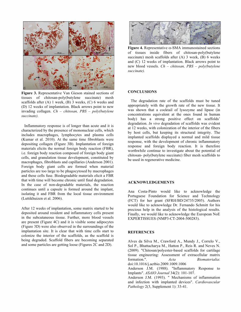
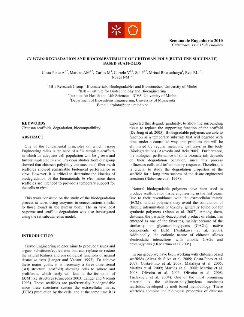
Figure 3. Representative Van Gieson stained sections of tissues of chitosan-poly(butylene succinate) mesh scaffolds after (A) 1 week, (B) 3 weeks, (C) 6 weeks and (D) 12 weeks of implantation. Black arrows point to new invading collagen. Ch – chitosan, PBS – poly(butylene succinate).
Inflammatory response is of longer than acute and it is characterized by the presence of mononuclear cells, which includes macrophages, lymphocytes and plasma cells (Kumar et al. 2010). At the same time fibroblasts were depositing collagen (Figure 3B). Implantation of foreign materials elicits the normal foreign body reaction (FBR), i.e. foreign body reaction composed of foreign body giant cells, and granulation tissue development, constituted by macrophages, fibroblasts and capillaries (Anderson 2001). Foreign body giant cells are formed when material particles are too large to be phagocytosed by macrophages and these cells fuse. Biodegradable materials elicit a FBR that with time will become chronic until final degradation. In the case of non-degradable materials, the reaction continues until a capsule is formed around the implant, isolating it and FBR from the local tissue environment (Luttikhuizen et al. 2006).
After 12 weeks of implantation, some matrix started to be deposited around resident and inflammatory cells present in the subcutaneous tissue. Further, more blood vessels are present (Figure 4C) and it is visible some adipocytes (Figure 3D) were also observed in the surroundings of the implantation site. It is clear that with time cells start to colonize the interior of the scaffolds, as the scaffold is being degraded. Scaffold fibers are becoming separated and some particles are getting loose (Figures 2C and 2D).
Figure 4. Representative α-SMA immunostained sections of tissues inside fibers of chitosan-poly(butylene succinate) mesh scaffolds after (A) 3 week, (B) 6 weeks and (C) 12 weeks of implantation. Black arrows point to new blood vessels. Ch – chitosan, PBS – poly(butylene succinate).
CONCLUSIONS The degradation rate of the scaffolds must be tuned appropriately with the growth rate of the new tissue. It was shown that a cocktail of lysozyme and lipase (in concentrations equivalent at the ones found in human body) has a strong positive effect on scaffolds’ degradation. In vivo degradation of scaffolds was evident at 12 weeks, with colonization of the interior of the fibers by host cells, but keeping its structural integrity. The implanted scaffolds displayed a normal and mild tissue response, with the development of chronic inflammatory response and foreign body reaction. It is therefore worthwhile continue to investigate about the potential of chitosan- poly(butylene succinate) fiber mesh scaffolds to be used in regenerative medicine. ACKNOWLEDGEMENTS Ana Costa-Pinto would like to acknowledge the Portuguese Foundation for Science and Technology (FCT) for her grant (SFRH/BD/24735/2005). Authors would like to acknowledge Dr. Fernando Schmitt for his precious help in the analysis of the histological results. Finally, we would like to acknowledge the European NoE EXPERTISSUES (NMP3-CT-2004-500283). REFERENCES Alves da Silva M., Crawford A., Mundy J., Correlo V., Sol P., Bhattacharya M., Hatton P., Reis R. and Neves N. (2009). "Chitosan/polyester-based scaffolds for cartilage tissue engineering: Assessment of extracellular matrix formation.". Acta Biomaterialia: doi:10.1016/j.actbio.2009.1009.1006 Anderson J.M. (1988). "Inflammatory Response to Implants". ASAIO Journal 34(2): 101-107. Anderson J.M. (1993). " Mechanisms of inflammation and infection with implanted devices". Cardiovascular Pathology 2(3, Supplement 1): 33-41.

Anderson J.M. (2001). "Biological responses to materials ". Annu. Rev. Mater. Res. 31: 81-110. Azad A.K. (2003). "Chitosan Membrane as a Wound-Healing Dressing:Characterization and Clinical Application". J Biomed Mater Res Part B: Appl Biomater 69(B): 216-222. Azevedo H. and Reis R. (2005). "Understanding the enzymatic degradation of biodegradable polymers and strategies to control their degradation rate". Biodegradable systems in tissue engineering and regenerative medicine. R. Reis and J. San Roman. Boca Raton, CRC Press: 177-201. Azevedo H.S., Gama F.M. and Reis R.L. (2003). "In Vitro Assessment of the Enzymatic Degradation of Several Starch Based Biomaterials". Biomacromolecules 4(6): 1703-1712. Babensee J., Anderson J., Mcintire L. and Mikos A. (1998). "Host response to tissue engineered devices". Advanced Drug Delivery Reviews 33: 111-139. Bahari K., Mitomo H., Enjoji T., Yoshi F. and Makuuchi K. (1998). "Degradability of poly (3-hydroxybutyrate) and its copolymer grafted with styrene by radiation". Polymer Degradation and Stability 62: 245-252. Balmayor E., Tuzlakoglu K., Marques A., Azevedo H. and Reis R. (2008). "A novel enzymatically-mediated drug delivery carrier for bone tissue engineering applications: combining biodegradable starch-based microparticles and differentiation agents". Journal of Materials Science: Materials in Medicine 19(4): 1617-1623. Cancedda R. (2003). "Tissue engineering and cell therapy of cartilage and bone". Matrix Biology 22: 81-91. Correlo V.M., Boesel L.F., Pinho E., Costa-Pinto A.R., Alves da Silva M.L., Bhattacharya M., Mano J.F., Neves N.M. and Reis R.L. (2009). "Melt-based compression-molded scaffolds from chitosan-polyester blends and composites: Morphology and mechanical properties". J Biomed Mater Res A 91(2): 489-504. Correlo V.M., Costa-Pinto A.R., Sol P., Covas J.A., Bhattacharya M., Neves N.M. and Reis R.L. (2010). "Melt Processing of Chitosan-Based Fibers and Fiber-Mesh Scaffolds for the Engineering of Connective Tissues". Macromolecular Bioscience: n/a-n/a. Costa-Pinto A.R., Correlo V.M., Sol P.C., Bhattacharya M., Charbord P., Delorme B., Reis R.L. and Neves N.M. (2009). "Osteogenic differentiation of human bone marrow mesenchymal stem cells seeded on melt based chitosan scaffolds for bone tissue engineering applications". Biomacromolecules 10(8): 2067-2073. Costa-Pinto A.R., Salgado A.J., Correlo V.M., Sol P., Bhattacharya M., Charbord P., Reis R.L. and Neves N.M. (2008). "Adhesion, proliferation, and osteogenic differentiation of a mouse mesenchymal stem cell line (BMC9) seeded on novel melt-based chitosan/polyester 3D porous scaffolds". Tissue Eng Part A 14(6): 1049-1057. De Jong W., Eelco B., Robinson J. and Bos R. (2005). "Tissue response to partially in vitro predegraded poly-L-lactide implants". Biomaterials 26(14): 1781-1791.
Di Martino A., Sittinger M. and Risbud M.V. (2005). "Chitosan: a versatile biopolymer for orthopaedic tissue-engineering". Biomaterials 26(30): 5983-5990. Gomes M.E., Azevedo H.S., Moreira A.R., Ellä V., Kellomäki M. and Reis R.L. (2008). "Starch–poly(ε-caprolactone) and starch–poly(lactic acid) fibre-mesh scaffolds for bone tissue engineering applications: structure, mechanical properties and degradation behaviour". Journal of Tissue Engineering and Regenerative Medicine 2(5): 243-252. Haiyan L., Jiang C., Amin C. and Junying W. (2005). "In vitro evaluation of biodegradable poly(butylene succinate) as a novel biomaterial". Macromol. Biosci 5: 433-450. Hankiewicz J. and Swierczek E. (1974). "Lysozyme in human body fluids". Clinica Chimica Acta 57(3): 205-209. Hasham S.N. and Pillarisetti S. (2006). "Vascular lipases, inflammation and atherosclerosis". Clinica Chimica Acta 372(1-2): 179-183. Hashitani T., Yano E. and Ando Y. (2002). "Biodegradable Packing Materials for LSIs.". Fujitsu Sci Tech J 38(1): 112-118. Hirano S., Tsuchida H. and Nagao N. (1989). "N-acetylation in chitosan and the rate of its enzymic hydrolysis". Biomaterials 10(8): 574-576. Hirotsu T., Tsujisaka T., Masuda T. and Nakayama K. (2000). "Plasma surface treatments and biodegradation of poly(butylene succinate) sheets". Journal of Applied Polymer Science 78(5): 1121-1129. Hubbell J.A. (1995). "Biomaterials in Tissue Engineering". Nat Biotech 13(6): 565-576. Ishihara M., Ono K., Sato M., Nakanishi K., Saito Y., Yura H., Matsui T., Hattori H., Fujita M., Kikuchi M. and Kurita A. (2001). "Acceleration of wound contraction and healing with a photocrosslinkable chitosan hydrogel". Wound Repair Regen 9(6): 513-521. Ishioka R., Kitakuni E. and Ichikawa Y. (2002). "Aliphatic polyesters: "Bionolle"". Biopolymers, Vol.4: polyesters III. Application and commercial products. Doi Y and S. A. Weinhem, Wiley-VCH Verlag GmbH. 4: 275-297. ISO I.O.f.S. (1994). Biological evaluation of medical devices. Part 6: tests for local efects after implantation. Geneva, International Organization for Standardization. Katti D.S., Lakshmi S., Langer R. and Laurencin C.T. (2002). "Toxicity, biodegradation and elimination of polyanhydrides". Advanced Drug Delivery Reviews 54(7): 933-961. Kumar V., Abbas K., Fausto N. and Aster j. (2010). "Acute and Chronic Inflammation". Robbins & Cotran Pathologic Basis of Disease, Saunders. Langer R. and Vacanti J. (1993). "Tissue engineering ". Science 260(5110): 920-926. Luttikhuizen D.l.T., Harmsen M.C. and Luyn M.J.A.V. (2006). "Cellular and Molecular Dynamics in the Foreign Body Reaction". Tissue Engineering 12(7): 1955-1970. Malafaya P., Pedro A., Peterbauer A., Gabriel C., Redl H. and Reis R. (2005). "Chitosan particles agglomerated

scaffolds for cartilage and osteochondral tissue engineering approaches with adipose tissue derived stem cells". Journal of Materials Science: Materials in Medicine 16: 1077-1085. Mano J., Silva G., Azevedo H., Malafaya P., Sousa R., Silva S., Boesal L., Oliveira J., Santos T., Marques A., Neves N. and Reis R. (2007). "Natural origin biodegradable systems in tissue engineering and regenerative medicine:present status and some moving trends". Journal of Royal Society Interface 4: 999-1030. Marten E., M¸ller R.-J. and Deckwer W.-D. (2003). "Studies on the enzymatic hydrolysis of polyesters I. Low molecular mass model esters and aliphatic polyesters". Polymer Degradation and Stability 80(3): 485-501. Martins A.M., Pereira R.C., Leonor I.B., Azevedo H.S. and Reis R.L. (2009). "Chitosan scaffolds incorporating lysozyme into CaP coatings produced by a biomimetic route: a novel concept for tissue engineering combining a self-regulated degradation system with in situ pore formation". Acta Biomater 5(9): 3328-3336. Martins A.M., Pham Q.P., Malafaya P.B., Raphael R.M., Kasper F.K., Reis R.L. and Mikos A.G. (2008). ""Smart'' and stimulus responsive chitosan-based scaffolds/cells for bone tissue engineering: Influence of lysozyme upon scaffold degradation and osteogenic differentiation of cultured marrow stromal cells induced by cap coatings". Tissue Engineering Part A 14(5): 795-795. Martins A.M., Santos M.I., Azevedo H.S., Malafaya P.B. and Reis R.L. (2008). "Natural origin scaffolds with in situ pore forming capability for bone tissue engineering applications". Acta Biomater 4(6): 1637-1645. Mori T., Okumura M., Matsuura M., Ueno K., Tokura S., Okamoto Y., Minami S. and Fujinaga T. (1997). "Effects of chitin and its derivatives on the proliferation and cytokine production of fibroblasts in vitro". Biomaterials 18(13): 947-951. Mukherjee M. (2003). "Human digestive and metabolic lipases--a brief review". Journal of Molecular Catalysis B: Enzymatic 22(5-6): 369-376. Muzzarelli R.A.A. (1993). "Biochemical significance of exogenous chitins and chitosans in animals and patients". Carbohydrate Polymers 20(1): 7-16. Nishikawa H., Ueno A., Nishikawa S., Kido J.-i., Ohishi M., Inoue H. and Nagata T. (2000). "Sulfated Glycosaminoglycan Synthesis and Its Regulation by Transforming Growth Factor-[beta] in Rat Clonal Dental Pulp Cells". Journal of Endodontics 26(3): 169-171. Nishimura K., Nishimura S., Nishi N., Saiki I., Tokura S. and Azuma I. (1984). "Immunological activity of chitin and its derivatives". Vaccine 2(1): 93-99. Nordtveit R.J., VÂrum K.M. and Smidsr¯d O. (1996). "Degradation of partially N-acetylated chitosans with hen egg white and human lysozyme". Carbohydrate Polymers 29(2): 163-167. Oliveira J.M., Rodrigues M.T., Silva S.S., Malafaya P.B., Gomes M.E., Viegas C.A., Dias I.R., Azevedo J.T., Mano J.F. and Reis R.L. (2006). "Novel hydroxyapatite/chitosan bilayered scaffold for osteochondral tissue-engineering applications: Scaffold design and its performance when
seeded with goat bone marrow stromal cells". Biomaterials 27(36): 6123-6137. Oliveira J.T., Correlo V.M., Sol P.C., Costa-Pinto A.R., Malafaya P.B., Salgado A.J., Bhattacharya M., Charbord P., Neves N.M. and Reis R.L. (2008). "Assessment of the suitability of chitosan/polybutylene succinate scaffolds seeded with mouse mesenchymal progenitor cells for a cartilage tissue engineering approach". Tissue Eng Part A 14(10): 1651-1661. Pangburn S.H., Trescony P.V. and Heller J. (1982). "Lysozyme degradation of partially deacetylated chitin, its films and hydrogels". Biomaterials 3(2): 105-108. Peluso G., Petillo O., Ranieri M., Santin M., Ambrosio L., Calabró D., Avallone B. and Balsamo G. (1994). "Chitosan-mediated stimulation of macrophage function". Biomaterials 15: 1215-1220. Prudden J.F., Migel P., Hanson P., Friedrich L. and Balassa L. (1970). "The discovery of a potent pure chemical wound-healing accelerator". The American Journal of Surgery 119(5): 560-564. Sashiwa H., Saimoto H., Shigemasa Y., Ogawa R. and Tokura S. (1990). "Lysozyme susceptibility of partially deacetylated chitin". International Journal of Biological Macromolecules 12(5): 295-296. Shigemasa Y. and Minami S. (1996). "Applications of chitin and chitosan for biomaterials". Biotechnol Genet Eng Rev 13: 383-420. Tietz N. and Shuey D. (1993). "Lipase in serum--the elusive enzyme: an overview". Clin Chem 39(5): 746-756. Tokiwa Y. and Suzuki T. (1977). "Hydrolysis of polyesters by lipases". Nature 270(5632): 76-78. Tomihata K. and Ikada Y. (1997). "In vitro and in vivo degradation of films of chitin and its deacetylated derivatives". Biomaterials 18: 261-268. Tuzlakoglu K., Alves C., Mano J. and Reis R. (2004). "Production and characterization of chitosan fibers and 3-D fiber mesh scaffolds for tissue engineering applications". Macromol Biosci 4(8): 811-819. Varum (1997). "In vitro degradation rates of partially N-acetylated chitosans in human serum". Carbohydrate res 299: 99-101. Williams D. and Remes A. (1992). "Immune response in biocompatibility". Biomaterials 13(11): 731-741. Wong H. and Schotz M.C. (2002). "The lipase gene family". J. Lipid Res. 43(7): 993-999. AUTHOR BIOGRAPHY
ANA COSTA-PINTO was born on December 22, 1980, in Felgueiras, Portugal. She obtained her Bachelor of Science in Applied Biology from the University of Minho in 2003.
In October 2005, she formally started her PhD at the 3B’s Research Group where she has been working on the development of a bone tissue engineering strategy, using human bone marrow stem cells and chitosan-based
scaffolds. During this time she have been abroad, under European projects collaborations, in Rome, Italy, under the supervision of Prof. Paolo Bianco and in Ankara, Turkey, under the supervision of Prof Erhan Piskin. She was also mentor of several undergraduate students, with different projects. During her PhD work, she has acquired a firm foundation of knowledge and experience with cell isolation, culture and characterization of stem cells form different sources, such as bone marrow and umbilical cord. She has also received training with in vivo animal models, related with tissue engineering strategies. As a result of her research she attended the most important international conferences in the field of Biomaterials, Tissue Engineering and Regenerative Medicine. Presently, she is the first author of 5 papers in international refereed journals (2 published and 3 submitted). She also received the outstanding student/young investigator award at the Asia-Pacific Chapter TERMIS meeting in 2007 She is currently a PhD candidate at the 3B’s Research Group (Biomaterials, Biodegradables and Biomimetics), University of Minho, Portugal. Her doctoral work has been supervised by Prof. Nuno Neves and Prof. Rui L. Reis (also the Director of 3B’s Research Group. She has just submitted her PhD thesis on Biomedical Engineering to the University of Minho. Her e-mail address is [email protected]