in vitro cytotoxic effects of gold nanoparticles coated ...aac.asm.org/content/59/2/763.full.pdf ·...
TRANSCRIPT
In Vitro Cytotoxic Effects of Gold Nanoparticles Coated withFunctional Acyl Homoserine Lactone Lactonase Protein from Bacilluslicheniformis and Their Antibiofilm Activity against Proteus Species
Gopalakrishnan Vinoj,a Rashmirekha Pati,b Avinash Sonawane,b Baskaralingam Vaseeharana
Crustacean Molecular Biology and Genomic Lab, Department of Animal Health and Management, Alagappa University, Karaikudi, Tamil Nadu, Indiaa; School ofBiotechnology, KIIT University, Bhubaneswar, Orissa, Indiab
N-acylated homoserine lactonases are known to inhibit the signaling molecules of the biofilm-forming pathogens. In this study,gold nanoparticles were coated with N-acylated homoserine lactonase proteins (AiiA AuNPs) purified from Bacillus lichenifor-mis. The AiiA AuNPs were characterized by UV-visible spectra, Fourier transform infrared spectroscopy (FTIR), transmissionelectron microscopy (TEM), and X-ray diffraction (XRD). The synthesized AiiA AuNPs were found to be spherical in shape and10 to 30 nm in size. Treatment with AiiA protein-coated AuNPs showed maximum reduction in exopolysaccharide production,metabolic activities, and cell surface hydrophobicity and potent antibiofilm activity against multidrug-resistant Proteus speciescompared to treatment with AiiA protein alone. AiiA AuNPs exhibited potent antibiofilm activity at 2 to 8 �M concentrationswithout being harmful to the macrophages. We conclude that at a specific dose, AuNPs coated with AiiA can kill bacteria withoutharming the host cells, thus representing a potential template for the design of novel antibiofilm and antibacterial protein drugsto decrease bacterial colonization and to overcome the problem of drug resistance. In summary, our data suggest that the com-bined effect of the lactonase and the gold nanoparticles of the AiiA AuNPs has promising antibiofilm activity against biofilm-forming and multidrug-resistant Proteus species.
Biofilm formation by Proteus species has been reported as asource of catheter-associated urinary tract infection that gives
rise to serious complications. Proteus vulgaris, Proteus mirabilis,and Proteus penneri are known to cause urinary tract infections inhumans (1). Among the Proteus spp., P. vulgaris is capable offorming crystalline biofilms and generating alkaline urine within24 h (2). Such biofilm-forming pathogenic bacteria are the majorcause of many chronic and recurrent infections, such as periodon-titis, endocarditis, chronic otitis, and urinary tract and woundinfections (3). Moreover, bacteria that adhere to indwelling med-ical devices such as intravenous catheters, artificial joints, and car-diac pacemakers or to damaged tissue can cause persistent infec-tions through biofilm formation (2, 4, 5). Mature biofilms ofProteus spp. develop on the surface of uroepithelial cells or cathe-ters; organisms within the mushroom-shaped structure regulate avariety of cellular functions, such as glutamine synthesis, autoin-ducer 2 (AI-2), cyclic dipeptides, and putrescine synthesis (6–8).Catheter-associated urinary tract infections (CAUTIs) affect morethan 400,000 patients per year in the United States (9). Antibioticsused to treat these biofilm-forming pathogens are not activeagainst the recalcitrant biofilms; rather, they target their plank-tonic counterparts, which create selective pressure on the bacteria,which develop resistance to a particular drug (10). Disrupting themulticellular structure of a Proteus biofilm was proposed as one ofthe most promising strategies for increasing the sensitivity ofpathogens to antibiotics and the host immune system (11, 12).Hence, effective therapeutic agents are required to control urinarytract infections caused by biofilm-forming Proteus spp. Recently,disruption of quorum sensing (QS) by quorum quenching hasbeen suggested as an anti-infective strategy to control pathogenicbacteria through interference with the colonization processes, in-cluding biofilm formation and invasion of host tissues (13–15).N-Acyl homoserine lactonase enzymes that degrade acyl homo-
serine lactone (AHL) might have commercial value as a means tomanipulate cell-to-cell signaling (16, 17).
N-Acyl homoserine lactonase plays a critical role in hydrolyz-ing the lactone bond within the acyl homoserine lactone moiety,thus changing the relative conformational structure of the signal-ing molecule; this prevents binding to the LuxR transcriptionalregulator, resulting in quorum-sensing inhibitory activity againstpathogens (18, 19). The first AHL-degrading enzyme was identi-fied in Bacillus strain 240B1 (20). To date, more than 20 AHL-degrading enzymes have been identified in bacteria, fungi, andmammals (15, 21, 22). Many Bacillus AHL lactonase-like enzymeshave been reported, which share approximately �90% sequenceidentity (22). These enzymes contain the conserved motif HXHXDH and a zinc binding motif and therefore are classified asmetallic �-lactamases (23). Recent developments in the field ofnanotechnology have helped to some extent to overcome prob-lems with drug resistance and biofilm formation through synthe-sis of bioactive materials (24). Chemically synthesized nanopar-ticles (NPs) give rise to toxicity, thereby limiting their biomedicalapplications (25). Microbially mediated nanoparticles may also
Received 15 April 2014 Returned for modification 16 May 2014Accepted 20 October 2014
Accepted manuscript posted online 17 November 2014
Citation Vinoj G, Pati R, Sonawane A, Vaseeharan B. 2015. In vitro cytotoxic effectsof gold nanoparticles coated with functional acyl homoserine lactone lactonaseprotein from Bacillus licheniformis and their antibiofilm activity against Proteusspecies. Antimicrob Agents Chemother 59:763–771.doi:10.1128/AAC.03047-14.
Address correspondence to Vaseeharan Baskaralingam, [email protected].
Copyright © 2015, American Society for Microbiology. All Rights Reserved.
doi:10.1128/AAC.03047-14
February 2015 Volume 59 Number 2 aac.asm.org 763Antimicrobial Agents and Chemotherapy
on June 9, 2018 by guesthttp://aac.asm
.org/D
ownloaded from

give rise to toxic substances such as endotoxin on the surface of thenanoparticles, which limits their use in the medical field (26).Therefore, the trend has shifted to the use of biologically synthe-sized nanoparticles. Gold can be stabilized with a wide variety ofmolecules, such as peptides, proteins, DNA, and polymers (27).The use of gold coating over magnetic nanoparticles to achieveboth a biocomposite surface and high magnetic properties hasdrawn intense scientific and technological interest for potentialapplications in biomedical field (28). These materials exhibitunique thermal, mechanical, and biological properties (29, 30)compared to other free enzymes and proteins. The performance ofbionanoparticles for the control of microbes is dependent on boththe chemical properties of the gold nanoparticles and the bioac-tivity of the enzymes and proteins. Colloidal gold nanoparticleshave flexible properties, such as plasmon resonance, optical prop-erties, and the presence of a bioconjugation surface for molecularprobes, compared to those of other inorganic nanoparticles (31).These properties have attracted attention with a view to develop anew approach to control biofilm-forming bacteria and will beused in the field of nanomedicine. In the present work, we havesynthesized AuNPs coated with AiiA protein (AiiA AuNPs) andstudied their AHL lactonase activity, antibiofilm activity againstProteus, and cytotoxic effects against macrophages. We found that2 �M AiiA AuNPs demonstrated maximum degradation of 1.8mg N-hexanoyl-L-homoserine lactone (C6-HSL) (liter�1 h�1)and inhibited the exopolysaccharide (EPS) production, hydro-phobicity, metabolic activity, and biofilm formation of the iso-lated Proteus strains DPr1, DPr2, and DPr3 and P. vulgaris ATCC49565. Furthermore, AiiA AuNPs did not show any significantcytotoxic effect against the macrophages tested at a 2 �M concen-tration. To our knowledge, this is the first study on AiiA AuNPs todetermine their antibiofilm potential against Proteus.
MATERIALS AND METHODSBacterial growth conditions. The clinical strains were isolated from spec-imens (urinary tract infection) from the Karaikudi Government Hospital,Tamil Nadu, India. Environmental strains were isolated from crustaceansof the Cuddalore coast, on the southeast coast of India, situated about 250km south of Chennai (11°27=N, 79°47= E). We selected only effective bio-film-forming and antibiotic-resistant environmental (DPr1 and DPr2)and clinical (DPr3 and DPr4) strains. The four selected strains were iden-tified at the genus level based on 16S rRNA gene sequencing. P. vulgarisATCC 49565 was used as the reference strain. Proteus strains were grownand maintained on tryptic soy agar (TSA) or tryptic soy broth (TSB) at37°C. Chromobacterium violaceum CV026 was used for the AHL lactonaseactivity bioassay (32). The reporter strain Chromobacterium violaceumATCC 12472 was used to determine quorum-sensing inhibitory activity,and strains were also cultured in Luria-Bertani (LB) broth at 33°C andstored at �80°C for the further studies.
Purification of AiiA protein from Bacillus licheniformis. The aiiAgene of Bacillus licheniformis isolated from the crustacean Fenneropenaeusindicus was amplified by PCR using modified primers incorporating NdeIand EcoRI restriction sites (underlined) (5= to 3=: AIF1, GGG AAT TCCATA TGA CAG TAA AAA AGC TTT ATT TC; AIR1, CCG GAA TTCCGG CTA TAT ATA CTC CGG GAA CTC) with 40 cycles of 5 min at95°C, 1 min at 50°C, and 2 min at 72°C and a final 10-min incubation at72°C. The PCR-amplified DNA was purified using a Wizard SV gel andPCR clean-up system (Promega), digested with NdeI and EcoRI (NEB),ligated into the expression plasmid pET-32a (Novogene), and then usedto transform Escherichia coli BL21 cells. Plasmid DNA was isolated fromrecombinant clones using a purification kit (Wizard SV Minipreps DNA;Promega, USA), and the insertion in one (pET-AiiA) was verified by se-
quencing by the dye termination method (Applied Biosystems, USA) us-ing vector-specific primers and a Li-Cor 4200 DNA sequencer. AiiA pro-tein was purified from IPTG (isopropyl-�-D-thiogalactopyranoside)-induced E. coli BL21 cultures using Ni-nitrilotriacetic acid (NTA)columns (Qiagen) following the method described by the manufacturer.Expression and isolation were monitored by SDS-PAGE, and the proteinconcentration was determined by the Bradford method (33). PurifiedAiiA protein showed a single 28-kDa band in the SDS-PAGE (data notshown). AiiA protein was lyophilized, stored at 4°C, and used for nano-particle preparation.
AiiA AuNPs. To prepare gold nanoparticles coated with AiiA protein(AiiA AuNPs), purified AiiA protein (2 mg) was incubated with a 0.5 mMconcentration of HAuCl4 in a 150-ml Erlenmeyer flask and agitated at37°C and 200 rpm. The pH of the final solution was adjusted to 5.0. Themixture was then incubated at 37°C until a colored solution was obtained.The reaction between AiiA protein and HAuCl4 was continued for 24 h toconfirm that the intensity of the color was stable.
Characterization of AiiA AuNPs. (i) UV-visible spectroscopic anal-ysis. For spectroscopic analysis, 1 ml of AiiA AuNPs was withdrawn fromthe reaction mixture and absorbance was measured with a UV-visiblespectrophotometer (UV-1800; Shimadzu, Japan) from 300 to 800 nm for100 days.
(ii) TEM. For transmission electron microscopy (TEM) analysis, asmall volume of AiiA AuNPs was loaded on a carbon-coated copper grid,and the solvent was allowed to evaporate for 30 min. TEM measurementswere performed on a JOEL 1200 EX TEM at an accelerating voltage of80 kV. The crystal structure of the AiiA AuNPs was determined using theselected area electron diffraction (SAED) pattern.
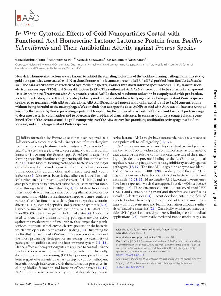
(iii) XRD and FTIR analyses. In order to determine their crystal na-ture, AiiA AuNPs were centrifuged, washed, and air dried. The air-driedpowder was coated on a glass surface and then subjected to X-ray diffrac-tion (XRD) analysis on a PAN analytical XRD analyzer (X’pert PRO)operating in the transmission mode at 40 kV and 30 mA with CuK radi-ation. Two milligram of the AiiA AuNPs was mixed with 200 mg KBr(FTIR grade) and pressed into a pellet. The AiiA AuNP pellet was placed inthe sample holder, and Fourier transform infrared (FTIR) spectra wererecorded with an FTIR spectrometer (Perkin-Elmer, Shelton, CT) at aresolution of 4 cm�1.
Biofilm assay. The biofilm inhibitory concentration (BIC) of AiiAAuNPs was determined as the lowest concentration that produced visibledisruption to biofilm formation. Examination of the BIC of AiiA AuNPson biofilm formation was performed in 24-well polystyrene plates (34).Briefly, overnight cultures of Proteus at 106 CFU/ml were inoculated with1 ml of fresh Zobell marine broth (ZMB) in the presence of AuCl2 (2 �M),AiiA protein alone (120 U ml�1), and AiiA AuNPs (0.5 to 4 �M). Theplates were incubated for 24 h at 37°C. After incubation, the cultures werediscarded, and the wells were gently rinsed twice with deionized water andallowed to air dry before staining with crystal violet (CV). The wells werestained with 210 �l of 0.1% (wt/vol) CV for 10 min before being rinsedtwice with deionized water and air dried. CV was then eluted with 210 �ldimethyl sulfoxide, and the A595 was measured using a Bio-Rad enzyme-linked immunosorbent assay (ELISA) reader.
AHL lactonase activity bioassay. The AHL lactonase activity of thebacteria treated with AuCl2 (2 �M), AiiA protein alone (120 U ml�1), andAiiA AuNPs (2 �M) was determined in a C6-HSL well diffusion bioassayusing C. violaceum CV026 as the reporter strain (31). A lawn of C. viola-ceum CV026 was prepared by mixing 3 ml of overnight culture on LB agarplates, and then wells of 5 mm in diameter were made in the agar plates.An AHL lactonase reaction mixture containing 10 �l sample plus 190 �lof solution containing 24 nM C6-HSL in 50 mM phosphate buffer (pH8.0) was prepared and incubated at 25°C for 45 min before the reactionwas terminated by the addition of 50 �l 10% (wt/vol) SDS. The reactionmixture was then transferred into the wells, and the radius of the C. viola-ceum CV026 nonpigmented zone was used to determine the residual C6-HSL levels after 2 days of incubation. The AHL lactonase activity of the
Vinoj et al.
764 aac.asm.org February 2015 Volume 59 Number 2Antimicrobial Agents and Chemotherapy
on June 9, 2018 by guesthttp://aac.asm
.org/D
ownloaded from

bacteria treated with AuCl2 (2 �M), AiiA protein alone (120 U ml�1), andAiiA AuNPs (2 �M) is reported here as the degradation rate of AHL (mgliter�1 h�1) or as the units of AHL-degrading activity mg�1 protein,where 1 U is defined as the amount of enzyme required to hydrolyze 1 nMC6-HSL per minute under the conditions described here. For the standardcurve, y � 24 nM � e0.42 � x/106 and r2 � 0.996 (y, in nmol, is theamount of C6-HSL, and x, in mm, is the distance of diffusion of C6-HSL).
Quantification of CSH by MATH assay. The effect of the AiiA proteinand AiiA AuNPs on cell surface hydrophobicity (CSH) of Proteus wasmeasured by the microbial adhesion to hydrocarbon (MATH) assay. Bac-teria were grown in tryptic soy broth at 37°C for 18 to 24 h, harvested bycentrifugation at 6,000 rpm for 5 min at 25°C, washed with and resus-pended in sterile distilled water, and adjusted to an optical density (OD) at600 nm of 0.6 � 0.04. The AiiA AuNPs/AiiA protein and toluene (1 ml)were added to 2 ml of the adjusted cell suspension (A600 � 0.6 � 0.04) ina test tube. After vortexing for 1 min, the cell suspensions were incubatedat room temperature overnight, and the OD of the aqueous phase wasmeasured (A600). The hydrophobicity index (HI) of Proteus was calcu-lated as HI � 100(E �100/E0) (35). The results were expressed as theproportion of the cells which were excluded from the aqueous phase,determined by the equation, where E0 is the initial optical density of thecell suspension and E is the optical density of the aqueous phase after itsseparation from the toluene phase.
Quantification of EPS. The level of exopolysaccharide (EPS) pro-duced by Proteus was determined by carbohydrate assay. Sterile catheterpieces were immersed in a Proteus culture containing 2 �M AiiA AuNPsor TSA broth (control) in 24-well polystyrene plates and were incubatedfor 24 h. The sterile catheters were vortexed with 0.9% NaCl solution. Cellsuspensions were transferred to test tubes to which an equal volume of 5%phenol and 5 volumes of sulfuric acid containing 0.2% hydrazine sulfatewere added. The mixture was incubated in dark for 1 h and centrifuged at10,000 � g for 10 min, and the optical density of the supernatant wasmeasured at 490 nm (36). The percentage of EPS was calculated as follows:EPS production (%) � [(control OD � test OD)/control OD] � 100.
Assessment of biofilm metabolic activity using XTT reduction as-say. Metabolic activities of cells during biofilm formation was assessedusing the XTT [2,3-bis(2-methyloxy-4-nitro-5-sulfophenyl)-2H-tetrazo-lium-5-carboxanilide] reduction assay, which measures the reduction of atetrazolium salt by metabolically active cells to a colored water-soluble form-azan derivative which can be quantified colorimetrically. AiiA AuNPs (2 �M)and AiiA protein (120 U ml�1) were added to the Proteus biofilms in a96-well polystyrene tissue culture plate containing ZMB and 2% (wt/vol)glucose, and then the plates were incubated as described above for thebiofilm assay. Following incubation, the biofilms were washed five timeswith phosphate-buffered saline (PBS), and then 100 �l PBS and 12 �lXTT-menadione solution (12.5:1, vol/vol) were added to the wells. Theplates were then incubated for 3 h in the dark at 37°C. Following incuba-tion, 100 �l of the solution was transferred to fresh wells, and the changein the color of the solution was measured at 450 nm spectrophotometri-cally (36).
Microscopic observation of Proteus biofilm inhibition by AiiAAuNPs. For visualization of biofilms by light microscopy, Proteus biofilmswere allowed to grow on a sterile catheter (1 by 1 cm) immersed in AiiAAuNPs (2 �M) in a 24-well polystyrene plate and incubated for 24 h at30°C. Catheter pieces were stained with crystal violet as described aboveand examined using a Nikon microscope (Eclipse Ti 100�) at a magnifi-cation of �40. Independently, another set of catheter pieces with biofilmswere washed with PBS stained with acridine orange (0.1%), and the bio-film formation was quantified using a confocal laser scanning microscope(CLSM) (Carl Zeiss LSM 710) with a 488-nm argon laser, a BP 500-640band pass emission filter, and Zen 2009 software (Carl Zeiss, Germany).CLSM biofilm observations were assessed using COMSAT software (37).
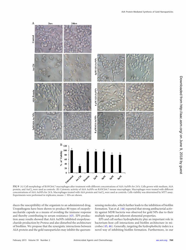
Cytotoxic effect of AiiA AuNPs on macrophages. To determine thecytotoxic activity of the gold nanoparticles coated with AiiA protein (AiiAAuNPs) on macrophages, RAW 264.7 cells (1 � 104 cells/ml) in Dulbecco
modified Eagle medium (DMEM) were grown in a 96-well plate at37°C with 5% CO2 for 24 h, followed by treatment of cells with differ-ent concentrations of AiiA AuNPs for another 24 h. To determine thecell viability, MTT [3-(4,5-dimethyl-2-thiazolyl)-2,5-diphenyl-2H-tetrazolium bromide] at a concentration of 2 to 8.0 �M was added tothe wells and incubated for 4 h at 37°C with 5% CO2 in the dark. Inmetabolically active cells, MTT was reduced to an insoluble dark pur-ple formazan. The formazan crystals were dissolved in dissolving buf-fer (11 g SDS in 50 ml of 0.02 M HCl and 50 ml isopropanol). Theabsorbance was read at 570 nm in an ELISA reader (Biotek, Germany)and compared with that of untreated cells, and the percentage of viablecells was calculated.
Statistical analysis. All assays were repeated at least three times, andall statistical analyses were performed using SPSS. Values are expressed asmean � standard deviation (SD). Mean values were compared using one-way ANOVA.
Nucleotide sequence accession numbers. The GenBank accessionnumbers of the clinical strains used in this study are as follows: Proteus sp.strain DPr1, HQ009354; P. vulgaris Dpr2, HQ116441; Proteus sp. strainDpr3, HQ640435; and P. vulgaris Dpr4, HQ640434.
RESULTSCharacterization of AiiA AuNPs. The reduction of aqueousHAuCl4 ions during the reaction with AiiA protein was monitoredby UV-visible spectroscopy. A strong resonance at 550 nm wasseen due to the excitation of surface plasmon vibrations in the goldnanoparticles (Fig. 1A). A change of the colorless solution to pur-ple (Fig. 1B and C) confirmed the synthesis of gold nanoparticlescoated with AiiA protein (AiiA AuNPs). The stability of the AiiA
FIG 1 (A) UV-visible spectral analysis of AiiA AuNPs. (B) Before reaction. (C)After reaction for 6 h. (D) Transmission electron microscopy images of AiiA-coated gold nanoparticles with a particle diameter of about 10 to 30 nm.
AiiA Protein-Mediated Synthesis of Gold Nanoparticles
February 2015 Volume 59 Number 2 aac.asm.org 765Antimicrobial Agents and Chemotherapy
on June 9, 2018 by guesthttp://aac.asm
.org/D
ownloaded from

AuNPs was determined by measuring the absorption spectrum atintervals of 24 h for 100 days. No significant changes in the absor-bance were observed during the storage, indicating that theAuNPs did not agglomerate and were stable during this period(data not shown). TEM analysis showed that AiiA AuNPs werespherical and monodispersed, and the average particle diameterwas about 10 to 30 nm. (Fig. 1D). The crystalline nature of AiiA-coated gold nanoparticles was determined by XRD. The charac-teristic XRD peaks at 2 of 38.2°, 44.3°, 64.6°, and 77.6° are in-dexed to the 111, 200, 220, and 311 crystallographic planes of thegold nanoparticles coated with AiiA protein, respectively (Fig.2A). The high crystallinity of AiiA AuNPs is evident from brightcircular spots in the SAED pattern (Fig. 2B). Fourier transforminfrared (FTIR) analysis was performed in order to identify thepossible biomolecules responsible for the reduction of the goldions and capping of the bioreduced gold nanoparticles. FTIR spec-tra obtained from the AiiA AuNPs showed the occurrence of threebands at 3,439 cm�1, 2,924 cm�1, and 2,853 cm�1, which wereassigned to the stretching vibrations of primary, secondary, andtertiary amines, respectively. The band at 1,592 cm–1 has beenidentified as an amide II band, which arose due to carbonyl stretch
and ONOHO stretch vibrations in the amide linkages of theproteins. The bands at 1,400 cm�1 and 1,043 cm�1 demonstrateCOC and CON stretching, respectively. The band at 1,743 cm�1
corresponds to the carbonyl stretching vibration in the CAO re-gion. The presence of amide linkages between amino acid residuesin the polypeptides showed a signature in the infrared region ofthe electromagnetic spectrum (Fig. 3). Major functional groupssuch as CAO and NOH are involved in the metal interaction withAu, which leads to the conjugate reduction of synthesized NPs,indicating the change in the secondary structure spectra. Thephysical characterization revealed that the synthesized AiiAAuNPs were monodispersed, crystalline, and capping AiiA pro-tein.
Biofilm inhibition and AHL lactonase assays. The effect ofAiiA AuNPs on Proteus biofilm formation was determined using acrystal violet (CV) staining method. AiiA AuNPs were found tosignificantly inhibit the biofilm formation by Proteus compared toAiiA alone. A 2 �M concentration of AiiA AuNPs was determinedas the biofilm inhibitory concentration, and therefore, all subse-quent experiments were performed with this concentration.Treatment for 24 h resulted in a decrease of more than 85% of the
FIG 2 (A) X-ray diffraction patterns of AiiA AuNPs. (B) SAED pattern of AiiA AuNPs.
Vinoj et al.
766 aac.asm.org February 2015 Volume 59 Number 2Antimicrobial Agents and Chemotherapy
on June 9, 2018 by guesthttp://aac.asm
.org/D
ownloaded from
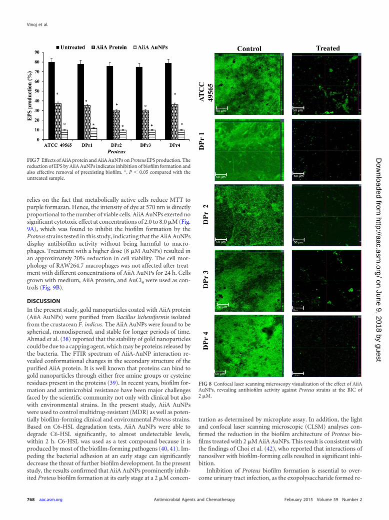
biofilm formation at a 2 �M concentration of AiiA AuNPs,whereas AiiA protein and AuCl2 alone resulted in approximately40% and 60% decreases in biofilm formation, respectively (Fig. 4).The AHL lactonase assay showed degradation rates of 0.3 � 0.1,0.9 � 0.3, and 1.8 � 0.12 mg liter�1 h�1 C6-HSL with AuCl2, AiiAprotein, and AiiA AuNP treatment, respectively (data not shown).
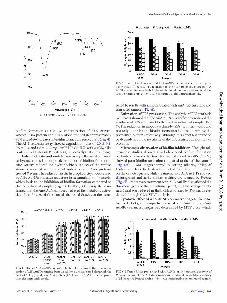
Hydrophobicity and metabolism assays. Bacterial adhesionto hydrocarbons is a major determinant of biofilm formation.AiiA AuNPs reduced the hydrophobicity indices of the Proteusstrains compared with those of untreated and AiiA protein-treated Proteus. The reduction in the hydrophobicity index causedby AiiA AuNPs indicates reduction in accumulation of bacteria,which leads to the inhibition of biofilm formation compared tothat of untreated samples (Fig. 5). Further, XTT assay also con-firmed that the AiiA AuNPs indeed reduced the metabolic activi-ties of the Proteus biofilms for all the tested Proteus strains com-
pared to results with samples treated with AiiA protein alone anduntreated samples (Fig. 6).
Estimation of EPS production. The analysis of EPS synthesisby Proteus showed that the AiiA Au NPs significantly reduced thesynthesis of EPS compared to that by the untreated sample (Fig.7). The reduction in exopolysaccharide (EPS) synthesis was foundnot only to inhibit the biofilm formation but also to remove thepreformed biofilms effectively, although this effect was found tobe dependent on the specificity of the EPS matrix composition ofbiofilms.
Microscopic observation of biofilm inhibition. The light mi-croscopic studies showed a well-developed biofilm formationby Proteus, whereas bacteria treated with AiiA AuNPs (2 �M)showed poor biofilm formation compared to that of the control(Fig. 8A). CLSM images showed the strong adhering ability ofProteus, which led to the development of dense biofilm formationon the catheter pieces, while treatment with AiiA AuNPs showeddisintegrated and labile biofilm architecture formed by Proteus(Fig. 8B). Moreover, treatment with AiiA AuNPs also affected thethickness (�m) of the biovolume (�m3), and the average thick-ness (�m) was reduced in the biofilms formed by Proteus, as evi-denced through COMSTAT analysis.
Cytotoxic effect of AiiA AuNPs on macrophages. The cyto-toxic effect of gold nanoparticles coated with AiiA protein (AiiAAuNPs) on macrophages was determined by MTT assay, which
FIG 3 FTIR spectrum of AiiA AuNPs.
FIG 4 Effect of AiiA AuNPs on Proteus biofilm formation. Different concen-tration of AiiA AuNPs ranging from 0.5 �M to 4 �M were used along with thecontrol AuCl2 (2 �M) and AiiA protein (120 U ml�1). *, P 0.05 comparedwith the untreated sample.
FIG 5 Effects of AiiA protein and AiiA AuNPs on the cell surface hydropho-bicity index of Proteus. The reduction of the hydrophobicity index in AiiAAuNP-treated bacteria leads to the inhibition of biofilm formation in all thetested Proteus strains. *, P 0.05 compared to the untreated sample.
FIG 6 Effects of AiiA protein and AiiA AuNPs on the metabolic activity ofProteus biofilm. The AiiA AuNPs significantly reduced the metabolic activityof all the tested Proteus strains. *, P 0.05 compared to the untreated sample.
AiiA Protein-Mediated Synthesis of Gold Nanoparticles
February 2015 Volume 59 Number 2 aac.asm.org 767Antimicrobial Agents and Chemotherapy
on June 9, 2018 by guesthttp://aac.asm
.org/D
ownloaded from
relies on the fact that metabolically active cells reduce MTT topurple formazan. Hence, the intensity of dye at 570 nm is directlyproportional to the number of viable cells. AiiA AuNPs exerted nosignificant cytotoxic effect at concentrations of 2.0 to 8.0 �M (Fig.9A), which was found to inhibit the biofilm formation by theProteus strains tested in this study, indicating that the AiiA AuNPsdisplay antibiofilm activity without being harmful to macro-phages. Treatment with a higher dose (8 �M AuNPs) resulted inan approximately 20% reduction in cell viability. The cell mor-phology of RAW264.7 macrophages was not affected after treat-ment with different concentrations of AiiA AuNPs for 24 h. Cellsgrown with medium, AiiA protein, and AuCl4 were used as con-trols (Fig. 9B).
DISCUSSION
In the present study, gold nanoparticles coated with AiiA protein(AiiA AuNPs) were purified from Bacillus licheniformis isolatedfrom the crustacean F. indicus. The AiiA AuNPs were found to bespherical, monodispersed, and stable for longer periods of time.Ahmad et al. (38) reported that the stability of gold nanoparticlescould be due to a capping agent, which may be proteins released bythe bacteria. The FTIR spectrum of AiiA-AuNP interaction re-vealed conformational changes in the secondary structure of thepurified AiiA protein. It is well known that proteins can bind togold nanoparticles through either free amine groups or cysteineresidues present in the proteins (39). In recent years, biofilm for-mation and antimicrobial resistance have been major challengesfaced by the scientific community not only with clinical but alsowith environmental strains. In the present study, AiiA AuNPswere used to control multidrug-resistant (MDR) as well as poten-tially biofilm-forming clinical and environmental Proteus strains.Based on C6-HSL degradation tests, AiiA AuNPs were able todegrade C6-HSL significantly, to almost undetectable levels,within 2 h. C6-HSL was used as a test compound because it isproduced by most of the biofilm-forming pathogens (40, 41). Im-peding the bacterial adhesion at an early stage can significantlydecrease the threat of further biofilm development. In the presentstudy, the results confirmed that AiiA AuNPs prominently inhib-ited Proteus biofilm formation at its early stage at a 2 �M concen-
tration as determined by microplate assay. In addition, the lightand confocal laser scanning microscopic (CLSM) analyses con-firmed the reduction in the biofilm architecture of Proteus bio-films treated with 2 �M AiiA AuNPs. This result is consistent withthe findings of Choi et al. (42), who reported that interactions ofnanosilver with biofilm-forming cells resulted in significant inhi-bition.
Inhibition of Proteus biofilm formation is essential to over-come urinary tract infection, as the exopolysaccharide formed re-
FIG 7 Effects of AiiA protein and AiiA AuNPs on Proteus EPS production. Thereduction of EPS by AiiA AuNPs indicates inhibition of biofilm formation andalso effective removal of preexisting biofilm. *, P 0.05 compared with theuntreated sample.
FIG 8 Confocal laser scanning microscopy visualization of the effect of AiiAAuNPs, revealing antibiofilm activity against Proteus strains at the BIC of2 �M.
Vinoj et al.
768 aac.asm.org February 2015 Volume 59 Number 2Antimicrobial Agents and Chemotherapy
on June 9, 2018 by guesthttp://aac.asm
.org/D
ownloaded from
duces the susceptibility of the organism to an administered drug.Uropathogens have been shown to produce 80 types of exopoly-saccharide capsule as a means of avoiding the immune responseand thereby contributing to serum resistance (43). EPS produc-tion assay results showed that AiiA AuNPs inhibited exopolysac-charide production by Proteus and also disturbed the architectureof biofilms. We propose that the synergistic interactions betweenAiiA protein and the gold nanoparticles may inhibit the quorum-
sensing molecules, which further leads to the inhibition of biofilmformation. Yan et al. (44) reported that strong antibacterial activ-ity against MDR bacteria was observed for gold NPs due to theirmultiple targets and inherent elemental properties.
EPS and cell surface hydrophobicity play an important role inbacterium-host cell interactions and biofilm architecture in mi-crobes (45, 46). Generally, targeting the hydrophobicity index is anovel way of inhibiting biofilm formation. Furthermore, in our
FIG 9 (A) Cell morphology of RAW264.7 macrophages after treatment with different concentrations of AiiA AuNPs for 24 h. Cells grown with medium, AiiAprotein, and AuCl4 were used as controls. (B) Cytotoxic activity of AiiA AuNPs on RAW264.7 mouse macrophages. Macrophages were treated with differentconcentrations of AiiA AuNPs for 24 h. Macrophages treated with AiiA protein and AuCl4 were used as controls. Cells viability was determined by MTT assay.Experiments were performed in triplicates; means � SDs are shown.
AiiA Protein-Mediated Synthesis of Gold Nanoparticles
February 2015 Volume 59 Number 2 aac.asm.org 769Antimicrobial Agents and Chemotherapy
on June 9, 2018 by guesthttp://aac.asm
.org/D
ownloaded from

study we did not see any significant changes in macrophage cellmorphology when treated with AiiA AuNPs at concentrations of 2to 8 �M. The cells were adherent to the surface, which furtherindicates that AiiA AuNPs have no cytotoxic effect on mouse mac-rophages. These results are consistent with previous investigationsperformed with HeLa cells (47, 48). The cell viability test showedno cytotoxic effects of AiiA AuNPs on macrophages at concentra-tions up to 6 �M AuNPs. A reduced cytotoxic effect may be due tothe surface coating of AiiA protein in AuNPs.
Conclusion. In the present study, gold nanoparticles coatedwith functional AHL lactonase protein from Bacillus licheniformiswere prepared and characterized by UV-visible spectra, FTIR,TEM, and XRD. AiiA AuNPs inhibited the in vitro biofilm forma-tion as well as the virulence factor (exopolysaccharide) produc-tion and metabolic activity of Proteus. The results of cytotoxicitystudies showed no changes in macrophage cell morphology whentreated with AiiA AuNPs at 2 to 8 �M. The outcome of theseresults may be development of potential biomaterials against uri-nary tract infection by MDR and biofilm-forming Proteus strains.
ACKNOWLEDGMENTS
This work was supported by the University Grants Commission (UGC),New Delhi, India, under Project Grants code F.No 36-5/2008(sr) and bygrant SR/NM/NS-1085/2011 from Department of Science and Technol-ogy, Government of India, to A.S. R.P. is grateful to the Department ofScience and Technology, Government of India, for a DST-INSPIRE fel-lowship.
REFERENCES1. Mobley HL, Jones BD, Penner JL. 1987. Urease activity of Proteus pen-
neri. J Clin Microbiol 25:2302–2305.2. Stickler DJ. 2008. Bacterial biofilms in patients with indwelling urinary
catheters. Nat Clin Pract Urol 5:598 – 608. http://dx.doi.org/10.1038/ncpuro1231.
3. Costerton JW, Stewart PS, Greenberg EP. 1999. Bacterial biofilms acommon cause of persistent infections. Science 284:1318 –1322. http://dx.doi.org/10.1126/science.284.5418.1318.
4. Vlastarakos PV, Nikolopoulos TP, Maragoudakis P, Tzagaroulakis A,Ferekidis E. 2007. Biofilms in ear, nose, and throat infections: how im-portant are they? Laryngoscope 117:4668 – 4673. http://dx.doi.org/10.1097/MLG.0b013e318030e422.
5. Fey PD, Olson ME. 2010. Current concepts in biofilm formation ofStaphylococcus epidermidis. Future Microbiol 5:917–933. http://dx.doi.org/10.2217/fmb.10.56.
6. Allison C, Lai HC, Gygi D, Hughes C. 1993. Cell differentiation ofProteus mirabilis is initiated by glutamine, a specific chemo attractant forswarming cells. Mol Microbiol 8:53– 60. http://dx.doi.org/10.1111/j.1365-2958.1993.tb01202.x.
7. Schneider R, Lockatell CV, Johnson D, Belas R. 2002. Detection andmutation of a luxS-encoded autoinducer in Proteus mirabilis. Microbiol-ogy 148:773–782.
8. Sturgill G, Rather PN. 2004. Evidence that putrescine acts as an extracel-lular signal required for swarming in Proteus mirabilis. Mol Microbiol51:437– 446. http://dx.doi.org/10.1046/j.1365-2958.2003.03835.x.
9. Klevens RM, Edwards JR, Richards CL, Horan TC, Gaynes RP, PollockDA, Cardo DM. 2007. Estimating health care associated infections anddeaths in U.S. hospitals. Public Health Rep 122:160 –166.
10. Hentzer M, Givskov M. 2003. Pharmacological inhibition of quorumsensing for the treatment of chronic bacterial infections. J Clin Invest112:1300 –1307. http://dx.doi.org/10.1172/JCI20074.
11. Reid G, Habash M. 2001. Oral fluoroquinolone therapy results in drug adsorp-tion on ureteral stents and prevention of biofilm formation. Int J AntimicrobAgents 17:317–320. http://dx.doi.org/10.1016/S0924-8579(00)00353-8.
12. Stewart PS, Costerton JW. 2001. Antibiotic resistance of bacteria in biofilms.Lancet 358:135–138. http://dx.doi.org/10.1016/S0140-6736(01)05321-1.
13. Rasmussen TB, Givskov M. 2006. Quorum sensing inhibitors: a bargin ofeffects. Microbiology 152:895–904. http://dx.doi.org/10.1099/mic.0.28601-0.
14. Defoirdt T, Boon N, Sorgeloos P, Verstraete W, Bossier P. 2008. Quorumsensing and quorum quenching in Vibrio harveyi: lessons learned from invivo work. ISME J 2:19 –26. http://dx.doi.org/10.1038/ismej.2007.92.
15. Czajkowski R, Jafra S. 2009. Quenching of acyl-homoserine lactone-dependent quorum sensing by enzymatic disruption of signal molecules.Acta Biochim Pol 56:1–16.
16. Dobretsov S, Teplitski M, Paul V. 2009. Quorum sensing in the marineenvironment and its relationship to biofouling. Biofouling 25:413– 427.http://dx.doi.org/10.1080/08927010902853516.
17. Teasdale ME, Liu J, Wallace J, Akhlaghi F, Rowley DC. 2009.Secondary metabolites produced by the marine bacterium Halobacillussalinus that inhibit quorum sensing-controlled phenotypes in gram-negative bacteria. Appl Environ Microbiol 75:567–557. http://dx.doi.org/10.1128/AEM.00632-08.
18. Kaufmann GF, Sartorio R, Lee SH, Rogers CJ, Meijler MM, Moss JA,Clapham B, Brogan AP, Dickerson TJ, Janda KD. 2005. Revisitingquorum sensing discovery of additional chemical and biological functionsfor 3-oxo-N-acylhomoserine lactones. Proc Natl Acad Sci U S A 102:309 –314. http://dx.doi.org/10.1073/pnas.0408639102.
19. Bai F, Han Y, Chen J, Zhang XH. 2008. Disruption of quorum sensing inVibrio harveyi by the AiiA protein of Bacillus thuringiensis. Aquaculture274:36 – 40. http://dx.doi.org/10.1016/j.aquaculture.2007.11.024.
20. Dong YH, Xu JL, Li XZ, Zhang LH. 2000. AiiA, an enzyme that inacti-vates the acylhomoserine lactone quorum sensing signal and attenuatesthe virulence of Erwinia carotovora. Proc Natl Acad Sci U S A 97:3526 –3531. http://dx.doi.org/10.1073/pnas.97.7.3526.
21. Dong YH, Zhang LH. 2005. Quorum sensing and quorum quenchingenzymes. J Microbiol 43:101–109.
22. Dong YH, Gusti AR, Zhang Q, Xu JL, Zhang LH. 2002. Identification ofquorum-quenching N-acyl homoserine lactonases from Bacillus species.Applied Environ Microbiol 68:1754 –1759. http://dx.doi.org/10.1128/AEM.68.4.1754-1759.2002.
23. Chen R, Zhou Z, Cao Y, Bai Y, Yao B. 2010. High yield expression of anAHL lactonase from Bacillus sp. B546 in Pichia pastoris and its applicationto reduce Aeromonas hydrophila mortality in aquaculture. Microb CellFact 9:39. http://dx.doi.org/10.1186/1475-2859-9-39.
24. Bandyopadhyay D, Prashar D, Luk YY. 2011. Anti-fouling chemistry ofchiral monolayers enhancing biofilm resistance on racemic surface. Lang-muir 27:6124 – 6131. http://dx.doi.org/10.1021/la200230t.
25. Kishen A, Shi Z, Shrestha A, Neoh KG. 2008. An investigation on theantibacterial and antibiofilm efficacy of cationic nanoparticulates for rootcanal disinfection. J Endod 34:1515–1520. http://dx.doi.org/10.1016/j.joen.2008.08.035.
26. Gurunathan S, Kalishwaralal K, Vaidyanathan R, Venkataraman D,Ram Kumar Pandian RS, Muniyandi J, Hariharan N, Eom SH. 2009.Biosynthesis, purification and characterization of silver nanoparticles us-ing Escherichia coli. Colloid Surf B 74:3283–3235.
27. Niemeyer CM, Angew. 2001. Nanoparticles, proteins, and nucleic acids: biotech-nology meets materials science. Chem Int ed 40:4128–4158. http://dx.doi.org/10.1002/1521-3773(20011119)40:224128::AID-ANIE4128�3.0.CO;2-S.
28. Devi R, Yadav S, Nehra R, Pundir CS. 2013. An amperometric hypo-xanthine biosensor based on Au@FeNPs for determination of hypoxan-thine in meat samples. Int J Biol Macromol 62:629 – 635. http://dx.doi.org/10.1016/j.ijbiomac.2013.10.009.
29. Balazs AC, Emrick T, Russell TP. 2006. Nanoparticle polymer compos-ites: where two small worlds meet. Science 314:1107–1110. http://dx.doi.org/10.1126/science.1130557.
30. Tang EJ, Cheng XL, Ma XL. 2006. Preparation of nano-Zno/PMMAcomposite particles via grafting of the copolymer on to the surface of zincoxide nanoparticles. Powder Technol 161:209 –214. http://dx.doi.org/10.1016/j.powtec.2005.10.007.
31. Hu M, Chen J, Li ZY, Au L, Hartland GV, Li X, Marquez M, Xia Y.2006. Gold nanostructures: engineering their plasmonic properties forbiomedical applications. Chem Soc Rev 35:1084 –1094. http://dx.doi.org/10.1039/b517615h.
32. McClean KH, Winson MK, Fish L, Taylor A, Chhabra SR, CamaraM, Daykin M, Lamb JH, Swift S, Bycroft BW, Stewart GS, WilliamsP. 1997. Quorum sensing and Chromobacterium violaceum: exploitationof violacein production and inhibition for the detection of N-acylhomoserine lactones. Microbiology 143:703–3711.
33. Bradford MM. 1976. A rapid and sensitive method for the quantitation ofmicrogram quantities of protein utilizing the principle of protein-dye
Vinoj et al.
770 aac.asm.org February 2015 Volume 59 Number 2Antimicrobial Agents and Chemotherapy
on June 9, 2018 by guesthttp://aac.asm
.org/D
ownloaded from
binding. Anal Biochem 72:248 –254. http://dx.doi.org/10.1016/0003-2697(76)90527-3.
34. Pratt LA, Kolter R. 1998. Genetic analysis of Escherichia coli biofilmformation: roles of flagella, motility, chemotaxis and type I pili. Mol Mi-crobiol 30:3052–3093.
35. Serebryakova EV, Darmov IV, Medvedev NP, Alekseev SM, Rybak SI.2002. Evaluation of the hydrophobicity of bacterial cells by measuringtheir adherence to chloroform drops. Mikrobiologiia 71:202–204.
36. Favre-Bonte S, Kohler T, Delden CV. 2003. Biofilm formation by Pseu-domonas aeruginosa role of the C4-HSL cell-to-cell signal and inhibitionby azithromycin. J Antimicrob Chemother 52:598 – 604. http://dx.doi.org/10.1093/jac/dkg397.
37. Heydorn A, Nielsen AT, Hentzer M, Sternberg C, Givskov M, ErsbollBK, Molin S. 2000. Quantification of biofilm structures by the novelcomputer program COMSTAT. Microbiology 146:2395–2407.
38. Ahmad P, Mukherjee S, Senapati D, Mandal MI, Khan R, Kumar MS.2003. Extracellular biosynthesis of silver nanoparticles using the fungusFusarium oxysporum. Colloid Surf B 28:313. http://dx.doi.org/10.1016/S0927-7765(02)00174-1.
39. Gole A, Dash C, Ramakrishnan V, Sainkar SR, Mandale AB, Rao M. 2001.Pepsin-gold colloid conjugates: preparation, characterization and enzymaticactivity. Langmuir 17:1674–1679. http://dx.doi.org/10.1021/la001164w.
40. Morohoshi T, Inaba T, Kato N. 2004. Identification of quorum-sensingsignal molecules and the LuxRI homologs in fish pathogen Edwardsiellatarda. J Biosci Bioeng 98:274 –281. http://dx.doi.org/10.1016/S1389-1723(04)00281-6.
41. Bruhn JB, Dalsgaard I, Nielsen KF, Buchholtz C, Larsen JL, Gram L.2005. Quorum sensing signal molecules (acylated homoserine lactones) inGram-negative fish pathogenic bacteria. Dis Aquat Org 65:43–52. http://dx.doi.org/10.3354/dao065043.
42. Choi O, Chang-Ping Yu, Fernandez G, Zhiqiang H. 2010. Interactions ofnanosilver with Escherichia coli cells in planktonic and biofilm cultures.Water Res 44:6095– 6103. http://dx.doi.org/10.1016/j.watres.2010.06.069.
43. Johnson JR. 1991. Virulence factors in Escherichia coli urinary tract infec-tion. Clin Microbiol Rev 4:80 –128.
44. Cui Y, Zhao Y, Tian Y, Zhang W, Lu X, Jiang X. 2012. The molecularmechanism of action of bactericidal gold nanoparticles on Escherichia coli. Bio-materials 33:2327–2333. http://dx.doi.org/10.1016/j.biomaterials.2011.11.057.
45. Swiatlo E, Champlin FR, Holman SC, Wilson WW, Watt JM. 2002.Contribution of choline-binding proteins to cell surface properties ofStreptococcus pneumoniae. Infect Immun; 70:412– 415. http://dx.doi.org/10.1128/IAI.70.1.412-415.2002.
46. Flemming HC, Wingender J. 2010. The biofilm matrix. Nat Rev Micro-biol 8:623– 633. http://dx.doi.org/10.1038/nrmicro2415.
47. Khan JA, Pillai B, Das TK, Singh Y, Maiti S. 2007. Molecular effects ofuptake of gold nanoparticles in HeLa cells. Chembiochem 8:1237–1240.http://dx.doi.org/10.1002/cbic.200700165.
48. Hauck TS, Ghazani AA, Chan WC. 2008. Assessing the effect of surfacechemistry on gold nanorod uptake, toxicity, and gene expression in mam-malian cells. Small 4:153–159. http://dx.doi.org/10.1002/smll.200700217.
AiiA Protein-Mediated Synthesis of Gold Nanoparticles
February 2015 Volume 59 Number 2 aac.asm.org 771Antimicrobial Agents and Chemotherapy
on June 9, 2018 by guesthttp://aac.asm
.org/D
ownloaded from