in vitro characterization of the bioconversion of...
TRANSCRIPT
1521-009X/43/5/756–761$25.00 http://dx.doi.org/10.1124/dmd.114.062893DRUG METABOLISM AND DISPOSITION Drug Metab Dispos 43:756–761, May 2015Copyright ª 2015 by The American Society for Pharmacology and Experimental Therapeutics
In Vitro Characterization of the Bioconversion of PomaglumetadMethionil, a Novel Metabotropic Glutamate 2/3 Receptor Agonist
Peptide Prodrug
Richard D. Moulton, Kenneth J. Ruterbories, David W. Bedwell, and Michael A. Mohutsky
Eli Lilly and Company, Indianapolis, Indiana (R.D.M., K.J.R., D.W.B., M.A.M.)
Received December 23, 2014; accepted March 9, 2015
ABSTRACT
To characterize the hydrolysis of the peptide prodrug pomaglumetadmethionil (LY2140023; (1R,4S,5S,6S)-4-(L-methionylamino)-2-thiabicyclo[3.1.0]hexane-4,6-dicarboxylic acid 2,2-dioxide), to the activedrug LY404039 [(1R,4S,5S,6S)-4-amino-2-thiabicyclo[3.1.0]hexane-4,6-dicarboxylic acid 2,2-dioxide], a series of in vitro studies wereperformed in various matrices, including human intestinal, liver,kidney homogenate, and human plasma. The studies were per-formed to determine the tissue(s) and enzyme(s) responsible for theconversion of the prodrug to the active molecule. This could enablean assessment of the risk for drug interactions, an evaluation ofpharmacogenomic implications, as well as the development of aPhysiologically Based Pharmacokinetic (PBPK) model for forma-tion of the active drug. Of the matrices examined, hydrolysis ofpomaglumetad methionil was observed in intestinal and kidneyhomogenate preparations and plasma, but not in liver homogenate.
Clearance values calculated after applying standard scaling factorssuggest the intestine and kidney as primary sites of hydrolysis. Studieswith peptidase inhibitors were performed in an attempt to identifythe enzyme(s) catalyzing the conversion. Near complete inhibition ofLY404039 formation was observed in intestinal and kidney homoge-nate and human plasmawith the selective dehydropeptidase1 (DPEP1)inhibitor cilastatin. Human recombinant DPEP1 was expressed andshown to catalyze the hydrolysis, which was completely inhibited bycilastatin. These studies demonstrate pomaglumetad methionil can beconverted to LY404039 via one or multiple enzymes completelyinhibited by cilastatin, likely DPEP1, in plasma, the intestine, andthe kidney, with the plasma and kidney involved in the clearanceof the circulating prodrug. These experiments define a strategyfor the characterization of enzymes responsible for the metabo-lism of other peptide-like compounds.
Introduction
Activation of prodrugs is often performed by hydrolytic enzymes,with the promoiety typically being an ester or a phosphoester. Theextent of characterization of the enzymes responsible for the hydrolyticcleavage of marketed prodrugs is variable. For example, studies havesuggested dabegatran etexilate, a diester prodrug, is sequentially hydrolyzedto its active form through the actions of both carboxylesterase 2 andcarboxylesterase 1 (Laizure et al., 2014). In contrast, the productlabel for tenofovir disoproxil fumarate is much less informative andsimply states the diester prodrug requires initial diester hydrolysisfor conversion to tenofovir (Viread, 2001).Although there are many examples of marketed ester prodrugs,
those containing an amide promoiety are much less common due to therelative stability of the amide bond compared with the ester. Midodrineis an example of a marketed amide prodrug that is deglycinated afteradministration to yield deglymidodrine, the active form of the drug. Theenzymes responsible for the conversion of midodrine to deglymidodrinehave not been identified. The determination of a specific enzyme can bedifficult and is often not performed when the enzyme(s) responsible arenot the well studied traditional xenobiotic metabolizing enzymes (e.g.,carboxylesterases) or the prodrug is not designed to be converted by
a specific enzyme, such as gludopa, which was designed to be cleavedby renal g-glutamyl transpeptidase (Simplicio et al., 2008).The amide bonds of xenobiotics can be hydrolyzed by many peptidases
and other hydrolases localized in various tissues, including the intestine,liver, kidney, and blood. Butyrylcholinesterase and acetylcholinesterasein the blood as well as carboxylesterases and paraoxonase in the liverare enzymes commonly associated with the hydrolysis of xenobiotics(Liederer and Borchardt, 2006), with many of these enzymes, especiallycarboxylesterases, involved in amide hydrolysis. For peptide xenobiotics,aminopeptidase A (AP-A), aminopeptidase N (AP-N), aminopeptidase P,aminopeptidase W (AP-W), dipeptidyl peptidase IV (DPP-IV), andmembrane dipeptidase (DPEP1) are enzymes that could be expected totake part in their metabolism (Howell et al., 1992). Many, but not all,of these enzymes are metalloproteases and require a divalent cation, suchas zinc, calcium, and magnesium, for catalytic activity. Therefore, thestudies described were performed with tissue homogenates preparedwithout added EDTA or protease inhibitors and with heparinized plasma.Peptide analogs have been used in published studies as model substratesto assess the activities of several aminopeptidases, DPEP1, and DPP-IV invarious matrices (Howell et al., 1992; Hooper et al., 1994). These analogswere used as model substrates to examine the amidase activity of thetissue homogenates employed in the present study.In the present manuscript, a series of in vitro experiments were
performed in the human liver, kidney, and intestinal homogenates anddx.doi.org/10.1124/dmd.114.062893.
ABBREVIATIONS: Ala-AMC, L-alanine-7-amido-4-methylcoumarin trifluoroacetate; AP-A, aminopeptidase A; AP-N, aminopeptidase N; AP-W,aminopeptidase W; DPEP1, dehydropeptidase 1, membrane dipeptidase; DPP-IV, dipeptidyl peptidase IV; Glu-pNA, glutamate-p-nitroaniline; Gly-Pro-AMC, glycine-proline-7-amido-4-methylcoumarin hydrobromide.
756
at ASPE
T Journals on M
ay 29, 2018dm
d.aspetjournals.orgD
ownloaded from
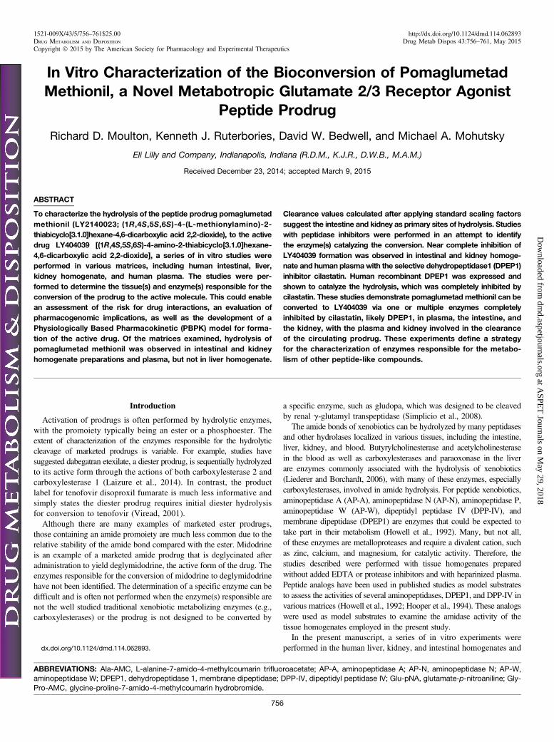
plasma to characterize the hydrolysis of pomaglumetad methionil,a methionil prodrug of the potent and selective metabotropic glutamate2/3 receptor agonist LY404039. The formation of the amine LY404039occurs via cleavage of the amide bond of pomaglumatad methionil (Fig. 1).An understanding of the sites of conversion, characteristics of the enzyme(s)involved, and the potential for the metabolism to be saturated or inhibitedcould enable the understanding of potential pharmacogenomic implicationsas well as the development of PBPK models and other In Vitro In VivoExtrapolation methods to assess the disposition of both the prodrug andthe active molecules (Posada M et al., manuscript in preparation). Theexperiments described used various inhibitors, expressed enzymes,and enzyme kinetic determinations to characterize the hydrolysis ofpomaglumetad methionil. Through these in vitro studies, an enzyme thatcatalyzes the conversion of pomaglumetad methionil to LY404039 wasidentified. In addition, these studies provide a template for a rigorousassessment of the hydrolysis of other peptide prodrugs.
Materials and Methods
Pomaglumetad methionil (LY2140023 monohydrate) and LY404039 wereobtained as reference standards from Eli Lilly and Company (Indianapolis, IN).Actinonin, amastatin, bestatin, cilastatin, 1,10-phenanthroline, flavin adeninedinucleotide (FAD), peroxidase, amino acid oxidase, glutamate-p-nitroaniline(Glu-pNA), L-alanine-7-amido-4-methylcoumarin trifluoroacetate (Ala-AMC),leucine-7-amido-4-methylcoumarin hydrochloride (Leu-AMC), and glycine-proline-7-amido-4-methylcoumarin hydrobromide (Gly-Pro-AMC) were purchasedfrom Sigma-Aldrich Chemical Company (St. Louis, MO). Glycine-D-Phenylalanine(Gly-D-Phe) was purchased from Bachem (Bubendorf, Switzerland). Sitagliptinwas obtained from the Eli Lilly and Company internal collection. Tissuehomogenates were prepared by Celsis/In Vitro Technologies (Baltimore, MD)without the addition of phenylmethanesulfonylfluoride (PMSF), EDTA, or otherprotease inhibitors. Homogenates were stored at approximately 270�C and werethawed only once prior to use. Fresh human blood treated with heparin as ananticoagulant was obtained from the Eli Lilly Research Biologic DonationProgram. Demographics of tissue homogenates and plasma are listed inTables 1–3. Human DPEP1 cDNA was purchased from Openbiosystems Co.Lafayette, CO (catalog number: IHS1380-97431793; clone identity: LIFE-SEQ1341660; accession: NM_004413). DPEP1 was expressed and purifiedby the TTx-Reagents-Proteins group at Eli Lilly and Company.
Incubations with Peptide Substrates. The activities of AP-A, AP-N, AP-W,DPP-IV, and DPEP1 were assessed in human intestinal homogenates usingGlu-pNA, Ala-AMC, Leu-AMC, Gly-Pro-AMC, and Gly-D-Phe as substrates,respectively. Peptide analogs were incubated at either 100 or 250 mM in 75 mg/mlhuman intestinal homogenate in 100 mM Tris-HCl, pH 7.4, containing 4 mMCaCl2. Formation of 7-amino-4-methylcoumarin was quantified by fluores-cence spectrophotometry at an excitation wavelength of 360 nm and an emissionwavelength of 470 nm. Formation of p-nitroaniline was quantified by UVspectrophotometry at a wavelength of 405 nm. DPEP1 activity was determinedby quantifying the hydrolysis of Gly-D-Phe using the method described by
Heywood and Hooper (1995). Briefly, a fluorometric assay mixture containingFAD, peroxidase, D-amino acid oxidase, and p-hydroxyphenylacetic acid wasprepared and incubated with the sample. The formation of D-Phe was quantifiedfluorometrically after several enzymatic steps involving D-amino acid oxidaseand peroxidase.
Incubation of PomaglumetadMethionil in Human Plasma. Fresh heparinizedhuman blood from four individual donors was centrifuged to separate plasma fromred blood cells. Preliminary experiments showed that pomaglumetad methionilturnover in plasma was linear for up to 60 minutes. For the determination of thekinetic parameters, pomaglumetad methionil was incubated at concentrationsranging from 16 to 2000 mM in plasma from each donor in a 96-well plate for10 minutes at 37�C in a shaking water bath. Incubations were terminated bytransferring an aliquot of plasma to methanol containing an internal standard([13C]3LY404039).
Incubation of Pomaglumetad Methionil in Tissue Homogenates andwith DPEP1. Protein and time linearity were determined through examinationof LY404039 formation rates in each of the matrices. These rates were calculatedfrom incubations of a range of tissue homogenate, DPEP1, and substrate con-centrations over a 30-minute time course, ensuring the formation of LY404039was linear over these times with increasing protein concentration. Conditionswere selected within this linear range to ensure accurate determination of kineticparameters and are described in the following paragraph.
For the determination of the kinetic parameters, pomaglumetad methionilwas incubated at concentrations ranging from 32 to 4000 mM in all of thematrices examined. The protein concentrations for human intestinal and kidneyhomogenates were 75 and approximately 300 mg/ml, respectively. Incubationsto determine kinetic parameters with DPEP1 were performed at a protein con-centration of 50 ng/ml. Each of the matrices was diluted in 100 mM Tris-HCl,pH 7.4, containing 4 mM CaCl2. Incubations were performed at 37�C in a shakingwater bath for up to 30 minutes depending on the matrix. All incubations wereterminated by transferring an aliquot of each incubation to methanol containing aninternal standard ([13C]3LY404039).
Incubations with Peptidase Inhibitors. Actinonin, amastatin, bestatin,cilastatin, and sitagliptin at a concentration of 100 mM and phenanthroline at aconcentration of 1 mM were incubated with 500 mM pomaglumetad methionilin each of the matrices at the protein concentrations described previously for thedetermination of kinetic parameters. These inhibitors and the concentrationsused were chosen from a published study, which examined the metabolism ofaspartame (Hooper et al., 1994). For the IC50 determination, 50 ng/ml recombinantDPEP1 was incubated with concentrations of cilastatin ranging from 0.05 to100 mM. Each matrix was preincubated with an inhibitor at least 15 minutes prior tothe addition of the substrate. Incubations were terminated by transferring an aliquotof each incubation to methanol containing an internal standard ([13C]3LY404039).
Sample Preparation and Analysis. Samples were centrifuged to removethe precipitated protein, and the formation of LY404039 was quantified by liquidchromatography tandem mass spectrometry (LC-MS/MS) via direct injection ofthe resulting supernatants. The analyses were performed in the positive ion modeusing selected reaction monitoring [M+H]+ transitions m/z 236.1 . 80.1 and m/z239.1 . 192.1 for LY404039 and [13C]3LY404039, respectively. The analyteswere chromatographically separated using a Thermo Hypersil SAX 5 mm 30 �2.1 mm, analytical column Thermo Scientific (Waltham, MA) and a gradient LCsystem composed of methanol (mobile phase A) and acetic acid/water (1:1, v/v)(mobile phase B). The gradient profile changed from 2% B at 0 minutes, 25% Bat 0.01–0.20 minutes, 75% B at 0.30–0.40 minutes, and 98% at 0.41–0.8 minutesat a flow rate of 1.5 ml/min. Chromatography was performed at ambient laboratoryconditions, and the flow was directed to the MS/MS instrument between 0.2 and0.6 minutes and to waste at all other times.
Standard curve concentrations were study dependent and ranged from 0.049to 200 mM, with each curve including a minimum of six concentrations ofLY404039. In addition, each batch included quality control samples at severalconcentrations within the standard curve range. The percentage CV and per-centage accuracy values for the standard curves ranged from 0.2 to 17.8 and78.6 to 123.0, respectively. The percentage CV and percentage accuracy valuesfor the quality control samples ranged from 0.6 to 15.5 and 78.2 to 123.2,respectively.
Data Analysis.Mass calibration, data acquisition, chromatographic analysis,mass spectral representation, and postacquisition quantitative analysis wereperformed via Analyst software version 1.4.2 (Sciex, Foster City, CA).Fig. 1. Conversion of pomaglumetad methionil to LY404039
Bioconversion of Pomaglumetad Methionil 757
at ASPE
T Journals on M
ay 29, 2018dm
d.aspetjournals.orgD
ownloaded from
Calculations. Concentration data were processed in Excel 2007 (version 12;Microsoft, Redmond, WA). Kinetic parameters were calculated using XLfit(version 5.3; ID Business Solutions Limited, Guildford, United Kingdom), andthe data were best fit to the one-enzyme Michaelis-Menten model. Intestinaland kidney clearance was scaled using the equation Vmax/Km * homogenate(mg)/tissue (g) * tissue (g)/body weight (kg) * 70 kg. Homogenate (mg)/tissue(g) values were provided by Celsis/In Vitro Technologies for each lot. Factorsof 30 and 4.4 g tissue/kg body weight for the intestine and kidney, respectively,were taken from Yamanaka et al. (2007). The scaled intrinsic clearance valuesfor the plasma were calculated using the equation Vmax/Km * 3 liters of plasma(Guyton, 1991). Inhibition data were fit to the four parameter Hill-Slope modelusing XLfit to determine an IC50 value.
Results
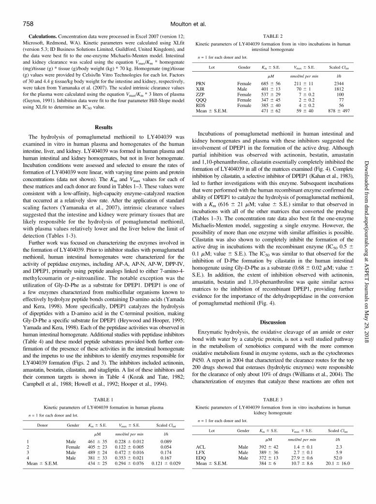
The hydrolysis of pomaglumetad methionil to LY404039 wasexamined in vitro in human plasma and homogenates of the humanintestine, liver, and kidney. LY404039 was formed in human plasma andhuman intestinal and kidney homogenates, but not in liver homogenate.Incubation conditions were assessed and selected to ensure the rates offormation of LY404039 were linear, with varying time points and proteinconcentrations (data not shown). The Km and Vmax values for each ofthese matrices and each donor are found in Tables 1–3. These values wereconsistent with a low-affinity, high-capacity enzyme–catalyzed reactionthat occurred at a relatively slow rate. After the application of standardscaling factors (Yamanaka et al., 2007), intrinsic clearance valuessuggested that the intestine and kidney were primary tissues that arelikely responsible for the hydrolysis of pomaglumetad methionil,with plasma values relatively lower and the liver below the limit ofdetection (Tables 1-3).Further work was focused on characterizing the enzymes involved in
the formation of LY404039. Prior to inhibitor studies with pomaglumetadmethionil, human intestinal homogenates were characterized for theactivity of peptidase enzymes, including AP-A, AP-N, AP-W, DPP-IV,and DPEP1, primarily using peptide analogs linked to either 7-amino-4-methylcoumarin or p-nitroaniline. The notable exception was theutilization of Gly-D-Phe as a substrate for DPEP1. DPEP1 is one ofa few enzymes characterized from multicellular organisms known toeffectively hydrolyze peptide bonds containing D-amino acids (Yamadaand Kera, 1998). More specifically, DPEP1 catalyzes the hydrolysisof dipeptides with a D-amino acid in the C-terminal position, makingGly-D-Phe a specific substrate for DPEP1 (Heywood and Hooper, 1995;Yamada and Kera, 1998). Each of the peptidase activities was observed inhuman intestinal homogenate. Additional studies with peptidase inhibitors(Table 4) and these model peptide substrates provided both further con-firmation of the presence of these activities in the intestinal homogenateand the impetus to use the inhibitors to identify enzymes responsible forLY404039 formation (Figs. 2 and 3). The inhibitors included actinonin,amastatin, bestatin, cilastatin, and sitagliptin. A list of these inhibitors andtheir common targets is shown in Table 4 (Kozak and Tate, 1982;Campbell et al., 1988; Howell et al., 1992; Hooper et al., 1994).
Incubations of pomaglumetad methionil in human intestinal andkidney homogenates and plasma with these inhibitors suggested theinvolvement of DPEP1 in the formation of the active drug. Althoughpartial inhibition was observed with actinonin, bestatin, amastatinand 1,10-phenanthroline, cilastatin essentially completely inhibited theformation of LY404039 in all of the matrices examined (Fig. 4). Completeinhibition by cilastatin, a selective inhibitor of DPEP1 (Kahan et al., 1983),led to further investigations with this enzyme. Subsequent incubationsthat were performed with the human recombinant enzyme confirmed theability of DPEP1 to catalyze the hydrolysis of pomaglumetad methionil,with a Km (616 6 21 mM; value 6 S.E.) similar to that observed inincubations with all of the other matrices that converted the prodrug(Tables 1–3). The concentration rate data also best fit the one-enzymeMichaelis-Menten model, suggesting a single enzyme. However, thepossibility of more than one enzyme with similar affinities is possible.Cilastatin was also shown to completely inhibit the formation of theactive drug in incubations with the recombinant enzyme (IC50 0.5 60.1 mM; value 6 S.E.). The IC50 was similar to that observed for theinhibition of D-Phe formation by cilastatin in the human intestinalhomogenate using Gly-D-Phe as a substrate (0.68 6 0.02 mM; value 6S.E.). In addition, the extent of inhibition observed with actinonin,amastatin, bestatin and 1,10-phenanthroline was quite similar acrossmatrices to the inhibition of recombinant DPEP1, providing furtherevidence for the importance of the dehydropeptidase in the conversionof pomaglumetad methionil (Fig. 4).
Discussion
Enzymatic hydrolysis, the oxidative cleavage of an amide or esterbond with water by a catalytic protein, is not a well studied pathwayin the metabolism of xenobiotics compared with the more commonoxidative metabolism found in enzyme systems, such as the cytochromesP450. A report in 2004 that characterized the clearance routes for the top200 drugs showed that esterases (hydrolytic enzymes) were responsiblefor the clearance of only about 10% of drugs (Williams et al., 2004). Thecharacterization of enzymes that catalyze these reactions are often not
TABLE 1
Kinetic parameters of LY404039 formation in human plasma
n = 1 for each donor and lot.
Donor Gender Km 6 S.E. Vmax 6 S.E. Scaled Clint
mM nmol/ml per min l/h
1 Male 461 6 35 0.228 6 0.012 0.0892 Female 405 6 23 0.122 6 0.005 0.0543 Male 489 6 24 0.472 6 0.016 0.1744 Male 381 6 33 0.353 6 0.021 0.167Mean 6 S.E.M. 434 6 25 0.294 6 0.076 0.121 6 0.029
TABLE 2
Kinetic parameters of LY404039 formation from in vitro incubations in humanintestinal homogenate
n = 1 for each donor and lot.
Lot Gender Km 6 S.E. Vmax 6 S.E. Scaled Clint
mM nmol/ml per min l/h
PRN Female 685 6 56 211 6 11 2344XJR Male 401 6 13 70 6 1 1812ZZP Female 537 6 29 7 6 0.2 100QQQ Female 347 6 45 2 6 0.2 77RDS Female 385 6 40 4 6 0.2 56Mean 6 S.E.M. 471 6 62 59 6 40 878 6 497
TABLE 3
Kinetic parameters of LY404039 formation from in vitro incubations in humankidney homogenate
n = 1 for each donor and lot.
Lot Gender Km 6 S.E. Vmax 6 S.E. Scaled Clint
mM nmol/ml per min l/h
ACL Male 392 6 42 1.4 6 0.1 2.3LFX Male 389 6 36 2.7 6 0.1 5.9EDQ Male 372 6 13 27.9 6 0.6 52.0Mean 6 S.E.M. 384 6 6 10.7 6 8.6 20.1 6 16.0
758 Moulton et al.
at ASPE
T Journals on M
ay 29, 2018dm
d.aspetjournals.orgD
ownloaded from

completed to the same extent as other enzyme systems, such as thecytochromes P450, due to a lack of previous experience with theseenzymes and appropriate in vitro tools. The cytochromes P450, forexample, have full arrays of recombinant enzymes, specific inhibitors,and specific substrates that are commercially available, enabling rigorouscharacterization of the involvement of these enzymes in the metabolismof xenobiotics. Also, many of the CYP enzymes have been quantifiedin various tissues to allow for the scaling of the activities. Hydrolyticenzymes, on the other hand, are limited to a relatively small numberof commercially available enzymes, with a relative dearth of specificsubstrates and inhibitors. Quantification of the enzymes in tissues is alsoin its relatively early stages. Making the characterization of hydrolysispathways more difficult, the commercially available tissue fractions alsooften contain EDTA, PMSF, or both, which are additives that will inhibitmany hydrolytic enzymes.Using the small number of available tools, the hydrolysis of
pomaglumetad methionil was characterized in human intestinal, kidney,and liver homogenates and plasma. The work presented provides themethodology for studying the hydrolysis of pomaglumetad and otherpeptide prodrugs. Assay conditions, such as buffer selection, the use oftissue homogenates without added protease inhibitors or EDTA, and theuse of heparin instead of EDTA as an anticoagulant for plasma, are
important considerations in the study of hydrolytic enzymes becausemany are metalloproteases. For example, no conversion of pomaglumetadmethionil was observed in two lots of human intestinal homogenates ininitial incubations performed in a sodium phosphate buffer. However,when the incubations were repeated using Tris buffer, activity wasobserved. The addition of calcium and magnesium to the incubationincreased the rate of conversion of pomaglumated methionil (Fig. 5).Chelation of calcium by the phosphate ion in the sodium phosphatebuffer is the likely explanation for the lack of conversion observedin preliminary studies. The use of phosphate buffers or other buffersthat may chelate metal ions could inhibit the activities of thesemetalloproteases. All results reported in this study were from incubationsperformed in Tris buffer with added calcium. Calcium was added inexcess to ensure the enzyme activity would not be affected by a lack ofdivalent cations. The effect of the addition of both calcium and magnesiumon the formation of LY404039 was investigated, with essentially nodifference observed in the rate of formation with each cation (datanot shown).Due to the limited knowledge of the enzymes involved in the
formation of LY404039, tissue homogenates were used in these studiesinstead of subcellular fractions to increase the possibility the enzymesinvolved would be represented. However, because these were humantissue preparations, the quality and treatment of the tissue, including thetime interval between receipt and processing of the tissue, may becritical to the activity of these enzymes and may explain the observedlarge differences in Vmax values between donors (Tables 2 and 3). Inaccordance with this concept, the human plasma was freshly preparedand the range in Vmax values was substantially reduced.Prior to incubations to establish kinetic parameters for pomaglumetad
methionil hydrolysis, linear rate conditions for LY404039 formation weredetermined for each of the matrices by incubating multiple concentrationsof pomaglumetad methionil, with varying protein concentrations of eachmatrix for increasing periods of time. Results from these initial incubations
TABLE 4
Peptidase inhibitors and their associated target enzyme(s)
Inhibitor Target Enzyme
Actinonin AP-NAmastatin AP-A, AP-N, and AP-WBestatin AP-W and DPEP1Cilastatin DPEP1Phenanthroline AP-A, AP-N, and DPEP1Sitagliptin DPP-IV
Fig. 2. Characterization of peptidase activity inhuman intestinal homogenate lot XJR. Formationof p-NA from Glu-pNA was substantially inhibitedby amastatin, as expected for a substrate of AP-A(A). Formation of AMC from Ala and leucine-7-amido-4-methylcoumarin hydrochloride (Leu-AMC)was substantially inhibited by amastatin, actinonin, andbestatin, as expected for substrates of AP-N (B) andAP-W (C). In addition, formation of AMC from Gly-Pro-AMC was completely inhibited by sitagliptin, asexpected for a substrate of DPP-IV (D). Each valuerepresents the mean 6 S.D. of triplicate wells.
Bioconversion of Pomaglumetad Methionil 759
at ASPE
T Journals on M
ay 29, 2018dm
d.aspetjournals.orgD
ownloaded from

enabled an appropriate selection of protein concentration and incubationtime for each matrix, where a linear rate of LY404039 formation wasmaintained. These conditions were critical to ensure first-order rateconditions for the determination of the Km and Vmax of pomaglumetadmethionil hydrolysis.The majority of the inhibitors used in these studies were selected
from a publication by Hooper et al. (1994) that describes in vitro workcharacterizing the metabolism of aspartame. Actinonin, amastatin, andbestatin are all naturally produced by actinomycetes and were originallyisolated for either their antibacterial or aminopeptidase inhibitory properties(Umezawa et al., 1976; Aoyagi et al., 1978; Chen et al., 2000). Actinonin isa known inhibitor of several metallo hydrolases and appears to act throughits hydroxamate group, which chelates metal ions required for enzymaticactivity (Chen et al., 2000). Amastatin and bestatin inhibit aminopeptidasesprimarily through slow, tight, and often time-dependent yet reversiblebinding to the enzymes (Wilkes and Prescott, 1985). Evidence suggeststhese inhibitors may act through the formation of a transition-state analog
complex with the enzyme (Wilkes and Prescott, 1985). In addition,amastatin and bestatin may interact with the metal ions required for activityof these enzymes, with spectral shifts for the metallo hydrolase Aeromonasaminopeptidase observed in the presence of these inhibitors (Wilkes andPrescott, 1985). Cilastatin was specifically designed as an inhibitor ofDPEP1 to be dosed with imipenem, an antibiotic whose b-lactam ring issusceptible to cleavage by the dehydropeptidase (Kahan et al., 1983).Coadministration of cilastatin with imipenem increases the urinary recoveryof the antibiotic, which is typically low when imipenem is dosed alone dueto extensive metabolism by DPEP1 in the renal brush border (Kahan et al.,1983). Sitagliptin is a marketed oral antihyperglycemic drug that acts bycompetitively inhibiting DPP-IV (Green et al., 2006). Although these
Fig. 3. Characterization of AP-N, AP-W, and DPP-IV activity across four lots of humanintestinal homogenate. The activities of AP-N, AP-W, and DPP-IV were assessed usingAla-AMC, Leu-AMC, and Gly-Pro-AMC as substrates, respectively. Each activity waspresent and varied to a similar degree across lots, with lots PRN and XJR showinga higher activity for each enzyme than lots QQQ and RDS. AP-N was inhibited byactinonin, AP-W was inhibited by amastatin, and DPP-IV was inhibited by sitagliptin,as expected.
Fig. 4. Effect of peptidase inhibitors on LY404039 formation. Actinonin, amastatin,bestatin, cilastatin, and sitagliptin were incubated at a concentration of 100 mM witheach matrix. Phenanthroline was incubated at a concentration of 1 mM with each matrix.Human intestinal homogenate, human kidney homogenate, and human recombinantmembrane dipeptidase were incubated at 75 mg/ml, 300 mg/ml, and 50 ng/ml,respectively. The concentration of LY2140023 in each incubation was 500 mM. Thepercentage of inhibition was determined by comparing LY404039 formation in thepresence of each inhibitor to the formation observed in a vehicle control for eachmatrix examined. Human plasma values represent data from an incubation of equalvolumes of plasma pooled from four individual donors. Human intestinal homogenatevalues (6S.E.M.) represent data from an incubation of four separate lots that were eachprepared from a single donor. Human kidney homogenate values (6S.E.M.) representdata from an incubation of three separate lots that were each prepared from a singledonor. Human DPEP1 values represent data from a single experiment.
Fig. 5. Effect of calcium addition on the hydrolysis of LY2140023 in humanintestinal homogenate lot RDS. The Km and Vmax values for the formation ofLY404039 in the human intestinal homogenate lot RDS with (circle; solid line) andwithout (square; dashed line) the addition of calcium were 3116 18 mM and 2.1560.04 nmol/mg per min and 396 6 25 mM and 0.64 6 0.01 nmol/mg per min.
760 Moulton et al.
at ASPE
T Journals on M
ay 29, 2018dm
d.aspetjournals.orgD
ownloaded from

studies do not preclude the involvement of other enzymes in the catalysis ofLY404039 formation, DPEP1 has been demonstrated to catalyze thehydrolysis of pomaglumetad methionil in human intestinal and kidneyhomogenates and human plasma. The fact that hydrolysis was not observedin liver homogenate suggested that the enzyme involved in the metabolismof the prodrug was not one of the common xenobiotic metabolizinghydrolysis enzymes, such as carboxylesterase 1 and 2 or paraoxonase. TheKm values for LY404039 formation in human intestinal homogenate,kidney homogenate, and plasma were similar to the Km value for humanrecombinant DPEP1-catalyzed formation. In addition, cilastatin was shownto essentially completely inhibit LY404039 formation in all of the matricesexamined. Partial inhibition of LY404039 formation was observed inall matrices in incubations with actinonin, amastatin, bestatin, and 1,10-phenanthroline (Fig. 4). Because several of these inhibitors act throughmetal ion chelation and DPEP1 has four zinc molecules involved incatalysis, partial inhibition by several inhibitors was not unexpected.Although some differences were observed in the extent of inhibition fora given inhibitor across matrices, in general, the extent of inhibition ofrecombinant DPEP1 by each inhibitor was similar to that observed in othermatrices. These results provide more evidence that DPEP1 is an importantenzyme involved in the formation of LY404039. DPEP1, also known asrenal dipeptidase or membrane dipeptidase (EC 3.4.13.19), is a glycosylphosphatidylinositol anchored cell-surface zinc metalloprotease that hasbeen characterized extensively in the microvillar membranes of the kidney(Littlewood et al., 1989). The enzyme is known to catalyze the hydrolysisof dipeptides with D-, L-, or dehydro-amino acids at the C-terminus(Liao et al., 2010). DPEP1 catalyzes the hydrolysis of beta lactamantibiotics, such as imipenem and carbapenem, as well as glutathioneand leukotriene D4. Cilastatin was designed as a selective inhibitor ofDPEP1 to be coadministered with imipenem, with the intent of improvingthe pharmacokinetic profile of the drug by inhibiting the DPEP1-catalyzed hydrolysis of its beta lactam ring (Kahan et al., 1983). Althoughan unlikely concomitant medication, Primaxin (imipenem/cilastatin) couldaffect the exposure of pomaglumetad methionil. Further studies would berequired to assess the possibility of a drug interaction with Primaxin.Pomaglumetad methionil was designed to be actively transported by
peptide transporter 1 and has been demonstrated through in vitro transportassays to be a substrate of this transporter (Pak YA et al., manuscript inpreparation.). Peptide transporter 1 is known to be located on the cellsurface of the enterocyte; however, the interplay between the conversionof the prodrug to the active drug and the transport of the prodrug in theintestine is not understood. Although DPEP1 is located on the apical sideof microvillar membranes in the kidney, the location of intestinal DPEP1is unknown, although evidence in this study suggests it may not only beon the apical side. Results of these in vitro studies, including affinities,metabolism via expressed enzymes, and inhibition, suggest DPEP1 mayplay a substantial role in the activation to LY404039. Knowledge of theenzyme identity, tissues involved in metabolism, and enzyme kinetics canallow for the development of PBPK models to assess the disposition of
prodrugs and active molecules as well as the potential for marketed drugsto inhibit the conversion. In addition, these studies provide a frameworkfor the characterization of peptidase-mediated hydrolysis.
Acknowledgments
The authors thank the TTx-Reagents-Proteins group at Eli Lilly and Companyfor expressing and purifying human DPEP1.
Authorship ContributionsParticipated in research design: Moulton, Ruterbories, Mohutsky.Conducted experiments: Moulton, Ruterbories, Bedwell.Performed data analysis: Moulton, Ruterbories, Bedwell, Mohutsky.Wrote or contributed to the writing of the manuscript:Moulton, Ruterbories,
Mohutsky.
References
Aoyagi T, Tobe H, Kojima F, Hamada M, Takeuchi T, and Umezawa H (1978) Amastatin, aninhibitor of aminopeptidase A, produced by actinomycetes. J Antibiot (Tokyo) 31:636–638.
Campbell BJ, Di Shih Y, Forrester LJ, and Zahler WL (1988) Specificity and inhibition studies ofhuman renal dipeptidase. Biochim Biophys Acta 956:110–118.
Chen DZ, Patel DV, Hackbarth CJ, Wang W, Dreyer G, Young DC, Margolis PS, Wu C, Ni ZJ,and Trias J, et al. (2000) Actinonin, a naturally occurring antibacterial agent, is a potentdeformylase inhibitor. Biochemistry 39:1256–1262.
Green BD, Flatt PR, and Bailey CJ (2006) Dipeptidyl peptidase IV (DPP IV) inhibitors: a newlyemerging drug class for the treatment of type 2 diabetes. Diab Vasc Dis Res 3:159–165.
Guyton AC (1991) Textbook of Medical Physiology, 8th ed, Saunders, Philadelphia, PA.Heywood SP and Hooper NM (1995) Development and application of a fluorometric assay formammalian membrane dipeptidase. Anal Biochem 226:10–14.
Hooper NM, Hesp RJ, and Tieku S (1994) Metabolism of aspartame by human and pig intestinalmicrovillar peptidases. Biochem J 298:635–639.
Howell S, Kenny AJ, and Turner AJ (1992) A survey of membrane peptidases in two humancolonic cell lines, Caco-2 and HT-29. Biochem J 284:595–601.
Kahan FM, Kropp H, Sundelof JG, and Birnbaum J (1983) Thienamycin: development ofimipenen-cilastatin. J Antimicrob Chemother 12 (Suppl D):1–35.
Kozak EM and Tate SS (1982) Glutathione-degrading enzymes of microvillus membranes. J BiolChem 257:6322–6327.
Laizure SC, Parker RB, Herring VL, and Hu ZY (2014) Identification of carboxylesterase-dependent dabigatran etexilate hydrolysis. Drug Metab Dispos 42:201–206.
Liao RZ, Himo F, Yu JG, and Liu RZ (2010) Dipeptide hydrolysis by the dinuclear zinc enzyme humanrenal dipeptidase: mechanistic insights from DFT calculations. J Inorg Biochem 104:37–46.
Liederer BM and Borchardt RT (2006) Enzymes involved in the bioconversion of ester-basedprodrugs. J Pharm Sci 95:1177–1195.
Littlewood GM, Hooper NM, and Turner AJ (1989) Ectoenzymes of the kidney microvillarmembrane. Affinity purification, characterization and localization of the phospholipase C-solubilizedform of renal dipeptidase. Biochem J 257:361–367.
Simplício AL, Clancy JM, and Gilmer JF (2008) Prodrugs for amines. Molecules 13:519–547.Umezawa H, Aoyagi T, Suda H, Hamada M, and Takeuchi T (1976) Bestatin, an inhibitor ofaminopeptidase B, produced by actinomycetes. J Antibiot (Tokyo) 29:97–99.
Viread. (2001) Package insert. Gilead Sciences, Foster City, CA.Wilkes SH and Prescott JM (1985) The slow, tight binding of bestatin and amastatin to amino-peptidases. J Biol Chem 260:13154–13162.
Williams JA, Hyland R, Jones BC, Smith DA, Hurst S, Goosen TC, Peterkin V, Koup JR, and BallSE (2004) Drug-drug interactions for UDP-glucuronosyltransferase substrates: a pharmacokineticexplanation for typically observed low exposure (AUCi/AUC) ratios. Drug Metab Dispos 32:1201–1208.
Yamada R and Kera Y (1998) D-amino acid hydrolysing enzymes. EXS 85:145–155.Yamanaka H, Nakajima M, Katoh M, and Yokoi T (2007) Glucuronidation of thyroxine in humanliver, jejunum, and kidney microsomes. Drug Metab Dispos 35:1642–1648.
Address correspondence to: Richard Moulton, Eli Lilly and Company, Indian-apolis, IN. E-mail: [email protected]
Bioconversion of Pomaglumetad Methionil 761
at ASPE
T Journals on M
ay 29, 2018dm
d.aspetjournals.orgD
ownloaded from