in vitro antiprotozoan activity and mechanisms of action
TRANSCRIPT

Received: 29 August 2017 Revised: 11 January 2018 Accepted: 24 March 2018
DOI: 10.1002/ptr.6093
R E S E A R CH AR T I C L E
In vitro antiprotozoan activity and mechanisms of action ofselected Ghanaian medicinal plants against Trypanosoma,Leishmania, and Plasmodium parasites
Mitsuko Ohashi1,2† | Michael Amoa-Bosompem1,2† | Kofi Dadzie Kwofie1,2† |
Jefferey Agyapong1† | Richard Adegle4 | Maxwell Mamfe Sakyiamah2,4 |
Frederick Ayertey4 | Kofi Baffuor‐Awuah Owusu1 | Isaac Tuffour1 | Philip Atchoglo1 |
Nguyen Huu Tung3 | Takuhiro Uto3 | Frederick Aboagye4 | Alfred Ampomah Appiah4 |
Regina Appiah-Opong1 | Alexander K. Nyarko1 | William Kofi Anyan1 | Irene Ayi1 |
Daniel Adjei Boakye1 | Kwadwo Ansah Koram1 | Dominic Edoh4 | Shoji Yamaoka2 |
Yukihiro Shoyama3 | Nobuo Ohta4
1Noguchi Memorial Institute for Medical Research, College of Health Sciences, University of Ghana, P.O. Box LG 581, Legon, Ghana
2Section of Environmental Parasitology, Faculty of Medicine, Tokyo Medical and Dental University, 1‐5‐45 Yushima, Bunkyo‐ku, Tokyo 113‐8510, Japan3Faculty of Pharmaceutical Sciences, Nagasaki International University, 2825‐7 Huis Ten Bosch, Sasebo, Nagasaki 859‐3298, Japan4Centre for Plant Medicine Research, P.O. Box 73, Mampong, Akuapem, Ghana
Correspondence
Mitsuko Ohashi, Noguchi Memorial Institute
for Medical Research, College of Health
Sciences, University of Ghana, P.O. Box LG
581, Legon, Ghana.
Email: [email protected]
Funding information
Japan Agency for Medical Research and
Development (AMED); Ministry of Education,
Culture, Sports, Science and Technology;
Japan Initiative for Global Research Network
on Infectious Diseases (J‐GRID); Japan Inter-
national Cooperation Agency (JICA); Japan
Science and Technology Agency (JST); Science
and Technology Research Partnership for
Sustainable Development (SATREPS)
Abbreviations: FACS, fluorescent activated cell so†Mitsuko Ohashi, Michael Amoa‐Bosompem, Kofi
Phytotherapy Research. 2018;32:1617–1630.
Trypanosomiasis, leishmaniasis, and malaria are protozoan infections of public health
importance with thousands of new cases recorded annually. Control of these infec-
tion(s) with existing chemotherapy is limited by drug toxicity, lengthy parenteral treat-
ment, affordability, and/or the emergence of resistant strains. Medicinal plants on the
other hand are used in the treatment of various infectious diseases although their chem-
ical properties are not fully evaluated. In this study, we screened 112 crude extracts
from 72 selected Ghanaian medicinal plants for anti‐Trypanosoma, anti‐Leishmania,
and anti‐Plasmodium activities in vitro and investigated their mechanisms of action.
Twenty‐three extracts from 20 plants showed significant antiprotozoan activity against
at least 1 of 3 protozoan parasites screened with IC50 values less than 20 μg/ml. Eleven
extracts showed high anti‐Trypanosoma activity with Bidens pilosa whole plant and
Morinda lucida leaf extracts recording the highest activities. Their IC50 (selectivity index
[SI]) values were 5.51 μg/ml (35.00) and 5.96 μg/ml (13.09), respectively. Nine extracts
had high anti‐Leishmania activity with Annona senegalensis and Cassia alata leaf extracts
as the most active. Their IC50 (SI) values were 10.8 μg/ml (1.50) and 10.1 μg/ml (0.37),
respectively. Six extracts had high anti‐Plasmodium activity with the leaf and stem‐bark
extracts of Terminalia ivorensis recording the highest activity. Their IC50 (SI) values were
7.26 μg/ml (129.36) and 17.45 μg/ml (17.17), respectively. Only M. lucida at 25 μg/ml
induced significant apoptosis‐like cell death inTrypanosoma parasites. Anti‐Leishmania
rting
Dadzie Kwofie, and Jefferey Agyapong contributed equally to this work.
Copyright © 2018 John Wiley & Sons, Ltd.wileyonlinelibrary.com/journal/ptr 1617

1618 OHASHI ET AL.
active extracts induced varying morphological changes in Leishmania parasites such as
multiple nuclei and/or kinetoplast, incomplete flagella division, or nuclear fragmenta-
tion. Active extracts may be potential sources for developing new chemotherapy
against these infections.
KEYWORDS
apoptosis, in vitro screening, Leishmania donovani, medicinal plants, morphology, Plasmodium
falciparum, Trypanosoma brucei brucei
1 | INTRODUCTION
Protozoan infections are a major health problem causing significant
morbidity and mortality in Africa, Asia, and Latin America (World
Health Organization [WHO], 2015b). Although chemotherapy is one
of the main forms of controlling protozoan infection, it is limited by
the accessibility, adverse side effects, and the emergence of resistant
parasites to available drugs.
African trypanosomiasis and leishmaniasis are protozoan infec-
tions caused by kinetoplastids. They are considered as the main path-
ogens of neglected tropical diseases. Trypanosomiasis is caused by
Trypanosoma brucei species, transmitted by the tsetse fly, and
threatens the lives of 50 million people in over 36 countries in sub‐
Saharan Africa with an estimated 30,000 new cases each year
(Adeyemi, Sykes, Akanji, & Avery, 2011; Centers for Disease Control
and Prevention, 2016; WHO, 2016a, 2016b). Chronic and acute forms
of human African trypanosomiasis are caused by Trypanosoma brucei
gambiense and Trypanosoma brucei rhodesiense, respectively, whereas
Trypanosoma brucei brucei causes Nagana in animals (Shi, Wei, Pan, &
Tabel, 2006). Leishmaniasis on the other hand is caused by over 20
different species of Leishmania and is transmitted by the sand fly.
Approximately 350 million people are at risk of infection in over 88
countries across the world (Bensoussan, Nasereddin, Jonas, Schnur,
& Jaffe, 2006). An estimated 1.3 million new cases and 20,000 to
30,000 deaths occur annually (WHO, 2016a). There are three forms
of leishmaniasis: cutaneous, mucocutaneous, and visceral leishmaniasis
(Alvar, Yactayo, & Bern, 2006; Desjeux, 2004; Herwaldt, 1999;
Lysenko, 1971; Murray, Berman, Davies, & Saravia, 2005). Last but
not least, malaria, a typical protozoan infection that affects millions of
people worldwide, is caused by Plasmodium species and transmitted
by the female Anopheles mosquito. Sub‐Saharan Africa alone accounts
for 89% of malaria cases with 78% of malaria fatality occurring in
children under 5 years old (WHO, 2015a).
The control of all three protozoan infections is limited by
drug toxicity, resistant strains of parasites, and economic/
financial factors. In the case of malaria for example, resistant malaria
is a very serious problem worldwide. Quinine was the drug of choice
for close to 100 years before it was replaced with artemisinin isolated
from Artemisia annua due to the emergence of resistant parasites.
There are however reports of artemisinin failure in South East Asia
making it necessary to develop new effective chemotherapy (Adeyemi
et al., 2011; Bacchi, 2009; Balunas & Kinghorn, 2005; Singh &
Sivakumar, 2004). Despite the use of medicinal plants in the treatment
of ailments including those caused by protozoan pathogens in Africa
(Ankrah et al., 2003; Barrett, Boykin, Brun, & Tidwell, 2007; Okpekon
et al., 2004; Rahmatullah et al., 2010; Trouiller et al., 2002), scientific
evidence of the medicinal properties of these plants have not been fully
evaluated (Abu & Uchendu, 2011; Fathuddin, 2011; M. A. Ibrahim et al.,
2010; N. Nweze, Anene, & Asuzu, 2011; Nweze, 2012; Ogbadoyi,
Kabiru, & Omotosho, 2011; Wurochekke & Anyanwu, 2012).
This study therefore aimed at screening selected Ghanaian medic-
inal plants, based on knowledge of their traditional use in treating
various infections/diseases, for anti‐Trypanosoma, anti‐Leishmania,
and anti‐Plasmodium properties in vitro.
2 | MATERIALS AND METHODS
2.1 | Plant materials and preparation ofcrude extracts
Based on the traditional knowledge of their medicinal use, extracts
from different parts (leaves, stem bark, fruits, seeds, or roots) of 72
plants were collected in Ghana by the Centre for Plant Medicine
Research, Mampong, Ghana, during the period of October 2010 to
November 2012. Authentication was done by one of the authors
(Y. S.). Voucher specimens have been deposited in Centre for Plant
Medicine Research. The air‐dried and pulverized plant samples
(200 g) were extracted by 50% aqueous EtOH 3 times under room
temperature. The accumulated solution was evaporated in a rotary
evaporator at 40 °C to obtain the crude extract. The extracts were
kept in sterile tubes and stored at 4 °C until use. Prior to drug‐sensitiv-
ity assays, 100‐mg/ml stock solutions were prepared with 50% EtOH
and filter sterilized.
2.2 | In vitro culture of parasitic protozoans
GUTat 3.1 strain of the bloodstream form of T. b. brucei was used for
this study. Parasites were cultured in vitro according to conditions
established previously (Yabu et al., 1998). Parasites were used for
assays when they reached a concentration of 1 × 10⁶ parasites/ml.
Estimation of parasitemia was done with the Neubauer's counting
chamber. Parasites were diluted to a concentration of 3 × 10⁵ para-
sites/ml with HM1‐9 medium and used for the various experiments.
For Leishmania parasites, promastigote forms of Leishmania donovani
(MHOM/NP/03/D10) cultures were used in this study with the
culture media previously established with slight modifications

OHASHI ET AL. 1619
(Mottram, 2008). The parasites were used for assays when they
reached a concentration of 1 × 10⁷ parasites/ml. Parasitemia was
estimated with the Neubauer's counting chamber. Parasites were
diluted to a concentration of 2.5 × 10⁶ parasites/ml with M199
medium for drug assays.
For the maintenance of malaria cultures, 3D7 strain of Plasmodium
falciparum was cultured based on previously established protocols
with modifications (Trager & Jensen, 1976). Culture media were
changed daily, and the level of parasitemia was determined by
counting red blood cells (RBCs) on a Giemsa‐stained thin blood smear
under a light microscope. Plasmodium culture, at 5% parasitemia, was
synchronized with 5% sorbitol to obtain ring stage, trophozoite
parasites, and incubated for an extra 48 hr to obtain trophozoites,
which were used in the screening of plant extracts.
2.3 | In vitro antiparasitic screening assays ofplant extracts
2.3.1 | Antikinetoplastid activity
The Alamar Blue assay (Alamar Blue®, Life Technologies™, USA) was
used to determine the antitrypanosomal and antileishmanial activities
of plant extracts. Assays were carried out in a 96‐well plate following
manufacturer's instructions with modifications (Kwofie et al., 2016).
Either 1.5 × 10⁴ Trypanosomaparasites perwell or 1.25 ×10⁵ Leishmania
parasites per well were seeded with varied concentrations of crude
extracts ranging from 0 to 200 μg/ml. Final concentration of EtOH was
kept under 1%, and a solvent control (negative control) was used in all
assays. Berberine and amphotericin B were used as positive controls
for Trypanosoma and Leishmania, respectively. After a 24‐hr incubation
of Trypanosoma and 44‐hr incubation of Leishmania parasites with or
without plant extracts, 10% Alamar Blue dye was added and incubated
for 24 and 4 hr in darkness, respectively. After 48 hr, the plate was read
for absorbance at a wavelength of 540 nm (reference wavelength of
595 nm) using a spectrophotometer (TECAN Sunrise Wako, Japan).
Trend curves were drawn to obtain IC50 values of plant extracts.
2.3.2 | Anti‐Plasmodium activity
Fluorescent activated cell sorting (FACS) was used to determine the
anti‐Plasmodium activity of the plant extracts. Synchronized parasites
at a packed volume of 2% hematocrit and 1% parasitemia were chal-
lenged with 0‐ to 25‐μg/ml plant extracts for 48 hr. Artesunate
(Sigma‐Aldrich, USA) was used as the positive control, whereas RBCs
at 2% hematocrit only and packed RBCs plus 2.5% EtOH served as
negative and vehicle controls, respectively. SYBR Green I solution
(0.25 μl of 10,000 × SYBR Green I/1 ml of 1 × phosphate buffered
saline [PBS]) was added to each well after the 48‐hr incubation period
and incubated for additional 30 min in the dark at 37 °C. Plates were
read using the Guava EasyCyte 5HT FACS machine (Millipore, USA)
following the manufacturer's instructions.
2.4 | In vitro cytotoxicity assay
Jurkat cells (human acuteT‐cell leukemia cells) were obtained from the
RIKEN BioResource Centre Cell Bank (Japan) and maintained in RPMI
supplemented with 10% fetal bovine serum and 1% penicillin–
streptomycin–L‐glutamine. The cells were incubated at 37 °C under
5% CO2 in fully humidified conditions. The toxicity of plant extracts
against the Jurkat cells was determined using a 3‐(4,5‐dimethyl-
thiazol‐2‐yl)‐2,5‐diphenyltetrazolium bromide (MTT) assay. Cells were
plated at a density of 3.0 × 10⁵ cells/ml into a 96‐well plate. Cells were
treated with various concentrations of each of the plant extracts and
incubated for 48 hr. MTT solution was added to each well, and the
cells were incubated for an extra 4 hr. The precipitated MTT‐formazan
product was dissolved in 0.04 N HCl–isopropanol, and the amount of
formazan was measured at a wavelength of 570 nm by a microplate
spectrophotometer (Tecan Infinite M200 Pro, Austria). Cytotoxicity
was calculated as the percentage of life cells relative to the control
culture. The selectivity index (SI) was expressed as the ratio of the
IC50 value obtained for mammalian cells to the IC50 values obtained
for parasites (Kwofie et al., 2016).
2.5 | Apoptosis assay
Nexin assay using EasyCyte 5HT FACS machine (Millipore, USA) was
performed to investigate apoptotic properties of active crude extracts
against T. brucei. Seeding and incubation of parasites with crude
extracts were done under the same conditions of in vitro antiparasitic
screening assay as described above. After 24 hr incubation, 10% Nexin
reagent (Millipore, USA) was added to the Trypanosome culture and
then subjected to FACS analysis (Guava EasyCyte 5HT, Millipore,
USA) following the manufacture's instruction (Kwofie et al., 2016).
2.6 | Effect of plant crude extract on Leishmaniaparasite morphology
To investigate the effect crude extracts with strong anti‐Leishmania
activity, IC50 less than 20 μg/ml had on parasite morphology, fluorescence
microscopy was performed with 4′,6‐diamidino‐2‐phenylindole (DAPI).
Parasites were incubated with or without each anti‐Leishmania active
extract at a concentration twice the IC50 value for 24 hr. Leishmania
parasites were harvested and fixed with 70% EtOH on eight well
chamber slides at −20 °C for 1 hr. After washing twice with PBS for
5 min each and 0.1% Triton X‐100 in PBS for 15 min at room
temperature, parasite nucleus and kinetoplasts were stained with
DAPI (5 μg/ml in PBS) for 10 min. Slides were then washed as
described above, mounted with a mounting reagent and covered with
cover slips. The slides were observed under the fluorescent microscope
(Olympus, BX‐530, Japan; Kwofie et al., 2016).
3 | RESULTS
3.1 | In vitro antiparasitic activity
Plants and plant parts used in this study are indicated in Table 1. One
hundred twelve plant extracts representing 72 plant species belonging
to 38 families were selected according to traditional knowledge desig-
nated as activities in Table 1. Table 1 also contains the botanical
names, families, and parts of the plants that were screened. Forty‐
three (38.4%) of the extracts were obtained from leaves, 33 (29.5%)
from stem barks, 14 (12.5%) from roots, and 9 (8.03%) from the whole

TABLE 1 List of selected Ghanaian medicinal plants and their known activities compiled by Centre for Plant Medicine Research
Plant species Family Plant part Components Activities
Acacia nilotica Fabaceae Stem bark Tannins, alkaloids, saponins Anti‐Trypanosoma, anti‐Plasmodium
Acanthospermumhispidum
Asteraceae Whole plant Essential oil, alkaloids Antiviral, anti‐Plasmodium, anti‐herpesvirus,anti‐pseudorabies virus
Aframomummelegueta
Zingiberaceae Seeds Tannins, saponin, flavonoids, steroid Anti‐HIV, antimicrobial
Afzelia africana Fabaceae Stem bark Alkaloids, tannins, flavonoids, saponins Anti‐Trypanosoma, anti‐Plasmodium,antibacterial
Alchornea cordifolia Euphorbiaceae Leaves Yohimbine, tannins, saponins, alkaloids Anti‐HIV‐1 (seed), anti‐Trypanosomacruzi (leaf)
Alstonia boonei Apocynaceae Leaves Indolealkaloids, triterpenoids, tannins Anti‐Plasmodium
Alstonia boonei Apocynaceae Stem bark Indolealkaloids, triterpenoids, tannins Anti‐Plasmodium
Annona senegalensis Annonaceae Leaves Alkaloids, flavonoids, tannins, terpenoids,saponins
Anti‐Trypanosoma
Annona senegalensis Annonaceae Stem cutting Flavonoids, tannins, alkaloids, saponins,glycosides
Anti‐Trypanosoma
Anogeissus schimperi Combretaceae Leaves Tannins, polysaccharide Anti‐Plasmodium, antihelminth
Anogeissus schimperi Combretaceae Stem bark Tannins, polysaccharide Anti‐Plasmodium, antihelminth
Anogeissus schimperi Combretaceae Root Tannins, polysaccharide Anti‐Plasmodium, antihelminth
Anthocleista nobilis Loganiaceae Leaves Glycosides, saponin, steroid Antihelminth, analgesic, antipyretic
Anthocleista nobilis Loganiaceae Stem bark Quinolone, alkaloid, monoterpene, glycoside Antimicrobial, anti‐inflammatory
Anthocleista nobilis Loganiaceae Roots Anthocleistol Anti‐Leishmania, hypoglycemic
Balanites aegyptiaca Balanitaceae Stem bark Tannin, saponin, Anti‐Plasmodium
Baphia nitida Fabaceae Stem bark Tannins, flavonoids, saponin glycosides Antiparasitic skin disease
Bidens pilosa Asteraceae Whole plant Essential oils, flavonoids, alkaloids,saponins, triterpenes
Anti‐Plasmodium
Bridelia ferruginea Euphorbiaceae Leaves Flavonoids, triterpenoids, tannins Antiviral, anti‐Plasmodium, anti‐Trypanosoma
Calotropis procera Asclepiadaceae Leaves Saponin, tannin, alkaloids Anti‐HIV
Carapa procera Meliaceae Stem bark Flavonoids, glycoside, tannins, saponin Anti‐HIV
Carica papaya Caricaceae Seeds Coumarins, alkaloids, flavonoids Anti‐Plasmodium, anti‐Entamoebaantiparasitic
Cassia alata Fabaceae Leaves Flavonoids, glycosides Analgesic, antihyperglycemic
Cassia siamea Fabaceae Stem bark Anthraquinones, flavonoids Anti‐Plasmodium
Cassia sieberiana Fabaceae Roots Galactosides, flavonoids Anti‐Trypanosoma, antiulceragentic
Cassia sieberiana Fabaceae Leaves Flavonoids, alkaloids Anti‐Trypanosoma
Cassia podocarpa Fabaceae Leaves Anthraquinone Anti‐Plasmodium
Cassia occidentalis Fabaceae Seeds Anthraquinone, flavonoids Antiparasitic, anti‐HIV
Cassia occidentalis Fabaceae Leaves Anthraquinone, flavonoids Antiparasitic, anti‐HIV
Cassia occidentalis Fabaceae Whole plant Anthraquinone, flavonoids Antiparasitic, anti‐HIV
Ceiba pentandra Bombacaceae Stem bark Isoflavones, sesquiterpenoid Antiparasitic
Cinnamomumzeylanicum
Lauraceae Leaves Essential oils, alkaloids, tannins,triterpenoids, coumarins
Antiviral, clinical trial for AIDS patients
Cinnamomumzeylanicum
Lauraceae Stem bark Essential oils, alkaloids, tannins,triterpenoids, coumarins
Antiviral, clinical trial for AIDS patients
Citrus aurantifolia Rutaceae Leaves Flavonoids, carotenoids Anti‐HIV, anti‐Plasmodium
Citrus aurantifolia Rutaceae Fruits Flavonoids, terpenes Anti‐HIV, anti‐Plasmodium, antiscurvy
Clausena anisata Rutaceae Roots Essential oil, indolealkaloids, coumarins Anti‐HIV‐1 and HIV‐2 (H2O ext.)
Cleistopholis patens Annonaceae Leaves Glycosides, terpenoids Anti‐Trypanosoma, anti‐Plasmodium,antihelminth
Cleistopholis patens Annonaceae Stem bark Flavonoids, saponins, alkaloids Anti‐Trypanosoma
Cola cordifolia Sterculiaceae Stem bark Tannin, phenols Anti‐Trypanosoma
Cola cordifolia Sterculiaceae Leaves Tannin, phenols Anti‐Trypanosoma
Cola acuminata Sterculiaceae Leaves andstem bark
Purine alkaloid, catechin, (tannin) Antipyrogenic, diarrhea treatment
(Continues)
1620 OHASHI ET AL.
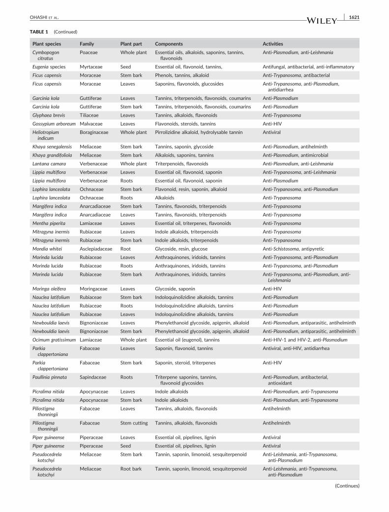
TABLE 1 (Continued)
Plant species Family Plant part Components Activities
Cymbopogoncitratus
Poaceae Whole plant Essential oils, alkaloids, saponins, tannins,flavonoids
Anti‐Plasmodium, anti‐Leishmania
Eugenia species Myrtaceae Seed Essential oil, flavonoid, tannins, Antifungal, antibacterial, anti‐inflammatory
Ficus capensis Moraceae Stem bark Phenols, tannins, alkaloid Anti‐Trypanosoma, antibacterial
Ficus capensis Moraceae Leaves Saponins, flavonoids, glucosides Anti‐Trypanosoma, anti‐Plasmodium,antidiarrhea
Garcinia kola Guttiferae Leaves Tannins, triterpenoids, flavonoids, coumarins Anti‐Plasmodium
Garcinia kola Guttiferae Stem bark Tannins, triterpenoids, flavonoids, coumarins Anti‐Plasmodium
Glyphaea brevis Tiliaceae Leaves Tannins, alkaloids, flavonoids Anti‐Trypanosoma
Gossypium arboreum Malvaceae Leaves Flavonoids, steroids, tannins Anti‐HIV
Heliotropiumindicum
Boraginaceae Whole plant Pirrolizidine alkaloid, hydrolysable tannin Antiviral
Khaya senegalensis Meliaceae Stem bark Tannins, saponin, glycoside Anti‐Plasmodium, antihelminth
Khaya grandifoliola Meliaceae Stem bark Alkaloids, saponins, tannins Anti‐Plasmodium, antimicrobial
Lantana camara Verbenaceae Whole plant Triterpenoids, flavonoids Anti‐Plasmodium, anti‐Leishmania
Lippia multiflora Verbenaceae Leaves Essential oil, flavonoid, saponin Anti‐Trypanosoma, anti‐Leishmania
Lippia multiflora Verbenaceae Roots Essential oil, flavonoid, saponin Anti‐Plasmodium
Lophira lanceolata Ochnaceae Stem bark Flavonoid, resin, saponin, alkaloid Anti‐Trypanosoma, anti‐Plasmodium
Lophira lanceolata Ochnaceae Roots Alkaloids Anti‐Trypanosoma
Mangifera indica Anarcadiaceae Stem bark Tannins, flavonoids, triterpenoids Anti‐Trypanosoma
Mangifera indica Anarcadiaceae Leaves Tannins, flavonoids, triterpenoids Anti‐Trypanosoma
Mentha piperita Lamiaceae Leaves Essential oil, triterpenes, flavonoids Anti‐Trypanosoma
Mitragyna inermis Rubiaceae Leaves Indole alkaloids, triterpenoids Anti‐Trypanosoma
Mitragyna inermis Rubiaceae Stem bark Indole alkaloids, triterpenoids Anti‐Trypanosoma
Mondia whitei Asclepiadaceae Root Glycoside, resin, glucose Anti‐Schistosoma, antipyretic
Morinda lucida Rubiaceae Leaves Anthraquinones, iridoids, tannins Anti‐Trypanosoma, anti‐Plasmodium
Morinda lucida Rubiaceae Roots Anthraquinones, iridoids, tannins Anti‐Trypanosoma, anti‐Plasmodium
Morinda lucida Rubiaceae Stem bark Anthraquinones, iridoids, tannins Anti‐Trypanosoma, anti‐Plasmodium, anti‐Leishmania
Moringa oleifera Moringaceae Leaves Glycoside, saponin Anti‐HIV
Nauclea latifolium Rubiaceae Stem bark Indoloquinolizidine alkaloids, tannins Anti‐Plasmodium
Nauclea latifolium Rubiaceae Roots Indoloquinolizidine alkaloids, tannins Anti‐Plasmodium
Nauclea latifolium Rubiaceae Leaves Indoloquinolizidine alkaloids, tannins Anti‐Plasmodium
Newbouldia laevis Bignoniaceae Leaves Phenylethanoid glycoside, apigenin, alkaloid Anti‐Plasmodium, antiparasitic, antihelminth
Newbouldia laevis Bignoniaceae Stem bark Phenylethanoid glycoside, apigenin, alkaloid Anti‐Plasmodium, antiparasitic, antihelminth
Ocimum gratissimum Lamiaceae Whole plant Essential oil (eugenol), tannins Anti‐HIV‐1 and HIV‐2, anti‐Plasmodium
Parkiaclappertoniana
Fabaceae Leaves Saponin, flavonoid, tannins Antiviral, anti‐HIV, antidiarrhea
Parkiaclappertoniana
Fabaceae Stem bark Saponin, steroid, triterpenes Anti‐HIV
Paullinia pinnata Sapindaceae Roots Triterpene saponins, tannins,flavonoid glycosides
Anti‐Plasmodium, antibacterial,antioxidant
Picralima nitida Apocynaceae Leaves Indole alkaloids Anti‐Plasmodium, anti‐Trypanosoma
Picralima nitida Apocynaceae Stem bark Indole alkaloids Anti‐Plasmodium, anti‐Trypanosoma
Piliostigmathonningii
Fabaceae Leaves Tannins, alkaloids, flavonoids Antihelminth
Piliostigmathonningii
Fabaceae Stem cutting Tannins, alkaloids, flavonoids Antihelminth
Piper guineense Piperaceae Leaves Essential oil, pipelines, lignin Antiviral
Piper guineense Piperaceae Seed Essential oil, pipelines, lignin Antiviral
Pseudocedrelakotschyi
Meliaceae Stem bark Tannin, saponin, limonoid, sesquiterpenoid Anti‐Leishmania, anti‐Trypanosoma,anti‐Plasmodium
Pseudocedrelakotschyi
Meliaceae Root bark Tannin, saponin, limonoid, sesquiterpenoid Anti‐Leishmania, anti‐Trypanosoma,anti‐Plasmodium
(Continues)
OHASHI ET AL. 1621

TABLE 1 (Continued)
Plant species Family Plant part Components Activities
Psidium guajava Myrtaceae Leaves Tannins, essential oil, triterpenoids, flavonoids Antimicrobial (all strains), anti‐Plasmodium,anti‐Leishmania
Pterocarpussantalinoides
Fabaceae Stem bark Alkaloids, flavonoids, tannins Anti‐HIV, antimicrobial
Pycnanthusangolensis
Myristicaceae Leaves Isoflavone, pycnanthuquinone Anti‐Plasmodium
Pycnanthusangolensis
Myristicaceae Stem bark Isoflavone, pycnanthuquinone Anti‐Plasmodium
Securidacalongepedunculata
Polygalaceae Leaves Saponins, tannins, cardiacglycoside, steroid
Anti‐Trypanosoma
Solanum torvum Solanaceae Leaves Steroidal sapogenins, steroidal alkaloids,isoflavonoid, steroidal glycosides
Antiviral, anti‐Plasmodium
Solanum torvum Solanaceae Stem bark Steroidal sapogenins, steroidal alkaloids,isoflavonoid, steroidal glycosides
Antiviral, anti‐Plasmodium
Sorghum bicolor Poaceae Leafstalk Alkaloids, saponins, tannins Anti‐HSV, antiviral
Spondias mombin Anacardiaceae Leaves Not available Antiviral
Tabernaemontanacrassa
Apocynaceae Leaves Tannins, saponins, alkaloids Antihelminth, antipyregic
Tabernaemontanacrassa
Apocynaceae Root Tannins, saponins, alkaloids Antipyretic, anti‐snake venom
Tamarindus indica Fabaceae Stem bark Saponins, tannins, glycoside Anti‐Trypanosoma
Tamarindus indica Fabaceae Leaves Phenols, flavonoid Anti‐Trypanosoma
Terminalia ivorensis Combretaceae Stem bark andleaves
Terminolic acid, quercetin, β‐glycyrrhetinic acid Anti‐Trypanosoma, anti‐Plasmodium
Terminalia ivorensis Combretaceae Leaves Terminolic acid, quercetin, β‐glycyrrhetinic acid Anti‐Trypanosoma
Theobroma cacao Serculiaceae Leaves Purine alkaloids, tannins, flavonoids Anti‐HIV
Theobroma cacao Serculiaceae Roots Purine alkaloids, tannins, flavonoids Anti‐HIV
Theobroma cacao Serculiaceae Stem bark Purine alkaloids, tannins, flavonoids Anti‐HIV
Thonningiasanguinea
Balanophoraceae Whole plant Alkaloids, tannins, flavonoids Anti‐Plasmodium, antifungal, antimicrobial
Treculia africana Moraceae Stem bark Catechin, cyaniding glycoside
Tridax procumbens Asteraceae Whole plant Flavonoids, alkyl esters, sterols Anti‐HIV
Vitex fosteri Verbenaceae Stem bark Essential oils, flavonoids Anti‐Trypanosoma
Vitex fosteri Verbenaceae Leaves Essential oils, flavonoids Anti‐Trypanosoma
Ximenia americana Olacaceae Stem andtwigs
Tannins, flavonoids, alkaloids Anti‐Trypanosoma
Ximenia americana Olacaceae Leaves Tannins, flavonoids, glycosides Anti‐Trypanosoma, antimicrobial
Zanthoxylumzanthoxyloides
Rutaceae Root Alkaloids (berberine), tannins,flavonoids, essential oil
Anti‐Leishmania
Zanthoxylumzanthoxyloides
Rutaceae Stem bark Alkaloids (berberine), tannins,flavonoids, essential oil
Anti‐Plasmodium
Zanthoxylumzanthoxyloides
Rutaceae Leaves Alkaloids (berberine), tannins,flavonoids, essential oil
Antiparasitic
1622 OHASHI ET AL.
plant. Together, 13 (11.6%) extracts were prepared from seeds, fruits,
stem cuttings, leafstalk, and twigs (Table 1).
Antikinetoplastid and anti‐Plasmodium properties of extracts
were investigated after a 48‐hr incubation period. The IC50 values
of the crude extracts against bloodstream T. b. brucei (GUTat 3.1),
promastigote L. donovani (MHOM/NP/03/D10), and P. falciparum
(3D7) strains are summarized in Table 2. Out of 112 crude extracts
screened, 61, 41, and 13 were found to have varying degrees of activ-
ity against T. b. brucei, L. donovani, and P. falciparum, respectively.
Eleven (9.82%) extracts had strong anti‐Trypanosoma activity with
IC50 values less than 20 μg/ml, whereas 30 (26.79%) and 20
(17.86%) extracts had moderate to fair activity with IC50 values in
the range of 21–50 and 51–100 μg/ml, respectively. Nine (8.04%)
extracts had high anti‐Leishmania activity with IC50 values less than
20 μg/ml, whereas 22 (19.64%) and 10 (8.93%) extracts had moderate
to fair activity with IC50 values ranging from 20 to 50 and 51 to
100 μg/ml, respectively. Six (5.36%) extracts had high anti‐Plasmodium
activity with IC50 values less than 20 μg/ml, whereas four (3.57%) and
five (4.46%) extracts had moderate to fair activity with IC50 values
ranging from 20 to 50 and 51 to 100 μg/ml, respectively. The IC50
values of the positive controls were 7.84, 0.1, and 0.01 μg/ml for ber-
berine, amphotericin B, and artesunate, respectively.
The SI values for individual pathogens obtained using Jurkat cells
are outlined inTable 2. The SI values of all but one crude extracts with
strong anti‐Trypanosoma activity, IC50 less than 20 μg/ml, were above
10.00. Acanthospermum hispidum whole plant extract was the only

TABLE 2 In vitro antiparasitic activity of screened crude extracts against Trypanosoma, Leishmania, and Plasmodium species, with cytotoxicityand SI values
Plant species Plant part
IC50 (μg/ml) Selectivity index (SI)
Jurkat T. b. brucei L. donovani P. falciparum T. b. brucei L. donovani P. falciparum
Acacia nilotica Stem bark 39.59 79.32 >1,000 208.33 0.49 <0.04 0.17
Acanthospermum hispidum Whole plant 55.50 7.57 32.1 >1,000 7.33 1.73 <0.06
Aframomum melegueta Seed 62.49 168.83 >1,000 509.68 0.37 <0.06 0.12
Afzelia africana Stem ‐bark 232.60 233.58 77.1 222.36 1.00 3.02 1.05
Alchornea cordifolia Leaves 73.01 219.80 443.2 17.44 0.33 0.16 4.19
Alstonia boonei Leaves 183.48 51.79 >1,000 >1,000 3.54 <0.18 <0.18
Alstonia boonei Stem bark 736.36 25.96 >1,000 >1,000 28.37 <0.74 <0.74
Annona senegalensis Leaves 273.49 182.23 10.8 >1,000 1.50 25.32 <0.27
Annona senegalensis Stem cutting 127.95 353.89 27.8 >1,000 0.36 4.60 <0.13
Anogeissus schimperi Leaves 51.72 34.44 >1,000 18.32 1.50 <0.05 2.82
Anogeissus schimperi Stem bark 38.29 >1,000 >1,000 25.86 <0.04 <0.04 1.48
Anogeissus schimperi Root 42.15 105.18 >1,000 50.46 0.40 <0.04 2.08
Anthocleista nobilis Leaves 245.59 24.68 41.5 >1,000 9.95 5.92 <0.25
Anthocleista nobilis Stem bark 761.33 410.39 843.7 >1,000 1.86 0.90 <0.76
Anthocleista nobilis Roots 716.41 33.32 79.0 >1,000 21.50 9.07 <0.72
Balanites aegyptiaca Stem bark 804.91 >1,000 173.6 >1,000 <0.80 4.64 <0.80
Baphia nitida Stem bark 990.55 >1,000 34.4 >1,000 <0.99 28.80 <0.99
Bidens pilosa Whole plant 192.85 5.51 28.9 >1,000 35.00 6.67 <0.19
Bridelia ferruginea Leaves 392.80 43.28 16.5 83.06 9.08 23.81 4.73
Calotropis procera Leaves 39.64 30.34 >1,000 >1,000 1.31 <0.04 <0.04
Carapa procera Stem bark 85.17 117.94 >1,000 >1,000 0.72 <0.09 0.09
Carica papaya Seed >1,000 >1,000 >1,000 519.38 1.0 1.0 >1.92
Cassia alata Leaves 371.46 >1,000 10.1 57.60 <0.37 36.78 6.44
Cassia siamea Stem bark 429.33 344.47 >1,000 >1,000 1.25 <0.43 <0.43
Cassia sieberiana Roots 917.32 289.69 142.6 432.48 3.17 6.43 2.12
Cassia sieberiana Leaves 48.48 45.87 62.9 >1,000 1.06 0.77 <0.05
Cassia podocarpa Leaves 453.87 54.05 >1,000 >1,000 8.40 <0.45 <0.45
Cassia occidentalis Seeds 926.63 >1,000 >1,000 >1,000 <0.93 <0.93 <0.93
Cassia occidentalis Leaves 329.94 >1,000 >1,000 >1,000 <0.33 <0.33 <0.33
Cassia occidentalis Whole plant 446.19 37.83 >1,000 >1,000 11.79 <0.45 <0.45
Ceiba pentandra Stem bark 100.52 98.93 31.1 >1,000 1.02 3.23 <0.1
Cinnamomum zeylanicum Leaves 273.98 50.89 >1,000 >1,000 5.38 <0.27 <0.27
Cinnamomum zeylanicum Stem bark 53.80 25.89 >1,000 >1,000 2.07 <0.05 <0.05
Citrus aurantifolia Leaves 520.06 217.31 542.9 >1,000 2.39 0.96 <0.52
Citrus aurantifolia Fruits 33.62 29.81 >1,000 34.83 1.13 <0.03 0.97
Clausena anisata Roots 293.14 29.50 12.1 487.56 9.94 24.23 0.60
Cleistopholis patens Leaves 484.76 214.57 >1,000 >1,000 2.26 <0.48 <0.48
Cleistopholis patens Stem bark 214.62 >1,000 60.2 >1,000 <0.21 3.57 <0.21
Cola cordifolia Stem bark 465.61 37.41 25.1 >1,000 12.45 18.55 <0.47
Cola cordifolia Leaves 465.61 10.08 18.2 730.34 46.19 25.58 0.64
Cola acuminata Stem bark 156.99 47.44 47.8 279.84 3.31 3.28 0.56
Cymbopogon citratus Whole plant 311.65 123.77 162.2 694.06 2.52 1.92 0.45
Eugenia species Seed 94.41 8.50 26.6 208.97 11.11 3.55 0.45
Ficus capensis Stem‐bark 56.66 36.10 37.0 >1,000 1.57 1.53 <0.06
Ficus capensis Leaves 258.00 159.62 88.9 >1000 1.62 2.90 <0.26
Garcinia kola Leaves 343.41 34.47 673.1 >1,,000 9.96 0.51 <0.34
Garcinia kola Stem bark 439.64 485.32 159.4 >1,000 0.91 2.76 <0.44
(Continues)
OHASHI ET AL. 1623

TABLE 2 (Continued)
Plant species Plant part
IC50 (μg/ml) Selectivity index (SI)
Jurkat T. b. brucei L. donovani P. falciparum T. b. brucei L. donovani P. falciparum
Glyphaea brevis Leaves 962.15 141.92 43.4 >1,000 6.78 22.17 <0.96
Gossypium arboreum Leaves 519.49 >1,000 >1,000 16.10 <0.52 <0.52 32.27
Heliotropium indicum Whole plant >1,000 >1,000 119.4 117.32 1.0 >8.38 8.52
Khaya senegalensis Stem bark 70.59 23.81 >1,000 >1,000 2.96 <0.07 <0.07
Khaya grandifoliola Stem bark 50.21 180.58 43.2 616.37 0.28 1.16 0.08
Lantana camara Whole plant 176.58 115.02 >1,000 33.39 1.54 <0.18 5.29
Lippia multiflora Leaves 249.86 83.59 >1,000 >1,000 2.99 <0.25 <0.25
Lippia multiflora Roots 497.66 837.72 >1,000 >1,000 0.59 <0.50 <0.50
Lophira lanceolata Stem bark 45.83 429.80 68.6 >1,000 0.11 0.67 <0.05
Lophira lanceolata Roots 38.63 288.66 66.0 >1,000 0.14 0.59 <0.04
Mangifera indica Stem bark 494.59 >1,000 >1,000 >1,000 <0.49 <0.49 <0.49
Mangifera indica Leaves 55.04 77.37 >1,000 14.25 0.71 <0.06 3.86
Mentha piperita Leaves 55.03 49.35 >1,000 566.88 1.11 <0.05 0.002
Mitragyna inermis Leaves 193.21 397.50 21.9 >1,000 0.49 8.82 <0.19
Mitragyna inermis Stem bark 424.52 362.02 28.0 >1,000 1.17 15.16 <0.42
Mondia whitei Root 433.19 35.10 31.0 >1,000 12.34 13.97 <0.43
Morinda lucida Leaves 78.07 5.96 >1,000 >1,000 13.09 <0.08 <0.08
Morinda lucida Roots 177.69 499.67 >1,000 >1,000 0.36 <0.18 <0.18
Morinda lucida Stem bark 939.12 28.04 >1,000 >1,000 33.49 <0.94 <0.94
Moringa oleifera Leaves 409.40 84.43 >1,000 >1,000 4.85 <0.41 <0.41
Nauclea latifolium Stem bark 544.29 549.78 784.0 >1,000 0.99 0.69 <0.54
Nauclea latifolium Roots 649.37 >1,000 138.9 >1,000 <0.65 4.68 <0.65
Nauclea latifolium Leaves 268.05 54.12 >1,000 >1,000 4.95 <0.27 <0.27
Newbouldia laevis Leaves 171.05 9.47 >1,000 593.79 18.06 <0.17 0.29
Newbouldia laevis Stem bark 171.05 147.66 >1,000 >1,000 1.16 <0.17 <0.17
Ocimum gratissimum Whole plant 420.17 22.47 >1,000 >1,000 18.70 <0.42 0.42
Parkia clappertoniana Leaves 114.73 58.30 17.3 501.23 1.97 6.63 0.23
Parkia clappertoniana Stem bark 42.37 99.99 17.6 >1,000 0.42 2.41 <0.04
Paullinia pinnata Roots 926.63 63.23 130.1 291.55 14.65 7.12 3.18
Picralima nitida Leaves 247.16 44.08 631.0 >1,000 5.61 0.39 <0.25
Picralima nitida Stem bark 583.53 268.68 >1,000 95.19 2.17 <0.58 6.13
Piliostigma thonningii Leaves 95.83 78.37 >1,000 >1,000 1.22 <0.10 <0.1
Piliostigma thonningii Stem cutting 156.09 135.25 >1,000 514.63 1.15 <0.16 0.3
Piper guineense Leaves 389.63 23.09 >1,000 621.7 16.87 <0.39 0.62
Piper guineense Seed 77.48 17.44 >1,000 266.7 4.44 <0.08 0.29
Pseudocedrela kotschyi Stem Bark 48.26 58.80 >1,000 510.96 0.82 <0.05 0.09
Pseudocedrela kotschyi Root bark 101.18 59.41 >1,000 414.01 1.70 <0.10 0.24
Psidium guajava Leaves 136.72 >1,000 >1,000 >1,000 <0.14 <0.14 <0.14
Pterocarpus santalinoides Stem bark 128.17 >1,000 >1,000 907.52 <0.13 <0.13 0.14
Pycnanthus angolensis Leaves 71.65 38.39 >1,000 >1,000 1.87 <0.07 <0.07
Pycnanthus angolensis Stem bark 218.66 9.76 150.0 >1,000 22.40 1.46 <0.22
Securidaca longepedunculata Leaves 45.83 237.43 >1,000 114.39 0.19 <0.05 0.40
Solanum torvum Leaves 38.63 >1000 137.0 50.53 <0.04 0.28 0.76
Solanum torvum Stem bark 494.59 >1000 601.4 >1,000 <0.49 0.82 <0.49
Sorghum bicolor Leafstalk 55.04 24.01 34.1 127.75 2.29 1.61 0.43
Spondias mombin Leaves 55.03 77.04 81.5 46.66 0.71 0.68 1.18
Tabernaemontana crassa Leaves 193.21 20.84 >1,000 >1,000 9.27 <0.19 0.19
Tabernaemontana crassa Root 424.52 163.16 >1,000 >1,000 2.60 <0.42 0.42
(Continues)
1624 OHASHI ET AL.

TABLE 2 (Continued)
Plant species Plant part
IC50 (μg/ml) Selectivity index (SI)
Jurkat T. b. brucei L. donovani P. falciparum T. b. brucei L. donovani P. falciparum
Tamarindus indica Stem bark 433.19 40.02 >1,000 >1,000 10.82 <0.43 0.43
Tamarindus indica Leaves 78.07 39.33 58.12 >1,000 1.98 1.34 0.08
Terminalia ivorensis Stem bark and leaves 177.69 17.45 23.2 10.35 10.18 7.66 17.17
Terminalia ivorensis Leaves 939.12 11.28 24.9 7.26 83.26 37.72 129.36
Theobroma cacao Leaves 409.40 58.37 >1,000 >1,000 7.01 <0.41 <0.41
Theobroma cacao Roots 544.29 334.65 >1,000 >1,000 1.63 <0.54 <0.54
Theobroma cacao Stem bark 649.37 65.95 >1,000 >1,000 9.85 <0.65 <0.65
Thonningia sanguinea Whole plant 268.05 139.89 18.6 133.59 1.92 15.38 2.00
Treculia africana Stem bark 171.05 53.24 44.8 614.95 3.21 3.84 0.28
Tridax procumbens Whole plant 171.05 310.74 >1,000 >1,000 0.55 <0.17 <0.17
Vitex fosteri Stem bark 420.17 146.75 49.8 >1,000 2.86 8.44 <0.42
Vitex fosteri Leaves 114.73 42.44 72.4 >1,000 2.70 1.58 <0.11
Ximenia americana Stem and twigs 42.37 85.54 36.1 >1,000 0.50 1.17 <0.42
Ximenia americana Leaves 926.63 180.30 >1,000 176.26 5.14 <0.93 5.25
Zanthoxylum zanthoxyloides Root 247.16 39.43 13.5 334.77 6.27 18.30 0.74
Zanthoxylum zanthoxyloides Stem bark 583.53 5.96 45.2 112.15 97.91 12.91 5.20
Zanthoxylum zanthoxyloides Leaves 95.83 27.73 >1,000 >1,000 3.46 <0.10 <0.96
Positive control 7.84 0.1 0.01
Note. Antiprotozoan activities and cytotoxicity are represented by IC50 values obtained from Alamar Blue (Trypanosoma brucei brucei and Leishmaniadonovani), SYBR Green (Plasmodium falciparum), and MTT (Jurkat) assays, respectively. The IC50 values are averages of three independent assays run foreach crude extract. SI was determined by dividing the IC50 values for the Jurkat cells by the IC50 values for each parasite tested. Berberine, amphotericinB, and artesunate were used as positive controls for anti‐Trypanosoma, anti‐Leishmania, and anti‐Plasmodium activities, respectively. Bold emphasis are forextracts with high activity against parasites.
OHASHI ET AL. 1625
anti‐Trypanosoma active extract with an SI value below 10, SI of 7.33.
With respect to anti‐Leishmania active extracts, seven out of nine had
SI values above 10.00. Parkia clappertoniana leaf extract and
P. clappertoniana stem bark extract were the two extracts with SI
values below 10, SI of 6.63 and 0.4, respectively. Three out of six
anti‐Plasmodium active extracts, Gossypium arboreum leaf and stem
bark extracts, and Terminalia ivorensis leaf extract showed SI values
greater than 10.00.
3.2 | Apoptosis inducing properties
The Nexin assay was performed to determine the apoptosis inducing
properties of the eight anti‐Trypanosoma active extracts. Parasites
were challenged with 25 μg/ml of the active extracts for 24 hr and
then subjected to apoptosis analysis. Morinda lucida induced the
highest level of apoptosis (69.8%) against Trypanosoma parasites
whereas the other extracts showed no significant induction of apopto-
tic cells (Figure 1). No extract was observed to induce significant
apoptosis‐like cell death in Leishmania parasites and Plasmodium para-
sites using the Nexin assay and the mitochondrial membrane potential
assays, respectively (data not shown).
3.3 | Morphological changes of L. donovanipromastigotes induced by active crude extracts
The morphological changes of Leishmania parasites induced by active
crude extracts were observed by fluorescence microscopy. Parasites
were fixed and stained with DAPI after 24 hr incubation with each crude
extract at a concentration 2 times the IC50 value. Parasites treated with
Annona senegalensis extracts were observed to have no kinetoplasts with
an intact nucleus. The A. senegalensis extracts also induced incomplete
parasite division with only a single observable flagellum, resulting in the
formation of paired daughter cells. Cola cordifolia induced nuclear frag-
mentation andmultiple flagella in parasites without cell division. Clausena
anisata‐treated parasites were aggregated with some abnormal morphol-
ogy. On the other hand, Bridelia ferruginea parasites were observed to
have fragmented nuclei and linked to each other with very prominent
flagella. Zanthoxylum zanthoxyloides‐treated parasites had no significant
change in the nucleus and kinetoplast.Cassia alata‐treated parasites how-
ever induced an increase in the number of nuclei and kinetoplast with
very severe aggregation of parasites, and most of the parasites showed
a short stumpy‐like form. Although there were short stumpy forms in
Z. zanthoxyloides‐treated parasites, the aggregations were not severe.
Parasites treated with both Z. zanthoxyloides and C. alata did not have
prominent flagella. P. clappertoniana‐treated parasites were observed to
have fragmented nuclei and aggregated into small groups. The parasites
appeared round shaped and stumpy like without prominent flagella
(Figure 2). Anti‐Trypanosoma active extracts were however not observed
to induce significant and/or variedmorphological changes inTrypanosoma
parasites as observed in Leishmania parasites (data not shown).
4 | DISCUSSION
Traditional knowledge stake holders (herbalists, farmers, and local indi-
genes) take advantage of the medicinal properties of plants and their

FIGURE 1 Signals of apoptosis induction in Trypanosoma parasites were detected based on the externalization of phosphatidylserine and DNAfragmentation. Data were obtained using the nexin assay and fluorescent activated cell sorting analysis. Results are represented by dot plots offour quadrants: Lower left = viable cells; lower right = early apoptotic cells; upper right = late apoptotic cells; upper left = necrotic cells [Colourfigure can be viewed at wileyonlinelibrary.com]
1626 OHASHI ET AL.
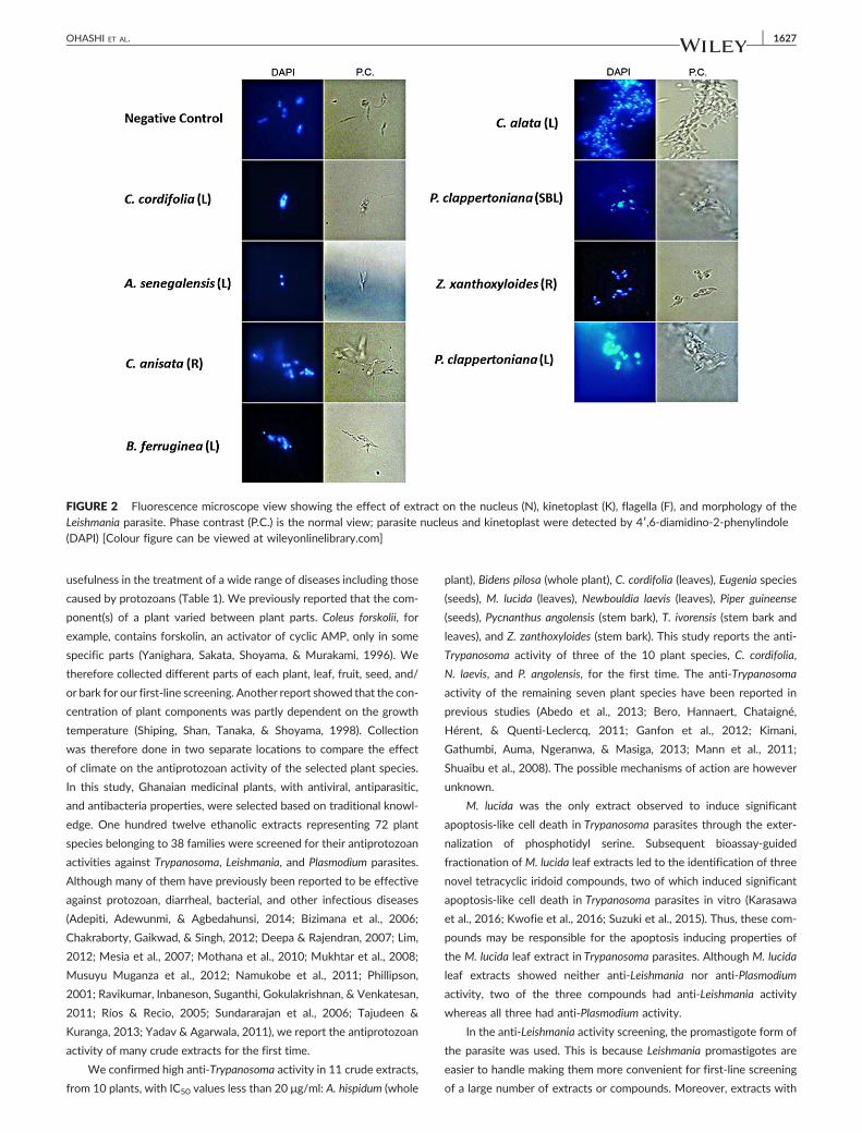
FIGURE 2 Fluorescence microscope view showing the effect of extract on the nucleus (N), kinetoplast (K), flagella (F), and morphology of theLeishmania parasite. Phase contrast (P.C.) is the normal view; parasite nucleus and kinetoplast were detected by 4′,6‐diamidino‐2‐phenylindole(DAPI) [Colour figure can be viewed at wileyonlinelibrary.com]
OHASHI ET AL. 1627
usefulness in the treatment of a wide range of diseases including those
caused by protozoans (Table 1). We previously reported that the com-
ponent(s) of a plant varied between plant parts. Coleus forskolii, for
example, contains forskolin, an activator of cyclic AMP, only in some
specific parts (Yanighara, Sakata, Shoyama, & Murakami, 1996). We
therefore collected different parts of each plant, leaf, fruit, seed, and/
or bark for our first‐line screening. Another report showed that the con-
centration of plant components was partly dependent on the growth
temperature (Shiping, Shan, Tanaka, & Shoyama, 1998). Collection
was therefore done in two separate locations to compare the effect
of climate on the antiprotozoan activity of the selected plant species.
In this study, Ghanaian medicinal plants, with antiviral, antiparasitic,
and antibacteria properties, were selected based on traditional knowl-
edge. One hundred twelve ethanolic extracts representing 72 plant
species belonging to 38 families were screened for their antiprotozoan
activities against Trypanosoma, Leishmania, and Plasmodium parasites.
Although many of them have previously been reported to be effective
against protozoan, diarrheal, bacterial, and other infectious diseases
(Adepiti, Adewunmi, & Agbedahunsi, 2014; Bizimana et al., 2006;
Chakraborty, Gaikwad, & Singh, 2012; Deepa & Rajendran, 2007; Lim,
2012; Mesia et al., 2007; Mothana et al., 2010; Mukhtar et al., 2008;
Musuyu Muganza et al., 2012; Namukobe et al., 2011; Phillipson,
2001; Ravikumar, Inbaneson, Suganthi, Gokulakrishnan, & Venkatesan,
2011; Ríos & Recio, 2005; Sundararajan et al., 2006; Tajudeen &
Kuranga, 2013; Yadav & Agarwala, 2011), we report the antiprotozoan
activity of many crude extracts for the first time.
We confirmed high anti‐Trypanosoma activity in 11 crude extracts,
from 10 plants, with IC50 values less than 20 μg/ml: A. hispidum (whole
plant), Bidens pilosa (whole plant), C. cordifolia (leaves), Eugenia species
(seeds), M. lucida (leaves), Newbouldia laevis (leaves), Piper guineense
(seeds), Pycnanthus angolensis (stem bark), T. ivorensis (stem bark and
leaves), and Z. zanthoxyloides (stem bark). This study reports the anti‐
Trypanosoma activity of three of the 10 plant species, C. cordifolia,
N. laevis, and P. angolensis, for the first time. The anti‐Trypanosoma
activity of the remaining seven plant species have been reported in
previous studies (Abedo et al., 2013; Bero, Hannaert, Chataigné,
Hérent, & Quenti‐Leclercq, 2011; Ganfon et al., 2012; Kimani,
Gathumbi, Auma, Ngeranwa, & Masiga, 2013; Mann et al., 2011;
Shuaibu et al., 2008). The possible mechanisms of action are however
unknown.
M. lucida was the only extract observed to induce significant
apoptosis‐like cell death in Trypanosoma parasites through the exter-
nalization of phosphotidyl serine. Subsequent bioassay‐guided
fractionation of M. lucida leaf extracts led to the identification of three
novel tetracyclic iridoid compounds, two of which induced significant
apoptosis‐like cell death in Trypanosoma parasites in vitro (Karasawa
et al., 2016; Kwofie et al., 2016; Suzuki et al., 2015). Thus, these com-
pounds may be responsible for the apoptosis inducing properties of
the M. lucida leaf extract in Trypanosoma parasites. Although M. lucida
leaf extracts showed neither anti‐Leishmania nor anti‐Plasmodium
activity, two of the three compounds had anti‐Leishmania activity
whereas all three had anti‐Plasmodium activity.
In the anti‐Leishmania activity screening, the promastigote form of
the parasite was used. This is because Leishmania promastigotes are
easier to handle making them more convenient for first‐line screening
of a large number of extracts or compounds. Moreover, extracts with

1628 OHASHI ET AL.
high anti‐Leishmania promastigote activity have been reported to have
higher activity against the amastigote form of the parasite
(Amoa‐Bosompemet al., 2016). Extensivework has however been done
on selected active fractions and compounds using both the
promastigote (Amoa‐Bosompem et al., 2016) and amastigote forms of
the parasite (unpublished). In our first‐line screening however, we
confirmed A. senegalensis (leaves), C. alata (leaves), C. anisata
(roots), B. ferruginea (leaves), C. cordifolia (leaves), Thonningia sanguinea
(whole plant), P. clappertoniana (stem bark and leaves), and
Z. zanthoxyloides (root) as extracts with strong anti‐Leishmania activity.
This study reports the anti‐Leishmania activity of six of the eight active
plant species for the first time with A. senegalensis and Z. zanthoxyloides,
the only ones previously reported to possess anti‐Leishmania
activity (Sahpaz et al., 1994). With respect to extract activity against
multiple parasites, C. cordifolia and Z. zanthoxyloides were found to
have activity against both Trypanosoma and Leishmania parasites. The
Z. zanthoxyloides stem bark extract however had only anti‐Trypanosoma
activity whereas its root extract had only anti‐Leishmania activity.
This differencemay be attributed to the differences in chemical compo-
nents and/or concentration in different plant parts. Both T. ivorensis
stem bark and leaf extracts were active against Trypanosoma and
Plasmodium parasites.
In addition to the anti‐Leishmania activity, we found A. senegalensis
to induce kinetoplast disintegration in deformed Leishmania parasites
although there was no observable effect on the nucleus. The shift
from the normal morphology may be due to the minor aggregation
of parasites resulting in the inhibition of parasite proliferation. Nuclear
fragmentation was observed in Leishmania parasites treated with
B. ferruginea, P. clappertoniana, or C. cordifolia extracts. C. alata‐treated
parasites formed the largest aggregation relative to all the test groups
with a significant increase in the number of parasites having a double
nuclei and kinetoplast. This phenotype may be due to the inhibition of
cytokinesis after nuclei and kinetoplast division, preventing the forma-
tion of two distinct daughter cells. Z. zanthoxyloides extract on the
other hand caused minor aggregation in Leishmania parasites with a
short and stumpy‐like morphology. All the anti‐Leishmania active
extracts did not induce significant apoptosis in Leishmania parasites
(data not shown).
With respect to the anti‐Plasmodium activity, six extracts from
five plant species were observed to have high anti‐Plasmodium
activity: Alchornea cordifolia (leaves), Anogeissus schimperi (leaves),
Gossypium arboretum (leaves), Mangifera indica (leaves), and T. ivorensis
(stem bark and leaves). Although all five plant species had previously
been reported to have anti‐Plasmodium activity, the plant parts,
extracts, differed in activity from previous reports. The leaves, flowers,
and bark of M. indica, for example, had previously been reported to
have anti‐Plasmodium activity (Bidla et al., 2004; H. A. Ibrahim et al.,
2012). Our study however found only the leaf extracts to have anti‐
Plasmodium activity (Table 2). This may be due to the effect of seasons
and/or habitats on the constituents of plants. Regarding the compo-
nents of mango, although it has been suggested that the most impor-
tant constituent in mango‐related antiparasitic activity is mangiferin
having the C‐glucosyl xanthone structure (Wauthoz & Balde, 2007),
the active component is still unknown. In this regard, we intend to
employ the bioassay‐guided fractionation techniques to determine
the active components of the promising anti‐Plasmodium active
extracts as in the case of M. lucida (Kwofie et al., 2016).
Moving forward, our aim is not only to determine the active
components but also to test the prospects of using active extracts to
develop herbal based treatment drugs at the Center for Plant
Medicine Research, Ghana. Work is currently ongoing to replicate
the efficacy of selected active extracts and compounds in vivo. Also,
although climate had no significant effect on the activity of active
extracts, there is the need to determine the seasonal activity of
each/selected active extracts. This study however shows the
prospects of Ghanaian medicinal plants in the development of new
chemotherapy against protozoan infections.
5 | CONCLUSION
In conclusion, all 23 extracts with high antiparasitic activity showed
high selectivity for at least one of the three parasites.
Anti‐Trypanosoma active compounds induced apoptosis in
Trypanosoma parasites whereas anti‐Leishmania active extracts caused
morphological changes in the Leishmania parasite.
Overall, the results obtained from the crude extracts screening,
especially all extracts with anti‐Trypanosoma, anti‐Leishmania, and
anti‐Plasmodium activities, suggest that these may be promising
sources for the development of new drugs for controlling African
trypanosomiasis, leishmaniasis, and malaria.
ETHICS APPROVAL AND CONSENT TO PARTICIPANTS
IRB approval was sought from the Noguchi Memorial Institute for
Medical Research, Ghana, IRB board before the start of the project.
CONSENT FOR PUBLICATION
Not applicable.
AVAILABILITY OF DATA AND MATERIAL
All data generated in this study have been included in this manuscript.
AUTHOR CONTRIBUTIONS
O. M. developed protocols, performed assays, analyzed data, and was
a major contributor in the writing of the manuscript. K. D. K., A. B. M.,
and A. J. performed antiparasitic assays, analyzed data, and contrib-
uted to the writing of the manuscript. U. T., A. R., S. M., A. F., A. F.,
and A. A. A. prepared the plant extracts. O. K. B. A., T. I., A. P., N. T.,
N. A., and A. O. R. performed toxicity assays and analyzed data. Y. S.
is responsible for the authentication of plant material and contributed
to the writing of the manuscript. W. K. A., A. I., B. D. A., K. K. A., E. D.,
S. Y., and O. N. analyzed data and contributed to the writing of the
manuscript. All authors have read and approved the final manuscript.
ACKNOWLEDGEMENTS
This research is supported by Science and Technology Research
Partnership for Sustainable Development (SATREPS) grant from the
Japan Science and Technology Agency (JST) and the Japan Interna-
tional Cooperation Agency (JICA) (2010 to 2015) and the Japan

OHASHI ET AL. 1629
Initiative for Global Research Network on Infectious Diseases (J‐GRID)
from Ministry of Education, Culture, Sports, Science and Technology
in Japan, and Japan Agency for Medical Research and Development
(AMED) (2015–present).
CONFLICT OF INTEREST
The authors have declared that there is no conflict of interest.
ORCID
Mitsuko Ohashi http://orcid.org/0000-0001-9329-7920
Yukihiro Shoyama http://orcid.org/0000-0001-7190-0258
REFERENCES
Abedo, J. A., Jonah, A. O., Mazadu, M. R., Abdullahi, R. S., Idris, H. Y.,Shettima, F. T., … Abdulmalik, U. (2013). In vitro, in vivo and phytochem-ical screening of extracts of Piper guineense for trypanocidal activitiesagainst Trypanosoma brucei brucei. International Journal of Biology,5(3), 1916–9671.
Abu, A. H., & Uchendu, C. N. (2011). In vivo trypanocidal activity ofhydroethanolic extract of Hymenocardia acida stem bark in rats. VetWorld, 4(3), 113–116.
Adepiti, O. A., Adewunmi, C. O., & Agbedahunsi, J. M. (2014).Antitrichomonal activity of Acanthospermum hispidum D. C.(Asteraceae). African Journal of Biotechnology, 13(11), 1303–1307.https://doi.org/10.5897/AJB2013.13064
Adeyemi, O S, M L Sykes, M A Akanji, and V M Avery. 2011. “Anti‐trypanosomal and cytotoxic activity of ethanolic extracts of Psidiumguajava leaves in Alamar Blue based assays.” Veterinarski Arhiv.http://www.vef.hr/vetarhiv.
Alvar, J., Yactayo, S., & Bern, C. (2006). Leishmaniasis and poverty. Trendsin Parasitology, 22(12), 552–557.
Amoa‐Bosompem, M., Ohashi, M., Mosore, M.‐T., Agyapong, J., Tung, N.H., Kwofie, K. D., … Ohta, N. (2016). In vitro anti‐Leishmania activityof tetracyclic iridoids from Morinda lucida, benth. Tropical Medicineand Health, 44, 25. https://doi.org/10.1186/s41182‐016‐0026‐5
Ankrah, N.‐A., Nyarko, A. K., Addo, P. G. A., Ofosuhene, M., Dzokoto, C.,Marley, E., … Ekuban, F. A. (2003). Evaluation of efficacy and safetyof a herbal medicine used for the treatment. Phytotherapy Research,701 (May 2002), 697–701.
Bacchi, C. J. (2009). Chemotherapy of human African trypanosomiasis.Interdisciplinary Perspectives on Infectious Diseases, 2009(January):195040. doi:https://doi.org/10.1155/2009/195040), 1–5.
Balunas, M. J., & Kinghorn, A. D. (2005). Drug discovery from medicinalplants. Life Sciences, 78, 431–441.
Barrett, M. P., Boykin, D. W., Brun, R., & Tidwell, R. R. (2007). HumanAfrican trypanosomiasis: Pharmacological re‐engagement with aneglected disease. British Journal of Pharmacology, 152(8),1155–1171. https://doi.org/10.1038/sj.bjp.0707354
Bensoussan, E., Nasereddin, A., Jonas, F., Schnur, L. F., & Jaffe, C. L. (2006).Comparison of PCR assays for diagnosis of cutaneous leishmaniasis.Journal of Clinical Microbiology, 44(4), 1435–1439. https://doi.org/10.1128/JCM.44.4.1435‐1439.2006
Bero, J., Hannaert, V., Chataigné, G., Hérent, M. F., & Quenti‐Leclercq, J.(2011). In vitro anti‐trypanosomal and anti‐leishmanial activity of plantsused in Benin in traditional medicine and bio‐guided fractionation ofthe most active extract. Journal of Ethnopharmacology, 137(2),998–1002.
Bidla, G., Titanji, V. P. K., Joko, B., El‐Ghazali, G., Bolad, A., & Berzins, K.(2004). Antiplasmodial activity of seven plants used in African folkmedicine. Indian Journal of Pharmacology, 36(4), 245–246.
Bizimana, N., Tietjen, U., Zessin, K. H., Diallo, D., Djibril, C., Melzig, M. F., &Clausen, P. H. (2006). Evaluation of medicinal plants from Mali for their
in vitro and in vivo trypanocidal activity. Journal of Ethnopharmacology,103(3), 350–356.
Centers for Disease Control and Prevention. 2016. “Parasites—Africantrypanosomiasis (also known as sleeping sickness).” CDC Home.http://www.cdc.gov/parasites/sleepingsickness/index.html.
Chakraborty, A. K., Gaikwad, A. V., & Singh, K. B. (2012).Phytopharmacological review on Acanthospermum hispidum. Journal ofApplied Pharmaceutical Science, 2(1), 144–148.
Deepa, N., & Rajendran, N. N. (2007). Anti‐bacterial and anti‐fungalactivities of various extracts of Acanthospermum hispidum DC. Journalof Natural Remedies, 7, 225–228.
Desjeux, P. (2004). Leishmaniasis: Current situation and new perspectives.Comparative Immunology, Microbiology and Infectious Diseases, 27(5),305–318. https://doi.org/10.1016/j.cimid.2004.03.004
Fathuddin, M. M. (2011). In vivo antitrypanosomal potentials of ethylacetate leaf extracts of Punica granatum against Trypanosoma bruceibrucei. Adv Agric Biotechnol, 1, 82–88.
Ganfon, H., Bero, J., Tchinda, A. T., Gbaguidi, F., Gbenou, J., Moudachirou,M., … Quentin‐Leclercg, J. (2012). Antiparasitic activities of twosesquiterpenic lactones isolated from Acanthospermum hispidum D.C.Journal of Ethnopharmacology, 14(1), 411–417.
Herwaldt, B. L. (1999). Leishmaniasis. Lancet, 354(9185), 1191–1199.
Ibrahim, H. A., Imama, I. A., Bello, A. M., Umar, U., Muhammad, S., &Abdullahi, S. A. (2012). The potential of Nigerian medicinal plants asantimalarial agent: A review. International Journal of Science andTechnology, 2(8), 600–605.
Ibrahim, M. A., Aliyu, A. B., Sallau, A. B., Bashir, M., Yunusa, I., & Umar, T. S.(2010). Senna occidentalis leaf extract possesses antitrypanosomalactivity and ameliorates the trypanosome‐induced anemia and organdamage. Pharmacognosy Research, 2(3), 175–180. https://doi.org/10.4103/0974‐8490.65513
Kimani, P. C., Gathumbi, P. K., Auma, J., Ngeranwa, J. J., & Masiga, D. K.(2013). In vitro activity of selected medicinal plants in Kenya onTrypanosoma evansi. Kenyan The Veterinarian, 37(1).
Kwofie, K. D., Tung, N. H., Suzuki‐Ohashi, M., Amoa‐Bosompem, M.,Adegle, R., Sakyiamah, M. M., … Ayi, I. (2016). Antitrypanosomal activ-ities and mechanisms of action of novel tetracyclic iridoids fromMorinda lucida Benth. Antimicrobial Agents and Chemotherapy, 60,3283–3290. https://doi.org/10.1128/AAC.01916‐15
Lim, T. K. (2012). Edible medicinal and non‐medicinal plants. In Ediblemedicinal and non‐medicinal plants 2 (pp. 867–878). Netherlands:Springer. http://www.springerlink.com/index/10.1007/978‐94‐007‐1764‐0
Lysenko, A. J. (1971). Distribution of leishmaniasis in the Old World.Bulletin of the World Health Organization, 44(4), 515–520.
Mann, A., Ifarajimi, O. R., Adewoye, A. T., Ukam, C., Udeme, E. E., Okorie, I.I., … Ogbadoyi, E. O. (2011). In vivo anti‐trypanosomal effects of someethnomedicinal plants from Nupeland of North Central Nigeria. AfricanJournal of Traditional, Complementary, and Alternative Medicines, 8(1),15–21.
Mesia, G. K., Tona, G. L., Nanga, T. H., Cimanga, R. K., Apers, S., Cos, P., …Vlietinck, A. J. (2007). Antiprotozoal and cytotoxic screening of 45plant extracts from Democratic Republic of Congo. Journal ofEthnopharmacology, 115(3), 409–415.
Suzuki, M., Tung, N. H., Kwofie, K. D., Adegle, R., Amoa‐Bosompen, M.,Sakyiamah, M., … Shoyama, Y. (2015). New anti‐trypanosomal activetetracyclic iridoid isolated from Morinda lucida Benth. Bioorganic &Medicinal Chemistry Letters, 25(15), 3030–3033. https://doi.org/10.1016/j.bmcl.2015.05.003
Mothana, R. A. A., Abdo, S. A. A., Hasson, S., Althawab, F. M. N., Alaghbari, S.A. Z., & Lindequist, U. (2010). Antimicrobial, antioxidant and cytotoxicactivities and phytochemical screening of some yemeni medicinal plants.Evidence‐Based Complementary and Alternative Medicine : eCAM, 7(3),323–330. https://doi.org/10.1093/ecam/nen004
1630 OHASHI ET AL.
Mottram, Laboratory. 2008. Protocols for handling and working withLeishmania species (Working with Leishmania for Dummies).
Mukhtar, M., Arshad, M., Ahmad, M., Pomerantz, R. J., Wigdahl, B., &Parveen, Z. (2008). Antiviral potentials of medicinal plants. VirusResearch, 131, 111–120.
Murray, H. W., Berman, J. D., Davies, C. R., & Saravia, N. G. (2005).Advances in Leishmaniasis. Lancet, 366, 1561–1577.
Musuyu Muganza, D., Fruth, B. I., Nzunzu Lami, J., Mesia, G. K., Kambu, O.K., Tona, G. L., … Pieters, L. (2012). In vitro antiprotozoal and cytotoxicactivity of 33 ethonopharmacologically selected medicinal plants fromDemocratic Republic of Congo. Journal of Ethnopharmacology, 141(1),301–308.
Namukobe, J., Kasenene, J. M., Kiremire, B. T., Byamukama, R.,Kamatenesi‐Mugisha, M., Krief, S., … Kabasa, J. D. (2011). Traditionalplants used for medicinal purposes by local communities around theNorthern sector of Kibale National Park, Uganda. Journal ofEthnopharmacology, 136(1), 236–245.
Nweze, N. E., Anene, B. M., & Asuzu, I. U. (2011). In vitro anti‐trypanosomalactivities of crude extracts, β‐sitosterol and α‐sulphur from Buchholziacoriacea seed. African Journal of Biotechnology, 10(69), 15626–15632.https://doi.org/10.5897/AJB11.865
Nweze, N. E. (2012). In vitro anti‐trypanosomal activity of Morinda lucidaleaves. African Journal of Biotechnology, 11(7), 1812–1817. https://doi.org/10.5897/AJB11.862
Ogbadoyi, E. O., Kabiru, A. Y., & Omotosho, R. F. (2011). Preliminary stud-ies of the antitrypanosomal activity of Garcinia kola nut extract in miceinfected with Trypanosoma brucei brucei. Journal of Medicine andMedical Sciences, 2(1), 628–631.
Okpekon, T., Yolou, S., Gleye, C., Roblot, F., Loiseau, P., Bories, C., …Hocquemiller, R. (2004). Antiparasitic activities of medicinal plantsused in Ivory Coast. Journal of Ethnopharmacology, 90, 91–97.https://doi.org/10.1016/j.jep.2003.09.029
Phillipson, J. D. (2001). Phytochemistry and medicinal plants. Phytochemis-try, 56(3), 237–243.
Rahmatullah, M., Samarrai, W., Jahan, R., Rahman, S., Emdad Ullah Miajee,Z. U. M., Chowdhury, M. H., … Ahsan, S. (2010). An ethnomedicinal,pharmacological and phytochemical review of some Bignoniaceae.Advances in Natural and Applied Science, 4(78), 236–253.
Ravikumar, S., Inbaneson, S. J., Suganthi, P., Gokulakrishnan, R., &Venkatesan, M. (2011). In vitro antiplasmodial activity of ethanolicextracts of seaweed macroalgae against Plasmodium falciparum.Parasitology Research, 108(6), 1411–1416.
Ríos, J. L., & Recio, M. C. (2005). Medicinal plants and antimicrobialactivity. Journal of Ethnopharmacology, 100, 80–84.
Sahpaz, S., Bories, C., Loiseau, P. M., Cortès, D., Hocquemiller, R., Laurens,A., & Cavé, A. (1994). Cytotoxic and antiparasitic activity from Annonasenegalensis seeds. Planta Medica, 60(6), 538–540. https://doi.org/10.1055/s‐2006‐959566
Karasawa, S., Yoza, K., Tung, N. H., Uto, T., Morinaga, O., Suzuki, M., …Shoyama, Y. (2016). Determination of the absolute configuration ofthe novel anti‐trypanosomal iridoid molucidin isolated from Morindalucida by X‐ray analysis. Tetrahedron Letters, 56, 7158–7160.
Shi, M., Wei, G., Pan, W., & Tabel, H. (2006). Experimental African trypano-somiasis: A subset of pathogenic, IFN‐γ‐producing, MHC class II‐restricted CD4+ T cells mediates early mortality in highly susceptiblemice. Journal of Immunology (Baltimore, Md. : 1950), 176(3),1724–1732.
Shiping, C., Shan, S. J., Tanaka, H., & Shoyama, Y. (1998). Effects of culturetemperature on microtuber formation of Aconitum carmichaelii Debx.and aconitinetype alkaloid contents. BIOTRONICS, 27, 15–20.
Shuaibu, M. N., Wuyep, P. T. A., Yanagi, T., Hirayama, K., Ichinose, A.,Tanaka, T., & Kouno, I. (2008). Trypanocidal activity of extracts andcompounds from the stem bark of Anogeissus leiocarpus and Terminaliaavicennoides. Parasitology Research, 102, 697–703. https://doi.org/10.1007/s00436‐007‐0815‐1
Singh, S., & Sivakumar, R. (2004). Challenges and new discoveries in thetreatment of leishmaniasis. Journal of Infection and Chemotherapy :Official Journal of the Japan Society of Chemotherapy, 10(6), 307–315.https://doi.org/10.1007/s10156-004-0348-9
Sundararajan, P., Dey, A., Smith, A., Doss, A. G., Rajappan, M., & Natarajan,S. (2006). Studies of anticancer and antipyretic activity of Bidens pilosawhole plant. African Health Sciences, 6(1), 27–30.
Tajudeen, O. O., & Kuranga, A. I. (2013). Antiplasmodial efficacy of metha-nolic extract of leaves of Morinda lucida. Science Focus, 18(1), 57–62.
Trager, W., & Jensen, J. B. (1976). Human malaria parasites in continuousculture. Science, 193(4254), 673–675.
Trouiller, P., Olliaro, P., Torreele, E., Orbinski, J., Laing, R., & Ford, N. (2002).Public health drug development for neglected diseases: A deficientmarket and a public‐health policy failure. Public Health, 359,2188–2194.
Wauthoz, N., & Balde, A. (2007). Ethnopharmacology of Mangifera indica L.bark and pharmacological studies of its main C‐glucosylxanthone,mangiferin. International Journal of Biomedical and PharmaceuticalSciences, 1(2), 112–119. https://doi.org/10.1093/jxb/erl055
World Health Organization. 2015a. “Malaria.” http://www.who.int/malaria/media/world‐malaria‐report‐2015/en/.
World Health Organization. 2015b. “Neglected tropical diseases.” http://www.who.int/neglected_diseases/diseases/en/.
World Health Organization. 2016a. “Leishmaniasis.” http://www.who.int/mediacentre/factsheets/fs375/en/.
World Health Organization. 2016b. “Trypanosomiasis, human African(sleeping sickness).” Media Center, WHO Fact Sheet. http://www.who.int/mediacentre/factsheets/fs259/en/Trypanosomiasis:
Wurochekke, A. U., & Anyanwu, G. O. (2012). Antitrypanosomal activity ofAnogeissus leiocarpus in rats infected with Trypanosoma brucei brucei.International Research Journal of Biotechnology, 3(1), 5–9.
Yabu, Y., Minagawa, N., Kita, K., Nagai, K., Honma, M., Sakajo, S., … Koide,T. (1998). Oral and intraperitoneal treatment of Trypanosoma bruceibrucei with a combination of ascofuranone and glycerol in mice.Parasitology International, 47, 131–137.
Yadav, R. N. S., & Agarwala, M. (2011). Phytochemical analysis of somemedicinal plants. Journal of Phytology, 3(12), 10–14. https://doi.org/10.1021/np800144q
Yanighara, H., Sakata, R., Shoyama, Y., & Murakami, H. (1996). Rapid anal-ysis of small samples containing forskolin using monoclonal antibodies.Planta Medica, 62(2), 169–172. https://doi.org/10.1055/s‐2006‐957844
How to cite this article: Ohashi M, Amoa‐Bosompem M,
Kwofie KD, et al. In vitro antiprotozoan activity and mecha-
nisms of action of selected Ghanaian medicinal plants against
Trypanosoma, Leishmania, and Plasmodium parasites.
Phytotherapy Research. 2018;32:1617–1630. https://doi.org/
10.1002/ptr.6093