in vitro and in vivo techniques for screening new natural
TRANSCRIPT
The University of Southern Mississippi The University of Southern Mississippi
The Aquila Digital Community The Aquila Digital Community
Dissertations
Spring 5-2008
In VitroIn Vitro and and In VivoIn Vivo Techniques for Screening New Natural Techniques for Screening New Natural
Product-Based Fungicides for Control of Strawberry Anthracnose Product-Based Fungicides for Control of Strawberry Anthracnose
Maritza Abril University of Southern Mississippi
Follow this and additional works at: https://aquila.usm.edu/dissertations
Part of the Agricultural Science Commons, Agriculture Commons, Agronomy and Crop Sciences
Commons, Biology Commons, Food Biotechnology Commons, and the Fruit Science Commons
Recommended Citation Recommended Citation Abril, Maritza, "In Vitro and In Vivo Techniques for Screening New Natural Product-Based Fungicides for Control of Strawberry Anthracnose" (2008). Dissertations. 1112. https://aquila.usm.edu/dissertations/1112
This Dissertation is brought to you for free and open access by The Aquila Digital Community. It has been accepted for inclusion in Dissertations by an authorized administrator of The Aquila Digital Community. For more information, please contact [email protected].

The University of Southern Mississippi
IN VITRO AND IN VIVO TECHNIQUES FOR SCREENING
NEW NATURAL PRODUCT-BASED FUNGICIDES
FOR CONTROL OF STRAWBERRY ANTHRACNOSE
by
Maritza Tatiana Abril-Castillo
A Dissertation
Submitted to the Graduate Studies Office of The University of Southern Mississippi in Partial Fulfillment of the Requirements
for the Degree of Doctor of Philosophy
Approved:
May 2008

COPYRIGHT BY
MARITZA TATIANA ABRIL-CASTILLO
2008

The University of Southern Mississippi
IN VITRO AND IN VIVO TECHNIQUES FOR SCREENING
NEW NATURAL PRODUCT-BASED FUNGICIDES
FOR CONTROL OF STRAWBERRY ANTHRACNOSE
by
Maritza Tatiana Abril-Castillo
Abstract of a Dissertation Submitted to the Graduate Studies Office of The University of Southern Mississippi in Partial Fulfillment of the Requirements
for the Degree of Doctor of Philosophy
May 2008

ABSTRACT
IN VITRO AND IN VIVO TECHNIQUES FOR SCREENING
NEW NATURAL PRODUCT-BASED FUNGICIDES
FOR CONTROL OF STRAWBERRY ANTHRACNOSE
by
Maritza Tatiana Abril-Castillo
May 2008
Seven plant pathogenic fungi (Botrytis cinerea, Colletotrichum acutatum, C.
fragariae, C. gloeosporioides, Fusarium oxysporum, Phomopsis obscurans, and P.
viticola) valuable in screening fungicide efficacy were tested. Optimal and reproducible
conditions for germination of these selected fungi were established by incorporating
Roswell Park Memorial Institute 1640 (RPMI) as a medium of known composition and
washing conidia to remove innate germination inhibitors. This step reduced average
fungal germination times between 3.5 h and 21.2 h. The natural product-based fungicide,
sampangine, seven sampangine analogs (4-bromo-sampangine, 4-methoxysampangine,
benzo[4,5]sampangine, liriodenine Mel AMC-XIII-103, onychine, cryptolepine, and
liriodenine CDH-II-37), plus seven conventional fungicides (benomyl, captan, cyprodinil,
fenbuconazole, fenhexamid, iprodione, and kresoxim-methyl) were tested in vitro for
their ability to inhibit germination and growth of the targeted fungal species. Microtiter
assays demonstrated that sampangine was more efficacious than any of its analogs and
than most of the commercial fungicides. In addition, I documented through
ii

microbioassays morphological anomalies in germ tube development caused by 4-
bromosampangine. Germ tubes of B. cinerea splayed and branched and C.fragariae
produced germ tubes that branched more frequently than the usual. These anomalies
indicate a physical mode of action where appresorial formation and hyphal growth, but
not conidial germination, are impaired, suggesting the possibility that 4-
bromosampangine may protect hosts after fungi have germinated. The natural product-
based fungicides, sampangine and CAY-1, and the commercial fungicide, azoxystrobin,
were tested using a detached leaf assay on strawberry {Fragaria x ananassa cv.
Chandler). All three fungicides reduced anthracnose lesions on the leaves when applied
to the host prior to inoculation with the fungal pathogen, but not after inoculation. In
planta screening procedures exhibited a consistent 10-fold reduction in the number of
conida landing on the plant surface. In vitro screening procedures demonstrated that
conidia adhered to the inner surfaces of spray delivery equipment in high numbers.
Practical implications of these findings include incorrectly classifying a fungicide as
efficacious or a cultivar as resistant when in reality the amount of inoculum was
insufficient to elicit disease.
iii

TO NICOLAS
My Sweet Little Angel
You Are the Greatest Miracle
I Have Ever Been Blessed With
iv

ACKNOWLEDGMENTS
I would like to thank the members of my committee Dr. Kenneth Curry, Dr.
Barbara Smith, Dr. David Wedge, Dr. Bryan Kreiser, and Dr. Raymond Scheetz for their
advice and support throughout the duration of this project. I especially would like to
thank them for providing me with the guidance required never to lose sight of my goals,
the inspiration I needed to embrace a whole new level of understanding, the wisdom to
perceive the world outside my preconceived conceptions, the strength to endure a painful
but rewarding learning experience, and the courage to defy my own fears and reach
beyond my own limitations. I thank them for the direction they provided me to strive
through new avenues. In particular I want to thank them for properly grooming me with
the highest ethical and moral values at the professional environment that I safeguard as
my motto in life. I cherish every opportunity they granted me to travel to national and
international meetings to get the exposure I so much needed to overcome my shy
personality and along the way savor the satisfaction of a well done job in reporting the
results of my research. I will always be grateful for the collaborative projects in which
they involved me because they have been a rewarding experience and the network of
scientists I have joined has allowed me to reach beyond geographical boundaries. I really
appreciate their confidence in me. I will always treasure the way they challenged my
thought processes, forcing that critical thinking that is at times so evasive. I have greatly
benefitted from the opportunities they gave me to excel in penmanship by taking part in
the creative process of writing grants and proposals that were fruitful ending in several
v

publications in international journals. I always will be grateful for their devotion in their
endeavor to make me a productive member of the international scientific community and
for being exemplary models for me to follow. But above all, I will be forever in debt
because they believed in me and willingly walked that extra mile with me and encouraged
me to work above and beyond the perception of my own capacity. I know I would had
been lost without their full support. I would like to thank Dr. Wedge because during the
summers I spent at Oxford he not only helped me built academic foundations, but also
long-lasting bridges of friendship in addition to making me feel at home "while away
from home." One of the fond memories of that experience was helping Dr. Wedge with
his hobby restoring antique furniture and wood crafting. I also would like to thank Dr.
Richard Bennett for giving me the opportunity to pursue my "second passion" by keeping
me involved in his marine sediment projects. My gratitude to Ms Melinda Butler-Miller
for helping me stride gracefully into new environments and situations by "counting my
blessings." I also want to thank the faculty and staff of the Department of Biological
Sciences of The University of Southern Mississippi for their assistance in the
administrative part of the scientifical enterprise.
I am exceptionally grateful to my family for standing by me unconditionally every
step of the way especially during the trying times. Finally, I would like to thank my
friends for their generous support in the biggest undertaking I ever faced, motherhood,
but above all for welcoming and embracing my son Nicolas with arms wide open.
vi

TABLE OF CONTENTS
ABSTRACT ii
ACKNOWLEDGMENTS v
LIST OF TABLES x
LIST OF ILLUSTRATIONS xi
CHAPTER
I. INTRODUCTION 1
Objectives 4
Objective la: Microbioassay 5 Objective lb: Microtiter Assay 5 Objective 2a: Detached Leaf Assays for Fungicide Evaluation 6 Objective 2b: Leaf Clearing Technique 6 Objective 3a: In Planta Screening Techniques 6 Objective 3b: In Vitro Screening Techniques 6
II. LITERATURE REVIEW 7
Fungal Physico-Chemical Conditions 7 Infection Strategies of Colletotrichum Species 12 Adhesion 13
Hydrophobins in Adhesion and Pathogenesis 13 Germination 22
Germ Tube Formation 24 Appressorium Differentiation 27
Fungicides 29 Studies of Mode of Action of Fungicides 29 Types of Fungicides by Function 31 Multi-site inhibitors 31 Single-site inhibitors 34 Types of Fungicides by Chemical Class 34
Need for Discovery, Synthesis, and Screening of New Fungicides 48
III. MATERIALS AND METHODS 51
In Vitro Testing 51 Fungal Isolates 51
vii

Conidial Suspension Preparation 51 Microbioassay 52 Microtiter Assay 54
In Planta Testing 55 Source of Strawberry Plants 55 Leaf Clearing Technique 56 Detached Leaf Assays for Fungicide Evaluation 56
In Planta Screening Techniques 59 Inoculation of Plants 59 Accounting for Conidia 59 Calculation of Conidia per Unit Area 60
In Vitro Screening Techniques 60 Simulated Spot Inoculation 60 Pipeting Experiments 61
VI. RESULTS 62
In Vitro Testing 62 Microbioassay 62 Microtiter Assay 64
In Planta Testing 66 Leaf Clearing Technique 66 Detached Leaf Assay for Fungicide Evaluation 66
In Planta Screening Techniques 67 Calculation of Conidia per Unit Area 67
In Vitro Screening Techniques 67 Simulated Spot Inoculation 67 Pipeting Experiments 68
V. DISCUSSION 90
In Vitro Testing 90 Microbioassay 90 Microtiter Assay 94
In Planta Testing 95 Leaf Clearing Technique 95 Detached Leaf Assay for Fungicide Evaluation 96
In Planta Screening Techniques 98 Calculation of Conidia per Unit Area 98
In Vitro Screening Techniques 99 Simulated Spot Inoculation 99 Pipeting Experiments 99
viii

REFERENCES 100
ix

LIST OF TABLES
Initial infection strategies exhibited by some Colletotrichum species 69
Mean conidial germination time (h)x of seven plant pathogenic fungal species on
24-well cell culture clusters following zero or three washes 70
Effects of sampangine, seven of its analogs, and seven commercial fungicides on
growth and development of selected plant pathogenic fungi in an in vitro
microbioassay 71
Sensitivity of fungi to fungicides 72
Sensitivity of fungi to natural product-based analog fungicides 75
Effects of natural product-based and commercial fungicides during pre- and post-
inoculation greenhouse treatments 78
Semi-quantitative analysis at the microscopic level of the effects of natural
product-based and commercial fungicides on fungal germination inplanta . . . . 80
Estimates of conidial inoculation of strawberry leaflets (cv. Chandler) with
Colletotrichum acutatum 82
Counts of conidia on photo-etched coverslips simulating spot inoculation of a
strawberry leaflet 83
The effect of repeated use of a single pipet to transfer conidia of Colletotrichum
acutatum isolate Goff to a hemocytometer 84
x

LIST OF ILLUSTRATIONS
Figure
1. Incipient conidial germination of Colletotrichum acutatum challenged with
iprodione at a concentration of 3.0 uM 85
2. Normal conidial germination of Colletotrichum gloeosporioides challenged with
captan at a concentration of 0.3 uM 85
3. Effect of 4-bromosampangine on germination of Colletotrichum fragariae at a
concentration of 0.3 uM 85
4. Effect of 4-bromosampangine on the germination oiBotrytis cinerea at a
concentration of 0.3 uM 85
5. Cleared strawberry leaflet from cv. Chandler inoculated with 1.5x10⁶ conidia/ml
of Colletotrichum fragariae isolate CF-75 showing ungerminated and germinated
conidia 86
6. Cleared strawberry leaflet from cv. Chandler inoculated with 1.5xl06 conidial/ml
of Colletotrichum fragariae isolate CF-75 showing an unidentified germinated
fungal conidium 86
7. Pre-inoculation treatment. Strawberry leaflet from cv. Chandler challenged with
CAY-1 at 1,250 ppm 86
8. Post-inoculation treatment. Strawberry leaflet from cv. Chandler challenged with
sampangine at 1,250 ppm 86
xi

9. Pre-inoculation treatment. Cleared strawberry leaflet from cv. Chandler
inoculated with 1.5xl06 conidia/ml of Colletotrichum fragariae isolate CF-75 and
challenged with sampangine at 625 ppm 87
10. Post-inoculation treatment. Cleared strawberry leaflet from cv. Chandler
challenged with sampangine at 625 ppm and inoculated with 1.5xl06 conidia/ml
of Colletotrichum fragariae isolate CF-75 87
11. Pre-inoculation treatment. Cleared strawberry leaflet from cv. Chandler
inoculated with 1.5xl06 conidia/ml ofColletotrichum fragariae isolate CF-75 and
challenged with CAY-1 at 625 ppm 87
12. Post-inoculation treatment. Cleared strawberry leaflet from cv. Chandler
challenged with CAY-1 at 2,500 ppm and inoculated with 1.5xl06 conidia/ml of
Colletotrichum fragariae isolate CF-75 87
13. Pre-inoculation treatment. Cleared strawberry leaflet from cv. Chandler
inoculated with 1.5xl06 conidia/ml ofColletotrichum fragariae isolate CF-75 and
challenged with azoxystrobin at 625 ppm 88
14. Post-inoculation treatment. Cleared strawberry leaflet from cv. Chandler
challenged with azoxystrobin at 1,250 ppm and inoculated with 1.5xl06
conidia/ml ofColletotrichum fragariae isolate CF-75 88
15. Pre-inoculation treatment. Strawberry leaflet from cv. Chandler challenged with
sampangine at 2,500 ppm 88
xu
16. Representative glass pipet tips showing the accumulation of conidia of
Colletotrichum acutatum isolate Goff on the inner wall through repeated aliquot
transfers 89
xiii

1
CHAPTER I
INTRODUCTION
The focus of my research is the improvement of techniques for reliable
assessment of in vitro and inplanta testing of the efficacy of new fungicides and
identification of potentially resistant strawberry germplasm. Most considerations of the
relationships between fungi and plants focus on the advantages and disadvantages of the
association to the plant while the significance of the role of the fungus is sometimes
overlooked. In addition, disease screening procedures are commonly assessed at the
macroscopic or whole plant level while frequently neglecting the microscopic or (whole)
fungal level.
Accurate characterization of the relationships between plants and fungi is essential
for understanding plant disease. In general terms, fungi that have partial or complete
nutritional dependance on their hosts are referred to as parasites (Bos and Parlevliet,
1995). Organisms that feed on plants from the outside are considered ectoparasites, i.e.,
insects feeding on leaves, while organisms that feed on plants from the inside are
considered endoparasites, i.e., fungi feeding on strawberry plants. Parasites become
pathogens when they incite enough damage to their hosts to produce disease symptoms
(Cooke, 1977). Even though parasitism often leads to disease, not all parasites are
pathogens, and likewise not all pathogens are parasites (Bos and Parlevliet, 1995). The
first scenario is illustrated by fungal latent infections since the fungi can penetrate the
plant and establish themselves within its host, but infection does not necessarily imply the
production of visible disease symptoms (Agrios, 2005). The latter scenario is illustrated
by plants or microorganisms that produce allelopathic substances (Bos and Parlevliet,

2
1995). Stakman and Harrar (1957) define inoculation as the "process by which spores are
brought into contact with plant surfaces." Infection is defined by Agrios (2005) as "the
process by which pathogens establish contact with susceptible cells or tissues of the host
and procure nutrients from them." Agrios (2005) defined incubation as "the time interval
between inoculation and the appearance of disease symptoms." For purposes of this
study, I shall address only those plant-pathogenic interactions initiated artificially in the
laboratory and greenhouse.
Variation within natural populations in disease levels is common but
unpredictable depending on environmental conditions. Therefore, a controlled,
experimental approach is needed to measure resistance, where a random sample of
individuals from a plant population is experimentally assessed for disease resistance.
Difficulties arise in deciding how to measure the resistance of the tested plants. Two
general approaches have been used: (1) measuring disease levels after plants have been
directly exposed to the pathogen by inoculation and (2) measuring disease levels in the
field on naturally randomized experimental plants (Alexander, 1992). Laboratory
screening provides pre-inoculation and post-inoculation environments, thereby
minimizing variation of the conditions. Inoculum densities are reproducible and different
species/isolates of a pathogen can be grown in pure culture. The method by which the
pathogen is inoculated onto the plant can be controlled, and ultimately, disease severity
can be assessed in laboratory screening procedures (Irwin et al., 1984). Greenhouse
studies, of interest here for the identification of efficacious fungicides and disease
resistant strawberry germplasm, rely on the artificial inoculation of plants with the
pathogen (Irwin et al., 1984). Among the common methods for testing resistance of plants

3
is the technique of exposing the crop species to an inoculum of the target pathogen
species (Niks and Rubiales, 2002). Since variation within treatments is reduced, only a
small number of plants are needed to test host resistance for a particular plant/pathogen
interaction. Another advantage of greenhouse testing is that single plants or even excised
leaves can be used to increase the number of plant populations, isolates, or fungicides that
can be tested at once (Alexander, 1992). Studies on variation in resistance have focused
on extensive data from inoculation studies of within- and between-population variation in
resistance over a broad geographic range or on localized field studies. A complementary
approach of greenhouse and field studies allows scientists to establish correlations
between resistance patterns measured in the greenhouse and those expressed in the field
(Alexander, 1992) since, ultimately, screening under field conditions is the final step in a
fungicide testing program.
Pathogenic fungi sprayed on plant surfaces are commonly assumed capable of
producing disease (Parbery, 1996). Several studies of Colletotrichum spp. have focused
on the initial stages of host-pathogen interactions (Perfect et al., 1999; Leandro et al.,
2001; Dieguez-Uribeondo et al., 2003). However, early interaction at the microscopic
(fungal) level is frequently not considered in screening experiments. Studies addressing
aggressiveness and virulence of Colletotrichum species may fail to distinguish between
the efficacy of the fungicide, the plant being resistant, or the fungi having the appropriate
conditions for causing disease.
In studies of the biology, ecology, resistance, biochemistry, and/or epidemiology
of Colletotrichum species, factors that may affect the physiology of the plant or the
physiology and mechanics of the fungal inoculum have not been explored adequately or

4
may have gone unnoticed since they were not the main focus of the study (Dieguez-
Uribeondo et al., 2003; Freeman et al., 2001; Leandro et al., 2003; Pangga et al., 2004;
Wharton et al., 2001). The apparent efficacy of a fungicide may depend on the
physiological state of the plant, the physiological state of the fungal inoculum, and the
mechanics of delivering the inoculum to the plant. While documenting the physiological
state of anything this complex in detail may be not possible, some aspects of plant and
fungal physiology that may be particularly important in testing fungicides is addressed in
the development of a protocol described in my research, thus improving the reliability of
fungicide testing in general.
Seemingly minor points, such as not washing the conidial inoculum, can lead to
misinterpreted results. The level of conidial germination when spores are not washed is
dramatically lower than after washing. But without knowing the level of germination in a
screening test, the efficacy of a developmental fungicide might be mistakenly rated high,
when in fact, the fungus just failed to germinate at infectious levels for other reasons. It
seems that a greater effort in studying the basic biology of the fungi upon landing on the
plant surface and monitoring the progress of germination events leading to disease
development is desirable.
I have identified several important criteria for reproducible inoculation and
germination of Colletotrichum acutatum and C. fragariae conidia. The next logical step
in a screening program for reliable assessment of in vitro and inplanta testing of the
efficacy of new natural product-based fungicides and potentially resistant strawberry
cultivars would include the application of those parameters.

5
Objectives
Two types of in vitro assays were conducted. Seven economically important plant
pathogens (Botrytis cinerea, Colletotrichum acutatum, C. fragariae, C. gloeosporioides,
Fusarium oxysporum, Phomopsis obscurans, and P. viticola) were tested using these
assays to derive complementary information. The new natural product-based fungicide
sampangine, seven of its analogs (4-bromosampangine, 4-methoxysampangine,
benzo[4,5]sampangine, liriodenine Mel AMC-XIII-103, onychine, cryptolepine, and
liriodenine CDH-II-37), and seven commercial fungicides (kresoxim-methyl,
azoxystrobin, fenhexamid, iprodione, benomyl, fenbuconazole, and cyprodinil) were
evaluated in these assays.
Objective la: Microbioassay. Determine the effects on the developmental morphology of
conidial germination, germ tube elongation, appressorial formation, and hyphal growth of
targeted fungi.
Objective lb: Microtiter Assay. Determine the sensitivity of selected plant pathogenic
fungi to the natural product-based fungicide, sampangine, its analogs, and seven
commercial fungicides.
In this manuscript, microtiter assay will mean assays conducted by challenging
fungi with concentrations of natural product-based and commercial fungicides in 96-well
cell culture clusters and evaluating fungal growth with a microplate photometer;
microbioassay will mean assays challenging fungi with concentrations of natural product-
based and commercial fungicides in 24-well cell culture clusters and evaluating growth
by direct, microscopic observation.

6
Inplanta assays were conducted in a greenhouse. The efficacy of natural product-
based fungicides sampangine and CAY-1 along with the commercial fungicide
azoxystrobin were tested.
Objective 2a: Detached Leaf Assays for Fungicide Evaluation. Determine the efficacy
and phytotoxicity of the natural product-based and commercial fungicides against C.
fragariae isolate CF-75 on strawberry leaves from cv. Chandler.
Objective 2b: Leaf Clearing Technique. Develop a tool to determine at the microscopic
level the efficacy of natural product-based and commercial fungicides as disease control
agents in support of routine greenhouse screening procedures to test for resistant
strawberry germplasm.
Variables that impact reproducibility of screening techniques were identified.
Emphasis was on accountibility of the conidial inoculum to determine levels of both
accuracy and precision in artificial inoculations of test plants with pathogenic fungi.
Objective 3a: In Planta Screening Techniques. To account for the totality of conidia
sprayed on the leaf surface of strawberry plants cv. Chandler.
Objective 3b: In Vitro Screening Techniques. To account for the totality of conidia
applied to an artificial glass surface simulating "spot inoculation."

7
CHAPTER II
LITERATURE REVIEW
Fungal Physico-Chemical Conditions
Colletotrichum is a genus of Ascomycete fungi containing many species that
cause anthracnose diseases on a wide range of important crops and ornamental plants and
represents an ideal model to study fungal-plant interactions in artificial, controlled
systems (Perfect et al., 1999). Several Colletotrichum species have been used in studies
based on traditional artificial inoculation procedures all over the world. These
procedures include spraying to run off, spot inoculation, direct application (by rubbing
with a swab), or dipping of the specific plant part of interest.
Administration of Colletotrichum inocula via spraying until run off has been
reported in several studies conducted on anthracnose disease in different pathosystems
(Curry et al., 2002; Freeman et al., 2001; Ntahimpera et al., 1999). Spot inoculation was
used less frequently (Curry et al., 2002; Vinijsanun et al., 1987). Dieguez-Uribeondo et
al. (2003) inoculated almond leaves with 10 ul droplets spaced 5 mm apart while
Denoyes-Rothan et al. (1999) inoculated strawberry fruits with 15 ul droplets. Mims and
Vaillancourt (2002) took conidia from the cultures by lightly rubbing the colony surface
with a sterile cotton swab and applied them directly to the upper surface of maize leaves.
Delp and Milholland (1980) injected strawberry crowns with 0.2 ml of the conidial
suspension. De Lapeyre de Bellaire et al. (2000) dipped banana fruit in the conidial
suspension.
Several factors have proven essential for inducing anthracnose symptoms
following inoculation with Colletotrichum species in artificial systems. Among those

controlled factors having an influence on disease development are temperature, growth
medium, conidial density, culture age, light regime, relative humidity, the use of
surfactants (e.g., Tween 20), and washing conidia. Optimum incubation temperature for
conidia of Colletotrichum species varies from 22 ± 2°C to 35 ± 2°C (Mould et al., 1991a,
1991b; Simpson et al., 1994; Smith et al., 1999; Wilson et al., 1990).
The media on which different strains of Colletotrichum species are grown include
potato dextrose agar (PDA), Mathur's medium (MS), strawberry fruit agar (SFA), or
modifications of them. PDA is the most common medium (Manaut and Maraite, 1997;
McRae and Stevens 1990; Simpson et al , 1994; Wilson et al., 1990; Yang et al, 1990;
Zulfiqar et al., 1996). A variation of 1:1 v/v mixture of oatmeal agar and PDA has been
included in some studies to increase conidial populations (Smith and Black, 1987; Smith
and Gupton, 1993). Modified MS medium was used by Freeman et al. (2001) and
Horowitz et al. (2002). SFA was used by Leandro et al. (2001). For long-term conidial
storage isolates were grown on PDA amended with streptomycin sulfate and tetracycline,
transferred to silica gel, and stored at 4°C (Khan and Hsiang, 2003; Leandro et al., 2001).
McRae and Auld (1988) grew their C orbiculare cultures on water agar sprinkled with
irradiated carnation leaf pieces. The size of conidia and appressoria in Colletotrichum
species may vary depending on the medium on which the fungus is grown (Baxter et al.,
1983; Dey, 1933; Gunnell and Gubler, 1992).
Conidial density used to inoculate plant tissues has been consistently reported
between 104 and 106 conidia/ml (Freeman et al., 2001; Khan and Hsiang, 2003; Yang et
al., 1990; Zulfiqar et al., 1996). Leandro et al. (2001) adjusted their conidial suspensions
to a 0.1 ml droplet of 5x10³ conidia/ml to inoculate coverslips and to obtain a similar

9
conidial density to that used on their strawberry leaves. The age of Colletotrichum
cultures used to inoculate plant tissues varied from 2 days to 4 weeks (McRae and
Stevens, 1990; Simpson et al., 1994; Smith and Gupton, 1993; Yang et al., 1990; Zulfiqar
etal., 1996).
The light regime to which isolates of Colletotrichum species have been exposed
varied from continuous low light (Khan and Hsiang, 2003), continuous fluorescent light
(Mims and Vaillancourt, 2002; Simpson et al., 1994; Smith and Black 1987; Smith and
Gupton, 1993), fluorescent light at 14/10 h photoperiod at 16.7 umol.m"2. sec"1 (Curry et
al., 2002), to complete darkness (Wilson et al., 1990). The relative humidity (RH) at
which Colletotrichum cultures are kept is rarely reported in the literature. Leandro et al.
(2001) studied conidial germination of C. acutatum on plastic coverslips within petri
dishes moist chambers and maintained 100% RH by sealing the petri dishes with
parafilm.
Only a few studies report the addition of any surfactant to the inoculation
procedure of their conidia. Tween 20, the most common surfactant, has been added in
different proportions to increase dispersion of the conidia to the plant surface (Horowitz
et al., 2002). The proportions reported include 0.1% v/v (Delp and Milholland, 1980),
0.02% v/v (Horowitz et al., 2002; Manaut and Maraite, 1997), and two drops of Tween
20 per liter (Smith and Black, 1987; Smith and Gupton, 1993). Other commercial
surfactants including Plus 50 have been added to conidial suspensions (80 ul per liter)
(McRae and Auld, 1988).
Washing conidia to remove natural germination inhibitors prior to inoculation is a
common part of the inoculation protocol. A conidial suspension is prepared by adding 10
10
ml of sterile distilled water to the culture plate, gently scraping the agar surface to
dislodge conidia, and filtering the resulting conidial suspension through one or two layers
of cheesecloth (Curry et al., 2002; Khan and Hsiang, 2003). McRae and Stevens (1990)
removed the conidial matrix from the inoculum by centrifuging twice at 11,000 g for 15
min at 15°C, discarding the supernatant, and resuspending the conidial pellet in distilled
water each time. Leandro et al. (2001) washed conidia twice by centrifuging at 3,020 g
for 2 min, discarding the supernatant, and resuspending the conidial pellet in sterile
deionized water each time. De Lapeyre de Bellaire et al. (2000) washed the conidia three
times by centrifuging at 4,000 rpm for 10 min, discarding the supernatant, and
resuspending the conidial pellet in sterile distilled water each time.
Pathogens may partially or completely loss their pathogenicity when maintained in
culture for prolonged periods of time. However some pathogens are capable of
recovering their virulence if transferred to their hosts under appropriate conditions
(Agrios, 2005). To maintain fungal pathogenicity, strawberry fruit was inoculated with
C. acutatum every two weeks and the fungus was then reisolated from infected fruits
(Ntahimpera et al., 1999; Wilson et al , 1990; Yang et al., 1990). Conidia were scraped
from infected fruit and streaked on PDA, and cultures were incubated in the dark for 2-7
days at 25 °C. To maintain fungal pathogenicity in their study, McRae and Stevens
(1990) inoculated Xanthium spinosum with C. orbiculare, reisolated it, and maintained it
on PDA at 25 °C in the dark. To produce conidia for inoculation, the culture was
established on water agar sprinkled with irradiated carnation leaf pieces in the dark at
25°C.

11
Some studies have addressed conidial germination and adhesion to artificial
substrates including teflon, mylar, polystyrene petri dishes, plastic coverslips, glass
coverslips, glass slides, silicone vacuum grease-coated plastic coverslips, teflon-coated
multiwell slides, polystyrene sheets, polystyrene-coated glass slides,
dimethyldichlorosilane-coated glass slides, polystyrene microtiter plate wells, 4%
agarose, 2% agar, chromatography paper, nylon filter membranes, cellulose acetate filter,
cellulose nitrate filter, teflon tape, parafilm, and wood tongue depressors (Chaky et al.,
2001; Egley, 1994; Mercure et al., 1994a; Young and Kauss, 1984). Young and Kauss
(1984) determined that adhesion of C. lindemuthianum conidia to polystyrene surfaces
was inhibited by sodium azide (respiratory inhibitor) and antimycin A (transcription
inhibitor) and reduced by cytochrome C, hemoglobin, ovalbumin, bovine serum albumin,
fetuin, immunoglobulin (proteins), and also by Tween 80. Condial binding was also
enhanced by sodium, potassium, calcium, and magnesium chloride (salts). Egley (1994)
determined that germination of C. truncatum was reduced when conidia were incubated
while suspended in water or when incubated on or in partially liquified agar, but
increased when conidia were incubated on firm agar. He also noted that more than 50%
of the conidia germinated on chromatography paper, cellulose acetate filter, or on plastic
cover slips, but very few germinated on cellulose nitrate filter and glass cover slips.
Mercure et al. (1994a) obtained the maximum percentage adhesion of ungerminated
conidia of C. graminicola on polystyrene petri dishes followed by dimethyldichlorosilane
(DMS) coated glass slides. They determined that sodium azide and antimycin A had no
effect on adhesion of C. graminicola conidia to polystyrene surfaces, but brefeldin A
(glycoprotein transport inhibitor) and cyclohexamide (protein synthesis inhibitor)

12
significantly reduced conidial adhesion. However, pronase E prevented adhesion
completely, and likewise conidia treated with the lectin Concanavalin A also failed to
adhere. Chaky et al. (2001) reported higher values of condial germination of C.
graminicola on hydrophobic surfaces such as corn leaf, teflon, plastic cover slip, mylar,
petri dish, and glass cover slip. Carbon sources including glucose, saccharose, maltose,
and yeast extract increased germination in comparison to nitrogen sources such as
glutamic acid and ammonium. They also reported that germination remained unaffected
by the addition of Concanavalin A, even though appressorial production was reduced.
Other studies have targeted the specific conditions required for optimal
development of specialized infection structures. The effects of temperature, carbon
dioxide, and metabolic inhibitors on appressoria of several Colletotrichum species have
been reported in the literature (Dey 1933; Miehle and Lukezic, 1972; Rahe and Kuc,
1970; Skoropad, 1967). Appressoria of C. lindemuthianum forms at 27°C (Rahe and
Kuc, 1970), appressoria of C. trifolii is reduced above 27 °C (Miehle and Lukezic, 1972),
appressoria of C. lagenarium forms between 20-26 °C (Ishida and Akai, 1969), but
appressoria of C. graminicola has a range of 15-35°C (Skoropad, 1967). Cell walls of
appressoria in C. trifolii produced in an atmosphere low in C02 exhibited thinner walls
and lacked the brown pigment common to "normal" appressoria (Miehle and Lukezic,
1972). The protein synthesis inhibitors cyclohexamine and/7-fluorophenylalanine
blocked germination and appresorium formation in C. trifolii (Miehle and Lukezic, 1972).
Infection Strategies of Colletotrichum Species
Colletotrichum species use two main strategies for invading host tissues,
intracellular hemibiotrophic and subcuticular intramural (Bailey et al., 1992; Perfect et

13
al., 1999) (Table 1). The initial stages of infection are similar for both strategies.
Conidia adhere to the plant surfaces, germinate, produce germ tubes, and form
appressoria. Upon penetration, subcuticular intramural pathogens develop a network of
hyphae under the cuticle before penetrating deeper into the tissue and obtaining nutrients
from dead host cells which have been killed in advance during a necrotrophic phase
(Bailey et al., 1992; Parbery, 1996). Intracellular hemibiotrophy is the most common
infection strategy among Colletotrichum species (Perfect et al., 1999). The length of the
biotrophic phase is variable (Curry et al., 2002; Latunde-Dada et al., 1996; Mould et al.,
1991a, 1991b; Wharton and Julian, 1996). Upon penetration, fungal hyphae grow within
living host cells, obtain nutrients from them and after colonizing one or more host cells,
biotrophic infection hyphae, infection vesicles, and secondary necrotrophic hyphae are
produced (Bailey et al., 1992; Latunde-Dada et al., 1996; O'Connell et al , 1985;
O'Connell 1987; O'Connell et al , 1993; Parbery, 1996; Wharton and Julian, 1996).
Regardless of the initial infection strategy used by Colletotrichum species to interact with
their hosts, it is significant that the fungi avoid being recognized by the host plant and that
there are no specific resistance responses, or if there are, that the fungi overcome them
with their specialized infection structures (Green et al., 1995; Mendgen et al., 1996;
Perfect etal., 1999).
Adhesion
Hydrophobins in Adhesion and Pathogenesis
Adhesion of conidia to their hosts is an essential component of the fungal
infection process and therefore considered a requirement for disease development since
adhesion prevents the displacement of the conidia from the plant surface (Hamer et al.,
14
1988; Mercure et al., 1994b; Nicholson and Epstein, 1991). Conidia of some
Colletotrichum species can adhere to both host (Curry et al., 2002; Smith et al., 1999;
Wharton et al., 2001;) and artificial substrates (Chaky et al., 2001; Egley, 1994;;
Mercure et al., 1994a; Young and Kauss, 1984), but the chemical and/or physical bases
for adhesion remain to be ascertained. Microscopic studies on conidial adhesion of
Colletotrichum species to plant surfaces are documented by Swinburne (1976) who
detected scars left on the host surface after conidia were removed, and O'Connell et al.
(1992) reported the presence of a mucilaginous material around germinating conidia. The
only direct evidence of conidial adhesion is provided by Young and Kauss (1984) who
demonstrated that conidia of C. lindemuthianum adhered to bean hypocotyls within one
hour after being suspended in water, and the fact that these conidia also adhered to
polystyrene surfaces suggests that adhesion is a non-specific event. Mercure et al.
(1994a) found that conidia of Colletotrichum graminicola adhere to artificial,
hydrophobic surfaces immediately after contact with the surface and hours before the
onset of germination.
Hydrophobins are small secreted proteins (-100 amino acids) that carry out a
broad spectrum of functions in fungal growth and development (Wessels, 1994). They
are involved in the formation of hydrophobic structures such as aerial hyphae, conidia,
and fruiting bodies (e.g., mushrooms) (Wessels, 1997). In addition they mediate
attachment of hyphae to hydrophobic surfaces (Wosten et al., 1994) resulting in
morphogenetic changes which are relevant in the initial steps of fungal pathogenesis
where the fungus must attach to the hydrophobic surface of the host before penetration
takes place (Talbot et al., 1996). Spores of Magnaporthe grisea excrete a hydrophobin

15
that allows them to adhere tightly to hydrophobic surfaces at the apical region (Hamer et
al., 1988). Even though surface hydrophobicity has been proven important for adhesion
of the conidia of several plant pathogenic fungi ( Sela-Buurlage et al., 1991; Young and
Kauss, 1984), adhesion may not always require a hydrophobic surface. That is the case of
conidia of Magnaporthe grisea that adhere to hydrophobic surfaces and also adhere to
hydrophilic surfaces although less efficiently (Hamer et al., 1988).
A remarkable characteristic of hydrophobins is the capacity for self-assembly at
any hydrophilic-hydrophobic interface (water-air, water-oil, and water-hydrophobic plant
surface) (Wessels, 1994; Wessels, 1997; Wosten et al., 1994; Wosten and Wessels,
1997). It seems that the confrontation of the monomer with a hydrophilic/hydrophobic
interface causes a conformational change in the protein and results in the formation of a
stable two-dimensional assemblage (amphipathic membrane) in which polar groups face
the hydrophilic phase and apolar groups face the hydrophobic phase (Wessels, 1997).
Once the hydrophobin is excreted by the fungus, self-assembly is expected to occur in
both the side of the spore facing the air and the side facing the hydrophobic plant surface.
The hydrophilic fungal wall becomes firmly glued to the hydrophobic plant surface by an
insoluble amphipathic membrane even when wet. The hydrophobicity of the surface is
sensed by a molecule at the outer wall surface that itself subsequently serves as an
adhesive to glue the two incompatible surfaces together (Wosten et al., 1994).
Several investigations support the fact that adhesion by means of hydrophobins
generate signals, e.g., via mechanosensitive ion channels in the fungal plasmalemma, that
trigger morphogenesis of infection structures (Nicholson, 1996). In addition, abundant
transcription of hydrophobin genes is concomitant with early stages of fungal infection

16
and during disease symptom development (Wessels, 1997; Wosten and Wessels, 1997).
Through gene disruption Talbot et al. (1993) demonstrated that mutants of the
hydrophobin gene exhibited reduced ability to cause disease symptoms that appeared to
result from an impaired ability to undergo appressorial formation caused by the absence
of a hydrophobin-mediated contact between the fungal wall and the hydrophobic surface
leading to the expression of a morphogenetic signal for appressorial formation.
Chaky et al. (2001), Egley (1994), and Young and Kauss (1984) focused their
studies on whether or not the hydrophobicity of the substratum influenced conidial
adhesion. Studies on the initial attachment of several Colletotrichum conidia to natural
and artificial substrates indicate hydrophobic interactions (Mercure et al., 1994a;
Nicholson and Epstein, 1991). These investigations also showed that, in addition to
surface hydrophobicity, adhesion might be affected by chemical substances and physical
parameters. Specifically cycloheximide (protein synthesis inhibitor), brefeldin A
(glycoprotein synthesis and transport inhibitor), and sodium azide and antimycin
(respiration inhibitors), significantly reduced conidial adhesion of C. lindemuthianum, C.
musae, and C. graminicola while pronase E prevented conidial adhesion of C.
graminicola completely (Mercure et al., 1994a; Sela-Buurlage et al., 1991;Young and
Kauss, 1984). The fact that conidial adhesion in C. musae and C. graminicola was
inhibited after treatment with a proteolytic enzyme suggests that, for these fungi,
preformed proteins on the surface of the spores are required for their initial attachment
(Mercure et al., 1994b; Sela-Buurlage et al., 1991). Exposure of conidia to different
temperatures delays their ability to adhere to hydrophobic surfaces (Mercure et al., 1994a;
Sela-Buurlage et al., 1991). Adhesion of spores to hydrophobic surfaces decreased

17
significantly as spores aged (Mercure et al., 1994a). These facts provide evidence that in
addition to hydrophobic interactions, conidial attachment in C. lindemuthianum, C.
musae, and C. graminicola requires an active metabolic process, including protein
synthesis (Mercure at al., 1994b; Sela-Buurlage et al., 1991;Young and Kauss, 1984).
Furthermore, Mercure et al. (1994a) determined that in C. graminicola neither the
extracellular mucilage (essential for conidial survival) nor a mucilage component affected
the ability of conidia to adhere to artificial surfaces.
Acervulus
Several species of Colletotrichum commonly produce conidia in acervuli
(Nicholson, 1992). This reproductive structure is formed from a hyphal stroma
developing within an intact epidermal and subepidermal layer despite the massive
destruction of host tissue underneath these layers (Curry et al., 2002; Sutton, 1966). Two
types of acervuli have been described. The most frequent type in Colletotrichum species
is the pulvinate type and the less common is the hypostromatic type (Bailey et al., 1992;
Sutton, 1966). Pulvinate acervuli are established in epidermal cells and seem to depend
on the mechanical force of the conidiophores, setae, and stromatic tissue to rupture the
epidermal host wall and to perforate the cuticle simultaneously. Development of
pulvinate acervuli in C. acutatum and C. fragariae was described by Curry et al. (2002).
Hypostromatic acervuli form when the epidermal host wall is ruptured by hyphae from
the stromatic tissue and the cuticle is perforated separately by setae and conidiophores
(Sutton, 1966). This is the case of C. sublinoleum where the production of acervuli
involves localized enzymatic penetration of the epidermal cell wall by narrow hyphae
while the cuticle appears to be ruptured mechanically, following the expansion of conidia,

18
conidiophores, and setae between the cell wall and the cuticle (Wharton et al., 2001).
Hypostromatic acervulus has been reported in all Colletotrichum species infecting grasses
(Sutton, 1966).
Lenne et al. (1984) reported observations made on conidial production by setae in
isolates of Colletotrichum species from Australia, U.S.A, and Colombia. In culture,
conidia produced from setae are of identical size as those produced in conidiophores,
while on the host, conidia produced from setae may be identical or even smaller than
those produced on conidiophores. Lenne et al. (1984) also pointed out the existence of
two types of setae produced in an acervulus: fertile setae that produce conidia and sterile
setae that do not produce conidia. Fertile setae are truncate bearing hyaline apices while
sterile setae exhibit darker pointed apices. Conidia produced by conidiophores embedded
in a matrix in the acervulus are dispersed by rain drops and insects while conidia
produced on setae are readily dislodge by air (Lenne et al., 1984).
Conidial Matrix
Whether produced in acervuli on infected plant tissue or in culture, conidia of
many Colletotrichum species are encased in a hydrophilic mucilaginous material, referred
to as a spore matrix (Curry et al., 2002; Kuo, 1999; McRae and Stevens, 1990; Mims et
al., 1995; Mould et al., 1991a; Nicholson, 1992). This water soluble mucilage may
completely surround conidia, or it may be confined to the spore apices (O'Connell et al.,
2000). This matrix is composed mostly of polysaccharides, glycoproteins, and enzymes
including invertase, polygalacturonases, cellulase, pectin lyase, protease, cutinase,
laccase, P-glucosidase, non-specific esterase, non-specific hydrolase, DNase, RNase, and
alkaline phosphatase (Bergstrom and Nicholson, 1981; Bonnen and Hammerschmidt,

19
1989a, 1989b; Louis and Cooke, 1985b; Louis et al., 1988; McRae and Stevens, 1990;
Nicholson and Moraes, 1980; Porter, 1969; Ramadoss et al., 1985). However, the
contribution of these enzymes to supply nutrients during germination, penetration of plant
surfaces, or subsequent growth within tissues is not supported by direct experimental
evidence. The principal component of the mucilage of C. graminicola is a mixture of
high molecular weight glycoproteins including mannose, rhamnose, galactose, and
glucose and relatively high levels of hydrophobic (aspartic acid, glutamic acid, proline,
glycine, alanine, valine, methionine, ilsoleucine, leucine, tyrosine, phenylalanine,
histidine, lysine, and arginine) and hydroxylic amino acids (threonine and serine)
(Ramadoss et al., 1985). The glycoprotein composition of this mucilage also includes an
unusually high concentration of proline (Ramadoss et al., 1985). These proline-rich
proteins are responsible for selective binding to toxic phenolic compounds and they
protect conidia from toxic secondary plant metabolites such as tannins (Nicholson et al.,
1986). By detoxifying phenolic compounds these proteins ensure that conidia are not
inhibited or killed as they are dispersed from the acervulus to new potential infection sites
(Nicholson et al., 1989).
The matrix has several roles. It is responsible for the antidesiccant properties of
mucilage and assists in the survival of conidia during periods of drought (Nicholson and
Moraes, 1980). The fact that spores lacking the matrix and exposed to high relative
humidity exhibited extensive disruption of their cytoplasm, such as lack of vacuolation,
suggests that the matrix is associated at least in part with preventing spore dessication
(Nicholson and Moraes, 1980; Louis et al., 1988). In addition, since spore dessication
does not occur at 100% relative humidity in spores devoid of matrix, the matrix may

20
suppress spore metabolism and prevent the use of nutritional reserves in an environment
not conducive to germination (Nicholson and Moraes, 1980). These findings question the
assumption that species of Colletotrichum only require wet conditions for germination
and dispersal and lead to the realization that as conidial masses become dry they may be
dispersed by wind as particulate matter (Nicholson, 1992). Therefore, as spore viability is
retained within the dry conidial masses by the matrix that binds spores together in the
acervulus, it seems that those conidial masses can act as a secondary inoculum for
spreading the pathogen and subsequent disease development even under field conditions
where relative humidity is variable and the availability of water is of periodic duration.
Gemination of these plant pathogens requires the release of conidia from conidial masses
and the dissemination of spores occurs readily in the presence of water, while dry spore
masses are distributed by the wind (Nicholson and Moraes, 1980). There is also evidence
that the matrix maintains viability of spores under conditions of low humidity, e.g.,
within dry acervuli or within dispersed dry spore masses by reducing spore metabolism to
the extent that intracellular nutrients essential for germination are not depleted (Nicholson
and Moraes, 1980). It may also protect spores from extremes of temperature and ultra
violet light (Nicholson et al., 1986).
Several studies have shown that the spores remain within acervuli, however, self-
inhibitory substances in the matrix prevent premature germination of conidia and ensure
that ungerminated, viable inoculum can be dispersed along the plant surface where
secondary infections can occur (Leite and Nicholson, 1992; Louis and Cooke, 1985a).
The nature of some inhibitors has been established (Arpin and Bouillant, 1981; Seebach
et al., 1989). A gloeosporone has been identified in C. gloeosporioides (Seebach et al.,

21
1989) and two inhibitors have been identified in the mucilage of C. graminicola (Arpin
and Bouillant, 1981; Leite and Nicholson, 1992). One of the inhibitors of C. graminicola
is a water soluble inhibitor of low molecular weight classified as a mycosporin-alanine
(Arpin and Bouillant, 1981; Leite and Nicholson, 1992). The principal effect of this
inhibitor appears to be to prevent germination within the acervulus and to inhibit spore
germination and appressorium formation until conditions that ensure dispersal of the
pathogen are favorable (Leite and Nicholson, 1992). The second inhibitor present in the
matrix of C. graminicola has a volatile nature and appears to influence the basal
metabolism of conidia by reducing the rate of lipid reserve depletion (Leite and
Nicholson, 1992). In the presence of this inhibitor, conidia of C. graminicola survive up
to 70 days at 25 °C after which they lose viability, but in the absence of such inhibitor,
their survival is limited to a few days, presumably because of the depletion of lipid
reserves (Leite and Nicholson, 1992). This fact is relevant when considering the long
term survival of plant pathogens that are poor competitors in the field (Vizvary and
Warren, 1982; Lipps, 1983).
Evidence that the conidial matrix directly influences the infection process is less
well documented, although McRae and Stevens (1990) showed that the addition of a
matrix to a conidial inoculum will expedite the onset of anthracnose symptoms in maize.
Invertase and hydrolase are two enzymes identified among the components of the
mucilage that may affect the ability of C. graminicola to survive and to function as a
sorghum and maize pathogen (Bergstrom and Nicholson, 1981). Even though the
functional significance of both enzymes for survival of conidia and their role in disease
development have yet to be ascertained, it has been suggested that invertase is involved in

the acquisition of carbon by the germinating spores and that hydrolase activity accounts
for the partial degradation of the maize leaf cuticle and therefore leads to more efficient
penetration (Bergstrom and Nicholson, 1981). Invertase and hydrolase enzymatic activity
in the spore matrix is not eliminated by desiccation of the matrix at relative low humidity
indicating that under drought conditions in the field these enzymes would be present but
they would be inactive (Nicholson and Moraes, 1980). The importance of cutinase
enzymes to fungal pathogenicity has been associated with the penetration of the host
cuticle (Kolattukudy, 1985). The assumption that cutinases are required by plant
pathogens remains controversial (Bonnen and Hammerschmidt, 1989a, 1989b).
However, the function of cutinases may be involved in surface recognition or adhesion
rather than in pathogenesis (Pascholati et al., 1993).
Germination
Even though Colletotrichum conidia respond to physical and chemical signals
from the plant surface to undergo germination and differentiation into appressoria
(Perfect et al., 1999), they also react to environmental signals such as light and
temperature (Emmett and Parbery, 1975). Thigmotrophic responses direct germ tubes on
host surfaces and enable plant pathogens to recognize an array of anticlinal walls or the
stomatal openings (Hoch and Staples, 1991). Germination and appressorium formation
are selectively triggered by chemical inducers such as ethylene produced by the host
which may occur at ripening as has been reported for C. gloeosporioides infecting
avocado and C. musae infecting banana (Podila et al., 1993; Kolattukudy et al., 1995).
Once conidia of C. gloeosporioides land on developing fruit they germinate and penetrate

23
the host cuticle after being triggered by the surface wax, but then remain latent until the
fruit ripens (Kolattukudy et al., 1995).
Ultrastructural studies have shown that conidia of diverse Colletotrichum species
have several features in common. Ungerminated, young conidium harvested from 5-7
day old cultures of C. gloeosporioides, C. graminicola, and C. truncatum are
characterized by lipid bodies that constitute the main endogenous nutritional reserve and
occupy most of the cytoplasm (Kerrigan and Mims, 1993; Mims et al., 1995; Schadeck et
al., 1998; Van Dyke and Mims, 1991). Mitochondria, microbodies, microtubules, multi
vesicular bodies, ribosomes, elements of smooth or tubular endoplasmatic reticulum
(ER), short strands of rough ER, simple cisternal elements, Woronin bodies, and
numerous small vacuoles with electrodense contents are also present in young conidia of
C. graminicola and C. truncatum (Van Dyke and Mims, 1991; Mims et al., 1995;
Schadeck et al., 1998). Most conidia are uninucleate with a centered nucleus and most of
the structures are oriented roughly parallel to the long axis of the conidium.
Older conidia of Colletotrichum species harvested from 30-35 day old cultures
exhibit significantly different changes in the ultrastructure. These changes are indicators
of physiological changes that affect the ability of the conidia to survive as they age. They
include a decrease in the ability of conidia to germinate in old cultures indicating the
onset of senescence in the population (Leite and Nicholson, 1992). This decreased
viability of older conidia seems to be related to the depletion of their primary energy
reserves, glycogen and lipids, as they contain fewer and smaller lipid bodies and lack the
large glycogen deposits. In addition, evidence of autophagic vacuolar activity indicates
that these conidia undergo nutrient stress (Mims et al., 1995; Schadeck et al., 1998,
2003). Older comma also are highly vacuolated and contain numerous, long strands of
rough ER and extensive arrays of tubular ER. Mitochondria are conspicuous in old
conidia along with numerous microbodies arranged in clusters (Van Dyke and Mims,
1991; Mims et al., 1995). Woronin bodies, multi-vesicular bodies, and microtubules are
present in old conidia, as well as simple cisternal elements even though fewer vesicles are
associated with these structures than in young conidia. The conidial wall is similar in
young and old conidia of C. graminicola, C. truncation, and C. fragariae and consists of
a thicker inner electron transparent layer and a very thin outer layer composed of fine
electron dense fibers (Curry et al., 2002; Mims et al., 1995; Van Dyke and Mims, 1991).
The inner wall layer labels with wheat germ agglutinin-gold (WGA) complexes
indicating the presence of chitin (Curry et al., 2002; Mims et al., 1995; Van Dyke and
Mims, 1991). The outer wall layer of the conidium of C. graminicola labels with «-
amylase-gold complexes specific for «-l,4 glucans (Mims et al., 1995).
Germ Tube Formation
The events leading to germ tube formation have been described for C. truncatum
by Van Dyke and Mims (1991). Most conidia of Colletotrichum species contain a single
nucleus that undergoes mitosis (Kuo, 1999; Skoropad, 1967; Staples et al., 1976; Van
Dyke and Mims, 1991). A septum develops near the middle of the conidium, resulting in
two uninucleate cells. Just prior to or during septum development, a germ tube emerges
laterally, usually near one end of the conidium. Upon septum formation, a central pore
and Woronin bodies are associated with it. Woronin bodies are also present in the tips of
developing germ tubes (Van Dyke and Mims, 1991). The inner wall of the septum laid
down by C. fragariae labels with WGA (Curry et al., 2002). After a couple of hours the
25
nucleus of the germinating cell moves into the germ tube and divides mitotically. One of
the resulting daughter nuclei moves back into the conidium and positions itself near the
base of the germ tube while the other moves into the germ tube tip which by this time has
curved sharply (Van Dyke and Mims, 1991).
During germination organelles move from the germinating conidial cell into the
developing germ tube. However, the germinating cell of C. truncatum never becomes
highly vacuolated or conspicuously devoid of organelles, even though some germ tubes
are larger than the parent conidium (Van Dyke and Mims, 1991). The cytoplasm of germ
tubes is highly organized. The cytoplasm of the germ tube of C. truncatum is very dense
and exhibits the same organelles present in germinating conidia. A cluster of vesicles is
located in the apical area and behind it several mitochondria are present. Endoplasmic
reticulum and the Golgi bodies are distributed throughout the cytoplasm except within the
apex. The shape of the hyphal tip wall is determined by a continuous apical secretion of
wall glycoproteins (processed in the Golgi apparatus) and polysaccharides, such as chitin
and glucans, that are assembled into microfibrils at the apex of the hyphae as a result of
hydrogen bonding and cross linking of adjacent polysaccharide chains that become rigid
as they move outward (Wessels, 1993, 1994). This process seems to be mediated by the
discharge of cytoplasmic vesicles along the walls that migrate from the Golgi apparatus to
the hyphal tip and interact in the middle of the apex with the Spitzenkorper (Hardham,
1992; Heath, 1990). A correlation between vesicle production and morphogenesis in
hyphae has been established since the accumulation of actin in the hyphal tip supports the
assumption that the movement of vesicles appear to be directed or mediated by the
cytoskeleton. At the hyphal tip, actin forms a net that appears to reach into the

Spitzenkorper (Bourett and Howard, 1991). Wessels (1990) suggested that this actin net
contributes to the firmness of the hyphal apex in the extension zone where vesicle fusion,
synthesis of wall polymers, and protein secretion occur. The Spitzenkorper seems to
function as a center of microtubule nucleation and organization (Roberson and Vargas,
1994), it provides directional mass transport of vesicles toward the hyphal apex (Lopez-
Franco et al., 1995), and it may also control vesicle secretion of enzymes required for host
cell wall degradation. The Spitzenkorper has in general a central position in the apical
dome of the hyphal tip, but in the germ tubes of Colletotrichum it is eccentric and is
located near the substrate where it might help recognize topographical features of the
plant surface (Roberson and Vargas, 1994).
Developing germ tubes and appressoria produce large amounts of additional
extracellular, material consisting of a distinct coating of electron dense, fibrillar material
(Kuo, 1999; Van Dyke and Mims, 1991). This material blends into the remnants of the
matrix initially surrounding the conidia to the point that they cannot be differentiated.
During germination some of the matrix persists around the conidia, although it changes
its appearance from finely granular to a loose network of interconnected strands (Van
Dyke and Mims, 1991).
Germ tubes grow to various lengths before forming appressoria (Van Dyke and
Mims, 1991). The length of the germ tube of C. acutatum is variable and it appears to
relate directly to moisture conditions, with wetter conditions favoring longer germ tubes
(Van Dyke and Mims, 1991). Apical growth cessation coincides with the sharp curving
of the tip of the germ tube and seems to be triggered by contact with an artificial or

natural surface. The tip of the germ tube begins to swell and subsequently differentiates
into an appressorium (Van Dyke and Minis, 1991).
During the germ tube and appressorial formation concomitantly with aging of the
conidia of C. graminicola, and C. gloeosporioides, lipid deposits are mobilized and
digested by vacuoles until they eventually disappear and the vacuoles become enlarged
(Kerrigan and Mims, 1993; Schadeck et al., 1998). The vacuolar system plays an
important role during the germination process of C. graminicola where the initial stages
of lipid metabolization take place. Large vacuoles have been also reported in the
appressoria of C. graminicola and C. lindemuthianum (O'Connell et al., 1985; Politis,
1976; Politis and Wheeler, 1973).
Appressorium Differentiation
Some Colletotrichum species form differentiated appressoria. Appressoria have
been described as swollen structures attached to conidia or at the ends of distinct germ
tubes or at the tips of mycelial branches that acquire different forms such as globose (C.
lindemuthianum and C. sublinoleum), and with or without lobes (Cox and Irwin, 1988;
Dey, 1933; Sutton, 1966; Wharton et al., 2001). Appressoria of different species differ in
size (O'Connell et al., 1992,1996).
Several experiments have been carried out to characterize stimuli and mechanisms
acting during appressorial formation. In Colletotrichum differentiation of appressoria
requires a specific sequence of nuclear events. During germination, a septum is produced
by conidia of C. graminicola (Skoropad, 1967) and C. truncatum (Staples et al., 1976;
Van Dyke and Mims, 1991) following mitosis. The septum delimits the tip that
eventually differentiates into a swollen appressoria (Van Dyke and Mims, 1991). One
28
nucleus migrates into the germ tube where a second mitosis occurs and a second septum
is formed. The result is the presence of a single nucleus in the cell that subsequently
develops into an appressorium. During maturation of appressoria, another mitosis takes
place generating a mature binucleate appressorium. Upon infection one nucleus moves
with the infection peg into the infected tissue, while the other remains in the
appressorium.
Mature appressoria of C. sublinoleum contain abundant mitochondria, lipid
globules, glycogen granules, polyribosomes, multi-vesicular bodies, and vacuoles.
Appressoria are surrounded by extracellular matrix material. Commonly, the area of
appressorial cell wall in contact with the host cuticle is small. The appressorial wall
consists of two distinct layers: an outer thin highly electron opaque and an inner thicker
moderately electron opaque. A third electron lucent layer is deposited at the base of the
appressorium, between the inner wall layer and the plasma membrane, forming a
thickened ring around the penetration pore that is continuous with the cell wall of the
infection peg which emerges through the pore and penetrates the host cuticle and
epidermal cell wall directly (Wharton et al., 2001).
During penetration of plant surfaces, the basal portion of the appressorial wall of
C. lindemuthianum (O'Connell et al., 1985; Xuei et al., 1988), C. gloeosporioides
(Brown, 1977), and C. trifolii (Mould et al., 1991a) in contact with the host cuticle forms
a pore. The pore wall is very thin but frequently has reinforced borders and the structure
becomes the appressorial cone (Landes and Hoffman, 1979a). This cone seems to be an
extension of the infection peg wall and probably directs hydrostatic pressure into the
penetration site (Landes and Hoffman, 1979b; Wolkow et al., 1983). This pore provides

29
an opening for the emerging penetration hyphae. The appressorial wall surrounding the
pore is prolonged inside the appressorium forming a funnel-shaped structure called a
collar. The cone is formed upon the collar and extends beyond the collar edge within the
appressorium to the pore where it is continuous with the wall of the emerging hypha
(Brown 1977; Landes and Hoffman, 1979b; Mould et al., 1991a; O'Connell and Bailey,
1991; Xuei et al., 1988). Colletotrichum graminicola and C. truncatum produce only a
pore from which the penetration hypha develops (Politis and Wheeler, 1973; Van Dyke
and Mims, 1991). The absence of an appressorial cone in several Colletotrichum species
including C. lindemuthianum 157 and C. graminicola (Politis and Wheeler, 1973)
suggests that these morphological variations could be related to the range of plants these
pathogens attack and with the different mechanisms used for successful pathogenesis.
The penetration pore of C. lagenarium, C. trifolii, and C. lindemuthianum is surrounded
by a funnel shaped elaboration of the appressorial wall called the appressorial cone
(Landes and Hoffman, 1979a, 1979b; Mercer et al , 1971; Mould et al., 1991a; Xuei et al.,
1988). The appressorial cone is absent in C. sublinoleum, C. graminicola, C.
destructivum, and C. truncatum and the penetration pore is surrounded by an annular wall
thickening called the pore wall overlay (Latunde-Dada et al., 1996; Mims, 1991; Politis,
1976; Politis and Wheeler, 1973; Wharton et al , 2001). Deformation of the cuticle and
localized changes in staining properties of the host cell wall caused by C. sublinoleum
around the infection peg suggests that penetration involves both mechanical force and
enzymatic dissolution (Wharton et al , 2001).
30
Fungicides
Studies of Mode of Action of Fungicides
Sisler (1996) points out several reasons for the importance of modes of action
studies of fungicides. They allow identification of pathways and target sites that support
the development of new and more effective fungicides. This is the case of the sterol
biosynthetic pathway where mode of action studies revealed the existence of several
potential target sites for various fungicides. Mode of action studies also identify systemic
fungicides useful for elucidation of biological and biochemical processess since such
fungicides are highly effective inhibitors of specific cellular processes. Benomyl
represents a great example since this fungicide selectively binds to fungal tubulin
(Davidse, 1986). In addition, mode of action studies provide useful information on
potential hazards of fungicides to nontarget organisms including plants, mammals, birds,
and insects (Lyr, 1995). The knowledge of the risks involved in the use of synthetic
compounds with toxic effects that are environmentally undesirable represents a subject of
particular concern among all the parties involved including growers, research
laboratories, and manufacturers. Mode of action studies provide the basis for
understanding fungal resistance. Mode of action studies allow identification of target
sites which enhance fungicide resistance management programs by guiding the selection,
combination, and/or alternation of fungicides with different target sites to minimized
cross-resistance (Dekker, 1995). These types of studies provide an understanding of
resistance mechanisms leading to appropriate measures such as the use of fungicide
analogs (Kato et al., 1984). In summary, mode of action studies provide the knowledge
required for the development of more effective fungicides and their improvement and

31
maintenance. Furthermore, potential target sites that remain unidentified can only be
detected by mode of action studies and screening techniques.
A distinction between a physical and a biochemical mode of action of a fungicide
has been addressed in the literature. The term physical mode of action is used to describe
"the observable effects of a fungicide on the host-pathogen interaction with respect to the
timing and/or placement of the fungicide application (e.g., protectant, post-infection
(curative), post-symptom, and vapor activities)" (Wong and Wilcox, 2001). On the other
hand, the term biochemical mode of action refers to the fungicide activity targeted at a
specific site in the pathogen. Genetic changes by the fungus or a naturally insensitive
fungus have a much greater chance to overcome the fungitoxic effect resulting in the
development of resistance since they only affect one biochemical function or pathway
(Rimelspach and Boehm, 2005).
Types of Fungicides by Function
Multi-site inhibitors. Multi-site inhibitors are non-systemic and are not
susceptible to the development of resistance (Koller, 1994). These organic fungicides
were introduced in the market between 1940 and 1950. Dithiocarbamates (ferbam and
thiram) were the first organic fungicides manufactured followed by the ethylene bis-
dithiocarbamates (zineb, maneb, and propineb). Organic fungicides containing N-
trichloromethylene thio groups included captan, captafol, folpet, tolyfluanid,
dichlofuaniol, and dithianon. All these fungicides are still in use although restrictions
have been applied to reduce toxicological effects on nontarget organisms and the
environment (Koller, 1994). Captan is one of the oldest R-S-CC13 phthalimide fungicides
32
that inhibits spore germination by inhibiting several enzymes involved in the energy
supply process (Leroux, 1996).
All these organic fungicides have a common biochemical mode of action
(Buchenauer, 1990). Numerous enzymes in many organisms contain a SH group as an
essential component of their biocatalytic activity. SH groups are highly reactive and are
chemically modified by the fungicide itself, or by a highly active conversion product
originated from the fungicide. The chemical modification leads to the deactivation of the
respective enzyme and the network of interconnected enzymatic reactions within a fungal
organism is disrupted at crucial points. As a result, all of these fungicides have a
biochemical mode of action where damage to the fungal cell is imminent and the
fungitoxic action of the fungicide leads to the death of the fungal cell (Buchenauer,
1990).
Unfortunately, enzymes with SH groups are abundant in nature and not restricted
to fungal organisms. Therefore, a multi-site biochemical mode of action implies that not
only fungal but also plant enzymes are susceptible to inhibition and plant tissue would be
severely damaged in the case when multi-site fungicides penetrate the plant (Buchenauer,
1990). Since phytotoxic effects are undesirable in crop production, multi-site fungicides
must be restricted to the cuticle (Koller, 1991). Consequently, multi-site inhibitors
exhibit protective activity only interfering with the pathogen before it becomes
established underneath the cuticle (Koller, 1994). Such protective activity is limited since
fungicides cannot stop disease development after the cuticle has been penetrated and
subcuticular hyphae are established (Koller, 1991). For this reason, the stages of fungal

33
development accessible to the inhibitory action of multi-site fungicides are spore
germination, germ tube elongation, and appressorial formation (Koller, 1994).
Measures of disease control promote the avoidance of unnecessary fungicide
applications and/or applications as required (Koller, 1994). At this point addressing how
long after infection has taken place treatment with protective fungicides should be
initiated becomes relevant. To address this issue the classification of fungicide activities
according their physical mode of action provided by Szkolnic (1981) and refined by
Koller (1994) becomes rather useful:
(a) Protectant activity: refers to the ability of a fungicide residue to prevent
conidia from penetrating the cuticle. Consequently, the residue must be on
the plant before infection is initiated.
(b) Post-infection or kick-back activity: refers to the ability of a fungicide
to completely prevent growth and development of the pathogen, if applied
within a given period after infection has occurred.
(c) Pre-symptom or curative activity: is defined as an extension of the
post-infection activity. A fungicide with curative properties will allow
small chlorotic lesions to develop when applied following an infection
period. However, it will reduce the production of conidia from those
lesions.
(d) Post-symptom or eradicant activity: refers to the ability of a fungicide
to prevent the continued production of spores when applied to an actively
sporulating lesion. Pre-symptom and post-symptom activity reduce the
spread of secondary inoculum. However, since the secondary inoculum

has already been produced, a post-symptom treatment should be followed
by a post-infection treatment.
(e) Translaminar activity has been reported for strobilurin fungicides.
Translaminar activity is defined as the ability of a fungicide to penetrate
through leaf tissue from a surface treated with a fungicide to an untreated
surface at adequate concentrations to provide disease control on the
untreated surface (Gold et al., 1996; Wong and Wilcox, 2001; Ypema and
Gold, 1999).
(f) Vapor activity has also been reported for strobilurin fungicides. Vapor
activity is defined as the vapor redistribution to tissues distal to the point
of application (Wong and Wilcox, 2001).
Single-site inhibitors. Single-site inhibitors are systemic and are prone to the
development of resistance (Roller, 1994). In addition, they are characterized by mobility
in plant tissue and selectivity against the pathogen at the infection site. Single-site
inhibitors represent an option for the use of fungicides "as needed" since they are able to
penetrate the plant and prevent the development of a disease even after colonization of
the tissue has taken place (Koller, 1994). Single-site inhibitors have a specific activity
that disrupts only one essential process in a fungal cell. Site-specific and systemic
fungicides are advantageous for management programs because they allow the use of
lower rates and longer spray intervals (Koller, 1994). However, single-site specific
fungicides are prone to the development of resistance (Dekker, 1995). It is widely
accepted that low frequencies of fungal genotypes with resistance to a site-specific
fungicide are present in pathogen populations before any fungicide has ever been applied.
35
Under selective pressure by the fungicide such populations build-up and become more
competitive and their frequency increases until control of the disease is no longer possible
(Dekker, 1995).
Types of Fungicides by Chemical Class
Kresoxim-methyl, azoxystrobin, benomyl, cyprodinil, fenbuconazole,
fenhexamid, and iprodione are highly fungal-specific and have various degrees of post
infection, pre-symptom, and/or post-symptom activities. The physical modes of action of
these fungicides have been previously documented (Duben et al., 2002; HanBler and
Pontzen, 1999; Latorre et al., 2002; Leroux and Gredt, 1995; Leroux et al., 1998;
Richmond and Pring, 1971; Rosslenbroich and Stuebler, 2000; Soto-Estrada et al., 2003;
Ypema and Gold, 1999).
Strobilurins. Strobilurins include all natural and synthetic compounds based on
the lead molecule, strobilurin A, derived from the basidiomycete Strobilurus tenacellus
(Ypema and Gold, 1999). Kresoxim-methyl and azoxystrobin are members of the
strobilurin class which includes broad spectrum fungicides that provide control against
oomycetes, ascomycetes, and basidiomycetes (Wong and Wilcox, 2001).
The biochemical mode of action of the strobilurins includes their ability to inhibit
mitochondrial respiration. Strobilurins bind to the ubiquinonone oxidation center of
cytochrome b and consequently hinder the transfer of electrons from ubiquinone to
cytochrome C! in fungi (Gold et al., 1996; Ypema and Gold, 1999). This reduces
drastically aerobic energy production, thereby inhibiting growth of the fungus (Leinhos et
al , 1997).

Azoxystrobm. Azoxystrobin was first registered in 1997 as a systemic fungicide
for control of black rot, powdery mildew, downey mildew, Phomopsis cane, leaf spot,
Botrytis bunch rot, rice blast, apple scab, and Septoria (Baldwin et al., 1996; Wong and
Wilcox, 2000, 2001). It has been used worldwide to control disease on cereals, rice,
potatoes, tomatoes, cucurbits, vegetables, peanuts, turf, ornamentals, stone fruit, and other
fruit crops such as citrus, grapes, bananas, and apples (Baldwin et al., 1996).
The physical mode of action of azoxystrobin has been determined in greenhouse,
field trials, and growth chamber conditions (Bertelsen et al., 2001). Azoxystrobin inhibits
spore germination and hyphal growth of Alternaria alter•nata and Cladosporium
macrocarpum on wheat (Bertelsen et al., 2001). Wong and Wilcox (2001) reported the
physical mode of action for azoxystrobin against Plasmopara viticola on grapevine
downey mildew as that of a protectant fungicide with strong eradicant activity and also
with effective translaminar activity. In addition, azoxystrobin exhibits protectant and
curative activity against Mycosphaerella spp. (Bertelsen et al., 2001) and protectant,
curative, and eradicant activity against Cercospora beticola and Erysiphe betae on sugar
beet (Anesiadis et al., 2003).
Although azoxystrobin is one of the first fungicides from the strobilurin group,
certain fungi that attack wheat developed resistance to it in Germany around 1998.
Azoxystrobin resistance spread throughout wheat crops in Europe the following year
(Ishii et al., 2001). In Japan resistance to azoxystrobin has spread to fungi on cucumber
crops (Ishii et al., 2001). Reduced disease control of potato blight caused by Alternaria
solani was reported in the U.S.A in 2000 (Pasche et al., 2004). In order to minimize the
risk of resistance development, azoxystrobin is used in limited number of times during a

growing season and in addition is rotated with fungicides that have different modes of
action (Wong and Wilcox, 2002).
Kresoxim-methyl. Ypema and Gold (1999) compiled extensive studies conducted
under laboratory, greenhouse, and field conditions that document the physical mode of
action of kresoxim-methyl. These studies identify the developmental stages inhibited by
this fungicide against phytopathogenic fungi. On detached leaf assays kresoxim-methyl
exhibits protectant effects on germination and sporulation of the rust fungi Puccinia
reconduct and Uromyces appendiculatus, and the powdery mildews Podosphaera
leucotricha on apple, U. necator on grape, Sphaerotheca fuliginea and Erysiphe
dehoracearum on cucumber, and Erysiphe graminis f. sp. tritici and hordei on wheat and
barley (Ypema and Gold, 1999). In addition, greenhouse and field studies have
confirmed the efficacy of kresoxim-methyl against germination and sporulation of
Venturia inaequalis, E. graminis f. sp. tritici, P. recondita, U. necator, U.
appendiculatus, P. leucotricha, E. cichoracearum, E. graminis f. sp. tritici and hordei,
and S. fuliginea (Ypema and Gold, 1999).
Greenhouse studies reporting the inhibitory effect of kresoxim-methyl on mycelial
growth have been documented for many powdery mildew fungi since their hyphae
develop exclusively on the leaf surface rendering all developmental stages accessible to
this fungicide (Ypema and Gold, 1999). Kresoxim-methyl provides protectant, post
infection, and/or eradicant activity against scab, powdery mildew, and downy mildew of
apple, grape, cucurbits, pear, rose, lettuce, broccoli, cherry, cereals, and pecan (Gold et
al., 1996; Leroux, 1996; Olaya and Koller, 1999;Ypema and Gold, 1999).
38
The translaminar activity of kresoxim-methyl has been observed in several host-
pathogen systems including U. appendiculatus on bean, E. graminis f. sp. tritici and
Puccinia recondita tritici on wheat, E. graminis f. sp. hordei on barley, V. inaequalis on
apple, U. necator and Plasmophora viticola on grape, and Eryphise chicoracearum on
cucumber (Gold et al., 1996). The vapor phase activity of kresoxim-methyl was observed
by its inhibitory effect on germination of V. inaequalis on apple (Gold et al., 1996).
The first report of field resistance to strobilurin fungicides was confirmed in
wheat powdery mildew in Germany in 1998 and a few years later in Japan (Ishii et al.,
2001). It has been determined that a single mutation in the cytochrome b gene, resulting
in the substitution of glycine by alanine, caused resistance in isolates of the downy
mildew Podosphaerafusca (Ishii et al., 2001).
Benzimidazoles. The group of benzimidazole fungicides includes benomyl which
was introduced as a systemic fungicide in the market in the early 1970s (Delp, 1987).
The physical mode of action of benomyl includes inhibition of several developmental
stages of important plant pathogenic fungi (Koller, 1994). Benomyl exhibits post
infection activity for the control of Botrytis blossom blight and Botrytis fruit rot in
blueberries (Smith, 1998), protectant activity for the control of rust in stone crops (Soto-
Estrada et al., 2003) and activity for the control of blue mold in apples (Rosenberg et al.,
1979).
The biochemical mode of action of benzimidazole fungicides is the specific
binding to fungal P-tubulin and consequent disruption of microtubule function (Hippe,
1991). Microtubules constitute the cytoskeleton of the cell and maintain the integrity of
the Spitzenkorper which contains the material necessary for cell wall synthesis and

indicates the direction of cell expansion (Howard and Aist, 1980). Benzimidazoles cause
a gradual disappearance and redistribution of the Spitzenkorper and as a consequence
microtubules no longer remain as ubiquitous components in the apical cytoplasm
(Richmond and Pring, 1971). Presumably, the reduction of cell expansion is a result of
the disruption of microtubule mediated transport of cell wall material (Howard and Aist,
1980). In addition, inhibition of microtubule assembly disturbs cellular processes that
depend on microtubules such as mitosis, meiosis, the intracellular transport of molecules,
and the movement of organelles such as chromosomes (Davidse, 1986).
Resistance to p-tubulin occurs by a mutational change of a single nucleotide that
results in a single amino acid substitution (Davidse, 1987). Development of field
resistance to benomyl has been reported in Australia and U.S.A for the blue mold in
apples and ornamental crops (Rosenberg et al., 1979; Wicks, 1977; Yourman and Jeffers,
1999). In addition numerous cases of benomyl resistance have been reported for
Colletotrichum spp., Mycosphaerella citri, and Elsinoe fawcetti on various crops (Goes
and Kimati, 1994; Hewitt, 1998; Peres et al., 2004; Whiteside, 1980a, 1980b).
Anilinopyrimidines. Cyprodinil is an anilinopyrimidine fungicide introduced in
the market by Ciba-Geigy and currently developed as a systemic fungicide for cereals,
grapes, vegetable crops, and deciduous fruits (Heye et al , 1994). On cereals cyprodinil
exhibits a broad spectrum of activity against Pseudocercosporella herpotrichoides,
Erysiphe graminis, Pyrenophora teres, Rhynchosporium secalis, and Septoria nodorum
(Heye et al., 1994; Knauf-Beiter et al., 1995). On grapes, vegetable crops, and
strawberries, cyprodinil exhibits excellent activity against Botrytis spp. On deciduous
fruit the target pathogens include Venturia, Alternaria, and Monilinia spp. (Heye et al.,

40
1994; Knauf-Beiter et al., 1995). In addition, cyprodinil is a promising product for
integrated disease control practices because its toxicological properties make it fully
compatible with modern human and environmental safety requirements (Heye et al.,
1994).
The toxicity of anilinopyrimidines depends on their ability to inhibit the secretion
of extracellular proteins (i.e., cutinases, pectinases, and cellulases) associated with
pathogenesis (Milling and Richardson, 1995; Miura et al., 1994; Rosslenbroich and
Stuebler, 2000). Studies carried out with Botrytis cinerea also indicate that
anilinopyrimidines interfere with the biosynthesis of methionine (Masner et al., 1994). In
greenhouse studies, the protectant and curative activity of cyprodinil was determined
against Erysiphe graminis f.sp. tritici on wheat, Drechslera teres on barley, Venturia
inaequalis on apples, and B. cinerea on grapes (Knauf-Beiter et al., 1995; Rosslenbroich
and Stuebler, 2000). Even though curative applications did not completely prevent
disease symptoms caused by E. graminis, sporulation was reduced 100%.
Histological studies have shown that secondary haustoria are the stage in the
infection cycle of E. graminis most sensitive to cyprodinil. Symptom development of V.
inaequalis is effectively minimized by cyprodinil through reduction in the growth of the
subcuticular stroma, and the inhibition of intra- and intercellular mycelial growth (Knauf-
Beiter etal., 1995).
Even though no cross-resistance of cyprodinil to any current market product has
been reported for North America or Europe (Foster and Staub, 1996; Heye et al., 1994),
one case of resistance in populations of B. cinerea in Chile (South America) to this

41
fungicide has been reported in an attempt to control gray mold of the grapevine Vitis
vinifera (Latorre et al., 2002).
Triazoles. Sterol-biosynthesis inhibiting fungicides contain functionally related
compounds that belong to different chemical groups including triazoles and pyrimidines
among others (Hippe, 1991). Sterols are required for growth and reproduction by
eukaryotic organisms and they play a crucial role in the structure and function of the
membranes of many fungi (Hickey and Yoder, 1981). Sterol biosynthesis is one aspect of
general lipid metabolism in which acetate is converted into mevalonate and takes place in
the smooth portion of the endoplasm reticulum (Siegel, 1981). The biochemical mode of
action of sterol inhibitors involves the inhibition of ergosterol biosynthesis (Siegel, 1981).
Even though ergosterol biosynthesis is inhibited by these compounds, mycelial growth
and various aspects of metabolism including respiration and protein and nucleic acid
synthesis are only moderately affected for a period of time after the reduction of the
synthesis of the sterol. Upon depletion of ergosterol biosynthesis signs of reduction of
the synthesis of fungal membrane are evident. Growth inhibition and changes in
morphology and metabolism and accumulation of sterol intermediates also ensue. In
addition, free fatty acids accumulate after cessation of membrane synthesis. This
accumulation results from continued de novo synthesis of fatty acids, a decline in the
utilization in the triglycerides and polar lipids, and from degradation of the existing
phospholipids in membranes (Siegel, 1981). Treatment with some sterol biosynthesis
inhibiting fungicides affects the morphogenesis and development of certain fungi. Some
of these modifications include disruption of the Spitzenkorper and cytoskeletal integrity
in germ tube tips, undulations of the plasmalemma, enlargement of the endoplasmic

reticulum, disturbances of the mitochondria, accumulation of lipids, vacuolization and
exocytosis, thickening of cell walls, and incomplete septa formation (Hippe, 1991). It has
been found that other sterol biosynthesis inhibiting fungicides prevent haustorial
formation in powdery mildew rather than germ tube development and/or formation of
appressoria. Degeneration of haustoria involved swelling of the sheath membrane,
damage and vacuolization of the cytoplasm, accumulation of electrodense material, and
thickening of the cell walls (Hippe, 1991). Two independent biochemical modes of
action for some sterol inhibitors have been described by Dahmen et al. (1988). It appears
that at low concentrations the physical strength of the germ tube wall is weakened, even
though permeability and turgor pressure remain unaltered. This leads to the bursting of
the hyphal tip. In addition, some sterol inhibitor fungicides at low dosage inhibit the
germ tube elongation on Puccinia graminis f. sp. tritici which constitutes the major
fungistatic effect caused by these compounds (Siegel, 1981). On the other hand, at higher
concentrations, rapid damage to the cell membranes caused by this fungicides also
induces cessation of germ tube elongation, but results in cytoplasmatic disarrangement at
the ultrastructural level and leakage of electrolytes, thus reducing turgor pressure and
preventing bursting of the germ tube tips (Dahmen et al., 1988).
Although sterol inhibitor fungicides differ in their effective dosage rate and ability
to control scab or powdery mildew, these fungicides are active in the control of a wide
range of diseases caused by Ascomycetes, Basidiomycetes, and Deuteromycetes (Hickey
and Yoder, 1981; Shabi et al., 1981; Siegel, 1981). Sterol inhibitor fungicides with
agricultural use have varying degrees of systemicity and have protectant, post-infection,
curative, and eradicative activity against apple diseases in the U.S.A and Israel. They
43
have been used for control of apple scab caused by Venturia inaequalis, powdery mildew
caused by Podosphaera leucotrida, and cedar-apple rust caused by Gymnosporangium
juniperi-virginianae (Hickey and Yoder, 1981; Jones, 1981; Shabi et al., 1981; Zuck and
MacHardy, 1981).
Resistance to sterol inhibitors appears to be mutagenic because the level of
fungicide required to inhibit growth of mutant strains varies considerably. Mutagenic
resistance could imply multiple sites of actions within the ergosterol biosynthetic pathway
and/or more than one mechanism of resistance (Siegel, 1981).
Hydroxyanilides. Fenhexamid is the only representative of this new chemical
class of hydroxyanilide fungicides (HanBler and Pontzen, 1999; Rosslenbroich and
Stuebler, 2000). It was introduced worldwide in the market in 1999 as Elevate®,
Teldor®, and Password® (Duben et al., 2002; Rosslenbroich and Stuebler, 2000).
The biochemical mode of action of fenhexamid has not been established, but since
it does not exhibit cross-resistance with benzimidazoles, dicarboximides, triazoles, or
anilinopyrimidines it is considered an option to prevent infection ofBotrytis spp.,
Monilinia spp., and Sclerotinia spp. (HanBler and Pontzen, 1999; Rosslenbroich and
Stuebler, 2000).
The physical mode of action of fenhexamid has been determined under laboratory
and greenhouse conditions (HanBler and Pontzen, 1999). The effect of fenhexamid in
vitro was determined on various developmental stages of Botrytis cinerea (HanBler and
Pontzen, 1999). Fenhexamid does not inhibit conidial germination of B. cinerea but it is
a strong inhibitor of subsequent developmental stages such as germ tube elongation and
mycelial growth (HanBler and Pontzen, 1999). Upon germination, germ tubes stop

growing and develop abnormalities that include bulges and excessive branching (HanBler
and Pontzen, 1999). In vivo fenhexamid exhibits protectant activity against B. cinerea
avoiding penetration of the pathogen into the host (HanBler and Pontzen, 1999). After
germination the cytoplasm coagulates, detaches from the walls, and retreats from the tip
of the germ tube causing conidia and germ tubes to collapse thus preventing infection
(HanBler and Pontzen, 1999). Fenhexamid also has an inhibitory effect when applied to
growing hyphae of B. cinerea. Hyphal tips show abnormal excretion of cytoplasm or cell
wall associated material suggesting that the integrity and function of the cytoplasmic
membrane and/or the cell wall are affected by fenhexamid (HanBler and Pontzen, 1999).
It is assumed that fenhexamid disrupts cell wall synthesis in B. cinerea allowing weak
points to develop in the tips of mycelia through which cytoplasm is discharged causing
them to collapse preventing infection (HanBler and Pontzen, 1999; Rosslenbroich and
Stuebler, 2000).
Dicarboximides. The biochemical mode of action of dicarboximides has been
described as a lipid peroxidation mechanism based on the generation of activated oxygen
products such as superoxide, free hydroxyl radicals, and hydrogen peroxide. Free radicals
are thought to be produced as a result of a blockage of the mitochondrial or microsomal
electron flow. A cascade process is subsequently induced leading to the peroxidation of
unsaturated fatty acids in the cellular membranes of sensitive fungi. Inhibition of fungal
growth in Mucor mucedo and Botrytis cinerea is correlated with the level of intracellular
lipid peroxidation responsible for the lysis of the cristae in mitochondria, damage to the
membranes of the endoplasmic reticulum and the nuclear envelope, and increased
coagulation of the cytoplasm (Edlich and Lyr, 1995).

45
Dicarboximide fungicides exhibit protectant, and some systemic and curative
systemic, activities against a variety of phytopathogenic fungi, particularly on grapes and
strawberry (Hippe, 1991; Legard et al., 2001; Pommer and Lorenz, 1982). The physical
mode of action of dicarboximides cause the inhibition of spore germination but more
efficiently inhibit growth of hyphae in Botrytis cinerea (Hisada et al., 1977; Pappas and
Fisher, 1979) and cause swelling and bursting of germ tubes and hyphal cells, followed
by extrusion of their cytoplasm (Hisada et al., 1977). These abnormalities suggest that
dicarboximides have a direct effect on cell wall synthesis.
The first resistant isolates to dicarboximide fungicides were found in vineyards in
Germany around 1978 and later in Switzerland and France (Pommer and Lorenz, 1982).
Pommer and Lorenz (1982) concluded that there are strong indications that resistant
isolates to dicarboximide fungicides were frequently less vigorous and/or less pathogenic
than sensitive ones and they also found that dicarboximide resistance was not stable.
They attributed to those elements the slow increase in resistant isolates in the field and
the limited dispersion of resistant strains to untreated plots.
Sampangine. The naturally occurring compound, sampangine, was first patented
by the University of Mississippi in 1990 as a therapeutic treatment for human fungal
infections. Sampangine is an alkaloid isolated from the root bark of Cleistopholis patens
(Benth.) Engl, and Diels (Annonacea), a tree endemic to West Africa. The stem bark of
C. patens is used against hepatitis and stomach disorders, while the root is used as a
vermifuge, and the leaves are employed to treat fever (Seidel et al., 1999). Phytochemical
studies conducted on the stem and root bark of this tree have resulted in the isolation of
monoterpenes, sesquiterpenes, and alkaloids (Atti et al., 1982; Waterman and

46
Muhammad, 1985). Among the alkaloids isolated and characterized are the
oxoaporphine alkaloids, isomoschatoline and liriodenine (Atti et al., 1982) and onychine
(Hufford et al., 1987; Waterman and Muhammad, 1985). Partially acetylated tri- and
tetrarhamnoside dodecanyl ether derivatives have been extracted from leaves of C. patens
(Seidel et al., 1999). Root bark extracts containing the alkaloids eupolauridine and
onychine have active compounds against Candida albicans (Hufford et al., 1987).
Most recently, D.E. Wedge from the USDA-ARS, Natural Products Utilization
Research Unit, located in the National Center for the Development of Natural Products,
and D.G. Nagle from the University of Mississippi have been issued a patent for
sampangine and seven of its analogs (4-bromosampangine, 4-methoxysampangine,
benzo[4,5]sampangine, liriodenine Mel AMC-XIII-103, onychine, cryptolepine, and
liriodenine CDH-II-37) as a group of new, promising chemicals with broad-spectrum and
low-toxicity on the agrochemical horizon for fungal control (Patent # 6,844,353B2;
Wedge and Nagle, 2005). Sampangine showed promising antifungal activity against
several economically important phytopathogenic fungi including B. cinerea, C. acutatum,
C. fragariae, C. gloeosporioides, and F. oxysporum (Patent # 6,844,353B2; Wedge and
Nagle, 2005). Data obtained from in vitro bioautography and 96-well microbioassay
indicated that sampangine and its analogs are active against benzimidazole and
dicarboximide resistant strains of B. cinerea (IC50 < 3.0 uM), C acutatum (IC50 < 3.0
uM), C. fragariae (IC50 < 3.0 uM), C. gloeosporioides (IC50 < 3.0 uM), and F.
oxysporum (IC50 < 3.0 uM) (Patent # 6,844,353B2; Wedge and Nagle, 2005).
CAY-1. CAY-1 is a steroidal saponin with a molecular weight of 1243.35 Da that
was first isolated by A.J. De Lucca from the fruit of cayenne pepper, Capsicum frutescens

47
(De Lucca et al., 2002). CAY-1 has fungicidal activity in vitro against germinating
conidia of the filamentous fungi Aspergillus flavus, A. fumigatus, A. parasiticus, and A.
niger. In addition, CAY-1 is also an effective fungicide against the opportunistic yeasts
Candida albicans, Pneumocystis carinii, and Cryptococcus neoformans (De Lucca et al.,
2002; Renault et al., 2003).
The biochemical mode of action of CAY-1 has been primarily associated to the
detergent properties of the saponins. It has been suggested that the antimycotic activity of
some saponins is a result of their ability to interact with membrane sterols leading to
changes in the membrane ultrastructure, followed by a leakage of cell components, and
ultimately leading to cell death (Polacheck et al., 1991; Segal and Schlosser, 1975). The
alteration of the cell wall permeability and the disruption of the integrity of the membrane
has been documented for a number of saponins and damaging effects have been reported
against a variety of fungi including Neurospora crassa, Candida albicans, Cryptococcus
neoformans, Trichoderma viride, Saccharomyces cerevisiae, Penicillum expansum,
Botrytis cinerea, Rhizoctonia solani, Bipolaris oryzae, and Acremonium spp. (Lalitha and
Venkataraman, 1991; Polacheck et al., 1991; Renault et al., 2003; Segal and Schlosser,
1975). In addition, Renault et al. (2003) also documented the inhibition of (l,3)B-glucan
synthase activity in A. fumigatus.
CAY-1 did not induce significant inhibition of cell growth when tested in
concentrations up to 100 |ig/ml at the National Cancer Institute against 55 panels of
human tissue culture lines including leukemia, melanoma, lung cancer, colon cancer,
CNS cancer, ovarian cancer, renal cancer, prostate cancer, and breast cancer (Renault et

48
al., 2003). De Lucca et al. (2002) also found that CAY-1 is not cytotoxic to A 549 lung
carcinoma cells or HeLa cells at effective fungicidal concentrations.
In addition to the antimicrobial properties, saponins have ample biological
properties. Commercial saponins from the bark tree of Quillaja saponaria are natural
larvicidal agents against Aedes aegypt (vector of dengue fever) and Culex pipiens (vector
for Western Nile virus) (Pelah et al , 2002). Saponins extracted from various plants,
fruits, and seeds have molluscicidal effects against several snails including the vectors of
schistosome, Biomphalaria glabrata and Oncomelania quadrasi (Abdel-Gawad et al.,
2000; Li et al., 1998; Mahfouz et al , 2000; Okunji et al, 1996). Saponins also exhibit
antimalarial and antileishmanial activity (Okunji et al., 1996).
Need for Discovery, Synthesis, and Screening of New Fungicides
One hundred and three fungicides are currently being used in agriculture
worldwide (Ehr et al., 2005) with an acceptable margin of safety to humans, wildlife,
crops, and the environment. However, in spite of the range of available fungicides, there
are several reasons for the agrochemical industry to continue fungicide discovery
programs. Often two or three different types of fungicides are required to control a
disease on a crop because of limitations with respect to their activity, spectrum,
selectivity, and persistence of action among other properties (Lyr, 1995). Furthermore,
some fungal crop diseases cannot be controlled by chemicals (such as the take-all disease
of wheat) or have only been partially controlled (such as the eyespot disease of cereals)
(Brent, 1996). In some instances, pathogens have acquired resistance to fungicides (i.e.,
acylalanines, benzimidazoles, thiophanates, carboxianilides, hydroxypyrimidines, and
organophosphates) making it impossible to control diseases (Dekker, 1995). This was the

49
case of benomyl (Benlate®) which on December 31, 2001 after 30 years on the market as
a fungicide used by growers worldwide was discontinued by DuPont (Hicks, 2001) due to
its lack of efficacy as many fungi developed resistance to it. Resistance to fungicides is
one of the major problems faced by disease management programs. It causes unexpected
crop loses for the grower, especially if no adequate substitutes are available. It also
affects the profits of the manufacturer who developed the fungicide at a high cost. In the
long run the development of resistance also has consequences for the extension officers,
the regulatory authorities, and eventually the consumer and the economy (Dekker, 1995).
Many fungicides including captafol, binapacryl, organomercurials, and ethylene
fe-dithiocarbamates have been banned from the market because they represent
environmental risks (Dekker, 1995; Leroux, 1996). In 1996 the Food Quality Protection
Act increased the requirements for pesticide safety to reduce infant and child exposure to
fungicides such as iprodione. In the past this fungicide could be applied to strawberries
in the field up until the day of harvest. These strawberries were then harvested directly
into consumer packs without washing, and often consumed without washing. In order to
comply with mitigation measures determined by the Environmental Protection Agency to
lessen human health risk and ecological effects, the iprodione's manufacturer, Rhone-
Poulenc, has restricted the use of iprodione on strawberries by not allowing it to be
applied after the first flowers are on the plant (Legard et al., 2001). The use of this
fungicide on crops such as strawberry is subject to re-registration of their product to abide
by today's standards (Smith, 1999). In the face of such adversity, there is no question that
the agrochemical industry is interested in investing in the development of natural product-
50
based fungicides as an alternative to the traditional arsenal of available disease control
agents.
For the reasons mentioned above, all the parties involved in fungicide discovery
will benefit from a greater diversity of available fungicides. The identification of a
promising lead molecule constitutes the direct path to the synthesis of any number of
potentially active analogs of chemical compounds (Brent, 1996). Moreover, the
discovery of new fungicides primarily isolated from natural sources is advantageous since
natural products represent a vast untapped reservoir of chemical compounds (Sisler,
1996). The principal advantage of this approach over chemical synthesis or modification
of existing compounds is the probability of discovering fungicides with different modes
of action and different chemical structures and, therefore, dissimilar toxicities and cross-
resistance to present fungicides (Dekker, 1995). Novel active molecules present in
natural product-based fungicides represent potential new groups of fungicides with broad
spectra for plant disease control employing different modes of action that make them
prime candidates to be included in plant disease management programs. Research
centers, universities and/or the industry can set up large scale, high-throughput test
programs that synthesize and screen between ten and twenty thousand new compounds
per year (Brent, 1996). However, less than 0.01% of the randomly synthesized chemicals
reach the market (Brent, 1996), and less than 1% of the natural product-based fungicides
and their chemical analogs synthesized at the USDA-ARS Natural Products Utilization
Research Unit leads to the identification of effective compounds (Wedge, unpublished
data).
51
CHAPTER III
MATERIALS AND METHODS
In Vitro Testing
Fungal Isolates
Seven fungal isolates were used in various aspects of this study. Colletotrichum
acutatum Simmonds isolate Goff, C. fragariae Brooks isolate CF-63, and C.
gloeosporioid.es (Penz.) Penz. & Sacc. in Penz. isolate CG-162 were provided by B.J.
Smith (Smith et al., 1990; Smith and Black, 1993a, 1993b), USDA-ARS, Small Fruit
Unit, Poplarville, MS; Phomopsis viticola Sacc. and P. obscurans (Ellis & Everh.) Sutton
were obtained from M. Ellis, Ohio State University, OH; and Fusarium oxysporum
Schlechtend.:Fr. and Botrytis cinerea Pers.:Fr. were provided by D.E. Wedge, USDA-
ARS, Natural Products Utilization Research Unit, University, MS. The strains of
Colletotrichum spp. and P. obscurans were isolated from strawberry (Fragaria x
ananassa Duchesne ex. Rozier). Phomopsis viticola and B. cinerea were isolated from
commercial grape (Vitis vinifera L.). Fusarium oxysporum was isolated from orchid
(Cycnoches sp.). Fungi were grown on potato dextrose agar (PDA, Difco, Detroit, MI) in
9-cm petri dishes and incubated in a growth chamber at 24 ± 2°C except B. cinerea at 18
± 2°C, and under cool-white fluorescent lights (55 ± 5 umols .m"2. sec"1) with a 12 h
photoperiod.
Conidial Suspension Preparation
Conidia were harvested from 7 to 10 day-old cultures by flooding plates with 10
ml of sterile distilled water and dislodging conidia by softly brushing the colonies with an
L-shaped plastic rod. Aqueous conidial suspensions were filtered through sterile

52
Miracloth (Calbiochem-Novabiochem Corp., La Jolla, CA) to remove hyphae. Conidial
concentrations were determined photometrically from a standard curve based on
absorbency at 625 nm (UV 3101PC Scanning Spectophotometer, Shimadzu Scientific
Instruments, Columbia, MD) (Espinel-Ingroff and Kerkering, 1991; Wedge and Kuhajek,
1998), and suspensions were then adjusted with sterile water to a concentration of 106
conidia/ml.
The effect of washing the conidia on germination time was determined for each
fungal species. The conidial matrix which inhibits germination was removed from the
conidial suspension by washing the spores three times. The procedure involved
centrifuging the conidial suspension at 3,000 rpm (Sorvall® RC 5C Plus Centrifuge,
Asheville, NC) for 10 min at 4°C, discarding the supernatant, and resuspending the
conidial pellet in sterile distilled water by vortexing for 10 sec (VWR Voxtexer 2,
Scientific Industries, Inc., Bohemia, NY). Conidial suspensions were refrigerated
overnight at 4°C (Kenmore, Hoffman Estatets, IL). Conidial germination times were
evaluated in trials with washed and unwashed conidia noting the time at which -90%
germination was achieved. Each species was replicated three times except F. oxysporum
that was replicated twice.
Conidial germination times were evaluated by analysis of variance (ANOVA)
using the general linear model procedure (GLM) of Statistical Analysis Systems (SAS)
software, Ver. 9.1 (SAS Institute, Inc., SAS Campus Drive, Cary, NC 27513-2414).
Means were compared by the least significant difference (LSD) test. A significance level
of P = 0.05 was used for LSD and pairwise comparisons.
Microbioassay

53
A 24-well cell culture cluster (Corning Inc., Corning, NY) was used to visualize
growth of B. cinerea, C. acutatum, C.fragariae, C. gloeosporioides, F. oxysporum, P.
viticola, and P. obscurans by direct observation. Technical grade natural product-based
fungicides, sampangine, seven sampangine analogs (4-bromosampangine, 4-methoxy-
sampangine, benzo [4,5] sampangine, liriodenine Mel AMC-XIII-103, onychine,
cryptolepine, and liriodenine CDH-II-37; National Center for Natural Products Research,
University, Mississippi) and seven commercial fungicides (benomyl, captan, cyprodinil,
fenbuconazole, fenhexamid, iprodione, and kresoxim-methyl; Chem Service, West
Chester, PA) were dissolved in 95% ethanol at 12 mM, sonicated for 10 min, and then
diluted 1:4 with Roswell Park Memorial Institute mycological liquid medium 1640
(RPMI, 16.2 g/1, Life Technologies™, Grand Island, NY) buffered with MOPS (3-[N-
morpholino] propanesulfonic acid] (34.5 g/L; Sigma, St. Louis, MO) to yield 1.2, 0.12,
and 0.012 uM concentrations. Eighty microliters of the fungicide solution, 320 ul of
RPMI buffer, and 400 ul of conidial suspension (prepared in RPMI) to yield fungicide
concentrations of 30, 3.0, and 0.3 uM respectively were added to each well. A positive
control containing the conidial suspension and RPMI buffer (but not the fungicide
solution) was also run to check for contamination. A sterilized 13 mm diameter cover
glass (Fisherbrand®, Pittsburgh, PA) was placed at the bottom of each well. Each 24-
well cell culture cluster was incubated in a growth chamber under the same conditions as
described in "Fungal Isolates." Conidial germination was assessed by estimating the time
at which 90% germination was achieved in a given well (based on the baseline
germination previously established during the conidial washing trials) by direct
observation of the wells through an IX 70 inverted system microscope (Olympus

America, Inc., Melville, NY). Starting experiments close to 90% germination times was
impossible due to the large number of replications handled simultaneously. Permanent
slides were made by removing the cover glass from each well, inverting it, placing it in a
drop of 1% lactophenol cotton blue (Stevens, 1981), and then sealing it to the microscope
slide (Fisherbrand®, Pittsburgh, PA) with transparent fingernail polish. Photographs of
the fungi to document changes in morphology were taken with a PM-35 DX Olympus
camera (Olympus America, Inc., Melville, NY). Each treatment was run in duplicate and
it was repeated independently three times for each fungal species.
Microtiter Assay
Treatments were arranged as a split-plot design where whole plots were fungal
isolates and sub-plots were fungicides and concentrations. Twenty microliters of the
fungicide solution, 80 ul of RPMI buffer, and 100 ul of the conidial suspension were
placed in each well of the 96-well cell culture clusters (Nunc Micro Well, untreated;
Roskilde, Denmark) at concentrations of 0.3, 3.0, and 30 uM. Each fungicide was
evaluated in duplicate against a negative control (reagent blank) containing the fungicide
solution and RPMI buffer (but not the conidial suspension) at each concentration. A
positive control containing the conidial suspension and RPMI buffer (but not the
fungicide solution; unamended media) was also run to check for contamination. Each 96-
well cell culture cluster was incubated in a growth chamber (as described in "Fungal
Isolates") for 72 h to allow time for mycelial development past the initial germination
times. Each treatment was run in duplicate and it was repeated independently three times
for each fungal species. Fungal growth was then evaluated by measuring absorbency of
each well at 620 nm using a microplate photometer (Packard Spectra Count, Packard

55
Instrument Co., Downers Grove, IL). Mean absorbency values and standard errors were
used to evaluate fungal growth of B. cinerea, Colletotrichum spp., and F. oxysporum at
48 and 72 h at each fungicide concentration. Mean absorbency values were expressed as
percent inhibition/stimulation using the formula:
% inhibition/stimulation = lYmean sample absorbency) - (mean unamended absorbency)! x 100
(mean unamended absorbency)
Positive numbers indicate stimulation of growth and negative numbers indicate inhibition
of growth compared to the growth of the fungus on unamended media. Differences in
fungal growth in vitro demonstrated the sensitivity of fungal plant pathogens to natural
product-based and commercial fungicides with known fungicidal modes of action
(Wedge etal., 2001).
Each concentration and response time was analyzed separately by ANOVA using
Statistical Analysis Systems (SAS) software (version 9.1). Mean separations were
determined based on least significant difference (LSD) test (^=0.05). Statistical
comparisons of fungal growth were made both at 48 and 72 h within each concentration
of fungicide and within each concentration of fungicide across fungal growth.
In Planta Testing
Source of Strawberry Plants
Strawberry plants (cv. Chandler) were purchased from a commercial nursery.
They were planted in 10 x 10 cm plastic pots containing a 1:1 (v/v) mixture of JiffyMix
(JPA, West Chicago, IL) and pasteurized sand. Strawberry plants were grown for a
56
minimum of 6 weeks in a greenhouse maintained at 30°C day/18°C night ± 6°C with a
16hphotoperiod.
Leaf Clearing Technique
Strips of inoculated middle leaflets from strawberry plants (cv. Chandler) were
excised on the center of either side of the midrib with a razor blade. Depending on the
size of the leaflet, two or three consecutive subsamples from each, the distal, median, and
proximal region (from the petiole), were cut with a razorblade. Each subsample
measured 3 x 6 mm. Subsamples were placed in a 1:2 absolute ethanol:glacial acetic acid
solution for 24 h to clear the leaf (Dhingra and Sinclair, 1995). In order to make conidial
counts, samples were stained with 0.05% acid fuchsin for 2 min, rinsed in distilled water,
and mounted in a drop of water on glass slides to be observed in a compound microscope
(Dieguez-Uribeondo et al., 2003). Photographs of selected sections of the leaflets were
taken with a DXC-151A color video camera (Hitachi Instruments, Inc., Houston, TX)
attached to a BH-2 Olympus light microscope (Olympus Corporation, Marietta, GA) and
captured with Bioquant 98® image analysis software package (R&M Biometrics, Inc.,
Nashville, TN).
Detached Leaf Assays for Fungicide Evaluation
Strawberry plants (cv. Chandler) maintained under standard greenhouse
conditions previously mentioned in "Source of Strawberry Plants" were used for this
assay. Whole leaves (with petioles) were removed from the plants no more than 4 h
before fungicide treatment and/or inoculation. Only the second or third youngest leaves
on a plant without visible signs of injury or symptoms of disease were collected. As
leaves were collected they were placed in a sealed container at 100% RH and 12 °C and
57
then transferred into sterile dH20 in 10 x 150 mm tissue culture tubes (Pyrex®, Sigma, St.
Louis, MO). All three leaflets on a leaf were inoculated by misting the adaxial surface to
the point of runoff with a hand pump with a conidial suspension of C. fragariae isolate
CF-75 prepared as described in "Conidial Suspension Preparation" and adjusted to a final
concentration of 7.5xl05 and 1.5xl06 conidia/ml with a hematocytometer (Baxter
Scientific Products, McGraw Park, IL) and three spore counts were made to obtain the
actual spore concentration. The C. fragariae isolate CF-75 was initiated from silica gel
cultures maintained by B.J. Smith at the USDA-ARS, Small Fruit Unit, Poplarville, MS.
Cultures were grown on potato dextrose agar in 9-cm petri dishes and maintained at room
temperature (-25 °C) under fluorescent light at 14 h photoperiod. The natural product-
based fungicides tested included sampangine and CAY-1 and were prepared at
concentrations of 0, 625,1250, and 2500 ppm and were applied with a vacuum/pressure
pump (Welch Pump, Model # 25228-01, Skokie, IL) at 5 psi and a 10 ml TLC sprayer
head (Kontes Glass Company, Vineland, NJ). In addition, CAY-1 was tested at 312 ppm.
These natural product-based fungicides were evaluated against a commercial fungicide
(azoxystrobin) and against two non-inoculated (non-treated) controls containing the
natural product-based and commercial fungicides at each concentration. The experiment
was repeated twice.
I conducted a pre-inoculation treatment and a post-inoculation treatment. In the
pre-inoculation treatment, leaflets were inoculated with the conidial suspension and
placed in racks in a dew chamber (Percival Scientific, Model I-60DL, Boone, IA) in
complete darkness for 24 h at 30°C. Then leaflets were allowed to air dry, sprayed with
the natural product-based and commercial fungicides, and returned to the dew chamber

58
for an additional 24 h. In the post-treatment, leaflets were sprayed with the natural
product-based and commercial fungicides and placed in racks in a dew chamber in
complete darkness for 24 h at 30°C. Then leaflets were allowed to air dry, inoculated
with the conidial suspension, and placed in the dew chamber for an additional 48 h.
Leaflets in both set of experiments were incubated in the dew chamber for a total of 48 h
after inoculation, then they were transferred to 18 gallon sealed plastic containers (65 x
36.1 x 53.1 cm, Rubbermaid®, Wooster, OH) at 100% RH, 28°C and continuous
fluorescent light (Sylvania Super Saver Lite White, 34 W, 16.7 umols.m"2. sec"1; OSRAM
Sylvania, Danvers, MA) for an additional three days before assessing disease symptoms.
Photographs of each leaflet for posterior macroscopic disease assessment (quantitative) as
well as photographs of selected sections of the leaflets for posterior microscopic
evaluation of fungal morphology (qualitative) were taken as described in "Leaf Clearing
Technique."
Disease severity on the leaflets was assessed as percentage area infected by the
fungi expressed in an arbitrary scale of 1 to 3. A score of 0 indicated no symptoms of
anthracnose and a score of 3 indicated the most severe anthracnose lesions (where the
plant was usually dead). Phytotoxicity was assessed as percentage of necrotic area
expressed in an arbitrary scale of 1 to 5. A score of 0 indicated no phytotoxicity and a
score of 5 indicated the most severe phytotoxicity (where the plant was usually dead).
Disease severity and phytotoxicity were evaluated by analysis of variance (ANOVA)
using the general linear model procedure (GLM) of Statistical Analysis Systems (SAS)
software (Version 9.1). Means were compared by least significant difference (LSD) test
(PO.05).
59
In Planta Screening Techniques
Inoculation of Plants
Colletotrichum acutatum isolate Goff maintained on silica gel was provided by
B.J. Smith, USDA-ARS, Small Fruit Unit, Poplarville, MS. Isolates were transferred and
grown on PDA for 14 to 30 days at 25 °C under fluorescent light at 14/10 h of light/dark
intervals. The cultures were located approximately 18 cm beneath four fluorescent tubes
(Sylvania Super Saver Lite White, 34 W, 16.7 umols .m"2. sec"1; OSRAM Sylvania,
Danvers, MA). Conidial suspensions were prepared as described in "Conidial
Suspension Preparation." Conidial concentrations were estimated from three counts
made with a hematocytometer (Baxter Scientific Products, McGraw Park, IL). Final
concentrations were adjusted to 7.5xl05 and 1.5xl06 conidia/ml.
Detached middle leaflets of strawberry cv. Chandler were inoculated by misting to
the point of runoff with 2.5 ml of conidial suspension on the adaxial surfaces. Leaflets
were incubated at 28° C in a sealed 10-gallon tank (ca. 100% relative humidity) for 24 h
and then at reduced relative humidity by removing half of the plastic tank cover for an
additional 12 h (Smith and Black, 1987, 1990). Following incubation, leaflets were
cleared for direct observation as described in "Leaf Clearing Technique." Counts of
ungerminated and germinated conidia of selected areas were registered.
Accounting for Conidia
The following procedure was used to account for all the conidia inoculated to
leaflets of strawberry cv. Chandler. The runoff excess was collected in an empty petri
dish (previously weighted) and the volume was determined by weight. The excess
conidial solution left in the sprayer was transferred to a falcon tube (Falcon®, Becton

60
Dickinson, NJ) and the volume was determined with a graduated pipet (Kimax®, Fischer
Scientific, Pittsburg, PA). The total volume sprayed on the leaflet surface was calculated
by subtracting the volume of the runoff and the volume left in the sprayer from the initial
volume of 2.5 ml. The volume sprayed on the leaf surface was then used to calculate the
expected number of conidia per unit area.
Calculation of Conidia per Unit Area
The total number of ungerminated and germinated conidia and appressoria was
counted in each of the 72 microscope fields (20X) observed on each subsample. These
fields were selected by tracing adjacent fields on each leaflet subsample. The area of the
entire leaflet was calculated with Bioquant 98® image analysis software package (R&M
Biometrics, Inc., Nashville, TN). The number of fields per leaflet was calculated by
dividing the area of the leaflet by the area of a 20X microscope field (738983 um2). The
actual number of conidia per leaflet was obtained by multiplying an average of the
conidia per field (from the right and left side of the leaflet) times the number of fields per
leaflet. The expected number of conidia per leaflet was calculated by multiplying the
initial conidial concentration times the total volume sprayed on the leaflet surface.
Between 26 and 40 subsamples were counted at each sampling time and the experiment
was repeated three times for each concentration (7.5xl05 and 1.5xl06 conidia/ml).
In Vitro Screening Techniques
Simulated Spot Inoculation
I tested an artificial system to account for the missing conidia in the inplanta
system I just described. Simulating spot inoculation, I placed 5 ul drops of approximately
7.5xl05 conidia/ml of C. acutatum isolate Goff on preweighed, photo-etched coverslips

61
(23 x 23 mm; Electron Microscopy Sciences, Washington, PA) and made exhaustive
counts of conidia. Conidial suspension was prepared as described in "Conidial
Suspension Preparation." I determined the weight of the drop by subtracting the weight
of the empty coverslip from the weight of the coverslip plus the drop. The expected
number of conidia was calculated by multiplying the concentration of conidia in the
starting inoculum times the weight of the drop. The experiment was repeated 20 times.
Pipeting Experiments
The consistent number of conidia unaccounted for led me to consider the degree
to which conidia would stick to the inside of the pipet I used to draw the conidial
suspension for the spot inoculation experiments. I designed a set of experiments where I
withdrew 50 aliquots from a stock solution (on sets of 5 aliquots) of C. acutatum isolate
Goff with the same glass pipet. I determined conidial concentration after each set of 5
aliquots by making three counts on the hemocytometer. The stock solution was vortexed
between sets of aliquots to homogenize the conidial suspension. Photographs of the pipet
tip containing conidia were taken as described in "Detached Leaf Assay for Fungicide
Evaluation." The experiment was repeated six times. Data were evaluated by analysis of
variance (ANOVA) using the general linear model procedure (GLM) of Statistical
Analysis Systems (SAS) software (Version 9.1) and means were compared by the least
significance (LSD) test (PO.05).
62
CHAPTER IV
RESULTS
In Vitro Testing
Microbioassay
Conidial washings to remove innate germination inhibitors considerably reduced
germination times for all seven species (Table 2). Germination times were assessed
against an unwashed control at the time when approximately 90% of the conidia in the
control had germinated. The reduction of conidial germination time was greatest for the
two Phomopsis spp. in which the washed conidia germinated more than 20 h earlier than
the unwashed conidia. Reducing germination times enhanced germination synchrony of
each fungal species, thus improving the precision of the microtiter assays.
The stages of fungal development I recognized included incipient germ tube,
germination, hyphal development, and mycelial development. For the purpose of this
study, an incipient germ tube is defined as a germ tube being largely surrounded by
parental conidial wall (Fig. 1), germination is defined as a germ tube being largely
composed of new wall material (Figs. 2-4), and hyphal development indicates a single,
possibly branched hypha, that grades into mycelial development with several hyphal
branches. Qualitative assessment of fungal development is presented in Table 3. Degrees
of development expressed in Table 3 cannot be compared across species, but were
assessed at the times of germination for each species with three washes (Table 2).
The most sensitive species were B. cinerea and P. obscurans with conidial
germination inhibited by all commercial fungicides except captan and fenheximide at
2:0.3 uM respectively (Table 3). The least sensitive species was P. viticola in which
63
conidial germination was only inhibited by sampagine at all rates and kresoxim-methyl at
^30 uM. Almost as insensitive was F. oxysporum in which conidial germination was
inhibited by kresoxim-methyl and sampangine (>3.0 uM) and by benomyl and captan
(>30 uM).
When comparing fungicide efficacy at the ^3.0 uM concentration, the most
effective commercial fungicide was kresoxim-methyl which prevented germination of all
fungal species except P. viticola. Benomyl and fenbuconazole were almost as effective
as kresoxim-methyl. Captan and fenhexamide prevented conidial germination of B.
cinerea, C. fragariae, C. gloeosporioides, and P. obscurans (Table 3). Iprodione was the
least effective commercial fungicides for inhibiting germination.
The natural product sampangine at S:3.0 uM inhibited conidial germination in all
fungal species except C. acutatum (Table 3), which was inhibited from further growth
when assessed in the microtiter assay at 48 hours. The analog, 4-bromosampangine, at
^3.0 uM only inhibited germination in P. obscurans, but it was the only fungicide
observed to affect subsequent germ tube development causing two morphological
anomalies in B. cinerea and C. fragariae during the time frame of this study. Germ tubes
in B. cinerea splayed and branched in the presence of this analog at approximately 18 h
(< 10% of the conidia, qualitative observation; Fig. 4B). In addition, Colletotrichum spp.
occasionally produce two germ tubes, but in the presence of this sampangine analog, two
or more germ tubes were observed more commonly than the usual for C. fragariae at
approximately 10 h (< 10% of the conidia, qualitative observation; Fig. 3B ).
Morphological anomalies were not observed in C. acutatum, C. gloeosporioides, F.
oxysporum, P. obscurans, or P. viticola.

64
The analogs of sampangine were generally less effective than sampangine itself or
the commercial fungicides (Table 3). Colletotrichum acutatum was the most resistant of
the seven species tested since only 4-methoxysampangine at ^30 uM and liriodenine
CDH-II-37 at ^3.0 uM inhibited conidial germination. Phomopsis viticola was the most
resistant of the species challenged with natural product-based fungicides since only
kresoxim-methyl at its highest concentration inhibited conidial germination. In contrast,
germination of P. obscurans was inhibited 100% by most sampangine analogs except 4-
methoxysampangine at ^0.3 uM and onychine at > 0.3 uM, suggesting that this species
was the most sensitive of those tested. Cryptolepine and liriodenine CDH-II-37 inhibited
germination of B. cinerea conidia at >3.0 uM. The most effective sampangine analog
was liriodenine CDH-II-37 which inhibited germination of B. cinerea, C. acutatum, C.
fragariae, C. gloeosporioides, and P. viticola at ^ 3.0 uM and completely inhibited
conidial germination of P. obscurans. Onychine was the least effective sampangine
analog since it only prevented conidial germination of B. cinerea at 30 uM.
Microtiter Assay
Data for fungicide efficacy of Phomopsis spp. against natural product-based and
commercial fungicides are not reported because the data exhibited unacceptably high
standard errors. Neither species achieved an acceptable level of synchronous growth or
achieved 100% germination during the time frame of this study. Among isolates of the
other species, there was a significant interaction between fungal isolate and fungicide
concentrations.
The sensitivity of the remaining five species to sampangine, 4-bromosampangine,
and seven commercial fungicides was assessed by measuring fungal growth in vitro

65
relative to their growth in unamended controls. After 72 h growth of the most sensitive
fungus, B. cinerea, was inhibited by benomyl, cyprodinil, fenbuconazole, fenheximid,
and kresoxim-methyl (Table 4). All three species of Colletotrichum were inhibited by
captan and sampangine. In addition, C. fragariae was sensitive to benomyl and
cyprodinil. Colletotrichum gloeosporioides was sensitive to 4-bromosampangine,
sampangine, and fenbuconazole. Fusarium oxysporum was sensitive to kresoxim-methyl.
Comparative efficacy of commercial and natural product-based fungicides on the
growth response of the plant pathogenic fungi were similar between 48 (data not shown)
and 72 h (Table 4), except for kresoxim-methyl which lost efficacy against the three
species of Colletotrichum and against F. oxysporum, benomyl which lost efficacy against
F. oxysporum, and iprodione which actually stimulated all Colletotrichum spp. and F.
oxysporum. Benomyl inhibited growth of B. cinerea, C. fragariae, and F. oxysporum. It
was less effective against C. gloeosporioides and actually stimulated C. acutatum.
Captan was not highly efficacious against growth of B. cinerea, but it inhibited growth of
F. oxysporum and the three species of Colletotrichum. Cyprodinil inhibited B. cinerea
and all three species of Colletotrichum. Fenbuconazole inhibited B. cinerea, C
fragariae, and C gloeosporioides. Fenhexamide, iprodione, and kresoxim-methyl
inhibited growth of B. cinerea. The sampangine analog 4-bromosampangine inhibited
growth of C gloeosporioides. Sampangine was effective against all fungi.
Sampangine analogs either inhibited or slightly stimulated fungal growth of each
species. Analysis of these analogs was, therefore, run separately to assess there efficacy
independently of the other fungicides (Table 5). Growth of C. gloeosporioides was
inhibited at >3.0 uM concentration by benzo[4,5]sampangine, 4-methoxysampangine,
and lmodenine CDH-II-37. Growth inhibition of the other four species by all the analogs
was significantly less than that experienced by C. gloeosporioides. All concentrations of
onychine caused stimulation in all Colletotrichum spp. and minimal growth inhibition in
F. oxysporum. Growth inhibition of B. cinerea was less than 50% for all analogs.
In Planta Testing
Leaf Clearing Technique
This technique also allowed me to explore the details of reproducibility that
accompany the commonly used techniques of spraying plants to the point of runoff and
spot inoculating specific parts of the plant (e.g., leaves, petioles, stems, etc). I was able to
determine the distribution pattern of the inoculum on selected areas of the leaf surface
using these techniques. For example, I found evidence that even three conidia in close
proximity are less likely to germinate than a solitary conidium on the leaf surface (Fig. 5).
I also found that conidia did not adhere to the abaxial surface of the leaves. In addition,
the development of the leaf clearing technique had a direct application at the microscopic
level as a tool for corroborating the efficacy as disease control agents of several natural
product-based fungicides among various research facilities or for greenhouse testing for
pathogen resistant strawberry germplasm. Another benefit provided by this technique is
that it allowed me to identify the presence of fungal conidia different from the ones that
were inoculated on the leaves (Fig. 6).
Detached Leaf Assay for Fungicide Evaluation
Sampangine, CAY-1, and azoxystrobin had negligible or no effect on the
incidence of anthracnose disease when applied 24 h after the fungal inoculum (pre-
inoculation treatment) (Table 6; Fig. 7). In contrast, the natural product-based fungicides

sampangine and CAY-1, and the commercial fungicide azoxystrobin provided protection
against anthracnose when applied 24 h before the fungal inoculum (post-inoculation
treatment; Table 6; Fig. 8). Direct observation of germination and appressorial formation
confirmed that the post-inoculation treatment provided protection (Table 7). Appressorial
formation was visible with sampangine (Fig. 9), CAY-1 (Fig. 11), and azoxystrobin (Fig.
13) at their lowest concentration, 625 ppm (Table 7 post-inoculation). Sampangine
prevented fungal germination even at its lowest concentration, 625 ppm (Fig. 10; Table 7
post-inoculation). CAY-1 prevented fungal germination at its medium and highest
concentration, 1,250 and 2,500 ppm (Fig. 13; Table 7 post-inoculation). Azoxystrobin
prevented fungal germination particularly at its medium concentration, 1,250 ppm (Fig.
14; Table 7 post-inoculation). Sporadic phytotoxicity was observed with sampangine,
CAY-1, and azoxystrobin on both the pre-inoculation and post-inoculation treatments at
all concentrations (Fig. 15).
In Planta Screening Techniques
Calculation ofConidiaper Unit Area
Conidia were selectively stained on the cleared leaf surface, which allowed me to
calculate conidia per unit area and provide a quantitative assessment of the conidia that
attached to the leaflet surface after spraying until run-off (Table 8). The number of
conidia unaccounted for range between 217,376 and 901,526 conidia/ml (Table 8).
In Vitro Screening Techniques
Simulated Spot Inoculation
Exhaustive conidial counts on the photo-etched coverslips showed a range of
unaccounted conidia (Table 9) between 73 and 1,955 conidia/ml. The potential for losing
68
conidia in runoff and for leaving conidia in the sprayer was eliminated in this experiment.
The "test for paired differences" test comparing the number of spores on the etched
coverslip indicated that the expected spore count (average 2,512) was significantly greater
(Table 9) than the actual spore count (average 1,647).
Pipeting Experiments
The inner wall of the pipet was saturated with conidia by 30 aliquot transfers as
determined by the relative stabilization of the number of conidia delivered in the
simulation of spot inoculation of leaflets (Table 10). Examination of the inner surface of
the pipet at the microscopic level confirmed that the tip was loaded with conidia after 25
aliquot transfers (Fig. 16). The density of conidia and the optics of viewing the inner
curved surface prohibited quantifying this experiment.

69
Table 1. Initial infection strategies exhibited by some Colletotrichum species*.
Infection strategy
Intracellular hemibiotrophic
Subcuticular intramural
Both infection strategies
Colletotrichum species
C. acutatum
C. destructivum
C. fragariae
C. gloeosporioides
C. graminicola
C. lindemuthianum 157
C. lindemuthianum
C. orbiculare C. lagenarium
C. sublineolum
C. trifolii
C. truncatum
C. capsici
C. circinans
C. gloeosporioides
C. musae
C. phomoides
C. gloeosporioides
Plant host
Fragaria x ananassa
Vigna unguiculata
Fragaria x ananassa
Citrus spp.
Medicago sativa
Populus tremuloides
Stylosanthes guianensis
Stylosanthes scabra
Zea mays
Vigna unguiculata
Phaseolus vulgaris
Cucumis sativus
Sorghum bicolor
Medicago sativa
Pisum sativum
Gossypium hirsutum
Vigna unguiculata
Allium cepa
Carica papaya
Muscadinia rotundifolia
Persea americana
Musa spp.
Lycopersicon esculentum
Citrus spp.
Stylosanthes spp.
Author
Curry et al. (2002)
Latunde-Dada et al. (1996)
Curry et al. (2002)
Brown (1977)
Churchill etal. (1988) Porto etal. (1988)
Marks etal. (1965)
Ogle etal. (1990)
Trevorrow et al. (1988)
Politis and Wheeler (1973)
Bailey etal. (1990)
Skipp and Deverall (1972) Mercer etal. (1975) Ellistonetal. (1976) Landes and Hoffman (1979b) O'Connell etal. (1985) O'Connell and Bailey (1986)
Anderson and Walker (1962) Dargent and Touze' (1974) Stumm and Gessler (1984) Xuei etal. (1988)
Wharton and Julian (1996)
Mould etal. (1991)
Uronu(1989)
Roberts and Snow (1984)
Bailey et al. unpublished
Walker (1921)
Chau and Alvarez (1983)
Daykin and Milholland (1984)
Pruskyetal. (1991)
Swinburne and Brown (1983)
Fulton (1948)
Brown (1977)
Irwin etal. (1984) Trevorrow etal. (1988) Vinijsanum et al. (1987)
* Compiled from data presented by Bailey et al., 1992; Perfect et al., 1999; and Curry et al., 2002.

70
Table 2. Mean conidial germination time (h)x of seven plant pathogenic fungal species on 24-well cell culture clusters following zero or three washes.
Fungus
Botrytis cinerea
Colletotrichum acutatum
C. fragariae
C. gloeosporioides
Fusarium oxysporium
Phomopsis obscurans
P. viticola
0 washes
22.3 ay
14.8 a
14.5 a
15.3 a
17.0 a
93.8 a
93.7 a
3 washes
18.8 b
8.8 b
10.8 b
9.2 b
13.0 b
68.7 b
72.5 b
F test1
#**
* **
* H = *
***
***
***
***
"Values are means of three studies with two replications each. Since there was not a significant difference between study and number of washings for any species, data from the three studies were combined for each species. {Fusarium oxysporium was run twice with two replications each). yValues in the same row that do not share a common letter were significantly different in a mean separation test using Fisher's protected LSD (P < 0.05). ZF test: *** significant at P < 0.0001.

Tab
le 3
. E
ffec
ts o
f sa
mpa
ngin
e, s
even
of
its a
nalo
gs, a
nd s
even
com
mer
cial
fun
gici
des
on g
row
th a
nd d
evel
opm
ent
of s
elec
ted
plan
t pa
thog
enic
fun
gi i
n an
in
vitr
o m
icro
bioa
ssay
.
Con
trol
"
Con
cent
ratio
n (u
M)y
Sam
pang
ine
4-br
omos
ampa
ngin
e
4-m
etho
xysa
mpa
ngin
e
Ben
zo[4
,5]s
ampa
ngin
e
Cry
ptol
epin
e
Lir
iode
nine
, A
MC
-XII
I-10
3
Lir
iode
nine
, C
DH
-II-
37
Ony
chin
e
Ben
omyl
Cap
tan
Cyp
rodi
nil
Fenb
ucon
azol
e
Fenh
exam
id
Ipro
dion
e
Kre
soxi
m-m
ethy
l
Bot
ryti
s ci
nere
a
+z
0.3 - +
+
+
++
+
+
++
- +
- - - -
3.0 - + +
+
- +
- +
- - - - - -
30
- - - +
- - - - - - - - - -
Col
leto
tric
hum
ac
utat
um
+
0.3 +
++
+
++
++
+
+
++
- +
+
+
+ -
3.0 +
++
+
++
+
+
- +
- - - + +
-
30
+
++
- ++
+
+
- + - - - - - -
Col
leto
tric
hum
fr
agar
iae
+
0.3 +
+
+
+
+ +
+
++
~ + ~ ~ + +
-
3.0 - +
+
+
+ - - ++
- - ~ ~ - +
-
30
- +
+ + ~ - - +
- - - ~ - ~ -
Col
leto
tric
hum
gl
oeos
pori
oide
s
++
0.3 +
+
++
+
++
+
++
++
~ ++
~ - +
+ -
3.0 - - - - +
- - ++
- - ~ - - +
-
30
- - - - - - - ++
- - - - - - -
Fus
ariu
m
oxys
poru
m
+
0.3 +
+ +
+
+
+
+
+
+
++
++
+
++
++
+
3.0 - +
+
+
+
- +
+ +
+
+
+
++
+
-
30
- +
- +
- - - +
- - +
+
+
+ -
Pho
mop
sis
obsc
uran
s
-H-+
0.3 - - +++
- - - -
+++
- - - - +
- -
3.0 - - - - - - -
++
+
- - - - - - -
30
- - - - - - - ~ - - - - - - -
Pho
mop
sis
viti
cola
++
0.3 - +
++
++
+
++
+
++
+
+
++
++
+
+
++
+
++
+
++
+
3.0 - +
+
+ +
+
- ++
+
++
+
+
++
++
+
30
- + +
+
+ +
- +
+
++
+
+
+ +
-
"Gro
wth
med
ium
with
out a
ny f
ungi
cide
. yC
once
ntra
tion
of f
ungi
cide
s in
corp
orat
ed i
nto
grow
th m
ediu
m.
zGro
wth
is
indi
cate
d by
the
follo
win
g sy
mbo
ls:
- =
no
germ
inat
ion,
~ =
ger
min
atio
n <
90%
, + =
ger
min
atio
n ap
prox
imat
ely
90%
, ++
= g
erm
inat
ion
> 9
0% b
ut l
ess
than
100
%,
+++
= v
irtu
ally
all
cond
ia g
erm
inat
ed.
Not
e th
at q
ualit
ativ
e le
vels
of
germ
inat
ion
cann
ot b
e co
mpa
red
acro
ss s
peci
es d
ue to
inh
eren
t sp
ecie
s di
ffer
ence
s in
gro
wth
rat
es a
nd
dela
yed
tim
es f
or b
egin
ning
obs
erva
tions
(du
e to
the
larg
e nu
mbe
r of
sim
ulta
neou
s ob
serv
atio
ns r
equi
red)
.

Tab
le 4
. Se
nsiti
vity
of
fung
i to
fun
gici
des.
C
ompa
rativ
e gr
owth
res
pons
ew o
f fi
ve p
lant
pat
hoge
ns i
n gr
owth
sol
utio
n am
ende
d w
ith th
ree
conc
entr
atio
n le
vels
x of
conv
entio
nal
and
natu
ral
prod
uct-
base
d fu
ngic
ides
com
pare
d to
gro
wth
in
unam
ende
d so
lutio
n af
ter
72 h
.
Con
cent
rati
on
Fun
gici
de
0.3
uM
4-B
rom
osam
pang
ine
Sam
pang
ine
Ben
omyl
Cap
tan
Cyp
rodi
nil
Fenb
ucon
azol
e
Fenh
exam
id
Ipro
dion
e
Kre
soxi
m-m
ethy
l
LS
DQ
.05
Bot
ryti
s ci
nere
a
-48c
y, Bz
-18b
, AB
-97d
, D
25a,
A
-90d
, C
-83d
, D
-98d
, C
24a,
A
-97d
, C
19
Col
leto
tric
h um
ac
utat
um
-led
, A
-6cd
, A
8bc,
A
10ab
c,B
-85e
, C
16ab
,A
20ab
,A
27a,
A
-18d
,A
18
Col
leto
tric
h um
fr
agar
iae
-15b
, A
-33b
c, B
-37c
, C
15a,
AB
-83d
, C
19a,
A
13a,
AB
17a,
A
-36c
, A
18
Col
leto
tric
h um
gl
oeos
pori
oide
s
-99d
, C
-33b
, B
-16b
, B
15a,
AB
-56c
, B
-59c
, C
11a,
AB
18a,
A
-59c
, B
20
Fus
ariu
m
oxys
poru
m
-3ab
, A
-5ab
,A
2a, A
B
5a, B
5a, A
-10b
,B
0ab,
B
2a, A
-30c
, A
2
LS
D00
5
24
22
18
14
9 19
17
35
18
-J

Tab
le 4
(con
t'd).
Con
cent
rati
on
Fun
gici
de
3.0
\M
4-B
rom
osam
pang
ine
Sam
pang
ine
Ben
omyl
Cap
tan
Cyp
rodi
nil
Fenb
ucon
azol
e
Fenh
exam
id
Ipro
dion
e
Kre
soxi
m-m
ethy
l
LS
D00
5
Bot
ryti
s ci
nere
a
-59c
, C
-79d
, B
-94e
, C
-2a,
A
-97e
, C
-93e
, C
-97e
, C
-40b
, C
-97e
, D
12
Col
leto
tric
hum
ac
utat
um
-25b
, AB
-90d
, BC
15a,
A
-99d
, C
-95d
, C
6a, A
11a,
A
22a,
A
-48c
, A
20
Col
leto
tric
h um
fr
agar
iae
-46c
, BC
-97f
, C
-93e
f, C
-98f
, C
-84e
, B
5b, A
10b,
AB
24a,
A
-72d
, B
12
Col
leto
tric
hum
gl
oeos
pori
oide
s
-99d
, D
-99d
, C
-60b
, B
-98d
, C
-84c
d, B
-98d
, C
16a,
A
31a,
A
-83c
, C
15
Fus
ariu
m
oxys
poru
m
-14b
c, A
-43e
, A
-la,
A
-24c
d, B
-6ab
, A
-29d
, B
-4ab
, B
4a, B
-66f
, B
12
LSD
005
23
11
19
20
7 20
14
17
9
-J
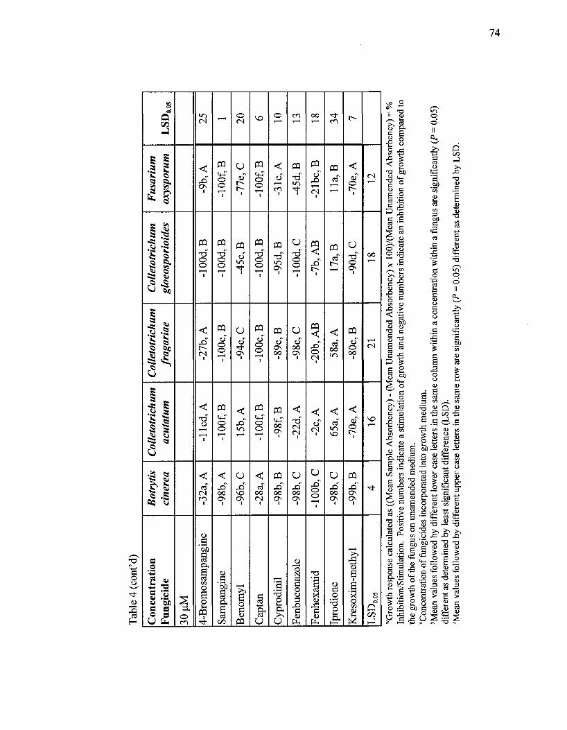
Tab
le 4
(co
nt'd
)
Con
cen
trat
ion
F
un
gici
de
30 n
M
4-B
rom
osam
pang
ine
Sam
pang
ine
Ben
omyl
Cap
tan
Cyp
rodi
nil
Fen
buco
nazo
le
Fen
hexa
mid
Ipro
dion
e
Kre
soxi
m-m
ethy
l
LkL
'0.0
5
Bot
ryti
s ci
nere
a
-32a
, A
-98b
, A
-96b
, C
-28a
, A
-98
b,B
-98
b,C
-10
0b
,C
-98
b,C
-99b
, B
4
Col
leto
tric
h um
ac
utat
um
-lie
d,
A
-10
0f,
B
15b,
A
-lOO
f, B
-98f
, B
-22d
, A
-2c,
A
65a,
A
-70e
, A
16
Col
leto
tric
h um
fr
agar
iae
-27
b,A
-100
c, B
-94c
, C
-100
c, B
-89c
, B
-98c
, C
-20b
, A
B
58a,
A
-80c
, B
21
Col
leto
tric
h um
gl
oeos
pori
oide
s
-lO
Od,
B
-lO
Od,
B
-45c
, B
-10
0d
,B
-95d
, B
-lO
Od,
C
-7b,
AB
17a,
B
-90d
, C
18
Fus
ariu
m
oxys
poru
m
-9b,
A
-lOO
f, B
-77e
, C
-lOO
f, B
-31c
, A
-45d
, B
-21b
c, B
11a,
B
-70e
, A
12
LS
D00
5
25
1 20
6 10
13
18
34
7
"Gro
wth
resp
onse
cal
cula
ted
as ((
Mea
n Sa
mpl
e A
bsor
benc
y) -
(Mea
n U
nam
ende
d A
bsor
benc
y) x
100
)/(M
ean
Una
men
ded
Abs
orbe
ncy)
= %
In
hibi
tion/
Stim
ulat
ion.
Pos
itive
num
bers
indi
cate
a st
imul
atio
n of
gro
wth
and
neg
ativ
e nu
mbe
rs in
dica
te a
n in
hibi
tion
of g
row
th c
ompa
red
to
the
grow
th o
f the
fung
us o
n un
amen
ded
med
ium
. "C
once
ntra
tion
of fu
ngic
ides
inc
orpo
rate
d in
to g
row
th m
ediu
m.
yMea
n va
lues
follo
wed
by
diff
eren
t lo
wer
cas
e le
tters
in th
e sa
me
colu
mn
with
in a
con
cent
ratio
n w
ithin
a fu
ngus
are
sign
ifica
ntly
(P
= 0
.05)
di
ffere
nt a
s de
term
ined
by
leas
t sig
nific
ant
diffe
renc
e (L
SD).
TVlea
n va
lues
follo
wed
by
diff
eren
t up
per
case
lette
rs in
the
sam
e ro
w a
re s
igni
fican
tly {
P =
0.0
5) d
iffer
ent
as d
eter
min
ed b
y L
SD.

Tab
le 5
. S
ensi
tivi
ty o
f fu
ngi
to n
atur
al p
rodu
ct-b
ased
ana
log
fung
icid
es.
Com
para
tive
gro
wth
res
pons
e™ o
f fi
ve p
lant
pa
thog
ens
in g
row
th s
olut
ion
amen
ded
wit
h th
ree
conc
entr
atio
n le
vels
x of
natu
ral
prod
uct-
base
d an
alog
fun
gici
des
com
pare
d to
gro
wth
in
unam
ende
d so
luti
on a
fter
72
h.
Con
cen
trat
ion
Fu
ngi
cid
e
0.3
uM
4-M
etho
xysa
mpa
ngin
e
Ben
zo[4
,5]S
ampa
ngin
e
Cry
ptol
epin
e
Lir
iode
nine
, C
DH
-II-
37
Lir
iode
nine
, M
el A
MC
-XII
-103
Ony
chin
e
LSD
0_05
Bot
ryti
s ci
nere
a
-5ay, A
z
-55b
, B
la,
A
-12a
, B
-15a
, B
-8a,
A
28
Col
leto
tric
h um
ac
utat
um
0c,
A
-26d
, A
18a,
A
20a,
A
lib
, A
20a,
A
7
Col
leto
tric
h um
fr
agar
iae
-3b,
A
-20c
, A
11a,
A
5ab,
AB
4ab
,AB
5ab
,AB
10
Col
leto
tric
h um
gl
oeos
pori
oide
s
-16b
, A
-100
c, C
22a,
A
-6b,
AB
-4b,
AB
-6b,
AB
14
Fus
ariu
m
oxys
poru
m
3a,
A
-17b
, A
0a,
A
la,A
B
0a,
AB
la,A
B
6
LS
D0
05
26
26
23
27
24
27

Tab
le 5
(co
nt'd
)
Con
cent
rati
on
Fun
gici
de
3.0
uM
4-M
etho
xysa
mpa
ngin
e
Ben
zo[4
,5]S
ampa
ngin
e
Cry
ptol
epin
e
Lir
iode
nine
, CD
H-I
I-37
Lir
iode
nine
, Mel
AM
C-X
II-1
03
Ony
chin
e
LkD
005
Bot
ryti
s ci
nere
a
-18a
, A
-47a
, A
-44a
, B
-59a
, B
-27a
, AB
-21a
, B
67
Col
leto
tric
h um
ac
utat
um
-44c
, B
-46c
, A
13a,
A
-12b
, A
-6ab
,A
13a,
A
22
Col
leto
tric
hum
fr
agar
iae
-44c
d, B
-55d
, A
-18a
b,A
B
-30b
cd, A
B
-27b
c, A
B
5a, A
B
27
Col
leto
tric
h um
gl
oeos
pori
oide
s
-91c
, C
-99c
, B
9a, A
-98c
, C
-54b
, C
8a, A
B
16
Fus
ariu
m
oxys
poru
m
-34b
, AB
-42b
, A
-6a,
A
-38b
, AB
-33b
, BC
-3a,
AB
14
LS
D00
5
24
41
33
29
22
33

Tab
le 5
(co
nt'd
)
Con
cen
trat
ion
F
un
gici
de
30 n
M
4-M
etho
xysa
mpa
ngin
e
Ben
zo[4
,5]S
ampa
ngin
e
Cry
ptol
epin
e
Lir
iode
nine
, C
DH
-II-
37
Lir
iode
nine
, M
el A
MC
-XII
-103
Ony
chin
e
kk.D
0.05
Bot
ryti
s ci
nere
a
19a,
A
-23
ab,A
-57b
cd, A
-92d
, A
-87c
d, A
-45b
c, B
45
Col
leto
tric
hum
ac
utat
um
-25b
, B
-39c
, A
-58d
, A
-99e
, B
-98e
, B
0a,
A
11
Col
leto
tric
hum
fr
agar
iae
-40b
, B
-29
ab,A
-97c
, B
-98c
, B
-93c
, A
B
-11a
, A
21
Col
leto
tric
h um
gl
oeos
pori
oide
s
-79
b,C
-100
c, B
-99c
, B
-99c
, B
-96c
, A
B
3a,
A
14
Fus
ariu
m
oxys
poru
m
-24
b,B
-27b
, A
-62c
, A
-99d
, B
-99d
, B
-11a
, A
7
LS
D0
05
23
30
31
3 10
21
wG
row
th re
spon
se c
alcu
late
d as
((M
ean
Sam
ple
Abs
orbe
ncy)
- (M
ean
Una
men
ded
Abs
orbe
ncy)
x 1
00)/(
Mea
n U
nam
ende
d A
bsor
benc
y) =
%
Inhi
bitio
n/St
imul
atio
n. P
ositi
ve n
umbe
rs in
dica
te a
stim
ulat
ion
of g
row
th a
nd n
egat
ive
num
bers
indi
cate
an
inhi
bitio
n of
gro
wth
com
pare
d to
the
grow
th o
f the
fung
us o
n un
amen
ded
med
ium
. "C
once
ntra
tion
of fu
ngic
ides
inc
orpo
rate
d in
to g
row
th m
ediu
m.
yMea
n va
lues
follo
wed
by
diff
eren
t lo
wer
cas
e le
tters
in th
e sa
me
colu
mn
with
in a
con
cent
ratio
n w
ithin
a fu
ngus
are
sig
nific
antly
(P
= 0
.05)
diff
eren
t as
det
erm
ined
by
leas
t sig
nific
ant
diff
eren
ce (
LSD
). zM
ean
valu
es fo
llow
ed b
y di
ffer
ent
uppe
r ca
se le
tters
in th
e sa
me
row
are
sign
ifica
ntly
(P
= 0
.05)
diff
eren
t as
det
erm
ined
by
LSD
.

Tab
le 6
. E
ffec
ts o
f na
tura
l pr
oduc
t-ba
sed
and
com
mer
cial
fun
gici
des
duri
ng p
re-
and
post
-ino
cula
tion
gre
enho
use
trea
tmen
ts.
Non
e
50%
Eto
h
625
ppm
1,25
0 pp
m
2,50
0 pp
m
LSD
Pr>
F
Azo
xyst
robi
n
Pre-
inoc
ulat
ion
Ant
hrac
nose
0.58
a
0.25
a
0.33
a
0.83
a
0.67
a
0.60
0.27
Phyt
otox
icity
0.08
b
0.33
a
0.08
b
0.08
b
0.08
b
0.17
0.14
Post
-ino
cula
tion
Ant
hrac
nose
0.75
a
0.83
a
0.42
ab
0.08
b
0.42
ab
0.54
0.06
Phyt
otox
icit
y
0.33
a
0.00
b
0.08
b
0.17
ab
0.00
b
0.23
0.04
Sam
pang
ine
Pre-
inoc
ulat
ion
Ant
hrac
nose
0.58
a
0.25
a
0.75
a
0.75
a
0.58
a
0.58
0.36
Phyt
otox
icit
y
0.08
b
0.33
ab
0.08
b
0.08
b
0.50
a
0.36
0.08
Post
-ino
cula
tion
Ant
hrac
nose
0.75
a
0.83
a
0.25
b
0.17
b
0.29
b
0.41
0.01
Phyt
otox
icit
y
0.33
a
0.00
b
0.00
b
0.00
b
0.00
b
0.17
0.00
oo

Tab
le 6
(co
nt'd
).
Non
e
50%
EtO
H
312
ppm
625
ppm
1,25
0 pp
m
2,50
0 pp
m
LS
D
Pr>
F
CA
Y-1
Pre-
inoc
ulat
ion
Ant
hrac
nose
0.58
abcx
0.25
c
0.42
bc
0.92
ab
1.00
a
0.83
ab
0.23
0.04
Phyt
otox
icity
0.08
b
0.33
ab
0.25
ab
0.58
a
0.25
ab
0.08
b
0.47
0.28
Post
-ino
cula
tion
Ant
hrac
nose
0.75
ab
0.83
a
0.42
abc
0.33
bc
0.21
c
0.21
c
0.42
0.02
Phyt
otox
icity
0.33
a
0.00
b
0.00
b
0.00
b
0.00
b
0.08
b
0.17
0.00
x Mea
n va
lues
follo
wed
by
diff
eren
t le
tters
in th
e sa
me
colu
mn
are
sign
ifica
ntly
(P
= 0
.05)
diff
eren
t as
de
term
ined
by
leas
t sig
nific
ant
diff
eren
ce (
LSD
).
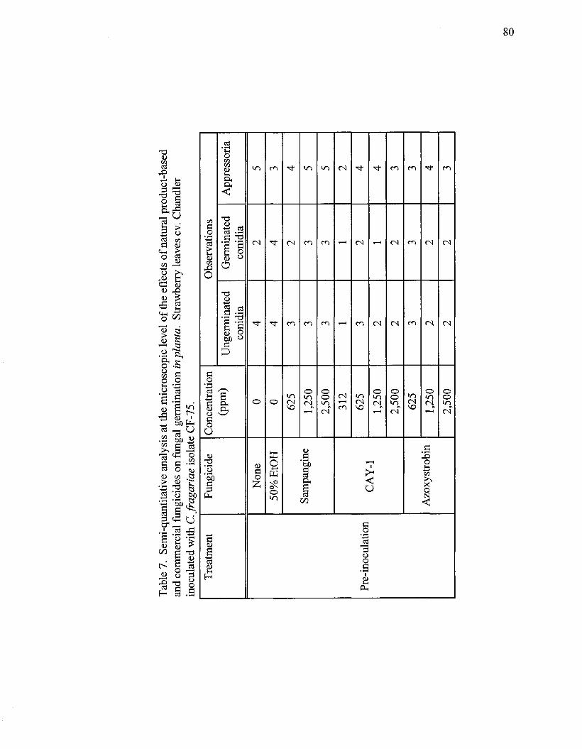
Tab
le 7
. Se
mi-
quan
titat
ive
anal
ysis
at t
he m
icro
scop
ic l
evel
of
the
effe
cts
of n
atur
al p
rodu
ct-b
ased
an
d co
mm
erci
al f
ungi
cide
s on
fung
al g
erm
inat
ion
inpl
anta
. St
raw
berr
y le
aves
cv.
Cha
ndle
r in
ocul
ated
with
C. f
raga
riae
isol
ate
CF
-75.
Tre
atm
ent
Pre-
inoc
ulat
ion
Fung
icid
e
Non
e
50%
EtO
H
Sam
pang
ine
CA
Y-1
Azo
xyst
robi
n
Con
cent
ratio
n (p
pm)
0 0 625
1,25
0
2,50
0
312
625
1,25
0
2,50
0
625
1,25
0
Obs
erva
tions
Ung
erm
inat
ed
coni
dia
4 4 3 3 3 1 3 2 2 3 2
Ger
min
ated
co
nidi
a
2 4 2 3 3 1 2 1 2 3 2
App
ress
oria
5 3 4 5 5 2 4 4 3 3 4
2,50
0 2
2 3

Tab
le 7
. (c
ont'd
)
Tre
atm
ent
Post
-ino
cula
tion
Fung
icid
e
Non
e
50%
EtO
H
Sam
pang
ine
CA
Y-1
Azo
xyst
robi
n
Con
cent
ratio
n (p
pm)
0 0 625
1,25
0
2,50
0
312
625
1,25
0
2,50
0
625
1,25
0
2,50
0
Obs
erva
tions
Ung
erm
inat
ed
coni
dia
3 3 4 2 2
Ger
min
ated
co
nidi
a
3 2 2 2 2
App
ress
oria
5 5
Scal
e:
1 =
ver
y fe
w; 2
= f
ew;
3 =
som
e; 4
= m
any;
5 =
lot
s

Tab
le 8
. E
stim
ates
of
coni
dial
ino
cula
tion
of s
traw
berr
y le
afle
ts (
cv. C
hand
ler)
wit
h C
olle
totr
ichu
m a
cuta
tum
.
Con
idia
l co
ne,
(con
idia
/ml)
600,
000
780,
000
650,
000
1,45
0,00
0
1,52
0,00
0
1,42
0,00
0
Inoc
ulum
de
liver
ed
(ml)
0.68
0.36
0.36
0.65
0.38
0.39
Con
idia
co
unte
d
2,22
6
1,01
4
1,15
5
2,96
7
2,54
4
1,52
2
Fiel
ds
coun
ted
162
144
126
162
144
135
Con
idia
/fie
ld
13.7
2
7.05
9.2
18.3
17.7
11.3
Tot
al l
eafl
et
area
(m2)a
1,82
8,45
1,34
5
1,63
6,60
1,48
3
1,33
5,17
8,00
9
1,65
4,55
8,25
3
1,44
5,38
8,23
1
1,66
3,73
4,20
6
Tot
al
fiel
ds/
leaf
letb
2,47
4
2,21
5
1,80
7
2,23
9
1,95
6
2,25
1
Act
ual
coni
dia/
le
afle
t0
34,0
18
15,6
16
16,6
24
40,9
74
34,6
21
25,4
36
Exp
ecte
d co
nidi
a/
leaf
letd
408,
000
280,
800
234,
000
942,
500
577,
600
553,
800
Una
cco
unte
d co
nidi
a6
373,
982
265,
184
217,
376
901,
526
542,
979
528,
364
"Cal
cula
ted
usin
g B
ioqu
ant 9
8®
"Tot
al fi
elds
/leaf
let =
Tot
al le
afle
t ar
ea/a
rea
of o
ne m
icro
scop
e fi
eld
(738
,982
um
2). °A
ctua
l con
idia
/leaf
let
= c
onid
ia/fi
eld
x to
tal
fiel
ds/le
afle
t dC
onc.
x in
ocul
um d
eliv
ered
eE
xpec
ted
coni
dia/
leaf
- a
ctua
l co
nidi
a/le
afle
t
to

83
Table 9. Counts of conidia on photo-etched coverslips simulating spot inoculation of a strawberry leaflet.
Actual conidial count3
2,528
2,332
1,400
1,441
2,760
3,451
1,475
1,318
1,343
1,441
1,333
1,461
1,282
1,125
1,593
1,659
1,346
1,239
1,107
1,305
Inoculum (750,000
conidia/ml)b
34,000
35,000
40,000
40,000
43,000
49,000
30,000
30,000
34,000
35,000
30,000
40,000
37,000
44,000
40,000
40,000
32,000
35,000
40,000
40,000
Conidial concentration (conidia/ml)
640,000
640,000
682,500
682,500
625,000
625,000
715,000
715,000
707,500
707,500
675,000
675,000
700,000
700,000
675,000
675,000
725,000
725,000
600,000
600,000
Expected conidial counta,c
2,176
2,240
2,730
2,730
2,687
3,066
2,145
2,145
2,405
2,476
2,025
2,700
2,590
3,080
2,700
2,700
2,320
2,537
2,400
2,400
Unaccounted conidiad
358
92
1,330
1,289
73
388
670
827
1,062
1,035
692
1,239
1,308
1,955
1,107
1,041
974
1,298
1,293
1,095 aMean of the difference (actual - expected) = 865.45; Std Dev 625.41; Std Error 139.85; t value 6.19; Pr< 0.0001 bOriginally measured by weight. Inoculum x conidial concentration dActual conidia/leaf - expected conidia/leaflet

Table 10. The effect of repeated use of a single pipet to transfer conidia of Colletotrichum acutatum isolate Goff to a hemocytometer. Counts were made after each set of five transfers.
Number of transfers
05
10
15
20
25
30
35
40
45
50
Number of observations
6
6
6
6
6
6
6
6
6
6
Average concentration in hemocytometer (conidia/ml)ab
487,500 a
417,500 a
287,500 b
245,000 b
217,500 b
107,500 c
112,500 c
100,000 c
62,500 c
87,500 c
Concentration in pipet
(conidia/ml)
262,500
332,500
462,500
505,000
532,500
642,500
637,500
650,000
687,500
662,500
"Starting concentration is 750,000 conidia/ml. bLSD = 77,500

Otr. •J. J
Figure 1. Incipient conidial germination of Colletotrichum acutatum challenged with iprodione at a concentration of 3.0 |iM. C = conidium, GT = germ tube. Bar =10 Sim
Figure 2. Normal conidial germination of Colletotrichum gloeosporioides challenged with captan at a concentration of 0.3 |iM. C = conidium, GT = germ tube. Bar =10 jxm
GT
Figure 3. Effect of 4-bromosampangine on germination of Colletotrichum fragariae at a concentration of 0.3 |J.M. A. Normal germ tube development. B. Anomalous germ tube branching. C = conidium, GT = germ tube. Bar = 10 (J.m
.1
GT
GT GT
Figure 4. Effect of 4-bromosampangine on the germination of Botrytis cinerea at a concentration of 0.3 |J,M. A. Normal germ tube. B. Splayed (upper) and branched (lower) germ tubes. C = conidium, GT = germ tube. Bar =10 |j,m

" / t
Figure 5. Cleared strawberry leaflet from cv. Chandler inoculated with 1.5xl06
conidia/ml of Colletotrichum fragariae isolate CF-75 showing ungerminated and germinated conidia.
Figure 7. Pre-inoculation treatment. Strawberry leaflet from cv. Chandler challenged with CAY-1 at 1,250 ppm.
Figure 6. Cleared strawberry leaflet from cv. Chandler inoculated with 1.5xl06
conidial/ml of Colletotrichum fragariae isolate CF-75 showing an unidentified germinated fungal conidium.
Figure 8. Post-inoculation treatment. Strawberry leaflet from cv. Chandler challenged with sampangine at 1,250 ppm.

87
'0
Figure 9. Pre-inoculation treatment. Cleared strawberry leaflet from cv. Chandler inoculated with 1.5xl06
conidia/ml of Colletotrichum fragariae isolate CF-75 and challenged with sampangine at 625 ppm. Arrows point to representative appressoria.
~".# Figure 10. Post-inoculation treatment. Cleared strawberry leaflet from cv. Chandler challenged with sampangine at 625 ppm and inoculated with 1.5xl06
conidia/ml of Colletotrichum fragariae isolate CF-75.
TjaV**1"
Figure 11. Pre-inoculation treatment. Cleared strawberry leaflet from cv. Chandler inoculated with 1.5xl06
conidia/ml of Colletotrichum fragariae isolate CF-75 and challenged with CAY-1 at 625 ppm. Arrows point to representative appressoria.
Figure 12. Post-inoculation treatment. Cleared strawberry leaflet from cv. Chandler challenged with CAY-1 at 2,500 ppm and inoculated with 1.5xl06
conidia/ml of Colletotrichum fragariae isolate CF-75.

88
r #>
• *
Figure 13. Pre-inoculation treatment. Cleared strawberry leaflet from cv. Chandler inoculated with 1.5xl06
conidia/ml of Colletotrichum fragariae isolate CF-75 and challenged with azoxystrobin at 625 ppm. Arrows point to representative appressoria.
Figure 14. Post-inoculation treatment. Cleared strawberry leaflet from cv. Chandler challenged with azoxystrobin at 1,250 ppm and inoculated with 1.5xl06
conidia/ml of Colletotrichum fragariae isolate CF-75.
Figure 15. Pre-inoculation treatment. Strawberry leaflet from cv. Chandler challenged with sampangine at 2,500 ppm.

Figure 16. Representative glass pipet tips showing the accumulation of conidia of Colletotrichum acutatum isolate Goff on the inner wall through repeated aliquot transfers. (A) 20 transfers, (B) 30 transfers, (C) 40 transfers, and (D) 50 transfers.

90
CHAPTER V
DISCUSSION
In Vitro Testing
Microbioassay
Several modifications to assay methods reduced variability in percent germination
and the time to maximum germination which proved beneficial for screening fungicides
in vitro. Conidial concentrations between 105 and 106 conidia/ml ensured the appropriate
amount of fungal inoculum to assess germination levels of selected plant pathogenic
fungi precisely and accurately. The incorporation of Roswell Park Memorial Institute
mycological liquid medium 1640 (RPMI) as the carbon source provided adequate
germination levels in a reasonable time plus it provided the advantage of knowing the
exact composition of the medium. My preliminary studies (data not presented) revealed
the importance of introducing the washing of conidia towards obtaining reproducible
germination levels of fungal conidia in screening experiments. Seemingly minor points
such as not washing the conidial inoculum can lead to misinterpreted results. The level of
conidial germination when spores are not washed is dramatically lower than after
washing. But without knowing the level of germination in a screening test, the efficacy
of a developmental fungicide might be mistakenly rated high, when in fact, the fungus
just failed to germinate at infectious levels for other reasons. Washing conidia and over
night refrigeration prior to using a conidial suspension as inoculum removed natural
germination inhibitors contained in the conidial matrix (Arpin and Bouillant, 1981; Leite
and Nicholson, 1992; Louis and Cooke, 1985a; Seebach et al., 1989), reduced
germination times, and increased cultural synchrony of all fungal species. This improved
91
the reliability and reproducibility of the microtiter assays and reduced experimental error
associated with growth measurement or error bias. Furthermore, fungal species were
selected in early testing of fungicides in microtiter assays based on observed response
patterns. Finally, the novel use of glass cover slips in each well of the 24-well cell culture
clusters provided a convenient method for achieving high quality microscopic
observations that allowed me to document the effect of fungicides on fungal
developmental morphology. Photometric readings are sensitive to percent germination
that resulted from increased biomass, but those readings cannot distinguish
developmental stages of fungal growth (i.e., conidial germination, germ tube elongation,
appressorial formation, hyphal growth, and mycelial development).
Sampangine is an effective natural product-based fungicide inhibiting germination
in all test fungi at 3.0 and 30 uM except C. acutatum. It out-performed all of its analogs
and was more effective against F. oxysporum and P. viticola than most of the commercial
fungicides tested. These data indicate that sampangine would be a welcome addition to
the arsenal against plant pathogenic fungi.
The physical mode of action of sampangine is apparently to inhibit conidial
germination in B. cinerea, C. fragariae, C. gloeosporioides, F. oxysporum, P. obscurans,
and P. viticola. This apparent physical mode of action is comparable to some of the
commercial fungicides tested including captan, iprodione, and kresoxim-methyl that also
inhibit conidial germination on several hosts (Edlich and Lyr, 1995; Leroux, 1996;
Pommer and Lorenz, 1982; Ypema and Gold, 1999). Koller (1994) describes captan as a
multi-site fungal inhibitor, consistent with my observations that captan (and sampangine)
92
did not inhibit conidial germination in C. acutatum at about 9 h, although both fungicides
prevented subsequent growth measured at 48 h (microtiter data not shown).
The sampangine analog, 4-bromosampangine, was the most interesting natural
product-based fungicide tested. Direct microscopic observations of the effects of this
sampangine analog indicated that germ tube development was affected not only by the
presence of distorted germ tubes that branched more frequently than those of untreated
conidia, but also by the emergence of multiple germ tubes suggesting the possibility that
this compound may protect the host after fungi have already established themselves in or
on the host. This analog provides a new avenue to further explore in detail its physical
mode of action given that it induces different effects in the germ tubes of two different
species suggesting a non-specific site to provide control of fungal pathogens.
Anomalies caused by 4-bromosampangine in germ tube development suggest a
physical mode of action similar to the action of commercial fungicides benomyl,
cyprodinil, and fenhexamid where conidial germination is not inhibited, but subsequent
developmental stages such as germ tube elongation and hyphal growth are inhibited
(Duben et al., 2002; HanBler and Pontzen, 1999; Latorre et al., 2002; Leroux et al., 1998;
Richmond and Pring 1971; Rosslenbroich and Stuebler, 2000). In addition, observations
of germinating conidia of B. cinerea (HanBler and Pontzen, 1999) and B. fabae
(Richmond and Pring 1971), when exposed to fenhexamid and benomyl respectively,
produced swollen, branched germ tubes. My observations of the effects of 4-
bromosampangine coincide with the results of those studies, and therefore, a testable
hypothesis for the physical mode of action would postulate that germ tube elongation and
hyphal growth, but not conidial germination, is inhibited by 4-bromosampangine.

93
Benomyl binds to p-tubulin disrupting microtubule function (Hippe, 1991). Microtubules
constitute the cytoskeleton of the cell and maintain the integrity of the Spitzenkorper
which contains the material necessary for cell wall synthesis and indicates the direction of
cell expansion (Howard and Aist, 1980; Richmond and Pring, 1971). My observations of
germ tube morphological anomalies may have resulted from the disruption of microtubule
mediated transport of cell wall material (Howard and Aist, 1980), and therefore, a
reasonable testable hypothesis for the biochemical mode of action would postulate that
microtubules in the cytoskeleton of growing fungal tips are disrupted by 4-
bromosampangine.
The most obvious effects of sampangine and the commercial fungicides tested
were to inhibit and/or delay germination which raises an interesting question, not tested in
the scope of this research, regarding the duration of inhibition. Richmond and Pring
(1971) reported that conidia of B. fabae that had been completely inhibited from
germinating in the presence of benomyl germinated normally when transferred to a
medium free of benomyl. In addition they reported that when exposed again to benomyl,
germinating conidia of B. fabae produced swollen and distorted germ tubes that branched
more frequently than the usual. However, conidia of B. fabae resumed normal growth
when transferred to a medium free of benomyl. Furthermore, the abnormal germ tubes
produced in the presence of benomyl formed a single hypha and grew normally in the
absence of the fungicide. The technique I developed allowed me to document not only
the presence of distorted germ tubes that branched more frequently than those of normal
conidia but also the emergence of multiple germ tubes caused by 4-bromosampangine in
B. cinerea and C. fragariae.

94
The interaction between fungal pathogens and fungicides described in this study is
the first detailed investigation on fungal germination of the two new natural product-
based synthesized fungicides sampangine and 4-bomosampangine against the anamorphs
of seven economically important Ascomycetes and may serve as a model for other similar
interactions. Microbioassays add a new dimension to the accurate, sensitive, and rapid
detection system of fungal sensitivities provided by the microtiter assays since it
incorporates visualization of fungal anomalies at various developmental stages caused by
the fungicides and would allow targeting specific compounds from a pool of tested
compounds. Moreover, since the microbioassay is corroborated by data obtained from
the microtiter assay, combining both assays provides a reasonable approach to better
understand fungicide-pathogen interactions and to investigate the physical and
biochemical modes of action of fungicides.
Microtiter Assay
Fusarium oxysporum was the least sensitive species challenged by the commercial
fungicides. In view of the increasing insensitivity exhibited by many fungi, sampangine
represents an alternative to commercial fungicides now used to control economically
important plant pathogenic fungi. The potential benefits and broad spectrum antifungal
activity exhibited by sampangine make it a promising candidate for further greenhouse
testing and even field studies. Results from preliminary greenhouse trials demonstrated
the protectant activity of sampangine by reducing leaf lesion severity on strawberry when
applied to the host prior to inoculation with C. fragariae (Wedge, unpublished data).
Phomopsis spp. were not included in the microtiter assay due to their
asynchronous germination behavior which prevented reliable absorbency readings by the
95
microplate photometer. Hyphal growth after conidial germination for these species
requires several days, and as a result, the sensitivity and reproducibility of the method
(absorbency) is diminished, since only a proportion of the growing population can be
measured. Phomopsis spp. proved not to be suitable organisms for testing the efficacy of
fungicides. On the other hand, B. cinerea, C. acutatum, C. fragariae, C. gloeosporioides,
and F. oxysporum represent excellent examples of organisms suitable for this type of
assay.
I focused through in vitro assays on the efficacy of the natural product-based
fungicide, sampangine, and its analogs, by observing their effects on fungal growth. The
antifungal activity of sampangine is comparable in efficacy to commercial fungicides at
inhibiting fungal growth and is more promising than any of its analogs. The inhibitory
effects of sampangine on fungal growth is relevant when considering the potential
reduction of the initial infection and dispersal of the pathogens (Ypema and Gold, 1999).
In Planta Testing
Leaf Clearing Technique
The development of this technique has proven valuable in support of the
greenhouse screening experiments I conducted for testing the efficacy of natural product-
based and commercial fungicides in planta. Microscopic observations of the effects of
sampangine and CAY-1 indicated that germ tube development was affected during the
post-inoculation treatments which is relevant regarding their potential use as fungicides.
Thus, the development of the leaf clearing technique confirms a proper direction for
exploring the mode of action of natural product-based fungicides suggested by the
detached leaf assays. Sampangine and CAY-1 exhibit a similar mode of action to that

96
documented for azoxystrobin to control anthracnose on the surface of treated strawberry
leaflets by preventing conidial germination, therefore acting as protectant fungicides
(Bertelsen et a l , 2001; Wong and Wilcox, 2001).
In addition, this technique allowed me to correlate disease symptoms at the
macroscopic level with fungal growth at the microscopic level ruling out the possibility of
"false-positive" results. "False- positive results" are due to the fact that the fungus might
not be on the leaf surface, or might be at such low concentrations that it does not cause
disease symptoms, or the fungus may fail to infect. Finally, this technique allowed me to
identify fungal spores different from the ones sprayed to the host and establish that the
presence of such unexpected spores may or may not contribute to some extent to the
severity of the disease. Therefore it let me directly relate anthracnose lesions with the
presence of the fungal pathogen C. fragariae isolate CF-75 that I had inoculated onto the
strawberry leaf.
Detached Leaf Assay for Fungicide Evaluation
A persistent problem for inplanta assays is the uncertainty of whether the
resistance of the plant or the efficacy of the fungicide is causally related to
"false-positive" results or to the failure of the fungus to perform on cue. Conducting in
planta tests to confirm fungal sensitivity in vitro is essential to reduce the level of
uncertainty generated by these variables. Furthermore, the fact that resistance to several
fungicide classes has occurred in some Colletotrichum especies (Peres et al., 2002, 2004)
makes an assessment of the level of the efficacy of new fungicides necessary. To
accomplish this, methods that allow the baseline sensitivity for these fungicides are
needed. However, current in vitro methods used to evaluate the effects of protectant
97
fungicides (radial growth measurements in the presence of different fungicide
concentrations and determination of the minimal inhibitory fungicide concentration,
among others) by inhibiting spore germination are inappropriate when the mechanism of
the fungicide to be tested is related to infection or growth within the plant (Kunz et al.,
1998).
To measure the inhibition of germination the plant host is not required and
sensitivity tests can be performed on microscopic slides, agar plates, or cell culture
clusters (Peres et al., 2004; Petsikos-Panayotarou et al., 2003; Wedge and Kuhajek, 1998;
Wedge et al., 2001). However, a disadvantage of this method is that inhibitory fungicide
concentrations in vitro are smaller than those used in the field and no conclusions can be
drawn regarding fungicide efficacy in the field. On the other hand, even though
sensitivity assays on whole plants or detached leaves are labor intensive inplanta assays
are particularly suited to analyze fungal sensitivity against protectant and curative
fungicides since fungal virulence and susceptibility of the plant tissue contribute to the
results (Kunz et al., 1998).
In vitro assays may not reflect field conditions and sensitive isolates could be
inaccurately classified as resistant since infection structures are not differentiated in vitro
(Birchmore et al., 1996). Colletotrichum species exhibit a targeted secretion of
cutinolytic enzymes only during differentiation of infection structures (Podila et al.,
1995). However, some fungi do not differentiate infection structures during growth on
agar plates and therefore, secretion of infection related enzymes is not required. As a
consequence, inplanta tests involving the plant host constitute an absolute necessity to
corroborate sensitivity of fungal populations to new fungicides in vitro.

98
My results show that the natural product-based fungicides sampangine, CAY-1,
and the commercial fungicide azoxystrobin provided protection against anthracnose when
applied 24 h before the fungal inoculum, but they had negligible or no effect on the
incidence of the disease when applied 24 h after the fungal inoculum. These results
support the findings of Wong and Wilcox (2001) who report protectant activity of
azoxystrobin when applied before fungal inoculation, but had little effect on the incidence
of disease when applied after fungal inoculation. These results obtained during the
greenhouse screening trials suggest a protectant physical mode of action for the natural
product-based fungicides sampangine and CAY-1 against C.fragariae isolate CF-75
similar to that documented for the commercial fungicide azoxystrobin which entails the
inhibition of conidial germination (Bertelsen et al., 2001; Wong and Wilcox, 2001).
Promising results from the greenhouse studies suggest that field studies of these two
natural product-based fungicides is warranted since ultimately screening under field
conditions is the final stage in a natural product discovery program before going into
commercial production.
In Planta Screening Techniques
Calculation of Conidia per Unit Area
The level of inoculum delivered to a leaf surface was consistently less than
expected. This consistent error can confound interpretation of in planta screening
studies. Without the microscopic analysis, an investigator would have expected their
inoculum to be about 10-fold higher than delivered. The significance of this finding has
practical applications. If a fungicide was being tested and the inoculum delivered was
insufficient to elicit disease symptoms, it would lead to the assumption that the fungicide

was effective. Likewise, if a susceptible cultivar of strawberry was being tested and the
inoculum was insufficient to elicit disease symptoms, it would lead to the assumption that
the cultivar was resistant. An estimate of the amount of inoculum that actually lands on
the host would be important for a correct interpretation of the results obtained during
screening trials.
In Vitro Screening Techniques
Simulated Spot Inoculation
The photo-etched coverslip experiments eliminated the potential experimental
errors from the inplanta experiments including conidia not attaching to the leaflet
surface, and thus in the runoff, and conidia settling on the inner tube of the sprayer.
Photo-etched coverslips allowed me to obtain an exhaustive count of the conidia.
Pipeting Experiments
Adsorbance to the pipet tips accounted for the missing conidia, suggesting that the
results of screening procedures should be interpreted with caution since the number of
conidia landing on the plant surface is consistently less than the initial inoculum
concentration. Inplanta assays will carry the uncertainty of whether the resistance of the
plant or the efficacy of the fungicide is causally related to "positive" results or to an
insufficient amount of inoculum delivered. A measure of the inoculum which actually
arrives on the plant is rarely assessed or reported which opens the possibility of
evaluating a fungicide as efficacious when in fact the amount of inoculum delivered to
the plant was insufficient to induce disease symptoms.

100
REFERENCES
Abdel-Gawad, M.M., S.M. El-Amin, H. Ohigashi, Y. Watanabe, N. Takeda, H.
Sugiyama, and M. Kawanaka. 2000. Molluscicidal saponins from Anagallis
arvensis against schistosome intermediate hosts. Japanese Journal of Infectious
Diseases 53:17-19.
Agrios, G.N. 2005. Plant Pathology. 5th ed., Elsevier Academic Press, San Diego, CA.
922 p.
Alexander, H.M. 1992. Evolution of disease resistance in natural plant populations.
Pages 326-344 in R.S. Fritz and E.L. Simms, eds., Plant Resistance to Herbivores
and Pathogens: Ecology, Evolution, and Genetics. The University of Chicago
Press, Chicago.
Anesiadis, T., G.S. Karaoglanidis, and K. Tzavella-Klonari. 2003. Protective, curative,
and eradicant activity of the strobilurin fungicide azoxystrobin against Cercospora
beticola and Erysiphe betae. Journal of Phytopathology 151:647-6 51.
Arpin, N., and M.L. Bouillant. 1981. Light and mycosporines. Pages 435-454 in G.
Turian and H.R. Hohl, eds., The Fungal Spore, Morphogenetic Controls.
Academic Press, London.
Atti, S.A-E., H.A Ammar, C.H. Phoebe, P.L. Schiff, and D.J. Slatkin. 1982. Journal of
Natural Products 45(4):476^80.

Bailey, J.A., R.J. O'Conell, RJ. Pring, and C. Nash. 1992. Infection strategies of
Colletotrichum species. Pages 88-120 in J.A. Bailey and M.J. Jegerm, eds.,
Colletotrichum: Biology, Pathology, and Control. CAB International Walling-
ford, United Kingdom.
Baldwin, B.C., J.M. Clough, C.R.A. Godfrey, J.R Godwin, and T.E. Wiggins. 1996. The
discovery and mode of action of ICIA 5504. Pages 69-78 in H. Lyr, P.E. Russel,
and H.D. Sisler, eds., Modern Fungicides and Antifungal Compounds. Intercept
Ltd., Andover, U.K.
Baxter, A.P., G.C.A. Van der Westhuizen, and A. Eric. 1983. Morphology and
taxonomy of South African isolates of Colletotrichum. South African Journal of
Botany 2:259-289.
Bergstrom, G.C., and R.L. Nicholson. 1981. Invertase in the spore matrix of
Colletotrichum graminicola. Phytopathology 102:139-147.
Bertelsen, J.R., E. de Neergaard, and V. Smedegaard-Petersen. 2001. Fungicidal effects
of azoxystrobin and epiconazole on phyllosphere fungi, senescence and yield of
winter wheat. Plant Pathology 50:190-205.
Birchmore, R.J., R.J. Williams, R.J. Milling, and P.E. Russell. 1996. Development of
methods for monitoring populations of Botrytis cinerea for sensitivity to
pyrimethanil. Pages 377-385 in H. Lyr, P.E. Russel, and H.D. Sisler, eds.,
Modern Fungicides and Antifungal Compounds. Intercept Ltd., Andover, U.K.
Bonnen, A.M., and R. Hammerschmidt. 1989a. Cutinolytic enzymes from
Colletotrichum lagenarium. Physiological and Molecular Plant Pathology
35:463^174.

Bonnen, A.M., and R. Hammerschmidt. 1989b. Role of cutinolytic enzymes in infection
of cucumber by Colletotrichum lagenarium. Physiological and Molecular Plant
Pathology 35:475-481.
Bos, L., and J.E. Parlevliet. 1995. Concepts and Terminology on Plant/Pest
Relationships: Toward consensus in plant pathology and crop protection. Annual
Review of Phytopathology 33:29-102.
Bourett, T.M., and R.J. Howard. 1991. Ultrastructural immunolocalization of actin in a
fungus. Protoplasm 163:199-202.
Brent, K.J. 1996. Pathways to success in fungicide research. Pages 3-15 in H. Lyr, P.E.
Russell, and H.D. Sisler, eds, Modern Fungicides and Antifungal Compounds,
Intercept Limited, Andover, Hants, U.K.
Brown, G.E. 1977. Ultrastructure of penetration of ethylene-degreened Robinson
tangerines by Colletotrichum gloeosporioides. Phytopathology 67:315-320.
Buchenauer, H. 1990. Physiological reactions in the inhibition of plant pathogenic fungi.
Pages 217-292 in G. Haug and H. Hoffmann, eds., Chemistry of Plant Protection.
Springer-Verlag, Berlin, Germany.
Chaky, J., K. Anderson, M. Moss, and L. Vaillancourt. 2001. Surface hydrophobicity
and surface rigidity induce spore germination in Colletotrichum graminicola.
Phytopathology 91:558-564.
Cooke, R. 1977. The Biology of Symbiotic Fungi. John Wiley & Sons. 282 p.
Cox, M.L., and J.A.G. Irwin. 1988. Conidium and appressorium variation in Australian
isolates of the Colletotrichum gloeosporioides group and closely related species.
Australian Systematic Botany 1:139-149.
103
Curry, K.J., M. Abril, J.B. Avant, and B.J. Smith. 2002. Strawberry anthracnose:
Histopathology of Colletotrichum acutatum and Colletotrichumfragariae.
Phytopathology 92:1055-1063.
Dahmen, H., H.C. Hoch, and T. Staub. 1988. Differential effects of sterol inhibitors on
growth, cell membrane permeability, and ultrastructure of two target fungi.
Phytopathology 78:1033-1042.
Davidse, L.C. 1986. Benzimidazole fungicides: Mechanism of action and biological
impact. Annual Review of Phytopathology 24:43-65.
Davidse, L.C. 1987. Biochemical aspects of benzimidazole fungicides - Action and
resistance. Pages 245-258 in H. Lyr, ed., Modern Selective
Fungicides-Properties, Applications, Mechanisms of Action. Gustav Fisher
Verlag, New York.
Dekker, J. 1995. Development of resistance to modern fungicides and strategies for its
avoidance. Pages 23-38 in H. Lyr, ed., Modern Selective Fungicides-Properties,
Applications, Mechanisms of Action. Gustav Fisher Verlag, New York.
Delp, C.J. 1987. Benzimidazoles and related fungicides. Pages 233-244 in H. Lyr, ed.,
Modern Selective Fungicides-Properties, Applications, Mechanisms of Action.
Gustav Fisher Verlag, New York.
Delp, B.R., and R.D. Milholland. 1980. Evaluating strawberry plants for resistance to
Colletotrichumfragariae. Plant Disease 64:1071-1073.
De Lapeyre de Bellaire, L., M. Chillet, and X. Mourichon. 2000. Elaboration of an early
quantification method of quiescent infections of Colletotrichum musae on
bananas. Plant Disease 84:128-133.

De Lucca, A.J., J.M. Bland, C.B. Vigo, M. Cushion, C.P. Selitrennikoff, J. Peter, and T.J.
Walsh. 2002. CAY-1, a fungicidal saponin from Capsicum sp. fruit. Medical
Mycology 40:131-137.
Denoyes-Rothan, B., M. Lafargue, G. Guerin, and M. Clerjeau. 1999. Fruit resistance to
Colletotrichum acutatum in strawberries. Plant Disease 83:549-553.
Dey, P.K. 1933. Studies in the physiology of the appressorium of Colletotrichum
gloeosporioides. Annals of Botany 47:305-312.
Dhingra, O.D., and Sinclair, J.B. 1995. Basic Plant Pathology Methods. Second edition.
Lewis Publishers, FL, USA. 434 p.
Dieguez-Uribeondo, J., H. Forster, and J.E. Adaskaveg. 2003. Digital image analysis of
internal light spots of appressoria of Colletotrichum acutatum. Phytopathology
93:923-930.
Duben, J., Rosslenbroich, H.J., and Jenner, G. 2002. Teldor® (Fenhexamid) - A new
specific fungicide for the control of Botrytis cinerea and related pathogens on
Rubus, Ribes, and other crops. Acta Horticulturae 585:325-329.
Edlich, W., and H. Lyr. 1995. Mechanism of action of dicarboximide fungicides. Pages
119-131 in H. Lyr, ed., Modern Selective Fungicides-Properties, Applications,
Mechanisms of Action. Gustav Fisher Verlag, New York.
Egley, G.H. 1994. Substrate influences upon germination of Colletotrichum truncatum
conidia. Canadian Journal of Botany 72:1758-1765.
Ehr, R.J., G. Kemmitt, and D. Ouimett. 2005. Periodic Table of Fungicides 2.1. Dow
Agrosciences LLC. Indianapolis, IN. L01-818-017 (3/05) BOD; 010-30143.
Emmett, R.W., and D.G. Parbery. 1975. Appressona. Annual Review of
Phytopathology 13:147-167.
Espinel-Ingrof, A., and Kerkering, T.M. 1991. Spectrophotometric method of inoculum
preparation for the in vitro susceptibility testing of filamentous fungi. Journal of
Clinic Microbiology 29:393-394.
Freeman, S., S. Horowitz, and A. Sharon. 2001. Pathogenic and non-pathogenic
lifestyles in Colletotrichum acutatum from strawberry and other plants.
Phytopathology 91:986-992.
Foster, B., and T. Staub. 1996. Basis for use strategies of anilinopyrimidine and
phenylpyrrole fungicides against Botrytis cinerea. Crop Protection
15(6):529-537.
Goes, A., and H. Kimati. 1994. Efeito de benomyl no crescimento micelial de
Colletrotrichum gloeosporioides, agente causal da queda prematura de frutos
jovens de citros. Summa Phytopathology 24:246-253.
Gold, R.E., E. Ammermann, H. Kohle, G.M.E. Leinhos, G. Lorenz, J.B. Speakman, M.
Stark-Urnau, and H. Sauter. 1996. The synthetic strobilurin BAS 490 F: Profile
of a modern fungicide. Pages 79-92 in H. Lyr, P.E. Russell, and H.D. Sisler, eds,
Modern Fungicides and Antifungal Compounds, Intercept Limited, Andover,
Hants, U.K.
106
Green, J.R., N.A. Pain, M.E. Cannell, G.L. Jones, C.L. Leckie, S. McCready, K.
Mendgen, A.J. Mitchell, J.A. Callow, and R.J. O'Connell. 1995. Analysis of
differentiation and development of the specialized infection structures formed by
biotrophic fungal plant pathogens using monoclonal antibodies. Canadian Journal
of Botany 73:S408-S417.
Gunnell, P.S., and W.D. Gubler. 1992. Taxonomy and morphology of Colletotrichum
species pathogenic to strawberry. Mycologia 84(2):157—165.
Hamer, J.E., R.J. Howard, F.G. Chumley, and B. Valent. 1988. A mechanism for
surfacce attachment in spores of a plant pathogenic fungus. Science 239:288-290.
HanBler, G., and R. Pontzen. 1999. Effect of fenhexamid on the development ofBotrytis
cinerea. Pflanzenschutz-Nachrichten Bayer 52(2):158—176.
Hardham, A.R. 1992. Cell biology of pathogenesis. Annual Review of Plant Physiology
and Plant Molecular Biology 43:491-526.
Heath, LB. 1990. The roles of actin in tip growth of fungi. International Review of
Cytology 123:95-127.
Hewitt, H.G. 1998. Fungicide resistance. Pages 155-181 mH.G. Hewitt, ed., Crop
Protection. Centre for Agriculture and Biosciences International, Wallingford,
UK.
Heye, U.J., J. Speich, H. Siegle, A. Steinemann, B. Forster, G. Knauf-Beiter, J. Herzog,
and A. Hubele. 1994. CGA 219417: a novel broad-spectrum fungicide. Crop
Protection 13:541-549.

Hickey, K.D., and K.S. Yoder. 1981. Field performance of sterol-inhibiting fungicides
against apple powdery mildew in the mid-Atlantic apple growing region. Plant
Disease 65:1002-1006.
Hicks, B.R. (ed.) 2001. DuPont abandoning Benlate. Crop Protection, April 28, 2001,
Issue # 137, page 11. Retrieved June 3, 2007, from Crop Protection Web site
http://www.crop-protection-monthly.co.Uk/Archives/CPMApr2001.doc#_
Toc65669388.
Hippe, S. 1991. Influence of fungicides on fungal fine structure. Pages 265-331 inK.
Mendgen and D.E. Lesemann, eds., Electron Microscopy of Plant Pathogens.
Springer-Verlag, Berlin, Germany.
Hisada, Y., T. Kato, and Y. Kawase. 1977. Systematic movement in cucumber plants
and control of cucumber grey mould by a new fungicide, S. 7131. Nether land
Journal of Plant Pathology 83(Sl):71-78.
Hoch, H.C., and R.C. Staples. 1991. Signaling for infection structure fromation in fungi.
Pages 25-46 in G.T. Cole and H.C. Hoch, eds., The Fungal Spore and Disease
Initiation in Plants and Animals. Plenum Press, New York.
Horowitz, S., S. Freeman, and A. Sharon. 2002. Use of green fluorescent protein-
transgenic strains to study pathogenic and nonpathogenic lifestyles in
Colletotrichum acutatum. Phytopathology 92:743-749.
Howard, R.J., and J.R. Aist. 1980. Cytoplasmic microtubules and fungal
morphogenesis: Ultrastructural effects of methyl benzimidazol-2-yl carbamate
determined by freeze-substitution of hyphal tip cells. Journal of Cellular Biology
87:55-64.

Hufford, CD., S. Liu, and A.M. Clark. 1987. Anticandidal activity of eupolauridine and
onychine, alkaloids from Cleistopholis patens. Journal of Natural Products
50(5):961-964.
Irwin, J.A.G., D.F. Cameron, and J.M. Lenne. 1984. Responses of Stylosanthes to
anthracnose. Pages 295-310 in H. Stace and L.A. Edye, eds., The Biology and
Agronomy of Stylosanthes. Academic Press, Australia.
Ishida, N., and S. Akai. 1969. Relation of temperature to germination of conidia and
appressorium formation in Colletotrichum lagenarium. Mycologia 61:382-386.
Ishii, H., B.A. Fraaije, T. Sugiyama, K. Noguchi, K. Nishimura, T. Takeda, T. Amano,
and D.W. Hollomon. 2001. Occurrence and molecular characterization of
strobilurin resistance in cucumber powdery mildew and downey mildew.
Phytopathology 91:1166-1171.
Jones, A.L. 1981. Fungicide resistance: Past experience with benomyl and dodine and
future concerns with sterol inhibitors. Plant Disease 65:990-992.
Kato, T., K. Suzuki, J. Takahashi, and K. Kamoshita. 1984. Negatively correlated cross-
resistance between benzimidazole fungicides and methyl N-(3,5-dichlorophenyl)
carbamate. Journal of Pesticide Science 9:489-495.
Kerrigan, J., and C.W. Mims. 1993. Ultrastructural and cytochemical investigations of
the fungus Colletotrichum gloeosporioides, causal organism of pecan anthracnose.
The BEAM Newsletter of the Southeastern Electron Microscopy Society 9:21-25.
Khan, A., and T. Hsiang. 2003. The infection process of Colletotrichum graminicola
and relative aggressiveness on four turfgrass species. Canadian Journal of
Microbiology 49:433-442.

Knauf-Beiter, G., H. Dahmen, U. Heye, and T. Staub. 1995. Activity of cyprodinil:
Optimal treatment timing and site of action. Plant Disease 79:1098-1103.
Kolattukudy, P.E. 1985. Enzymatic penetration of the plant cuticle by fungal pathogens.
Anuual Review of Phytopathology 23:223-250.
Kolattukudy, P.E., L.M. Rogers, D. Li, C-S Hwang, and M. Flaishman. 1995. Surface
signalling in pathogenesis. Proceedings of the National Academy of Sciences of
the United States 92:4080^087.
Koller, W. 1991. The plant cuticle - A barrier to be overcome by fungal plant pathogens.
Pages 219-246 in G.T. Cole and H.C. Hoch, eds., The Fungal Spore and Disease
Initiation in Plants and Animals. Plenum Press, New York.
Koller, W. 1994. Chemical control of apple scab- status quo and future. Norwegian
Journal of Agricultural Sciences S17:149—170.
Kunz, S., B. Lutz, H. Deising, and K. Mendgen. 1998. Assessment of sensitivities to
anilinopyrimidine- and strobilurin-fungicides in populations of the apple scab
fungus Venturia inaequalis. Journal of Phytopathology 146:231-238.
Kuo, K.C. 1999. Germination and appressorium formation in Colletotrichwn
gloeosporioides. Proceedings of the National Science Council, Republic of
China, Part B 23:126-132.
Lalitha, T., and L.V. Venkataraman. 1991. Antifungal activity and mode of action of
saponins from Madhuca butyracea Mach. Indian Journal of Experimental
Biology 29:558-562.

110
Landes, M., and G.M. Hoffmann. 1979a. Germination and infection behaviour of
Colletotrichum lindemuthianum on Phaseolus vulgaris. Phytopathologische
Zeitschrift 95:259-273.
Landes, M., and G.M. Hoffmann. 1979b. Ultrahistological investigations of the
interactions in compatible and incompatible systems of Phaseolus vulgaris and
Colletotrichum lindemuthianum. Phytopathologische Zeitschrift 96:330-351.
Latorre, B.A., I. Spadaro, and M.E. Rioja. 2002. Occurrence of resistant strains of
Botrytis cinerea to anilinopyrimide fungicides in table grapes in Chile. Crop
Protection 21:957-961.
Latunde-Dada, A.O., R.J. O'Connell, C. Nash, R.J. Pring, J.A. Lucas, and J.A. Bailey.
1996. Infection process and identity of the hemibiotrophic anthracnose fungus
(Colletotrichum destructivum O'Gara) from cowpea (Vigna unguiculata (L.)
Walp.). Mycological Research 100:1133-1141.
Leandro, L.F.S., M.L. Gleason, F.W. Nutter, Jr., S.N. Wegulo, and P.M. Dixon. 2001.
Germination and sporulation of Colletotrichum acutatum on symptomless
strawberry leaves. Phytopathology 91:659-664.
Leandro, L.F.S., M.L. Gleason, F.W. Nutter, Jr., S.N. Wegulo, and P.M. Dixon. 2003.
Strawberry plant extracts stimulate secondary conidiation by Colletotrichum
acutatum on symptomless strawberry leaves. Phytopathology 93:1285-1291.
Legard, D.E., C.L. Xiao, J.C. Mertely, and C.K. Chandler. 2001. Management of
Botrytis fruit rot in annual winter strawberry using captan, thiram, and iprodione.
Plant Disease 85:31-39.
I l l
Leinhos, M.E., R.E. Gold, M. Duggelin, and R. Guggenheim. 1997. Development and
morphology of Uncinula necator following treatment with the fungicides
kresoxim-methyl and penconazole. Mycological Research 101:1033-1046.
Leite, B., and R.L. Nicholson. 1992. Mycosporine-Alanine: A self-inhibitor of
germination from the conidial mucilage of Colletotrichum graminicola.
Experimental Mycology 16:76-86.
Lenne, J.M., R.M. Sonoda, and D.G. Parbery. 1984. Production of conidia by setae of
Colletotrichum species. Mycologia 76:359-362.
Leroux, P. 1996. Recent developments in the mode of action of fungicides. Pesticide
Science 47:191-197.
Leroux, P., and M. Gredt. 1995. Etude in vitro de la resistance de Botrytis cinerea aux
fungicides anilinopyrimidines. Agronomie 15:367-370.
Leroux P., F.Chapeland, A. Arnold, and M. Gredt. 1998. Resistance de Botrytis cinerea
aux fungicides. Du laboratoire au vignoble et vice versa. Phytoma 504:62-67.
Li, G., C. He, X. Wang, and X. Wang. 1998. Extraction of Phytolacca acinose and its
mulloscacidal effects. Journal Tongji Medical University 18:69-71.
Lipps, P.E. 1983. Survival of Colletotrichum graminicola in infested corn residues in
Ohio. Plant Disease 67:102-104.
Lopez-Franco, R., R.J. Howard, and C.E. Bracker. 1995. Satellite Spitzenkorper in
growing hyphal tips. Protoplasma 188:85-103.
Louis, I., and R.C. Cooke. 1985a. Conidial matrix and spore germination in some plant
pathogens. Transactions of the British Mycological Society 84:661-667.
112
Louis, I., and R.C. Cooke. 1985b. Enzymes in conidial matrix of Colletotrichum
gloeosporioides and Mycosphaerellapinod.es. Transactions of the British
Mycological Society 84:742-745.
Louis, I., A. Chew, and G. Lim. 1988. Influence of spore density and extracellular
conidial matrix on spore germination in Colletotrichum capsici. Transactions of
the British Mycological Society 91:694-697.
Lyr, H. 1995. Selectivity in modern fungicides and its basis. Pages 13-22 in H. Lyr, ed.,
Modern Selective Fungicides-Properties, Applications, Mechanisms of Action.
Gustav Fisher Verlag, New York.
Mahfouz, M., S. El-Amin, H. Ohigashi, Y. Watanabe, N. Takeda, H. Sugiyama, and M.
Kawanaka. 2000. Mulliscicidal saponins from Anagallis arvensis against
Schistosome intermediate hosts. Japanese Journal of Infectious Diseases
53:17-19.
Manaut, F., and H. Maraite. 1997. Conidium germination and appressorium penetration
of Colletotrichum gloeosporioides on Stylosanthes guianensis. Journal of
Phytopathology 146:19-26.
Masner, P., P. Muster, and J. Schmid. 1994. Possible methionine biosynthesis inhibition
by pyrimidineamine fungicides. Pesticide Science 42:163-166.
McRae, C.F., and B.A. Auld. 1988. The influence of environmental factors on
anthracnose of Xanthium spinosum. Phytopathology 78:1182-1186.
McRae, C.F., and G.R. Stevens. 1990. Role of conidial matrix of Colletotrichum orbi-
culare in pathogenesis of Xanthium spinosum. Mycological Research
94:890-896.

113
Mendgen, K., M. Hahn, and H. Deisign. 1996. Morphogenesis and mechanisms of
penetration by plant pathogenic fungi. Annual Review of Phytopathology
34:367-386.
Mercer, P.C., R.K.S. Wood, and A.D. Greenwood. 1971. Initial infection ofPhaseolus
vulgaris by Colletotrichum lindemuthianum. Pages 381-389 in T.F. Preece, and
C.H. Dickinson, eds., Ecology of Leaf Surface Micro-organisms. Academic Press,
London.
Mercure, E.W., B. Leite, and R.L. Nicholson. 1994a. Adhesion of ungerminated conidia
of Colletotrichum graminicola to artificial hydrophobic surfaces. Physiological
and Molecular Plant Pathology 45:421^140.
Mercure, E.W., H. Kunoh, and R.L. Nicholson. 1994b. Adhesion of Colletotrichum
graminicola to corn leaves: A requirement for disease development.
Physiological and Molecular Plant Pathology 45:407-420.
Miehle, B.R., and F.L. Lukezic. 1972. Studies on conidial germination and appressorium
formation by Colletotrichum trifolii. Canadian Journal of Microbiology
18:1263-1269.
Milling, R.J., and C.J. Richardson. 1995. Mode of action of the anilino-pyrimidine
fungicide pyremethanil. 2. Effects on enzyme secretion of Botrytis cinerea.
Pesticide Science 45:34-^18.
Miura, L, T. Kamakura, S. Maeno, T. Nagata, S. Hayashi, and I. Yamaguchi. 1994.
Effect of mepanipyrim on uptake of various substrates and macromolecular
biosynthesis in Botrytis cinerea. Pesticide Science 19:103-109.
114
Mims, C.W. 1991. Using electron microscopy to study plant pathogenic fungi.
Mycologia 83:1-19.
Mims, C.W., E.A. Richardson, R.P. Clay, and R.L. Nicholson. 1995. Ultrastructure of
conidia and the conidium aging process in the plant pathogenic fungus
Colletotrichum graminicola. International Journal of Plant Sciences 156:9-18.
Mims, C.W., and L.J. Vaillancourt. 2002. Ultrastuctural characterization of infection
and colonization of maize leaves by Colletotrichum graminicola, and by a C.
graminicola pathogenicity mutant. Phytopathology 92:803-812.
Mould, M.J.R., G.J. Boland, and J. Robb. 1991a. Ultrastructure of the Colletotrichum
trifolii-Medicago sativa pathosystem. I. Pre-penetration events. Physiological
and Molecular Plant Pathology 38:179-194.
Mould, M.J.R., G.J. Boland, and J. Robb. 1991b. Ultrastructure of the Colletotrichum
trifolii-Medicago sativa pathosystem. II. Post-penetration events. Physiological
and Molecular Plant Pathology 38:195-210.
Nicholson, R.L. 1992. Colletotrichum graminicola and the anthracnose disease of corn
and sorghum. Pages 186-202 in J.A. Bailey and M.J. Jeger, eds., Colletotrichum:
Biology, Pathology and Control. CAB International, Wallingford, UK.
Nicholson, R.L. 1996. Adhesion of fungal propagules. Pages 117-134 in M. Nicolle
and V. Gianinazzi-Pearson, eds., Histology, Ultrastructure and Molecular
Cytology of Plant-microorganisms Interactions. Kluwer Academic Publishers,
Dordrecht/Norwell, MA.

115
Nicholson, R.L., and L. Epstein. 1991. Adhesion of fungi to the plant surface:
prerequisite for pathogenesis. Pages 3-23 in G.T. Cole, and H.C. Hoch, eds., The
Fungal Spore and Disease Initiation in Plants and Animals. New York, Plenum
Press.
Nicholson, R.L., and W.B.C. Moraes. 1980. Survival of Colletotrichum graminicola:
Importance of the spore matrix. Phytopathology 70:255-261.
Nicholson, R.L., L.G. Butler, and T.N. Asquith. 1986. Glycoproteins from
Colletotrichum graminicola that bind phenols: Implications for survival and
virulence of phytopathogenic fungi. Phytopathology 70:1315-1318.
Nicholson, R.L., J. Hipskind, and R.M. Hanau. 1989. Protection against phenol toxicity
by the spore mucilage of Colletotrichum graminicola, an aid to secondary spread.
Physiological and Molecular Plant Pathology 35:243-252.
Niks, R.E., and D. Rubiales. 2002. Potentially durable resistance mechanisms in plants
to specialized fungal pathogens. Euphytica 124:201-216.
Ntahimpera, N., L.L. Wilson, M.A. Ellis, and L.V. Madden. 1999. Comparison of rain
effects on splash dispersal of three Colletotrichum species infecting strawberry.
Phytopathology 89:555-563.
O'Connell, R.J. 1987. Absence of a specialized interface between intracellular hyphae of
Colletotrichum lindemuthianum and cells of Phaseolus vulgaris. The New
Phytologist 107:725-734.
O'Connell, R.J., and J.A. Bailey. 1991. Hemibiotrophy in Colletotrichum
lindemuthianum. Pages 211-222 in K. Mendgen, and E.D. Lesseman, eds.,
Electron Microscopy of Plant Pathogens, Springer-Verlag, Berlin, Heildelberg.

116
O'Connell, R.J., J.A. Bailey, and D.V. Richmond. 1985. Cytology and physiology of
infection of Phaseolus vulgaris by Colletotrichum lindemuthianum. Physiological
Plant Pathology 27:75-98.
O'Connell, R.J., C. Nash, and J.A. Bailey. 1992. Lectin cytochemistry: a new approach
to understanding cell differentiation, pathogenesis and taxonomy in Colleto
trichum. Pages 67-87 in J.A. Bailey, and M.J. Jeger, eds., Colletotrichum:
Biology, Pathology, and Control, CAB International, Wallingford.
O'Connell, R.J., A.B. Uronu, G. Waksman, C. Nash, J.P.P. Keon, and J.A. Bailey. 1993.
Hemibiotrophic infection oiPisum sativum by Colletotrichum truncatum. Plant
Pathology 42:774-783.
O'Connell, R.J., N.A. Pain, K.A. Hutchison, G.L. Jones, and J.R. Green. 1996.
Ultrastructure and composition of the cell surfaces of infection structures formed
by the fungal plant pathogen Colletotrichum lindemuthianum. Journal of
Microscopy 181:204-212.
O'Connell, R.J., S. Perfect, B. Hughes, R. Carzaniga, J. Bailey, and J. Green. 2000.
Dissecting the cell biology of Colletotrichum infection processes. Pages 57-77 in
D. Prusky, S. Freeman, and M. Dickman, eds., Colletotrichum: Host Specificity,
Pathology, and Host-Pathogen Interaction. APS Press, St. Paul, MN.
Okunji, CO., M.M. Iwu, J.E. Jackson, and J.D. Tally. 1996. Biological activity of
saponins from two Dracaena species. Advances in Experimental Medicine and
Biology 404:415-428.
Olaya, G. and Koller, W. 1999. Baseline of Venturia inaequalis populations to the
strobilurin fungicide kresoxim-methyl. Plant Disease 83:274-278.

117
Pangga, I.B., S. Chakraborty, and D. Yates. 2004. Canopy size and induced resistance in
Stylosanthes scabra determine antrachnose severity at high C02. Phytopathology
94:221-227.
Pappas, A.C., and D.H. Fisher. 1979. A comparison of the mechanism of action of
vinclozolin, procymidone, iprodioine, and prochloraz against Botrytis cinerea.
Pesticide Science 10:239-246.
Parbery, D.G. 1996. Trophism and the ecology of fungi associated with plants.
Biological Reviews 71:473-527.
Pasche, J. S., C. M. Wharam, and N. C. Gudmestad. 2004. Shift in Sensitivity of
Alternaria solani in Response to Q(o)I Fungicides. Plant Disiase 88:181-187.
Pascholati, S.F., H. Deising, B. Leite, D. Anderson, and R.L. Nicholson. 1993. Cutinase
and non-specific esterease activities in the conidial mucilage of Colletotrichum
graminicola. Physiological and Molecular Plant Pathology 42:37-51.
Pelah, D., Z. Abramovich, A. Markus, and Z. Wiesman. 2002. The use of commercial
saponin from Quillaja saponaria bark as a natural larvicidal agent against A edes
aegypti and Culexpippiens. Journal of Ethnopharmacology 81:407-409.
Peres, N.A.R., N.L. Souza, S.E. Zitko, and L.W. Timmer. 2002. Activity of benomyl
for control of postbloom fruit drop of citrus caused by Colletotrichum acutatum.
Plant Disease 86:125-130.
Peres, N.A.R., N.L. Souza, T.L. Peever, and L.W. Timmer, 2004. Benomyl sensitivity of
isolates of Colletotrichum acutatum and C. gloeosporioides from citrus. Plant
Disease 88:125-130.

118
Perfect, S.E., H.B. Hughes, R.J. O'Connell, and J.R. Green. 1999. Colletotrichum: A
model genus for studies on pathology and fungal-plant interactions. Fungal
Genetics and Biology 27:186-198.
Petsikos-Panayotarou, N., E. Markellou, and A.E. Kalamarakis. 2003. In vitro and in
vivo activity of cyprodinil and pyrimethanil on Botrytis cinerea isolates resistant
to other botrycides and selection for resistance to pirymethanil in a greenhouse
population in Greece. European Journal of Plant Pathology 109:173-182.
Podila, G.K., L.M. Rogers, and P.E. Kolattukudy. 1993. Chemical signals from avocado
surface wax trigger germination and appressorium formation in Colletotrichum
gloeosporioides. Plant Physiology 103:267-272.
Podila, G.K., E. Rosen, M.J.D. San Francisco, and P.E. Kolattukudy. 1995. Targeted
secretion of cutinase in Fusarium solani f. sp. pisi and Colletotrichum
gloeosporioides. Phytopathology 85:238-242.
Polacheck, I., M. Levy, M. Guizie, U. Zehavi, M. Nairn, and R. Evron. 1991. Mode of
action of the antimycotic agent G2 isolated from alfalfa roots. International
Journal of Medical Microbiology 275:504-512.
Politis, D.J. 1976. Ultrastructure of penetration by Colletotrichum graminicola of highly
resistant oat leaves. Physiological Plant Pathology 8:117-122.
Politis, D.J., and H. Wheeler. 1973. Ultrastructural study of penetration of maize leaves
by Colletotrichum graminicola. Physiological Plant Pathology 3:465-471.
Pommer. E.H., and G. Lorenz. 1982. Resistance of Botrytis cinerea Pers. to
dicarboximide fungicides - a literature review. Crop Protection 1:221-230.
119
Porter, F.M. 1969. Protease, cellulase, and differential localization of endo- and
exopolygalacturonase in conidia and conidial matrix of Colletotrichum orbi-
culare. Phytopathology 59:1209-1213.
Rahe, J.E., and J. Kuc. 1970. Metabolic nature of the infection limiting effect of heat on
bean anthracnose. Phytopathology 60:1005-1009.
Ramadoss, C.S., J. Uhlig, D.M. Carlson, L.G. Butler, and R.L. Nicholson. 1985.
Composition of the mucilaginous spore matrix of Colletotrichum graminicola, a
pathogen of corn, sorghum, and other grasses. Journal of Agricultural Food
Chemistry 33:728-732.
Renault, S., A.J. DeLucca, S. Boue, J.M. Bland, C.B. Vigo, and C.P. Selitrennikoff.
2003. CAY-l, a novel antifungal compound from cayenne pepper. Medical
Mycology 41:1-7.
Richmond, D.V., and R.J. Pring. 1971. The effect of benomyl on the fine structure of
Botrytisfabae. Journal of General Microbiology 66:79-94.
Rimelspach, J.W., and M.J. Boehm. 2005. Terms and Concepts for Fungicides.
Retrieved February 7, 2007, from The Ohio State University, Department of Plant
Pathology Web site http://www.turf.msu.edu/docs/74th_Conference/Boehm_
Fungicides_Concepts.pdf
Roberson, R.W., and M.M. Vargas. 1994. The tubulin cytoskeleton and its sites of
nucleation in hyphal tips of Allomycetes macrogynus. Protoplasma 182:19-31.
Rosenberg, D.A., F.W. Meyer, and C.V. Cecilia. 1979. Fungicide strategies for control
of benomyl-tolerant Penicillum expansum in apple storages. Plant Disease
Reporter 63:1033-1037.

Rosslenbroich, H.J., and D. Stuebler. 2000. Botrytis cinerea - History of chemical
control and novel fungicides for its management. Crop Protection 19:557-561.
Schadeck, R.J.G., B. Leite, and D.F. Buchi. 1998. Lipid mobilization and acid
phosphatase activity in lytic compartments during conidium dormancy and
appressorium formation of Colletotrichum graminicola. Cell Structure and
Function 23:333-340.
Schadeck, R.J.G., M.A.F. Randi, D.F. Buchi, and B. Leite. 2003. Vacuolar system of
ungerminated Colletotrichum graminicola cinidia: convergence of autophagic and
endocytic pathways. FEMS Microbiology Letters 218:277-283.
Seebach, D., G. Adam, C. von dem Bussche-Hunnefeld, U. Gisi, and H. Binder. 1989.
EPC-Synthesis and fungistatic activity of a gloeosporone analog with an ©-
hydroxy-butyl instead of the pentyl side chain on the macrocyclic ring. Liebigs
Annalen der Chemie (12):1007-1012.
Segal, R., and E. Schlosser. 1975. Role of glycosidases in the membranolytic, antifungal
action of saponins. Arch. Microbiol. 104:147-150.
Seidel, V., F. Bailleul, and P.G. Waterman. 1999. Partially acetylated tri- and
tetrarhamnoside dodecanyl ether derivatives from Cleistopholis patens.
Phytochemistry 52:465-472.
Sela-Buurlage, M.B., L. Epstein, and R.J. Rodriguez. 1991. Adhesion of ungerminated
Colletotrichum musae conidia. Physiological and Molecular Plant Pathology
39:345-352.
Shabi, E., S. Elisha, and Y. Zelig. 1981. Control of pear and apple disease in Israel with
sterol-inhibiting fungicides. Plant Disease 65:992-994.

121
Siegel, M.R. 1981. Sterol-inhibiting fungicides: Effects on sterol biosynthesis. Plant
Disease 65:986-989.
Simpson, D.W., C.Q. Winterbottom, J.A. Bell, and M.L. Maltoni. 1994. Resistance to a
single UK isolate of Colletotrichum acutatum in strawberry germplasm from
Northern Europe. Euphytica 77:161-164.
Sisler, H.D. 1996. Why study modes of action of fungicides? Pages 35-41 in H. Lyr,
P.E. Russell, and H.D. Sisler, eds, Modern Fungicides and Antifungal
Compounds, Intercept Limited, Andover, Hants, U.K.
Skoropad, W.P. 1967. Effect of temperature on the ability of Colletotrichum
graminicola to form appressoria and penetrate barley leaves. Canadian Journal of
Plant Science 47:431-434.
Smith, B.J. 1998. Botrytis blossom blight of southern blueberries: Cultivar susceptibility
and effect of chemical treatments. Plant Disease 82:924-927.
Smith, B.J., and L.L. Black. 1987. Resistance of strawberry plants to Colletotrichum
fragariae affected by environmental conditions. Plant Disease 71:834—837.
Smith, B.J., and L.L. Black. 1990. Morphological, cultural, and pathogenic variation
among Colletotrichum species isolated from strawberry. Plant Disease 74:69-76.
Smith, B.J., and L.L. Black. 1993a. In vitro activity of fungicides against Colletotrichum
fragariae. Acta Horticulturae 348:5 09-512.
Smith, B.J., and L.L. Black. 1993b. In vitro fungicide studies show the occurrence of
benomyl-resistant Colletotrichum spp. from strawberry. Advances of Strawberry
Research 12:42-48.
Smith, B.J., L.L. Black, and G.J. Galleta. 1990. Resistance to Colletotrichum.fragariae
in strawberry affected by seedling age and inoculation method. Plant Disease
74:1016-1021.
Smith, B J., and C.L. Gupton. 1993. Calcium applications before harvest affect the
severity of anthracnose fruit rot on greenhouse-grown strawberries. Acta
Horticulturae 348:477-482.
Smith, K. (ed.). 1999. Iprodione (Rovral) RED. USDA Pesticide re-registration
notification network, Volume 9, Number 1, January 21, 1999. Retrieved June 3,
2007, from Purdue University, The Center for Environmental and Regulatory
Information Systems Web site:http://ceris.purdue.edu/info/usda_rnn/rnn5-7.html.
Smith, J.E., L. Korsten, and T.A.S. Aveling. 1999. Infection process of Colletotrichum
dematium on cowpea stems. Mycological Research 103:230-234.
Soto-Estrada, A., H. Forster, J. Hasey, and J.E. Adaskaveg. 2003. New fungicides and
application strategies based on inoculum and precipitation for managing stone
fruit rust on peach in California. Plant Disease 87:1094-1101.
Stakman, E.C., and J.G. Harrar. 1957. Principles of Plant Pathology. The Ronald Press,
Co., New York. 581 p.
Staples, R.C., L. Laccetti, and Z. Yaniv. 1976. Appressorium formation and nuclear
division in Colletotrichum truncatum. Archives of Microbiology 109:75-84.
Stevens, R.B., (ed.). 1981. Mycology Guidebook. Washington University Press,
Washington. 712 pp.
Sutton, B.C. 1966. The appressoria of Colletotrichum graminicola and Colletotrichum
falcatum. Canadian Journal of Botany 44:887-897.

Swinburne, T.R. 1976. Stimulants of germination and appressoria formation by
Colletotrichum musae (Berk, and Curt.) Arx in banana leachate.
Phytopathologische Zeitschrift 87:74-90.
Szkolnic, M. 1981. Physical modes of action of sterol-inhibiting fungicides against
apple diseases. Plant Disease 65:981-985.
Talbot, N.J., D.J. Ebbole, and J.E. Hamer. 1993. Identification and characterization of
MPG1, a gene involved in pathogenicity from the rice blast fungus Magnaporthe
grisea. Plant Cell 5:1575-1590.
Talbot, N.J., M. Kershaw, G.E. Wakley, O.M.H. de Vries, J.G.H. Wessels, and J.E.
Hamer. 1996. MPG1 encodes a fungal hydrophobin involved in surface
interactions during infection-related development of the rice blast fungus
Magnaporthe grisea. Plant Cell 8:985-999.
Van Dyke, C.G., and C.W. Mims. 1991. Ultrastructure of conidia, conidium
germination, and appressorium development in the plant pathogenic fungus
Colletotrichum truncatum. Canadian Journal of Botany 69:2455-2467.
Vinijsanun, T., J.A.G. Irwin, and D.F. Cameron. 1987. Host range of three strains of
Colletotrichum gloeosporioides from tropical pasture legumes, and comparative
histological studies of interactions between type B disease-producing strains and
Stylosanthes scabra (non host) and S. guianensis (host). Australian Journal of
Botany 35:665-677.
Vizvary, M.A., and H.L. Warren. 1982. Survival of Colletotrichum graminicola in soil.
Phytopathology 72:522-525.
Waterman, P.G., and I. Muhammad. 1985. Sesquiterpenes and alkaloids from
Cleistopholispatens. Phytochemistry 24:523-527.
Wedge, D.E., and M.A. Kuhajek. 1998. A microbioassay for fungicide discovery.
SAAS Bulletin of Biochemistry and Biotechnology 11:1-7.
Wedge, D.E., and D.G. Nagle. 2005. Fungicidal properties of sampangine and its
analogs to agriculturally important fungal plant pathogens. United States Patent #
6,844,353B2; January 18, 2005.
Wedge, D.E., KJ. Curry, J. Boudreaux, P. Pace, and B.J. Smith. 2001. A microtiter
assay demonstrates sensitivity and resistance profiles for Botrytis cinerea isolates
from Louisiana strawberry farms. Advances in Strawberry Research 20:27-33.
Wessels, J.G.H. 1990. Role of the cell wall architecture in fungal tip growth generation.
Pages 1-29 in LB. Heath, ed., Tip Growth in Plant and Fungal Cells. San Diego,
Academic Press, Inc.
Wessels, J.G.H. 1993. Wall growth, protein excretion and morphogenesis in fungi. New
Phytopathology 123:397-^13.
Wessels, J.G.H. 1994. Developmental regulation of fungal cell wall formation. Annual
Review of Phytopathology 32:413-437.
Wessels, J.G.H. 1997. Hydrophobins: proteins that change the nature of the fungal
surface. Advances in Microbial Physiology 38:1-45.
Wharton, P.S., and A.M. Julian. 1996. A cytological study of compatible and
incompatible interactions between Sorgum bicolor and Colletotrichum
sublineolum. New Phytologist 14:25-34.
Wharton, P.S., A.M. Julian, and R.J. O'Connell. 2001. infrastructure of the infection of
Sorghum bicolor by Colletotrichum sublineolum. Phytopathology 91:149-158.
Whiteside, J.O. 1980a. Tolerance of Mycosphaerella citri to benomyl in Florida citrus
groves. Plant Disease 64:300-302.
Whiteside, J.O. 1980b. Detection of benomyl tolerant strains of Elsinoe fawcettii in
Florida citrus groves and nurseries. Plant Disease 64:871-872.
Wicks, T. 1977. Tolerance to benzimidazole fungicides in blue mold (Penicillum
expansum) on pears. Plant Disease Reporter 61:447-449.
Wilson, L.L., L.V. Madden, and M.A. Ellis. 1990. Influence of temperature and wetness
duration on infection of immature and mature strawberry fruit by Colletotrichum
acutatum. Phytopathology 80:111-116.
Wolkow, P.M., H.D. Sisler, and E.L. Vigil. 1983. Effect of inhibitors of melanin biosyn
thesis on structure and function of appressoria of Colletotrichum lindemuthianum.
Physiological Plant Pathology 22:55-71.
Wong, F.P., and W.F. Wilcox. 2000. Distribution of baseline sensitivities to
azoxystrobin among isolates of Plasmopara viticola. Plant Disease 84:275-281.
Wong, F.P., and W.F. Wilcox. 2001. Comparative physical modes of action of
azoxystrobin, mancozeb, and metalaxyl against Plasmopar a viticola (grapevine
downy mildew). Pant Disease 85:649-656.
Wong, F.P., and W.F. Wilcox. 2002. Sensitivity to azoxystrobin among isolates of
Uncinula necator: Baseline distribution and relationship to myclobutanil
sensitivity. Plant Disease 86:394^104.

Wosten, H.A.B., S.A. Asgeirsdottir, J.H. Krook, J.H. Drenth, and J.G.H. Wessels. 1994.
The fungal hydrophobin Sc3p self-assembles at the surface of aerial hyphae as a
protein membrane constituing the hydrophobic rodlet layer. European Journal of
Cell Biology 63:122-129.
Wosten, H.A.B., and J.G.H. Wessels. 1997. Hydrophobins, from molecular structure to
multiple functions in fungal development. Mycoscience 38:363-374.
Xuei, X.L., U. Jarlfors, and J. Kuc. 1988. Ultrastructural changes associated with
induced systemic resistance of cucumber to disease: host response and
development of Colletotrichum lagenarium in systemically protected leaves.
Canadian Journal of Botany 66:1028-1038.
Yang, X., L.L. Wilson, L.V. Madden, and M.A. Ellis. 1990. Rain splash dispersal of
Colletotrichum acutatum from infected strawberry fruit. Phytopathology
80:590-595.
Young, D.H., and H. Kauss. 1984. Adhesion of Colletotrichum lindemuthianum spores
to Phaseolus vulgaris hypocotyls and to polystyrene. Applied and Environmental
Microbiology 47:616-619.
Yourman, L., and S.N. Jeffers. 1999. Resistance to benzimidazoles and dicarboximide
fungicides in greenhouse isolates of Botrytis cinerea. Plant Disease 83:569-567.
Ypema, H.L., and R.E. Gold. 1999. Kresoxim-methyl: Modification of a neturally
occurring compound to produce a new fungicide. Plant Disease 83:4-19.
Zuck, M.G., and W.E. MacHardy. 1981. Recent experience in timing sprays for control
of apple sacb: Equipment and test results. Plant Disease 65:995-998.

127
Zulfiqar, M , R.H. Brlansky, and L.W. Timmer. 1996. Infection of flower and vegetative
tissues of citrus by Colletotrichum acutatum and C. gloeosporioides. Mycologia
88:121-128.
