in silico modelling of cancer nanomedicine, across scales
TRANSCRIPT
REVIEW ARTICLE OPEN
In silico modelling of cancer nanomedicine, across scales andtransport barriersNamid R. Stillman1, Marina Kovacevic2, Igor Balaz3 and Sabine Hauert 1✉
Nanoparticles promise to improve the treatment of cancer through their increasingly sophisticated functionalisations and ability toaccumulate in certain tumours. Yet recent work has shown that many nanomedicines fail during clinical trial. One issue is the lack ofunderstanding of how nanoparticle designs impact their ability to overcome transport barriers in the body, including theircirculation in the blood stream, extravasation into tumours, transport through tumour tissue, internalisation in the targeted cells,and release of their active cargo. Increased computational power, as well as improved multi-scale simulations of tumours,nanoparticles, and the biological transport barriers that affect them, now allow us to investigate the influence of a range of designsin biologically relevant scenarios. This presents a new opportunity for high-throughput, systematic, and integrated design pipelinespowered by data and machine learning. With this paper, we review latest results in multi-scale simulations of nanoparticle transportbarriers, as well as available software packages, with the aim of focussing the wider research community in building a commoncomputational framework that can overcome some of the current obstacles facing efficient nanoparticle design.
npj Computational Materials (2020) 6:92 ; https://doi.org/10.1038/s41524-020-00366-8
INTRODUCTIONFor over three decades, nanomedicine has held the potential toprevent, delay, control, or even cure cancer. Over two-thirds ofnanomedical research has been focused on oncology1–3. Startingwith Doxil in 1995, there are now around a dozen FDA-approvedanti-cancer nanoparticles4. Nanoparticles, typically between 1 and100 nm in size5, have been shown to be effective drug-vectorsthanks to their ability to shield therapeutic cargos throughouttransport, accumulate in certain tumour types6, activate throughenvironmental (e.g. pH, enzymatic activity) or external stimuli(such as magnetic resonance, ultrasound, or infrared light7–9), ortarget specific cells10. Active targeting includes designs where thenanoparticle surface is functionalized to bind to target receptorsor other proteins expressed on the membrane of the cancer cells,the extracellular matrix (ECM), or tumour vasculature. On the otherhand, passive targeting is mediated by the size of nanoparticles,which is thought to favour their accumulation at certain tumoursites due to the EPR effect6. Increasingly sophisticated nanopar-ticle functionalisation, or combination strategies, are beingexplored as mechanisms to make treatments both smarter andmore effective11.The versatility of nanomedicine is a result of the vastness of the
design space for nanoparticles. Changing the behaviour ofnanoparticles can be achieved by altering the conventional 4Sparameters, namely size, shape, surface functionalisation andstiffness12. The surface functionalisation in particular contributesto the charge of the particle, its stability in (and clearance from)the bloodstream, as well as active targeting. Further specificationsinclude the material used (for example, energy receptive orreactive to the environment), and the loading of the drug (such aschemotherapy).Key to predicting clinical impact, such as the correct distribution
of nanoparticles over a tumour, relies on an accurate under-standing of the interaction between individual nanoparticles and
their environment. These interactions can motivate coordinatedstrategies, where sub-populations of nanoparticles might altertheir local environment (such as degrading the ECM13), amplifysignals to other nanoparticles14, create anchored binding sites forother nanoparticles, or self-assemble/disassemble to improvetransport15,16. Such coordinated strategies, sometimes inspiredby swarm intelligence11, demonstrate the high degree ofcustomisability inherent to nanoparticle design. While thiscustomisability can lead to novel nanoparticle strategies, theirdiscovery and validation can be a challenge.Despite, or perhaps because of, the many design parameters
that can alter the functioning of nanoparticles, many prototypesfail to realise success as an alternative (or supplement) toconventional treatment techniques. Though many pre-clinicaltrials demonstrate increased specificity, only 5% of the dosetypically reaches the tumour site during clinical trials17. Transportbarriers to the target site are expected to play a key role in thisdisappointing result, including travel through the circulatorysystem and avoiding clearance, extravasation to the tumour site,tissue penetration within the tumour, and delivery to the relevantpart of the cell.Recent advances in computational power, as well as improved
simulations of nanoparticles, and the biological transport barriersthat affect them, allow for multi-scale simulations that caninvestigate the influence of a range of parameters in biologicallyrealistic scenarios. This presents a new opportunity for high-throughput, systematic, and integrated nanoparticle-design pipe-lines. Such future pipelines may enable general design principleswhich, when combined with patient-specific data, could providepersonalised treatment and care. Furthermore, utilisation of insilico models can minimise the costs associated with moreconventional trial and error approaches in the laboratory,especially when combined with recent machine learning techni-ques such as ‘active learning’18.
1Department of Engineering Mathematics, University of Bristol, Bristol, UK. 2Department of Chemistry, Biochemistry, and Environmental Protection, Faculty Of Sciences, Universityof Novi Sad, Novi Sad, Serbia. 3Laboratory for Meteorology, Physics, and Biophysics, Faculty of Agriculture, University of Novi Sad, Novi Sad, Serbia. ✉email: [email protected]
www.nature.com/npjcompumats
Published in partnership with the Shanghai Institute of Ceramics of the Chinese Academy of Sciences
1234567890():,;
There are now many computational models and results thathave been reported on different stages of tumour initiation,growth, and the interaction of nanoparticles within the body andtumour. Below, we summarise some of the most recent andrelevant in silico models for overcoming nanoparticle transportbarriers. We focus predominantly on cancer nanomedicine, butnote that many of the models we describe are relevant to othercancer therapies that may not use nanoparticles, such asimmunotherapy or chemotherapy, as highlighted in severalreviews19–21.Many of the in silico models that we describe have been
integrated with in vitro and in vivo experiments, as well as withmachine learning techniques. We believe that a systematic andintegrated framework for drug development, building on theseexamples, can minimise costly trial-and-error approaches andaccelerate the development of effective nanoparticle cancertherapies. However, such a framework requires a shared modellinglanguage and a multidisciplinary approach, with ongoing colla-boration between mathematical and computational modellers,experimentalists, and clinicians. This paper is an attempt toprovide a comprehensive review that sets the stage for such apipeline, as we detail in our concluding remarks.
IN SILICO MODELS OF TUMOURSCentral to in silico modelling of nanoparticle treatments is havingrealistic models of the tumours. Malignant tumours caused by theuncontrolled proliferation of faulty cells, can be separated intothree groups; carcinomas, leukomas, and sarcomas. Of the threegroups, carcinomas are the most common (accounting for 90% ofreported cases22) and predominantly appear in certain cell typessuch as breast, lung, prostrate, and colon/rectum. These cell typesalone accounted for more than half of all cancers reported in theUSA in 201823. Carcinomas are typically large, greater than 1 cm3
when detected, and fatal if left untreated22. They are the maintopic of this paper.Existing mathematical approaches24 can be separated into
three groups: continuum, discrete, and hybrid. The continuummodels, which utilise ordinary and partial differential equations,are typically faster to compute and are better suited to capturingglobal changes to a tumour, such as availability of oxygen andnutrients (e.g. to predict the development of a hypoxic core in asolid tumour). However, they are limited in their ability to recreateheterogeneity, single-cell interactions and other features bettersuited to a discrete modelling approach such as agent-based (AB),cellular potts (CP), and cellular automata (CA). Discrete modelsfocus on the individual units (such as the cell) that follow a simpleset of rules guiding their growth, death, and interaction with thelocal environment and other agents. This could be used to modelthe realistic heterogeneous development of a tumour over time,
how cellular resistance emergence, or the growth of angiogenicvessels.Both approaches have their strengths and weaknesses, and this
has led some researchers to combine both continuum anddiscrete approaches to gain the benefits of both, so-called hybridmethods. Hybrid models build on discrete models but combinethem with gradients of variables, modelled using continuumequations25. These models are also able to monitor individual cellbehaviour such as mutation as well as the influence of localenvironmental variables. For a review of discrete and hybridmodels and their application to cancer, see, for example work byKim et al. and An et al.26,27, for multi-scale cancer modelling, seework by Deisboeck et al. and Norton et al.28,29, and for agent-based cancer models, see work by Metzcar et al.30.
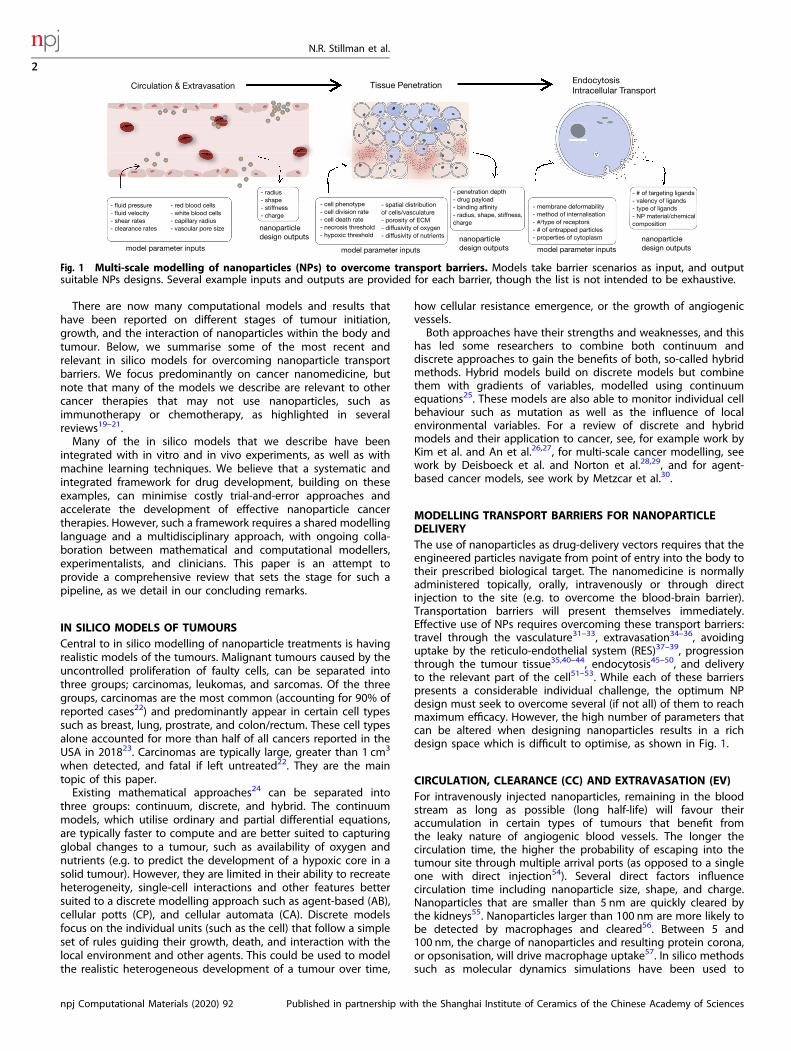
MODELLING TRANSPORT BARRIERS FOR NANOPARTICLEDELIVERYThe use of nanoparticles as drug-delivery vectors requires that theengineered particles navigate from point of entry into the body totheir prescribed biological target. The nanomedicine is normallyadministered topically, orally, intravenously or through directinjection to the site (e.g. to overcome the blood-brain barrier).Transportation barriers will present themselves immediately.Effective use of NPs requires overcoming these transport barriers:travel through the vasculature31–33, extravasation34–36, avoidinguptake by the reticulo-endothelial system (RES)37–39, progressionthrough the tumour tissue35,40–44, endocytosis45–50, and deliveryto the relevant part of the cell51–53. While each of these barrierspresents a considerable individual challenge, the optimum NPdesign must seek to overcome several (if not all) of them to reachmaximum efficacy. However, the high number of parameters thatcan be altered when designing nanoparticles results in a richdesign space which is difficult to optimise, as shown in Fig. 1.
CIRCULATION, CLEARANCE (CC) AND EXTRAVASATION (EV)For intravenously injected nanoparticles, remaining in the bloodstream as long as possible (long half-life) will favour theiraccumulation in certain types of tumours that benefit fromthe leaky nature of angiogenic blood vessels. The longer thecirculation time, the higher the probability of escaping into thetumour site through multiple arrival ports (as opposed to a singleone with direct injection54). Several direct factors influencecirculation time including nanoparticle size, shape, and charge.Nanoparticles that are smaller than 5 nm are quickly cleared bythe kidneys55. Nanoparticles larger than 100 nm are more likely tobe detected by macrophages and cleared56. Between 5 and100 nm, the charge of nanoparticles and resulting protein corona,or opsonisation, will drive macrophage uptake57. In silico methodssuch as molecular dynamics simulations have been used to
Fig. 1 Multi-scale modelling of nanoparticles (NPs) to overcome transport barriers. Models take barrier scenarios as input, and outputsuitable NPs designs. Several example inputs and outputs are provided for each barrier, though the list is not intended to be exhaustive.
N.R. Stillman et al.
2
npj Computational Materials (2020) 92 Published in partnership with the Shanghai Institute of Ceramics of the Chinese Academy of Sciences
1234567890():,;
examine the influence of these factors (such as protein coronaformation and pH-stability) on nanoparticle transport58–60. For afurther review of factors that affect circulation time for nanopar-ticles, see, for example, work by Yoo et al.61.There are other influencing factors that can affect nanoparticle
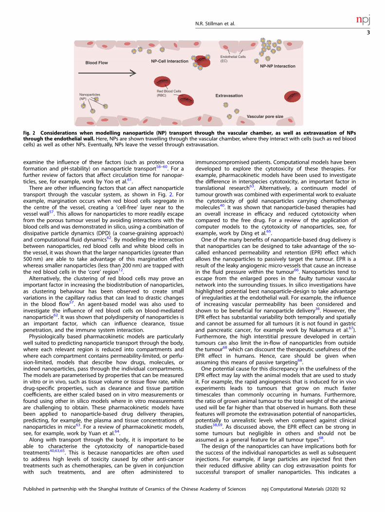
transport through the vascular system, as shown in Fig. 2. Forexample, margination occurs when red blood cells segregate inthe centre of the vessel, creating a ‘cell-free’ layer near to thevessel wall57. This allows for nanoparticles to more readily escapefrom the porous tumour vessel by avoiding interactions with theblood cells and was demonstrated in silico, using a combination ofdissipative particle dynamics (DPD) (a coarse-graining approach)and computational fluid dynamics62. By modelling the interactionbetween nanoparticles, red blood cells and white blood cells inthe vessel, it was shown that the larger nanoparticles (greater than500 nm) are able to take advantage of this margination effectwhereas smaller nanoparticles (less than 200 nm) are trapped withthe red blood cells in the ‘core’ region12.Alternatively, the clustering of red blood cells may prove an
important factor in increasing the biodistribution of nanoparticles,as clustering behaviour has been observed to create smallvariations in the capillary radius that can lead to drastic changesin the blood flow57. An agent-based model was also used toinvestigate the influence of red blood cells on blood-mediatednanoparticle62. It was shown that polydispersity of nanoparticles isan important factor, which can influence clearance, tissuepenetration, and the immune system interaction.Physiologically based pharmacokinetic models are particularly
well suited to predicting nanoparticle transport through the body,where each relevant region is reduced into compartments andwhere each compartment contains permeability-limited, or perfu-sion-limited, models that describe how drugs, molecules, orindeed nanoparticles, pass through the individual compartments.The models are parameterised by properties that can be measuredin vitro or in vivo, such as tissue volume or tissue flow rate, whiledrug-specific properties, such as clearance and tissue partitioncoefficients, are either scaled based on in vitro measurements orfound using other in silico models where in vitro measurementsare challenging to obtain. These pharmacokinetic models havebeen applied to nanoparticle-based drug delivery therapies,predicting, for example, the plasma and tissue concentrations ofnanoparticles in mice63. For a review of pharmacokinetic models,see, for example, work by Yuan et al.64.Along with transport through the body, it is important to be
able to characterise the cytotoxicity of nanoparticle-basedtreatments40,63,65. This is because nanoparticles are often usedto address high levels of toxicity caused by other anti-cancertreatments such as chemotherapies, can be given in conjunctionwith such treatments, and are often administered to
immunocompromised patients. Computational models have beendeveloped to explore the cytotoxicity of these therapies. Forexample, pharmacokinetic models have been used to investigatethe difference in interspecies cytotoxicity, an important factor intranslational research63. Alternatively, a continuum model oftumour growth was combined with experimental work to evaluatethe cytotoxicity of gold nanoparticles carrying chemotherapymolecules40. It was shown that nanoparticle-based therapies hadan overall increase in efficacy and reduced cytotoxicity whencompared to the free drug. For a review of the application ofcomputer models to the cytotoxicity of nanoparticles, see, forexample, work by Ding et al.65.One of the many benefits of nanoparticle-based drug delivery is
that nanoparticles can be designed to take advantage of the so-called enhanced permeability and retention (EPR) effect whichallows the nanoparticles to passively target the tumour. EPR is aresult of the leaky angiogenic micro-vessels that cause an increasein the fluid pressure within the tumour66. Nanoparticles tend toescape from the enlarged pores in the faulty tumour vascularnetwork into the surrounding tissues. In silico investigations havehighlighted potential best nanoparticle-design to take advantageof irregularities at the endothelial wall. For example, the influenceof increasing vascular permeability has been considered andshown to be beneficial for nanoparticle delivery36. However, theEPR effect has substantial variability both temporally and spatiallyand cannot be assumed for all tumours (it is not found in gastricand pancreatic cancer, for example work by Nakamura et al.67).Furthermore, the high interstitial pressure developed in certaintumours can also limit the in-flow of nanoparticles from outsidethe tumour68 which can discount the therapeutic usefulness of theEPR effect in humans. Hence, care should be given whenassuming this means of passive targeting68.One potential cause for this discrepancy in the usefulness of the
EPR effect may lay with the animal models that are used to studyit. For example, the rapid angiogenesis that is induced for in vivoexperiments leads to tumours that grow on much fastertimescales than commonly occurring in humans. Furthermore,the ratio of grown animal tumour to the total weight of the animalused will be far higher than that observed in humans. Both thesefeatures will promote the extravasation potential of nanoparticles,potentially to unrealistic levels when compared against clinicalstudies38,69. As discussed above, the EPR effect can be strong insome tumours but negligible in others and should not beassumed as a general feature for all tumour types68.The design of the nanoparticles can have implications both for
the success of the individual nanoparticles as well as subsequentinjections. For example, if large particles are injected first thentheir reduced diffusive ability can clog extravasation points forsuccessful transport of smaller nanoparticles. This indicates a
Fig. 2 Considerations when modelling nanoparticle (NP) transport through the vascular chamber, as well as extravasation of NPsthrough the endothelial wall. Here, NPs are shown travelling through the vascular chamber, where they interact with cells (such as red bloodcells) as well as other NPs. Eventually, NPs leave the vessel through extravasation.
N.R. Stillman et al.
3
Published in partnership with the Shanghai Institute of Ceramics of the Chinese Academy of Sciences npj Computational Materials (2020) 92

possible two-phase extravasation profile where fast extravasationis followed by slow extravasation, highlighting additional designconsiderations when engineering nanoparticles69. The structure ofthe vascular network will also be influential, especially around thetumour site. For example, spatial irregularities within the networkcan lead to uneven distribution of drugs. This has been consideredin work by Sefidgar et al.32 where the concentration of drug withinthe interstitial flow was investigated using a multi-scale model of aheterogeneous and dynamic vascular network around a tumour.By allowing feedbacks between hemodynamic and metabolicstimuli and the capillary network, a more irregular capillarynetwork was generated with high interstitial fluid pressure (IFP)within the tumour, as typically observed in vivo. It was shown thatthe elevated IFP within this irregular network led to aheterogeneous distribution of drugs around the tumour region,highlighting that static or regular vascular networks may be anoverly restrictive assumption when modelling drug delivery.As extravasation is central to nanoparticle penetration and
accumulation, it is important that general design principles areknown to optimally extravasate into the tumour. Again, in silicomodels are of use in testing various designs. For example,Brownian dynamics were used to investigate the influence ofnanoparticle diameter and aspect ratio on extravasation anddemonstrated that larger aspect ratio increases extravasation byalignment with the streamlines exiting the pore34. Alternatively,models that describe nanoparticle vascular transport also includeconsiderations of extravasation to improve model accuracy36,70.Both vascular transport and extravasation at the tumour site canbe modelled using agent-based models33. This has been used tosimulate tumour growth with a realistic vessel network structure(when compared against 3D intravital image). Though this has yetto be coupled with a nanoparticle design framework, this mayoffer a further tool for bioengineers to trial designs prior to in vitroand/or vivo testing.
TISSUE PENETRATION (TP)For nanoparticle-based therapies, a substantial challenge ispredicting the depth that the NPs are able to penetrate into thetumour and where they accumulate, as shown in Fig. 3. In healthytissue, the IFP leads to the necessary pressure gradient totransport nanoparticles away from vessels. However, IFP withintumour environments can lead to additional barriers for drug
delivery as well as driving the growth of the tumour. Experimentsin vivo have shown that IFP is uniform across much of the tumourbut drops significantly at the edge71. This uniform pressure createsa diffusive environment across much of a tumour and a steepoutward flow around the edge. The diffusive environment withinthe tumour means that extravasated particles move slowly andcan diffuse back into the capillary system rather than enteringdeeper into the tumour tissue35,72. Near the edge of a tumour,there is a risk that the nanoparticles will not be retained, insteadbeing irreversibly pushed by the steep pressure gradient37.In silico models offer a means of understanding the obstacles to
tumour penetration. The influence of the IFP on the blood andlymphatic systems and their influence on drug-delivery has beeninvestigated using a combination of fluid dynamics and agent-based modelling73. This work demonstrated how drug distributionincreases as the lymphatic response decreases, due to a reductionin the clearance of drug-delivery vectors. It also demonstrated thatthe dual normalising of both the vasculature and interstitial isrequired to improve drug efficacy in order to simultaneouslyreduce the IFP within the tumour as well as minimise theheterogeneity of the drug distribution within the tumour tissue.The heterogeneous distribution of nanoparticles has recently
been considered using in silico models that combines the growthof a tumour with angiogenesis and drug delivery74. This work useda three-dimensional model continuum approach to highlightconditions of the tumour for enhanced nanoparticle drug deliverysuch as high interstitial porosity (to enhance nanoparticletransport). Alternatively, the connection between tumour growthand avascular network generation was modelled using a multi-scale approach that coupled tumour growth with nanoparticletransport75. The binding affinity of nanoparticles was shown toplay a role in the accumulation within the tumour, where thosewith high binding affinity concentrate near the vessels and thatslow nanoparticles failed to penetrate at lethal levels throughoutthe tumour tissue76.This coupled model of tumour growth, vasculature system and
nanoparticle adhesion has since been used as a tool for optimisingnanoparticle design77. Here, the model was used to find theoptimal nanoparticle diameter for accumulation and penetration.Alternatively, a combination of deterministic and stochasticmathematical methods were used to find the optimum size andbinding affinity for nanoparticle penetration into a tumour76. Suchapproaches demonstrate the feasibility of integrating in silicomethods with nanoparticle design. Other work has also exploredbroad biodistribution and tissue distribution of drug-antibodyconjugates78,79. Here, simulations compared the difference intransport profiles between small molecules and larger macro-molecular drugs. Four fundamental classes of drug delivery agentswere highlighted (those limited by blood flow, vessel permeability,interstitial diffusion, and local binding and metabolism) each ofwhich have strengths and weaknesses.Along with optimising, in silico models have sought to explain
why, in some instances, novel drug delivery methods lead tobetter accumulation and penetration than free-drug alternatives.For example, a general mathematical model was developed toinvestigate why experimental data showed a 3-fold drop intumour growth when using nano-vectored drug delivery43. Thismathematical model combined the cell cycle, vasculature net-work, and the drug diffusion rates, and was validated using in vivomethods. It demonstrated improved outcome for nanoparticle-based therapies, thought to be driven by the geometry of theparticles. In vitro models have also confirmed improved tissuepenetration and retention of nanoparticle-based therapies17,80.These improvements are thought to be in part due to tumour-scale phenomena such as the vascular network, which preventscertain cells from being reached. Hence, the combination oftherapy with vasculature normalisation may provide a furtherimprovement in treatment efficacy.
Fig. 3 Considerations when modelling NP transport through thetumour tissue, as well as interactions with cells of different typesor other NPs. NPs (shown as black dots) are shown leaving thevascular system and diffusing through towards endothelial cells. NPsare required to penetrate deep enough through the tumour tissuein order to be effective while aiming to be internalised by specificcell types (cancer cells and cancer stem cells) while avoiding othercell types (such as healthy cells).
N.R. Stillman et al.
4
npj Computational Materials (2020) 92 Published in partnership with the Shanghai Institute of Ceramics of the Chinese Academy of Sciences
ENDOCYTOSIS (END) AND INTRACELLULAR TRANSPORT (IT)Endocytosis can be performed by various cellular uptakemechanisms46,81,82. The rate and mechanism of cellular uptakedepends on nanoparticle geometry, coating, and other physico-chemical properties. Yet the specific design of a nanoparticle cangreatly improve efficacy. Nanoparticles can be designed toincrease cellular uptake (endocytosis) as well as to improvespecificity by preferentially targeting cancer cells (throughtargeting receptors which are found to be upregulated in varioustumour cells). Most in silico approaches consider a singlenanoparticle or several (no more than ten) nanoparticles on thecell membrane and use either atomistic simulations or coarse-grain approaches such as DPD.These studies have shown that the size and shape83, ligand
multi-valency47 and the role of the protein corona aroundnanoparticles45 can all have considerable influence on cellularuptake. For example, super-selectivity was found to occur inmultivalent nanoparticles, as they demonstrate ‘on-off’ bindingprofiles that is particularly well suited for receptor-concentrationselective targeting47. This super-selectivity occurs when thefraction of bound particles sharply increases with receptorconcentration. In this work, an analytical model was developedand compared against Monte Carlo simulations of nanoparticleswith various coatings and general design principles were derived,once again demonstrating the use of in silico models innanoparticle design. Other work has considered a thermodynamicmodel for nanoparticle–cell interactions84 where uptake rateswere shown to be strongly related to the size of the nanoparticleand the interaction of multiple nanoparticles with the cellmembrane85. Both works used coarse-grain approaches. For acomprehensive review of nanoparticle endocytosis see work byAngioletti-Uberti, and Zhang et al.46,49.Having successfully been taken up by the cell, nanoparticles
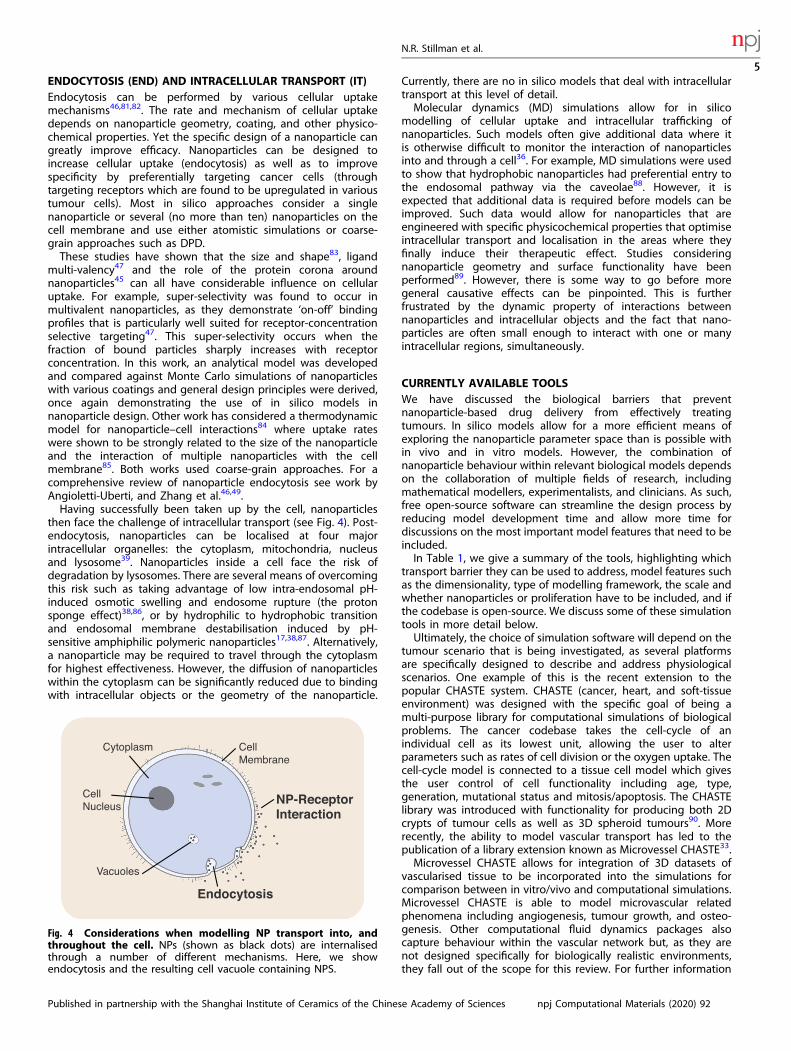
then face the challenge of intracellular transport (see Fig. 4). Post-endocytosis, nanoparticles can be localised at four majorintracellular organelles: the cytoplasm, mitochondria, nucleusand lysosome39. Nanoparticles inside a cell face the risk ofdegradation by lysosomes. There are several means of overcomingthis risk such as taking advantage of low intra-endosomal pH-induced osmotic swelling and endosome rupture (the protonsponge effect)38,86, or by hydrophilic to hydrophobic transitionand endosomal membrane destabilisation induced by pH-sensitive amphiphilic polymeric nanoparticles17,38,87. Alternatively,a nanoparticle may be required to travel through the cytoplasmfor highest effectiveness. However, the diffusion of nanoparticleswithin the cytoplasm can be significantly reduced due to bindingwith intracellular objects or the geometry of the nanoparticle.
Currently, there are no in silico models that deal with intracellulartransport at this level of detail.Molecular dynamics (MD) simulations allow for in silico
modelling of cellular uptake and intracellular trafficking ofnanoparticles. Such models often give additional data where itis otherwise difficult to monitor the interaction of nanoparticlesinto and through a cell36. For example, MD simulations were usedto show that hydrophobic nanoparticles had preferential entry tothe endosomal pathway via the caveolae88. However, it isexpected that additional data is required before models can beimproved. Such data would allow for nanoparticles that areengineered with specific physicochemical properties that optimiseintracellular transport and localisation in the areas where theyfinally induce their therapeutic effect. Studies consideringnanoparticle geometry and surface functionality have beenperformed89. However, there is some way to go before moregeneral causative effects can be pinpointed. This is furtherfrustrated by the dynamic property of interactions betweennanoparticles and intracellular objects and the fact that nano-particles are often small enough to interact with one or manyintracellular regions, simultaneously.
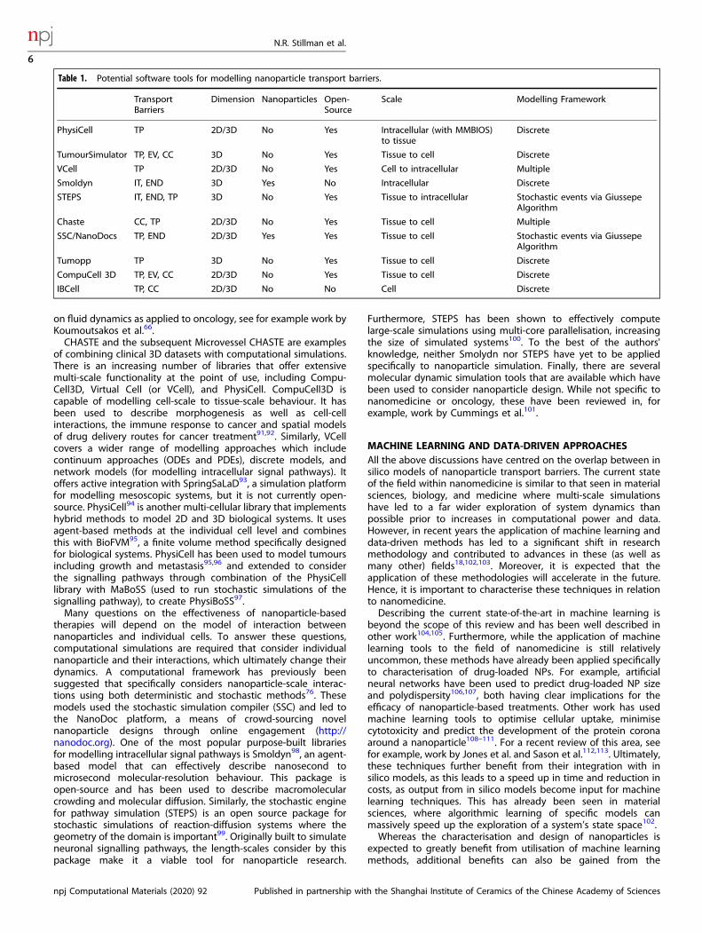
CURRENTLY AVAILABLE TOOLSWe have discussed the biological barriers that preventnanoparticle-based drug delivery from effectively treatingtumours. In silico models allow for a more efficient means ofexploring the nanoparticle parameter space than is possible within vivo and in vitro models. However, the combination ofnanoparticle behaviour within relevant biological models dependson the collaboration of multiple fields of research, includingmathematical modellers, experimentalists, and clinicians. As such,free open-source software can streamline the design process byreducing model development time and allow more time fordiscussions on the most important model features that need to beincluded.In Table 1, we give a summary of the tools, highlighting which
transport barrier they can be used to address, model features suchas the dimensionality, type of modelling framework, the scale andwhether nanoparticles or proliferation have to be included, and ifthe codebase is open-source. We discuss some of these simulationtools in more detail below.Ultimately, the choice of simulation software will depend on the
tumour scenario that is being investigated, as several platformsare specifically designed to describe and address physiologicalscenarios. One example of this is the recent extension to thepopular CHASTE system. CHASTE (cancer, heart, and soft-tissueenvironment) was designed with the specific goal of being amulti-purpose library for computational simulations of biologicalproblems. The cancer codebase takes the cell-cycle of anindividual cell as its lowest unit, allowing the user to alterparameters such as rates of cell division or the oxygen uptake. Thecell-cycle model is connected to a tissue cell model which givesthe user control of cell functionality including age, type,generation, mutational status and mitosis/apoptosis. The CHASTElibrary was introduced with functionality for producing both 2Dcrypts of tumour cells as well as 3D spheroid tumours90. Morerecently, the ability to model vascular transport has led to thepublication of a library extension known as Microvessel CHASTE33.Microvessel CHASTE allows for integration of 3D datasets of
vascularised tissue to be incorporated into the simulations forcomparison between in vitro/vivo and computational simulations.Microvessel CHASTE is able to model microvascular relatedphenomena including angiogenesis, tumour growth, and osteo-genesis. Other computational fluid dynamics packages alsocapture behaviour within the vascular network but, as they arenot designed specifically for biologically realistic environments,they fall out of the scope for this review. For further information
Fig. 4 Considerations when modelling NP transport into, andthroughout the cell. NPs (shown as black dots) are internalisedthrough a number of different mechanisms. Here, we showendocytosis and the resulting cell vacuole containing NPS.
N.R. Stillman et al.
5
Published in partnership with the Shanghai Institute of Ceramics of the Chinese Academy of Sciences npj Computational Materials (2020) 92
on fluid dynamics as applied to oncology, see for example work byKoumoutsakos et al.66.CHASTE and the subsequent Microvessel CHASTE are examples
of combining clinical 3D datasets with computational simulations.There is an increasing number of libraries that offer extensivemulti-scale functionality at the point of use, including Compu-Cell3D, Virtual Cell (or VCell), and PhysiCell. CompuCell3D iscapable of modelling cell-scale to tissue-scale behaviour. It hasbeen used to describe morphogenesis as well as cell-cellinteractions, the immune response to cancer and spatial modelsof drug delivery routes for cancer treatment91,92. Similarly, VCellcovers a wider range of modelling approaches which includecontinuum approaches (ODEs and PDEs), discrete models, andnetwork models (for modelling intracellular signal pathways). Itoffers active integration with SpringSaLaD93, a simulation platformfor modelling mesoscopic systems, but it is not currently open-source. PhysiCell94 is another multi-cellular library that implementshybrid methods to model 2D and 3D biological systems. It usesagent-based methods at the individual cell level and combinesthis with BioFVM95, a finite volume method specifically designedfor biological systems. PhysiCell has been used to model tumoursincluding growth and metastasis95,96 and extended to considerthe signalling pathways through combination of the PhysiCelllibrary with MaBoSS (used to run stochastic simulations of thesignalling pathway), to create PhysiBoSS97.Many questions on the effectiveness of nanoparticle-based
therapies will depend on the model of interaction betweennanoparticles and individual cells. To answer these questions,computational simulations are required that consider individualnanoparticle and their interactions, which ultimately change theirdynamics. A computational framework has previously beensuggested that specifically considers nanoparticle-scale interac-tions using both deterministic and stochastic methods76. Thesemodels used the stochastic simulation compiler (SSC) and led tothe NanoDoc platform, a means of crowd-sourcing novelnanoparticle designs through online engagement (http://nanodoc.org). One of the most popular purpose-built librariesfor modelling intracellular signal pathways is Smoldyn98, an agent-based model that can effectively describe nanosecond tomicrosecond molecular-resolution behaviour. This package isopen-source and has been used to describe macromolecularcrowding and molecular diffusion. Similarly, the stochastic enginefor pathway simulation (STEPS) is an open source package forstochastic simulations of reaction-diffusion systems where thegeometry of the domain is important99. Originally built to simulateneuronal signalling pathways, the length-scales consider by thispackage make it a viable tool for nanoparticle research.
Furthermore, STEPS has been shown to effectively computelarge-scale simulations using multi-core parallelisation, increasingthe size of simulated systems100. To the best of the authors'knowledge, neither Smolydn nor STEPS have yet to be appliedspecifically to nanoparticle simulation. Finally, there are severalmolecular dynamic simulation tools that are available which havebeen used to consider nanoparticle design. While not specific tonanomedicine or oncology, these have been reviewed in, forexample, work by Cummings et al.101.
MACHINE LEARNING AND DATA-DRIVEN APPROACHESAll the above discussions have centred on the overlap between insilico models of nanoparticle transport barriers. The current stateof the field within nanomedicine is similar to that seen in materialsciences, biology, and medicine where multi-scale simulationshave led to a far wider exploration of system dynamics thanpossible prior to increases in computational power and data.However, in recent years the application of machine learning anddata-driven methods has led to a significant shift in researchmethodology and contributed to advances in these (as well asmany other) fields18,102,103. Moreover, it is expected that theapplication of these methodologies will accelerate in the future.Hence, it is important to characterise these techniques in relationto nanomedicine.Describing the current state-of-the-art in machine learning is
beyond the scope of this review and has been well described inother work104,105. Furthermore, while the application of machinelearning tools to the field of nanomedicine is still relativelyuncommon, these methods have already been applied specificallyto characterisation of drug-loaded NPs. For example, artificialneural networks have been used to predict drug-loaded NP sizeand polydispersity106,107, both having clear implications for theefficacy of nanoparticle-based treatments. Other work has usedmachine learning tools to optimise cellular uptake, minimisecytotoxicity and predict the development of the protein coronaaround a nanoparticle108–111. For a recent review of this area, seefor example, work by Jones et al. and Sason et al.112,113. Ultimately,these techniques further benefit from their integration with insilico models, as this leads to a speed up in time and reduction incosts, as output from in silico models become input for machinelearning techniques. This has already been seen in materialsciences, where algorithmic learning of specific models canmassively speed up the exploration of a system’s state space102.Whereas the characterisation and design of nanoparticles is
expected to greatly benefit from utilisation of machine learningmethods, additional benefits can also be gained from the
Table 1. Potential software tools for modelling nanoparticle transport barriers.
TransportBarriers
Dimension Nanoparticles Open-Source
Scale Modelling Framework
PhysiCell TP 2D/3D No Yes Intracellular (with MMBIOS)to tissue
Discrete
TumourSimulator TP, EV, CC 3D No Yes Tissue to cell Discrete
VCell TP 2D/3D No Yes Cell to intracellular Multiple
Smoldyn IT, END 3D Yes No Intracellular Discrete
STEPS IT, END, TP 3D No Yes Tissue to intracellular Stochastic events via GiussepeAlgorithm
Chaste CC, TP 2D/3D No Yes Tissue to cell Multiple
SSC/NanoDocs TP, END 2D/3D Yes Yes Tissue to cell Stochastic events via GiussepeAlgorithm
Tumopp TP 3D No Yes Tissue to cell Discrete
CompuCell 3D TP, EV, CC 2D/3D No Yes Tissue to cell Discrete
IBCell TP, CC 2D/3D No No Cell Discrete
N.R. Stillman et al.
6
npj Computational Materials (2020) 92 Published in partnership with the Shanghai Institute of Ceramics of the Chinese Academy of Sciences

combination of machine learning with experiment design. Activelearning uses machine learning techniques (such as reinforcementlearning114 or surrogate-assisted optimisation115) to guide experi-ment design by selecting for only the most promising candidatesthat require testing. This approach, distinct from traditional trial-and-error, has already shown promise within the fields of clinicalcancer trials, drug discovery and material discovery18,116,117. Webelieve that designing nanoparticles to overcome transportbarriers is a rich future area of study118. Furthermore, theintegration of in silico models with active learning will allow forboth the automatic exploration and nanoparticle designs, and away to test them, often a pre-requisite when implementing anactive learning approach102.With recent advances in multi-scale simulations of tumours and
nanomedicines and their combination with machine learningtechniques, the application of in silico methods in a clinical settingis beginning to become reality. However, establishing the clinicalrelevance of computational models requires industry-gradeevidence gained through verification and validation, sensitivityanalysis, and uncertainty quantification, amongst other things119.The level of rigour of these evidences will depend on both theirintended use and the clarity of such use at the outset. Hence, theexistence of guidance documents is critical in the successful andsafe application of such methods to the clinical setting120. In 2013,the International Medical Device Regulators Forum (IMDRF)published the first in a series of guidance documents on ‘Softwareas a Medical Device’ (SaMD). They covered key definitions121,categorisation122, quality management system123 and planningthe process for clinical evaluation of a SaMD124.Taken together those documents provide a risk-based frame-
work where the level of clinical evidence depends on the riskprofile of a SaMD, where the risk profile is characterised by theseverity of the underlying health condition and the significance ofthe information provided by the SaMD to the healthcare decisionmaker. Since computational models of physiological systems areusually nonlinear, highly complex, and contain high numbers ofparameters and time-dependent properties, their proper assess-ment can be very challenging. On top of this, the introduction ofmachine learning into simulation models opens up a new set ofproblems including lack of transparency, automation bias (wherethere is a tendency of users to non-critically accept computerrecommendations) and changes in input/output relations as analgorithm learns from real-world use and experience. In 2019, theFDA published a discussion paper which calls for medical machinelearning algorithms to include the types of anticipated modifica-tions125. In light of this, we advise developers, as early as possiblein the development pipeline, to get familiar with the possible risksassociated with their simulation platform and take into accountrelevant sources of evidence (such as verification, validation, anduncertainty quantification) that are likely to be required in clinicalsettings.
CONCLUSIONS AND FUTURE PERSPECTIVEOvercoming nanoparticle transport barriers, specifically travelthrough the vasculature, extravasation, tissue penetration, endo-cytosis and the delivery of a therapeutic cargo, is paramount tothe effective use of nanoparticles or anti-cancer treatments. Insilico tools allow for the fast and systematic exploration of thenanoparticle design space to select nanoparticles with thepotential to deliver their cargo to the right place. Generalguidelines extracted from such tools could prove useful in makingmore effective treatments. Specific solutions could also beprovided to tailor nanoparticles to patient needs and towardspersonalised medicine, or to produce sufficient amounts of datafor machine learning. Building useful in silico tools will requireclose validation with in vitro and in vivo results. The overarchingaim is to create a systems approach to nanomedicine whichaccounts for multi-scale phenomena, which is repeatedly vali-dated, and which exists within a shared modelling framework. Wedescribe these elements in more detail below.To become effective, nanoparticle cancer therapies require a
systematic approach to prototyping. In silico modelling has nowadvanced to the point of being an effective tool that can minimisecostly trial-and-error design methods (see, for example, the reviewby Karolak et al.50). However, such modelling cannot exist inisolation and a collaboration between mathematical modellers,experimentalists, and clinicians will be required to inform the besttransfer of knowledge. As a first step, this method will help identifyguidelines for the design of suitable nanoparticles for a class ofproblems, say a specific tumour type. In the future, integration ofpatient data such as MRI scans or biopsies can inform computa-tional models of tumour growth and offer a route to personalisednanoparticles126,127. We envisage a pipeline where theoreticalpredictions are checked against clinical outcomes and thenreturned to inform future simulations, as shown in Fig. 5.Tumour progression and nanoparticle transport exhibit clear
multi-scale behaviour. This can be understood within a systemsbiology framework128,129; individual microscopic effects are morethan the sum of their parts when viewed in aggregate. It is notstraightforward to determine the probability distribution ofemergent behaviour using microstate characterisation, as thevariables in complex biological systems are interdependent andunlikely to be normally distributed27. Yet, by testing multiple insilico models, a sufficient sampling of the model outputs allows usto project microstate knowledge higher up the hierarchy of scales.This is possible using in silico models as system parameters can beclosely controlled for and many thousands of possible modelscenarios can be investigated in a systematic and efficient way.Using a systems approach to cancer modelling, covering topicssuch as tissue complexity, cell heterogeneity, targeted therapy,and drug resistance, has been reviewed in, for example, work byWerner et al.130.This system approach highlights three immediate advantages of
using multi-scale in silico models: improved hypothesis testing,strategy generation, and clinical relevance. Brute force simulation
Fig. 5 Integrated pipeline for optimised anti-cancer nanoparticle design. A clinical challenge is identified, in silico models are used todesign NPs for overcoming this challenge, and iterative synthesis and testing of NPs leads to the development of effective translationmedicine.
N.R. Stillman et al.
7
Published in partnership with the Shanghai Institute of Ceramics of the Chinese Academy of Sciences npj Computational Materials (2020) 92

experiments allow for much wider characterisation and samplingof a systems state-space73. These allows the simulations to testwhether microstate phenomena have a verifiable causal relationto emergent macroscale observables. This is the hypothesistesting benefit. For example, PhysiCell has been used to runlarge-scale parallelised simulations that generate data-driven errormetrics and create in silico validations of biomedicalhypotheses131.Second, the increased sample rate gained from running many
thousands of simultaneous simulations generates much largerdata. Increased data, combined with optimisation and machinelearning, will lead to novel, more efficient, strategy generation76.Furthermore, by combining computer simulations with patientdata and experimental results, these novel strategies can beimplemented and checked to explore new medical applications.This motivates the third advantage of silico models. In silico
models validated and optimised by in vivo and/or in vitro modelshave both better explanatory and predictive power. The strengthof multi-scale in silico models is the ability to bridge length-scaleswhich have traditionally isolated causal factors. By integratingacross scales, the predictive power of in silico modellingapproaches can be increased with a corresponding expectedpayoff to clinical relevance. Furthermore, the combination ofsimulating across scales with parameters gained from in vitro and/or in vivo can expand causal relations beyond scale ofobservations (such as relating intercellular growth models to theobserved change in the size of a tumour). Finally, the combinationof these methods with machine learning techniques can be usedto build causal models from correlations inferred from big data103.The three advantages of in silico models (improved hypothesis
testing, strategy generation, and clinical relevance) will requirestandardisation between computational models such that relevantbenchmarks can be used to compare various in silico approaches.Furthermore, a common design framework which includes shareddatatypes will allow for expedited comparison between modelsthat have a shared common input. This has been advocatedbefore132–134, but only time will tell whether such a standardisa-tion becomes successful.
DATA AVAILABILITYAll relevant data are available from the authors.
Received: 29 October 2019; Accepted: 12 June 2020;
REFERENCES1. Etheridge, M. L. et al. The big picture on nanomedicine: the state of investiga-
tional and approved nanomedicine products Nanomedicine 9, 1–14 (2013).2. Su, Y. L. & Hu, S. H. Functional nanoparticles for tumor penetration of ther-
apeutics. Pharmaceutics 10, 1–21 (2018).3. Tran, S., DeGiovanni, P.-J., Piel, B. & Rai, P. Cancer nanomedicine: a review of
recent success in drug delivery. Clin. Transl. Med. 6, 44 (2017).4. Hare, J. I. et al. Challenges and strategies in anti-cancer nanomedicine devel-
opment: an industry perspective. Adv. Drug Deliv. Rev. 108, 25–38 (2017).5. Strambeanu, N., Demetrovici, L., Dragos, D. & Lungu, M. in Nanoparticles’ Pro-
mises and Risks: Characterization, Manipulation, and Potential Hazards toHumanity and the Environment (eds Lungu, M., Neculae, A., Bunoiu, M. & Biris, C.)(Springer, 2015).
6. Roberts, W. G. & Palade, G. E. G. Increased microvascular permeability andendothelial fenestration induced by vascular endothelial growth factor. J. CellSci. 108, 2369–2379 (1995).
7. Tong, R., Hemmati, H. D., Langer, R. & Kohane, D. S. Photoswitchable nano-particles for triggered tissue penetration and drug delivery. J. Am. Chem. Soc.134, 8848–8855 (2012).
8. Kong, S. D. et al. Magnetically vectored nanocapsules for tumor penetration andremotely switchable on-demand drug release. Nano Lett. 10, 5088–5092 (2010).
9. Wang, B. et al. Simultaneously overcome tumor vascular endothelium andextracellular matrix barriers via a non-destructive size-controlled nanomedicine.J. Control. Release 268, 225–236 (2017).
10. Bazak, R. et al. Cancer active targeting by nanoparticles: a comprehensive reviewof literature. J. Cancer Res. Clin. Oncol. 141, 769–784 (2015).
11. Hauert, S. & Bhatia, S. N. Mechanisms of cooperation in cancer nanomedicine:towards systems nanotechnology. Trends Biotechnol. 32, 448–455 (2014).
12. Li, Y. et al. Cell and nanoparticle transport in tumour microvasculature: the role ofsize, shape and surface functionality of nanoparticles. Interface Focus 6, 1–15 (2016).
13. Park, J.-H. et al. Cooperative nanomaterial system to sensitize, target, and treattumors. Proc. Natl Acad. Sci. 107, 981–986 (2010).
14. Von Maltzahn, G. et al. Nanoparticles that communicate in vivo to amplifytumour targeting. Nat. Mater. 10, 545–552 (2011).
15. Fu, Y. et al. A feasible strategy for self-assembly of gold nanoparticles: via dithiol-PEG for photothermal therapy of cancers. RSC Adv. 8, 6120–6124 (2018).
16. Xiao, Z. et al. DNA self-assembly of targeted near-infrared-responsive goldnanoparticles for cancer thermo-chemotherapy. Angew. Chem.—Int. Edn. 51,11853–11857 (2012).
17. Bae, Y. H. & Park, K. Targeted drug delivery to tumors: myths, reality and pos-sibility. J. Control. Release 153, 198–205 (2011).
18. Lookman, T., Balachandran, P. V., Xue, D. & Yuan, R. Active learning in materialsscience with emphasis on adaptive sampling using uncertainties for targeteddesign. npj Comput. Mater. 5, 1–17 (2019).
19. Deisboeck, T. S., Zhang, L., Yoon, J. & Costa, J. In silico cancer modeling: is itready for prime time? Nat. Clin. Practice Oncol. 6, 34–42 (2009).
20. Michor, F. & Beal, K. Improving cancer treatment via mathematical modeling:surmounting the challenges is worth the effort. Cell 163, 1059–1063 (2015).
21. Dogra, P. et al. Mathematical modeling in cancer nanomedicine: a review.Biomed. Microdevices 21, 40 (2019).
22. Talmadge, J. E. & Fidler, I. J. The biology of cancer metastasis: historical per-spective. Cancer Res. 70, 5649–5669 (2010).
23. Siegel, R., Miller, K. D. & Ahmedin, J. Cancer Statistics, 2017. CA: Cancer J. Clin-icians 67, 7–30 (2017).
24. Chauviere, A. H. et al. Mathematical oncology: how are the mathematical andphysical sciences contributing to the war on breast cancer? Curr. Breast CancerRep. 2, 121–129 (2010).
25. Deisboeck, Z. & Yoon, C. In silico modelling—is it ready for prime time. Program6, 34–42 (2011).
26. Rejniak, K. A. & Anderson, A. R. Hybrid models of tumor growth. Wiley Inter-disciplinary Rev.: Syst. Biol. Med. 3, 115–125 (2011).
27. An, G. & Mi, Q. Agent based models in translational systems biology. Syst. Biol.Med. 1, 159–171 (2009).
28. Deisboeck, T. S. & Stamatakos, G. S. Multiscale Cancer Modeling (CRC Press, 2010).29. Norton, K.-A., Gong, C., Jamalian, S. & Popel, A. Multiscale agent-based and
hybrid modeling of the tumor immune microenvironment. Processes 7, 1–23(2019).
30. Metzcar, J., Wang, Y., Heiland, R. & Macklin, P. A review of cell-based compu-tational modeling in cancer biology. JCO Clin. Cancer Inform. 3, 1–13 (2019).
31. Zhu, X., Zhou, X., Lewis, M. T., Xia, L. & Wong, S. Cancer stem cell, niche and EGFRdecide tumor development and treatment response: a bio-computationalsimulation study. J. Theor. Biol. 269, 138–49 (2011).
32. Sefidgar, M. et al. Numerical modeling of drug delivery in a dynamic solid tumormicrovasculature. Microvascular Res. 99, 43–56 (2015).
33. Grogan, J. A. et al. Microvessel chaste: an open library for spatial modeling ofvascularized tissues. Biophys. J. 112, 1767–1772 (2017).
34. Shah, P. N. et al. Extravasation of Brownian spheroidal nanoparticles throughvascular pores. Biophys. J. 115, 1103–1115 (2018).
35. Owen, M. R. et al. Mathematical modeling predicts synergistic antitumor effectsof combining a macrophage-based, hypoxia-targeted gene therapy with che-motherapy. Cancer Res. 71, 2826–2837 (2011).
36. Chou, C. Y., Huang, C. K., Lu, K. W., Horng, T. L. & Lin, W. L. Investigation of thespatiotemporal responses of nanoparticles in tumor tissues with a small-scalemathematical model. PLoS ONE 8, e59135 (2013).
37. Nehoff, H., Parayath, N. N., Domanovitch, L., Taurin, S. & Greish, K. Nanomedicinefor drug targeting: strategies beyond the enhanced permeability and retentioneffect. Int. J. Nanomed. 9, 2539–55 (2014).
38. Chrastina, A., Massey, K. A. & Schnitzer, J. E. Overcoming in vivo barriers totargeted nanodelivery. Wiley Interdisciplinary Rev.: Nanomed. Nanobiotechnol. 3,421–37 (2011).
39. Barua, S. & Mitragotri, S. Challenges associated with penetration of nanoparticlesacross cell and tissue barriers: a review of current status and future prospects.Nano Today 9, 223–243 (2014).
40. Curtis, L. T., England, C. G., Wu, M., Lowengrub, J. & Frieboes, H. B. An inter-disciplinary computational/experimental approach to evaluate drug-loadedgold nanoparticle tumor cytotoxicity. Nanomedicine 11, 197–216 (2016).
N.R. Stillman et al.
8
npj Computational Materials (2020) 92 Published in partnership with the Shanghai Institute of Ceramics of the Chinese Academy of Sciences

41. Shah, A. B., Rejniak, K. A. & Gevertz, J. L. Limiting the development of anti-cancerdrug resistance in a spatial model of micrometastases. Math. Biosci. Eng. 13,1185–1206 (2016).
42. Al-Obaidi, H. & Florence, A. T. Nanoparticle delivery and particle diffusion inconfined and complex environments. J. Drug Deliv. Sci. Technol. 30, 266–277(2015).
43. Wang, Z. et al. Theory and experimental validation of a spatio-temporal modelof chemotherapy transport to enhance tumor cell kill. PLoS Comput. Biol.12,e1004969 (2016).
44. Hamis, S., Nithiarasu, P. & Powathil, G. G. What does not kill a tumour may makeit stronger: In silico insights into chemotherapeutic drug resistance. J. Theor. Biol.454, 253–267 (2018).
45. ming Ding, H. & qiang Ma, Y. Computer simulation of the role of protein coronain cellular delivery of nanoparticles. Biomaterials 35, 8703–8710 (2014).
46. Zhang, S., Gao, H. & Bao, G. Physical principles of nanoparticle cellular endo-cytosis. ACS Nano 9, 8655–8671 (2015).
47. Martinez-Veracoechea, F. J. & Frenkel, D. Designing super selectivity in multi-valent nano-particle binding. Proc. Natl Acad. Sci. 108, 10963–10968 (2011).
48. Pascal, J. et al. Mechanistic modeling identifies drug-uptake history as predictorof tumor drug resistance and nano-carrier-mediated response. ACS Nano 7,11174–11182 (2013).
49. Angioletti-Uberti, S. Theory, simulations and the design of functionalizednanoparticles for biomedical applications: a soft matter perspective. npj Comput.Mater. 3, 1–48 (2017).
50. Karolak, A., Markov, D. A., McCawley, L. J. & Rejniak, K. A. Towards personalizedcomputational oncology: from spatial models of tumour spheroids, to orga-noids, to tissues. J. Roy. Soc. Interface 15, 20170703 (2018).
51. Afonin, K. A. et al. In silico design and enzymatic synthesis of functional RNAnanoparticles. Acc. Chem. Res. 47, 1731–1741 (2014).
52. Finley, S. D., Angelikopoulos, P., Koumoutsakos, P. & Popel, A. S. Pharmacoki-netics of Anti-VEGF Agent aflibercept in cancer predicted by data-driven,molecular-detailed model. CPT: Pharmacometrics Syst. Pharmacol. 4, 641–649(2015).
53. Wang, Z., Bordas, V., Sagotsky, J. & Deisboeck, T. S. Identifying therapeutic tar-gets in a combined EGFR-TGFβ R signalling cascade using a multiscale agent-based cancer model. Math. Med. Biol. 29, 95–108 (2012).
54. Blanco, E., Shen, H. & Ferrari, M. Principles of nanoparticle design for overcomingbiological barriers to drug delivery. Nature Biotechnol. 33, 9 (2015).
55. Longmire, M., Choyke, P. L. & Kobayashi, H. Clearance properties of nano-sizedparticles and molecules as imaging agents: considerations and caveats. Nano-medicine 3, 703–717 (2008).
56. Gustafson, H. H., Holt-Casper, D., Grainger, D. W. & Ghandehari, H. Nanoparticleuptake: the phagocyte problem. Nano Today 10, 487–510 (2015).
57. Fedosov, D. A., Noguchi, H. & Gompper, G. Multiscale modeling of blood flow:from single cells to blood rheology. Biomech. Model. Mechanobiol. 13, 239–258(2014).
58. Lopez, H. & Lobaskin, V. Coarse-grained model of adsorption of blood plasmaproteins onto nanoparticles. J. Chem. Phys. 143, 12B620_1 (2015).
59. Shao, Q. & Hall, C. K. Protein adsorption on nanoparticles: model developmentusing computer simulation. J. Phys.: Condens. Matter 28, 414019 (2016).
60. Maleki, R. et al. ph-sensitive loading/releasing of doxorubicin using single-walled carbon nanotube and multi-walled carbon nanotube: a moleculardynamics study. Comput. Methods Programs Biomed. 186, 105210 (2020).
61. Yoo, J.-W., Chambers, E. & Mitragotri, S. Factors that control the circulation timeof nanoparticles in blood: challenges, solutions and future prospects. Curr.Pharm. Des. 16, 2298–2307 (2010).
62. Müller, K., Fedosov, D. A. & Gompper, G. Margination of micro- and nano-particles in blood flow and its effect on drug delivery. Sci. Rep. 4, 1–8 (2014).
63. Lin, Z., Monteiro-Riviere, N. A. & Riviere, J. E. A physiologically based pharma-cokinetic model for polyethylene glycol-coated gold nanoparticles of differentsizes in adult mice. Nanotoxicology 10, 162–172 (2016).
64. Yuan, D., He, H., Wu, Y., Fan, J. & Cao, Y. Physiologically based pharmacokineticmodeling of nanoparticles. J. Pharm. Sci. 108, 58–72 (2019).
65. Ding, H.-m & Ma, Y.-q Computational approaches to cell–nanomaterial inter-actions: keeping balance between therapeutic efficiency and cytotoxicity.Nanoscale Horizons 3, 6–27 (2017).
66. Koumoutsakos, P., Pivkin, I. & Milde, F. The fluid mechanics of cancer and itstherapy. Annu. Rev. Fluid Mech. 45, 325–355 (2013).
67. Nakamura, Y., Mochida, A., Choyke, P. L. & Kobayashi, H. Nanodrug delivery: isthe enhanced permeability and retention effect sufficient for curing cancer?Bioconjugate Chem. 27, 2225–2238 (2016).
68. Björnmalm, M., Thurecht, K. J., Michael, M., Scott, A. M. & Caruso, F. Bridging bio-nano science and cancer nanomedicine. ACS Nano 11, 9594–9613 (2017).
69. Nichols, J. W. & Bae, Y. H. Odyssey of a cancer nanoparticle: from injection site tosite of action. Nano Today 7, 606–618 (2012).
70. Fullstone, G., Wood, J., Holcombe, M. & Battaglia, G. Modelling the transport ofnanoparticles under blood flow using an agent-based approach. Sci. Rep. 5,1–13 (2015).
71. Ferretti, S., Allegrini, P. R., Becquet, M. M. & McSheehy, P. M. Tumor interstitialfluid pressure as an early-response marker for anticancer therapeutics. Neoplasia11, 874–881 (2015).
72. Lameijer, M. A., Tang, J., Nahrendorf, M., Beelen, R. H. & Mulder, W. J. Monocytesand macrophages as nanomedicinal targets for improved diagnosis and treat-ment of disease. Expert Rev. Mol. Diagnostics 13, 576–580 (2013).
73. Wu, M. et al. The effect of interstitial pressure on therapeutic agent transport:coupling with the tumor blood and lymphatic vascular systems. J.Theor. Biol.335, 194–207 (2014).
74. Wijeratne, P. A. & Vavourakis, V. A quantitative in silico platform for simulatingcytotoxic and nanoparticle drug delivery to solid tumours. Interface Focus 9,20180063 (2019).
75. Frieboes, H. B., Wu, M., Lowengrub, J., Decuzzi, P. & Cristini, V. A computationalmodel for predicting nanoparticle accumulation in tumor vasculature. PLoS ONE8, 1–11 (2013).
76. Hauert, S., Berman, S., Nagpal, R. & Bhatia, S. N. A computational framework foridentifying design guidelines to increase the penetration of targeted nano-particles into tumors. Nano Today 8, 566–576 (2013).
77. Chamseddine, I. M., Frieboes, H. B. & Kokkolaras, M. Design optimization oftumor vasculature-bound nanoparticles. Sci. Rep. 8, 17768 (2018).
78. Thurber, G. M. & Weissleder, R. A systems approach for tumor pharmacokinetics.PLoS ONE 6, e24696 (2011).
79. Cilliers, C., Guo, H., Liao, J., Christodolu, N. & Thurber, G. M. Multiscale modelingof antibody-drug conjugates: Connecting tissue and cellular distribution towhole animal pharmacokinetics and potential implications for efficacy. The AAPSJ. 18, 1117–1130 (2016).
80. Liu, J. et al. Design of nanocarriers based on complex biological barriers in vivofor tumor therapy. Nano Today 15, 56–90 (2017).
81. Daum, N., Tscheka, C., Neumeyer, A. & Schneider, M. Novel approaches for drugdelivery systems in nanomedicine: Effects of particle design and shape. WileyInterdisciplinary Rev.: Nanomed. Nanobiotechnol. 4, 52–65 (2012).
82. Anselmo, A. C. et al. Elasticity of nanoparticles influences their blood circulation,phagocytosis, endocytosis, and targeting. ACS Nano 9, 3169–3177 (2015).
83. Bao, G. et al. USNCTAM perspectives on mechanics in medicine. J. Roy. Soc.Interface 11, 20140301 (2014).
84. Zhang, S., Li, J., Lykotrafitis, G., Bao, G. & Suresh, S. Size-dependent endocytosisof nanoparticles. Adv. Mater. 21, 419–424 (2009).
85. Yue, T. & Zhang, X. Cooperative effect in receptor-mediated endocytosis ofmultiple nanoparticles. ACS Nano 6, 3196–3205 (2012).
86. Stewart, M. P., Lorenz, A., Dahlman, J. & Sahay, G. Challenges in carrier-mediatedintracellular delivery: moving beyond endosomal barriers. Wiley InterdisciplinaryRev.: Nanomed. Nanobiotechnol. 8, 465–478 (2016).
87. Martens, T. F., Remaut, K., Demeester, J., De Smedt, S. C. & Braeckmans, K.Intracellular delivery of nanomaterials: How to catch endosomal escape in theact. Nano Today 9, 344–364 (2014).
88. Chiu, Y.-L. et al. The characteristics, cellular uptake and intracellular trafficking ofnanoparticles made of hydrophobically-modified chitosan. J. Control. Release146, 152–159 (2010).
89. Chithrani, B. D. & Chan, W. C. Elucidating the mechanism of cellular uptake andremoval of protein-coated gold nanoparticles of different sizes and shapes.Nano Lett. 7, 1542–1550 (2007).
90. Pitt-Francis, J. et al. Chaste: a test-driven approach to software development forbiological modelling. Comput. Phys. Commun. 180, 2452–2471 (2009).
91. Winner, K. R. et al. Spatial modeling of drug delivery routes for treatment ofdisseminated ovarian cancer. Cancer Res. 76, 1320–1334 (2016).
92. Li, J. F. & Lowengrub, J. The effects of cell compressibility, motility and contactinhibition on the growth of tumor cell clusters using the Cellular Potts Model. J.Theor. Biol. 343, 79–91 (2014).
93. Michalski, P. J. & Loew, L. M. SpringSaLaD: a spatial, particle-based biochemicalsimulation platform with excluded volume. Biophys. J. 110, 523–529 (2016).
94. Ghaffarizadeh, A., Heiland, R., Friedman, S. H., Mumenthaler, S. M. & Macklin, P.PhysiCell: an open source physics-based cell simulator for 3-D multicellularsystems. PLoS Comput. Biol. 14, e1005991 (2018).
95. Ghaffarizadeh, A., Friedman, S. H. & MacKlin, P. BioFVM: an efficient, parallelizeddiffusive transport solver for 3-D biological simulations. Bioinformatics 32,1256–1258 (2016).
96. Juarez, E. F., Garri, C., Ghaffarizadeh, A., Macklin, P. & Kani, K. Quantification ofcancer cell migration with an integrated experimental-computational pipeline.F1000Research 7, 1296 (2018).
97. Letort, G. et al. PhysiBoSS: a multi-scale agent-based modelling frameworkintegrating physical dimension and cell signalling. Bioinformatics 35, 1188–1196(2019).
N.R. Stillman et al.
9
Published in partnership with the Shanghai Institute of Ceramics of the Chinese Academy of Sciences npj Computational Materials (2020) 92

98. Andrews, S. S., Addy, N. J., Brent, R. & Arkin, A. P. Detailed simulations of cellbiology with Smoldyn 2.1. PLoS Comput. Biol. 6, e1000705 (2010).
99. Hepburn, I., Chen, W., Wils, S. & De Schutter, E. STEPS: efficient simulation of sto-chastic reaction-diffusion models in realistic morphologies. BMC Syst. Biol. 6, 36(2012).
100. Chen, W. & De Schutter, E. Parallel STEPS: large scale stochastic spatial reaction-diffusion simulation with high performance computers. Frontiers Neuroinform.11, 1–15 (2017).
101. Cummings, P. T. & Gilmer, J. B. Open-source molecular modeling software inchemical engineering. Curr. Opin. Chem. Eng. 23, 99–105 (2019).
102. Schmidt, J., Marques, M. R., Botti, S. & Marques, M. A. Recent advances andapplications of machine learning in solid-state materials science. npj Comput.Mater. 5, 1–36 (2019).
103. Alber, M. et al. Integrating machine learning and multiscale modeling-per-spectives, challenges, and opportunities in the biological, biomedical, andbehavioral sciences. npj Digital Med. 2, 1–11 (2019).
104. Bishop, C. M. Pattern Recognition and Machine Learning (springer, 2006).105. Alpaydin, E. Introduction to Machine Learning (MIT press, 2020).106. Asadi, H., Rostamizadeh, K., Salari, D. & Hamidi, M. Preparation of biodegradable
nanoparticles of tri-block pla–peg–pla copolymer and determination of factorscontrolling the particle size using artificial neural network. J. Microencapsulation28, 406–416 (2011).
107. Shalaby, K. S. et al. Determination of factors controlling the particle size andentrapment efficiency of noscapine in peg/pla nanoparticles using artificialneural networks. Int. J. Nanomed. 9, 4953 (2014).
108. Liu, R. et al. Classification nanosar development for cytotoxicity of metal oxidenanoparticles. Small 7, 1118–1126 (2011).
109. Hataminia, F., Noroozi, Z. & Eslam, H. M. Investigation of iron oxide nanoparticlecytotoxicity in relation to kidney cells: a mathematical modeling of data mining.Toxicol. in Vitro 59, 197–203 (2019).
110. Labouta, H. I., Asgarian, N., Rinker, K. & Cramb, D. T. Meta-analysis of nanoparticlecytotoxicity via data-mining the literature. ACS Nano 13, 1583–1594 (2019).
111. Findlay, M. R., Freitas, D. N., Mobed-Miremadi, M. & Wheeler, K. E. Machinelearning provides predictive analysis into silver nanoparticle protein coronaformation from physicochemical properties. Environ. Sci.: Nano 5, 64–71 (2018).
112. Jones, D. E., Ghandehari, H. & Facelli, J. C. A review of the applications of datamining and machine learning for the prediction of biomedical properties ofnanoparticles. Comput. Methods Programs Biomed. 132, 93–103 (2016).
113. Sason, H. & Shamay, Y. Nanoinformatics in drug delivery. Israel J. Chem. https://doi.org/10.1002/ijch.201900042 (2019).
114. Sutton, R. S. & Barto, A. G. Reinforcement Learning: An Introduction (MIT press, 2018).115. Jin, Y. Surrogate-assisted evolutionary computation: recent advances and future
challenges. Swarm Evolut. Comput. 1, 61–70 (2011).116. Zhao, Y., Kosorok, M. R. & Zeng, D. Reinforcement learning design for cancer
clinical trials. Statistics Med. 28, 3294–3315 (2009).117. Warmuth, M. K. et al. Active learning with support vector machines in the drug
discovery process. J. Chem. Inform. Comput. Sci. 43, 667–673 (2003).118. Yamankurt, G. et al. Exploration of the nanomedicine-design space with high-
throughput screening and machine learning. Nature Biomed. Eng. 3, 318–327(2019).
119. Parvinian, B. et al. Credibility evidence for computational patient models used inthe development of physiological closed-loop controlled devices for critical caremedicine. Frontiers Physiol. 10, 220 (2019).
120. Morrison, T. M. et al. Assessing computational model credibility using a risk-based framework: application to hemolysis in centrifugal blood pumps. ASAIO J.65, 349 (2019).
121. Software as a Medical Device (SaMD): Key Definitions (2013). Available at: http://www.imdrf.org/docs/imdrf/final/technical/imdrf-tech-131209-samd-key-definitions-140901.pdf.
122. Software as a Medical Device- (SaMD): Possible Framework for Risk Categorizationand Corresponding Considerations (2014). Available at: http://www.imdrf.org/docs/imdrf/final/technical/imdrf-tech-140918-samd-framework-risk-categorization-141013.pdf.
123. Software as a Medical Device- (SaMD): Application of Quality Management Sys-tems. (2014). Available at: http://www.imdrf.org/docs/imdrf/final/technical/imdrf-tech-151002-samd-qms.pdf.
124. Software as a Medical Device- (SaMD): Clinical Evaluation (2017). Available at:http://www.imdrf.org/docs/imdrf/final/technical/imdrf-tech-170921-samd-n41-clinical-evaluation_1.pdf.
125. Proposed regulatory framework for modifications to artificial intelligence/machine learning (AI/ML)-based software as a medical device (SaMD) (2019).Available at: https://www.fda.gov/media/122535/download.
126. Weis, J. A. et al. Predicting the response of breast cancer to neoadjuvant therapyusing a mechanically coupled reaction-diffusion model. Cancer Res. 10,4333–4347 (2015).
127. Macklin, P., Edgerton, M. E., Thompson, A. M. & Cristini, V. Patient-calibratedagent-based modelling of ductal carcinoma in situ (DCIS): From microscopicmeasurements to macroscopic predictions of clinical progression. J. Theor. Biol.301, 122–140 (2012).
128. Korsunsky, I. et al. Systems biology of cancer: a challenging expedition forclinical and quantitative biologists. Frontiers Bioeng. Biotechnol. 2, 27 (2014).
129. Faratian, D., Bown, J. L., Smith, V. A., Langdon, S. P. & Harrison, D. J. CancerSystems Biology 245–263 (Humana Press, Totowa, NJ, 2010).
130. Werner, H. M., Mills, G. B. & Ram, P. T. Cancer systems biology: a peek into thefuture of patient care? Nat. Rev. Clin. Oncol. 11, 167–176 (2014).
131. Kim, M. Y. et al. Tumor self-seeding by circulating cancer cells. Cell 139,1315–1326 (2009).
132. Koutsoukas, A. et al. From in silico target prediction to multi-target drug design:current databases, methods and applications. J. Proteomics 74, 254–2574 (2011).
133. Macklin, P. et al. Progress Towards Computational 3-D Multicellular SystemsBiology 225–246 (Springer International Publishing, 2016).
134. Rockne, R. C. et al. The 2019 mathematical oncology roadmap. Physical Biology16, 041005 (2019).
ACKNOWLEDGEMENTSThis project has received funding from the European Union’s Horizon 2020 researchand innovation programme under grant agreement No 800983.
AUTHOR CONTRIBUTIONSAll authors contributed to the content of the paper. N.S. led the creation and writingof the manuscript with input from M.K., I.B., and S.H. S.H. defined the structure andscope of the paper, supervised its writing, and provided feedback on all stages of themanuscript.
COMPETING INTERESTSThe authors declare no competing interests.
ADDITIONAL INFORMATIONCorrespondence and requests for materials should be addressed to S.H.
Reprints and permission information is available at http://www.nature.com/reprints
Publisher’s note Springer Nature remains neutral with regard to jurisdictional claimsin published maps and institutional affiliations.
Open Access This article is licensed under a Creative CommonsAttribution 4.0 International License, which permits use, sharing,
adaptation, distribution and reproduction in anymedium or format, as long as you giveappropriate credit to the original author(s) and the source, provide a link to the CreativeCommons license, and indicate if changes were made. The images or other third partymaterial in this article are included in the article’s Creative Commons license, unlessindicated otherwise in a credit line to the material. If material is not included in thearticle’s Creative Commons license and your intended use is not permitted by statutoryregulation or exceeds the permitted use, you will need to obtain permission directlyfrom the copyright holder. To view a copy of this license, visit http://creativecommons.org/licenses/by/4.0/.
© The Author(s) 2020
N.R. Stillman et al.
10
npj Computational Materials (2020) 92 Published in partnership with the Shanghai Institute of Ceramics of the Chinese Academy of Sciences