in and out of the minor groove: interaction of an at-rich
TRANSCRIPT

research papers
1614 doi:10.1107/S139900471400697X Acta Cryst. (2014). D70, 1614–1621
Acta Crystallographica Section D
BiologicalCrystallography
ISSN 1399-0047
In and out of the minor groove: interaction of anAT-rich DNA with the drug CD27
Francisco J. Acosta-Reyes,a
Christophe Dardonville,b
Harry P. de Koning,c Manal
Natto,c Juan A. Subiranaa and
J. Lourdes Camposa*
aDepartament d’Enginyeria Quımica, Universitat
Politecnica de Catalunya, Diagonal 647,
08028 Barcelona, Spain, bInstituto de Quımica
Medica, IQM–CSIC, Juan de la Cierva 3,
28006 Madrid, Spain, and cInstitute of Infection,
Immunity and Inflammation, College of
Medical, Veterinary and Life Sciences,
University of Glasgow, 120 University Place,
Glasgow G12 8TA, Scotland
Correspondence e-mail:
The DNA of several pathogens is very rich in AT base pairs.
Typical examples include the malaria parasite Plasmodium
falciparum and the causative agents of trichomoniasis and
trypanosomiases. This fact has prompted studies of drugs
which interact with the minor groove of DNA, some of which
are used in medical practice. Previous studies have been
performed almost exclusively with the AATT sequence. New
features should be uncovered through the study of different
DNA sequences. In this paper, the crystal structure of the
complex of the DNA duplex d(AAAATTTT)2 with the
dicationic drug 4,40-bis(imidazolinylamino)diphenylamine
(CD27) is presented. The drug binds to the minor groove of
DNA as expected, but it shows two new features that have not
previously been described: (i) the drugs protrude from the
DNA and interact with neighbouring molecules, so that they
may act as cross-linking agents, and (ii) the drugs completely
cover the whole minor groove of DNA and displace bound
water. Thus, they may prevent the access to DNA of proteins
such as AT-hook proteins. These features are also expected for
other minor-groove binding drugs when associated with all-AT
DNA. These findings allow a better understanding of this
family of compounds and will help in the development of new,
more effective drugs. New data on the biological interaction of
CD27 with the causative agent of trichomoniasis, Trichomonas
vaginalis, are also reported.
Received 4 February 2014
Accepted 28 March 2014
PDB reference:
d(AAAATTTT)2–CD27, 4ocd
1. Introduction
Enormous progress has been achieved in the past in the study
of small-molecule ligands that have affinity for the DNA
minor groove, as recently reviewed by Sheng et al. (2013).
More complex types of drug binding to DNA have also been
reviewed by Boer et al. (2009). Dervan and coworkers have
carried out an extensive series of studies (Sheng et al., 2013;
Chenoweth & Dervan, 2009) aimed at developing ligands that
recognize specific DNA sequences. Some intercalating drugs
also favour binding through the minor groove (Niyazi et al.,
2012). The main group of studies has concentrated on the
interaction of different drugs with AT-rich DNA regions,
mainly with the Dickerson–Drew dodecamer d(CGCGAA-
TTCGCG), which easily provides crystals with high resolution
suitable for X-ray analysis. However, there is no evidence that
AATT is the preferred sequence of interaction in vivo. In fact,
little is known of the eventual DNA-sequence selectivity. We
therefore decided to study the interaction of the all-AT DNA
sequence d(AAAATTTT) with the dication 4,40-bis(imidazo-
linylamino)diphenylamine (CD27; shown in Fig. 1).
CD27 is chemically related to diamidines, a class of
dicationic DNA minor-groove binders with a long history of
clinical success as antiprotozoal agents (Paine et al., 2010;

Soeiro et al., 2005). In the last few years, our group has
discovered a set of similar related compounds (i.e. bisimida-
zolinium diphenyl compounds) that kill African trypano-
somes, the aetiological agent of sleeping sickness, very
efficiently in vitro (Dardonville & Brun, 2004). In addition,
some of these compounds were also very active in vitro against
the malaria parasite Plasmodium falciparum (Rodrıguez et al.,
2008). CD27, in particular, proved to be a potent inhibitor of
Trypanosoma brucei growth in vitro (Dardonville & Brun,
2004) and in vivo. This compound was able to cure 100% of
mice in the STIB900 murine model of stage 1 sleeping sickness
(Dardonville et al., 2006), but was not effective in the late
(CNS) stage of the illness owing to poor blood–brain barrier
permeability (Nieto et al., 2011). Here, we provide additional
evidence of its antiprotozoal activity by demonstrating a
growth-inhibiting effect on Trichomonas vaginalis parasites,
the pathogens responsible for the most common sexually
transmitted infection in the world (Johnston & Mabey, 2008).
T. vaginalis is a monogenetic, anaerobic, amitochondrial
parasite, and as such is very different from kinetoplastid
parasites such as Trypanosoma species, which are digenetic,
aerobic and have functional mitochondria that perform
essential functions. Despite these differences, the genomes of
these parasites have in common a high content of AT base
pairs. Thus, it was of interest to assess the effects of bis-
imidazolines on T. vaginalis and compare this with the effects
on trypanosomes, apart from the inherent interest in new drug
leads against this major human pathogen (Johnston & Mabey,
2008).
The DNA-binding properties of CD27 have previously been
studied using different techniques such as thermal melting
curves [Tm = 38.5� for poly(dAdT)2; Dardonville et al., 2006)],
fluorescence intercalator displacement (FID) and biosensor
surface plasmon resonance (SPR; Glass et al., 2009). The
crystal structure of CD27 bound to the self-complementary
nucleotide d(CTTAATTCGAATTAAG)2 has previously
been determined using a host–guest approach (Glass et al.,
2009). The compound was found to interact with the two
central AATT sequences in a similar way to that found in
other drugs which interact with the Dickerson–Drew dode-
camer.
In the current paper, we describe a completely different
DNA interaction behaviour of CD27: the compound
completely covers the minor groove of the two A-tracts of the
oligonucleotide d(AAAATTTT)2. Moreover, we found that
the drug may interact with a neighbouring DNA molecule.
These results show the need to study the interaction of drugs
with the minor groove of different AT-rich sequences. In this
sense, we have recently reported striking results for the
interaction of pentamidine with an alternating AT oligo-
nucleotide (Moreno et al., 2010).
2. Materials and methods
2.1. Synthesis
The deoxyoligonucleotide d(AAAATTTT) was synthesized
at the Pasteur Institute as the ammonium salt on an automatic
synthesizer by the phosphoramidite method. It was purified by
gel filtration and reverse-phase HPLC.
The synthesis of N1-(4,5-dihydro-1H-imidazol-2-yl)-N4-
{4-[(4,5-dihydro-1H-imidazol-2-yl)amino]phenyl}benzene-1,4-
diamine (CD27) was performed following a procedure
described previously (Dardonville et al., 2000). Two equiva-
lents of N,N0-di(tert-butoxycarbonyl)imidazolidine-2-thione
were reacted with one equivalent of 4,4-diaminodiphenyl-
amine in the presence of mercuric chloride and an excess of
triethylamine in dimethylformamide to give, after purification
by silica chromatography, the corresponding fully Boc-
protected derivative of CD27: tetra-tert-butyl 2,20-{[azane-
diylbis(4,1-phenylene)]bis(azanediyl)}bis(imidazolidine-1,3-di-
carboxylate). Removal of the Boc protecting groups was
carried out with an HClgas-saturated solution of dioxane at
room temperature overnight. Recrystallization from
methanol/diethyl ether yielded pure CD27 as the dihydro-
chloride salt.
2.2. Biological assays
The assays used in order to determine the effect of CD27
and related drugs on Trichomonas species are given in the
Supporting Information1. In brief, compound susceptibility
was tested using either the fluorescent dye resorufin as an
indicator of viability (only live cells metabolize it to the
nonfluorescent dihydroresorufin) or the fluorophore propi-
dium iodide (to measure cell numbers based on the binding of
propidium iodide to their DNA and RNA) (Natto et al., 2012).
2.3. Crystallization
The crystals were grown by vapour diffusion at 15�C using
the hanging-drop method. We explored various divalent
cations for crystallization by DLS (dynamic light scattering)
and found that Mn2+ was the most appropriate. A detailed
report is given in the Supporting Information. Thus, we used
the following conditions: a pre-incubated DNA–CD27
complex in sodium cacodylate buffer was added to a drop with
final concentrations of 0.25 mM DNA duplex, 0.75 mM CD27,
40 mM sodium cacodylate buffer pH 6.5, 8 mM MnCl2,
0.5 mM spermine and 5% MPD and equilibrated against a
30% MPD reservoir. MPD acts both as a precipitant and a
cryoprotectant. After two weeks, polyhedral crystals appeared
(shown in the Supporting Information).
research papers
Acta Cryst. (2014). D70, 1614–1621 Acosta-Reyes et al. � Interaction of an AT-rich DNA with CD27 1615
Figure 1Chemical structure of CD27.
1 Supporting information has been deposited in the IUCr electronic archive(Reference: DZ5324).

2.4. Data collection and structure determination
The crystals were flash-cooled at �173�C. A PILATUS 6M
detector on beamline BL13-XALOC at the ALBA synchro-
tron was used for data collection, at a wavelength of 0.979 A,
to a maximum resolution of 2 A. A summary of crystal data
and refinement statistics is given in Table 1. The data were
integrated using XDS (Kabsch, 2010) and were scaled using
SCALA (Evans, 2006). The space group turned out to be
hexagonal, as confirmed using POINTLESS (Evans, 2006),
which indicated P6122 and P6522 as possible space groups. A
theoretical B-DNA model was constructed using TURBO-
FRODO (http://www.afmb.univ-mrs.fr/-TURBO-), with a
base-pair stacking of 3.25 A, a uniform base-pair twist of 36�
and Watson–Crick base-pair bonding. It was used as a starting
search model for molecular replacement, but without success
even after an exhaustive search in different hexagonal and
monoclinic space groups. Since the diffraction pattern showed
three orientations of stacked oligonucleotides crossed by
about 60� to their neighbours, we generated a possible
arrangement of the oligonucleotides in the crystal with these
requirements: we built another theoretical search model
formed by a column of three B-DNA duplexes which had a
�26� virtual twist between terminal base pairs (Campos et al.,
2006). A final solution was only obtained after using the
column of three duplexes as a search model in the monoclinic
space group C121 (a = 135.29, b = 78.10, c = 90.54 A,
� = 90.02�), where the asymmetric unit is formed by three
columns of duplexes. In the first round of molecular replace-
ment we could place one column using Phaser (McCoy et al.,
2007). This replacement was refined with REFMAC5
(Murshudov et al., 2011). Firstly, a rigid-body refinement was
performed up to 3.4 A resolution with duplexes defined as
groups. After a few cycles of maximum-likelihood isotropic
restrained refinement with Watson–Crick hydrogen-bond
distances restrained, the obtained model was used as a search
model for molecular replacement with Phaser. Thus, it was
possible to place the three columns (nine duplexes) of the full
asymmetric unit in the correct position. In this model the
hexagonal screw axis symmetry and its direction were clear.
Thus, the correct space group is P6122 (a = b = 78.05,
c = 91.66 A). Before translating the solution to the hexagonal
space group the drug was placed in the minor groove with two
drugs per duplex using Coot (Emsley et al., 2010). The drug
coordinates from a previous structure (PDB entry 3fsi; Glass
et al., 2009) were not suitable since they contained several
anomalous bonds and angles. Therefore, a stereochemical
restraint dictionary was generated for CD27 with the help of
the Grade web server (http://grade.globalphasing.org). The
values obtained were confirmed by an ab initio calculation
(at B3LYP/6-311++G*) and by comparison with related
compounds in the Cambridge Crystallographic Database
(http://www.ccdc.cam.ac.uk). The final model was formed by
one and a half DNA duplexes and three CD27 molecules.
Using this model, molecular replacement with Phaser led us to
the correct placement of the final model. A stereochemical
restraint dictionary was generated for CD27 with the help of
the Grade web server. Several cycles of maximum-likelihood
isotropic restrained refinement were performed using
REFMAC5 to 2.1 A resolution, with Watson–Crick hydrogen-
bond distances restrained. Noncrystallographic symmetry
(NCS) was defined between single strands of DNA, jelly body
set to 0.01. The external Grade CIF dictionary was used. For
the last round of refinement the NCS was turned off and the
correlation factors decreased to final values of Rwork = 0.236
and Rfree = 0.251 in the resolution range 20–2.1 A, with a
completeness of 97% (a 5% set of free reflections was used as
an independent cross-validation indicator of the progress of
refinement). No divalent ions were detected. Solution coor-
dinates have been deposited in the Protein Data Bank as PDB
entry 4ocd. The DNA structural parameters were analyzed
with the help of the 3DNA software (http://x3dna.org/).
Drawings were prepared with PyMOL (http://www.pymol.org).
3. Results
3.1. Effects of bisimidazolines on T. vaginalis
Compound CD27 and two closely related analogues were
tested in vitro against the human pathogen T. vaginalis using
two different protocols: the resorufin and the propidium
iodide (PI) assays, respectively (Natto et al., 2012). In general,
these compounds showed weak or no activity against
T. vaginalis. However, CD27 displayed the lowest EC50 of the
series, whereas its guanidine analogue CD25 was approxi-
mately twofold less active against this pathogen. The opposite
results were obtained against Trypanosoma brucei rhode-
siense, as shown in Table 2. Taken together, the high anti-
research papers
1616 Acosta-Reyes et al. � Interaction of an AT-rich DNA with CD27 Acta Cryst. (2014). D70, 1614–1621
Table 1Data-collection and refinement statistics.
Values in parentheses are for the highest resolution shell.
Data collectionBeamline BL13-XALOC, ALBAWavelength (A) 0.97949Resolution range (A) 39–2.1 (2.21–2.10)Space group P6122Unit-cell parameters (A, �) a = b = 78.01, c = 91.55,
� = � = 90, � = 120Total reflections 114934 (9823)Unique reflections 10091 (1433)Multiplicity 11.4 (6.9)Completeness (%) 99.8 (99.8)hI/�(I)i 30.2 (3.0)Wilson B factor (A2) 59.64Rmerge† 0.036 (0.578)
RefinementRwork‡/Rfree§ 0.2361/0.2510No. of reflections 9432No. of non-H atoms 622DNA duplexes per asymmetric unit 1.5Ligands 3 CD27Waters 64R.m.s.d., bond lengths (A) 0.0238R.m.s.d., angles (�) 1.6525Average B factor (A2) 60.32
† Rmerge =P
hkl
Pi jIiðhklÞ � hIðhklÞij=
Phkl
Pi IiðhklÞ. ‡ Rwork and Rfree were calcu-
lated as R =P
hkl
��jFobsj � jFcalcj
��=P
hkl jFobsj. § Rfree is the R factor evaluated for thereflections (5%) used for cross-validation during refinement.

T. brucei activity and the weak anti-trichomonal activity of
these compounds are completely consistent with kinetoplastid
DNA targeting (or at least a mitochondrial target) being more
important than nuclear DNA. The AT-rich minicircles in
kinetoplasts (Jensen & Englund, 2012) appear to be the target
for drug interaction, given their unique structural features.
This is in part driven by the strong accumulation of cations in
the mitochondria of trypanosomes because of the mitochon-
drial membrane potential (Ibrahim et al., 2011).
3.2. Structure of the complex
The drug–DNA complex crystallized in a P6122 unit cell
with an asymmetric unit which contained three drugs plus one
and a half duplexes, as shown in Fig. 2(a). Views of the unit cell
are given in the Supporting Information.
The duplexes are stacked and organized as infinite contin-
uous columns which cross in space at 60�. The drug molecules
completely fill the minor groove of the DNA duplexes. No
water molecules remain in the minor groove. Thus, the
complex appears as a pseudo-continuous triple helix with one
drug and two phosphodiester strands.
As shown in the Supporting Information, the DNA–drug
columns cross in space and are surrounded by large solvent
channels. Crossings are stabilized in part by the interaction of
the drug molecules with neighbouring DNA phosphates. Such
interactions are shown in Fig. 3. A network of associated water
molecules is also present in this region (not shown) and
research papers
Acta Cryst. (2014). D70, 1614–1621 Acosta-Reyes et al. � Interaction of an AT-rich DNA with CD27 1617
Figure 2(a) View of the different crystallographic units of the complex. The blacklozenge indicates one of the dyad axes. There are three independentoligonucleotide chains. One of them (green) forms a duplex with anidentical symmetric chain. The other two (blue and magenta) formanother DNA duplex. Three crystallographically independent drugmolecules are indicated in different colours. (b) Hydrogen bonds formedby one CD27 drug with minor-groove atoms of the DNA duplex. N atomsof the drug are shown in dark blue. All three drugs show similarinteractions.
Table 3Hydrogen bonds formed by CD27 in the minor groove ofd(AAAATTTT)2 and external interactions with neighbouring phos-phates.
All values are given in A. A spatial representation of drugs D and E is shownin Figs. 2(b) and 3(b). The hydrogen bonds are ordered from the centre to theend of the duplex.
Atoms involved Drug D Drug E Drug F
N3(A4)–N4(CD27) 3.06 2.88 2.77O2(T60)–N4(CD27) 3.08 3.24 3.26N3(A2)–N3(CD27) 3.28 3.36 3.34O2(T80)–N3(CD27) 2.90 3.00 2.80N1(CD27)–OP1(A2 oligo B0) 3.27 — —N7(CD27)–OP2(A3 oligo B0) 3.14 — —N7(CD27)–OP1(A3 oligo B0) 3.24 — —N2(CD27)–OP1(A3 oligo B0) — 3.01 —
Table 2In vitro activity of CD27 and related analogues against T. vaginalis.
T. vaginalis† T. brucei rhodesiense‡
EC50 (mM) EC50 (mM)
Compound StructureResorufinassay
Propidiumiodide assay
AlamarBlue assay Tm§ (�C)
CD27 36.8 � 6.9 25.1 � 6.5 0.069 38.5
CD25 65.9 � 9.7 48.7 � 3.9 0.022 29.6
CD29 >100 71.7 � 6.1 32.4 12.1
Reference drug Metronidazole 0.66 � 0.20 0.20 � 0.03 —
† Trichomonas vaginalis trophozoites of the metronidazole-susceptible strain G3. ‡ Bloodstream-form trypomastigotes of Trypanosoma brucei rhodesiense strain STIB900. Thesedata have previously been reported (Dardonville & Brun, 2004; Rodrıguez et al., 2008) and are given here for comparative purposes. § Thermal melting for complexes with thepoly(dAdT) oligonucleotide (Rodrıguez et al., 2008).
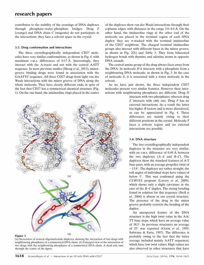
contributes to the stability of the crossings of DNA duplexes
through phosphate–water–phosphate bridges. Drug F
(orange) and DNA chain C (magenta) do not participate in
the interactions: they face a solvent space in the crystal.
3.3. Drug conformation and interactions
The three crystallographically independent CD27 mole-
cules have very similar conformations, as shown in Fig. 4, with
maximum r.m.s. differences of 0.17 A. Interestingly, they
interact with the A-tracts and not with the central AATT
sequence. In most previous studies (Sheng et al., 2013), minor-
groove binding drugs were found in association with the
GAATTC sequence. All three CD27 drugs form tight van der
Waals interactions with the minor groove of DNA along the
whole molecule. They have clearly different ends, in spite of
the fact that CD27 has a symmetrical chemical structure (Fig.
1). On the one hand, the imidazoline rings placed in the centre
of the duplexes show van der Waals interactions through their
coplanar edges, with distances in the range 3.6–4.0 A. On the
other hand, the imidazoline rings at the other end of the
molecule are placed in the terminal region of each DNA
duplex: they are �-stacked with the terminal imidazoline
of the CD27 neighbour. The charged terminal imidazoline
groups also interact with different bases in the minor groove,
as shown in Fig. 2(b) and Table 3. They form bifurcated
hydrogen bonds with thymine and adenine atoms in opposite
DNA strands.
The central amino group of the drug always faces away from
the DNA. In molecule D it interacts with a phosphate from a
neighbouring DNA molecule, as shown in Fig. 3. In the case
of molecule E, it is associated with a water molecule in the
solvent.
As we have just shown, the three independent CD27
molecules present very similar features. However their inter-
actions with neighbouring phosphates are different. Drug D
interacts with two phosphates, whereas drug
E interacts with only one. Drug F has no
external interactions. As a result the latter
has higher B factors and is more disordered,
as can be appreciated in Fig. 4. These
differences are mainly owing to their
different positions in the crystal. Molecule F
faces a solvent region and no external
interactions are possible.
3.4. DNA structure
The two crystallographically independent
duplexes in the structure are very similar,
with an r.m.s. difference of 0.49 A between
the two duplexes (A–A and B–C). The
duplexes show the standard features of A�T
base pairs, with an average propeller twist of
�13.6�. The duplexes are rather straight; the
roll angles of individual steps have values of
below 5�. This was confirmed using the
CURVES program (Lavery et al., 2009),
which shows only a slight curvature in the
case of the B–C duplex. The strong bending
found in solution for this sequence (Stefl et
al., 2004) is absent in our crystal structure.
The presence of the drug in the minor
groove probably restricts the bending of the
duplex.
An unexpected feature of the DNA
structure is the high twist value in the AA/
TT base steps, which have an average value
of 38.5�. In previous structures an average
of 35� was reported (Gorin et al., 1995;
Subirana & Faria, 1997). The difference is
probably owing to the fact that the latter
average included mainly AATT sequences,
which have low twist values. High values are
also observed in other structures that have
research papers
1618 Acosta-Reyes et al. � Interaction of an AT-rich DNA with CD27 Acta Cryst. (2014). D70, 1614–1621
Figure 3(a) Stereoview of crossed oligonucleotide duplexes, showing the interaction of two drugs withneighbouring phosphates of a symmetrical DNA chain. (b) Enlarged view of the interaction oftwo drugs with the neighbouring phosphates of a symmetrical DNA chain. A dyad axis runsthrough the centre of the figure.

AAA sequences (Edwards et al., 1992; Valls et al., 2005). Thus,
we can conclude that long adenine stretches will have a high
value of twist, which may explain some of the anomalous
features observed in A tracts.
The duplexes in our structure are organized as infinite
columns, as shown in Fig. 5. They are similar to those
described for other all-AT octamer duplexes (Valls et al., 2005;
Campos et al., 2006). Thus, the value of twist in the base step
TA between terminal bases of neighbouring duplexes is
negative (�26�). In our case there are three duplexes in the
repeating unit, so that the average rotation angle � between
neighbouring duplexes is 240�. In other octamers (Valls et al.,
2005; Campos et al., 2006) it is smaller at close to 230�.
Another feature of these columns is that the angle of the axis
of each duplex with respect to the overall axis of the column is
9�. Thus, the duplexes are organized as a smooth coiled coil
(De Luchi et al., 2011), as shown in Fig. 5.
research papers
Acta Cryst. (2014). D70, 1614–1621 Acosta-Reyes et al. � Interaction of an AT-rich DNA with CD27 1619
Figure 5Stereoview of the helical organization of the duplex columns in thecrystal. The axis of each individual duplex is also indicated (calculatedwith CURVES). The drug is not shown.
Figure 4OMIT 2Fo� Fc electron-density map of the three drugs in the complex atthe 1� level. D is at the top, followed by E and F below. The bottom twoframes show a superposition of the three drugs in two perpendicularviews.

4. Discussion
In the present study, we have found that the CD27 molecule
completely covers the entire minor groove of the DNA
duplexes. Since the duplexes are stacked in columns, the
complex appears as a continuous triple helix formed by two
single strands of DNA and one strand of CD27 molecules
arranged end to end (Fig. 2a). To our knowledge, complete
coverage of the minor groove has not been described
previously, with the exception of the complex of the unusual
duplex d(CCCCCIIIII)2 with netropsin (Chen et al., 1998).
Another unique feature of the complex is the interaction of
CD27 with the phosphates of neighbouring molecules in the
crystal, as shown in detail in Fig. 3. Interactions are found both
in the terminal charged groups of CD27 and in its central N1
atom. Similar features have been observed (Moreno et al.,
2010) in the complex formed by pentamidine and the alter-
nating duplex d(ATATATATAT)2. A scheme of the inter-
actions is presented in Fig. 6. Such interactions are allowed by
crystal packing. In contrast, in the studies performed with the
conventional Dickerson dodecamer d(CGCGAATTCGCG)2
(Sheng et al., 2013) no external interactions are found since
the drug is always completely buried inside the minor groove.
In this case, crystal packing also prevents interaction of the
drugs with neighbouring molecules. The interactions with
neighbouring phosphates that we have described are certainly
a feature of this sequence, and demonstrate that minor-groove
binding drugs may interact with neighbouring molecules,
including other DNA duplexes. It is likely that other drugs
might show similar interactions when bound to appropriate
DNA sequences. The formation of cross-links may be a
feature related to their biological action.
5. Conclusions
Our studies show two new features of DNA complexes with
minor-groove binding drugs: (i) the drugs completely fill the
minor groove and displace water in the AT-rich minor groove
of DNA and (ii) the drugs protrude from the DNA and
interact with neighbouring molecules. These findings demon-
strate that further studies of oligonucleotides with different
sequences are required in order to fully understand the
structural features of the interaction of DNA with drugs.
This work was supported through research fellowships
BFU-2009-10380 and SAF-2009-10399 from MICINN, Spain
and AQU-2009-SGR-1208 from the Catalan Government,
grant No. S8306 from the Government of Saudi Arabia and
a Doctoral Fellowship to FAR (CONACYT: 212993). The
authors are also grateful to Dr Anthonius Eze for technical
assistance and to J. Juanhuix and F. Gil at the ALBA
Synchrotron BL13-XALOC beamline. We also thank
Professor Isabel Rozas for providing the optimized structure
of CD27 and Professor G. Murshudov, Drs F. Long and J. Pous
for advice during refinement.
References
Boer, D. R., Canals, A. & Coll, M. (2009). Dalton Trans., pp. 399–414.
Campos, L., Valls, N., Urpı, L., Gouyette, C., Sanmartın, T., Richter,M., Alechaga, E., Santaolalla, A., Baldini, R., Creixell, M., Ciurans,R., Skokan, P., Pous, J. & Subirana, J. A. (2006). Biophys. J. 91,892–903.
Chen, X., Mitra, S. N., Rao, S. T., Sekar, K. & Sundaralingam, M.(1998). Nucleic Acids Res. 26, 5464–5471.
Chenoweth, D. M. & Dervan, P. B. (2009). Proc. Natl Acad. Sci. USA,106, 13175–13179.
Dardonville, C., Barrett, M. P., Brun, R., Kaiser, M., Tanious, F. &Wilson, W. D. (2006). J. Med. Chem. 49, 3748–3752.
Dardonville, C. & Brun, R. (2004). J. Med. Chem. 47, 2296–2307.Dardonville, C., Goya, P., Rozas, I., Alsasua, A., Martın, M. I. &
Borrego, M. J. (2000). Bioorg. Med. Chem. 8, 1567–1577.De Luchi, D., Urpı, L., Subirana, J. A. & Campos, L. (2011). Ind. Eng.
Chem. Res. 50, 5218–5224.Edwards, K. J., Brown, D. G., Spink, N., Skelly, J. V. & Neidle, S.
(1992). J. Mol. Biol. 226, 1161–1173.Emsley, P., Lohkamp, B., Scott, W. G. & Cowtan, K. (2010). Acta
Cryst. D66, 486–501.Evans, P. (2006). Acta Cryst. D62, 72–82.Glass, L. S., Nguyen, B., Goodwin, K. D., Dardonville, C., Wilson,
W. D., Long, E. C. & Georgiadis, M. M. (2009). Biochemistry, 48,5943–5952.
Gorin, A. A., Zhurkin, V. B. & Olson, W. K. (1995). J. Mol. Biol. 247,34–48.
Ibrahim, H. M. S., Al-Salabi, M. I., El Sabbagh, N., Quashie, N. B.,Alkhaldi, A. A. M., Escale, R., Smith, T. K., Vial, H. J. & de Koning,H. P. (2011). J. Antimicrob. Chemother. 66, 111–125.
Jensen, R. E. & Englund, P. T. (2012). Annu. Rev. Microbiol. 66,473–491.
Johnston, V. J. & Mabey, D. C. (2008). Curr. Opin. Infect. Dis. 21,56–64.
Kabsch, W. (2010). Acta Cryst. D66, 125–132.Lavery, R., Moakher, M., Maddocks, J. H., Petkeviciute, D. &
Zakrzewska, K. (2009). Nucleic Acids Res. 37, 5917–5929.McCoy, A. J., Grosse-Kunstleve, R. W., Adams, P. D., Winn, M. D.,
Storoni, L. C. & Read, R. J. (2007). J. Appl. Cryst. 40, 658–674.Moreno, T., Pous, J., Subirana, J. A. & Campos, J. L. (2010). Acta
Cryst. D66, 251–257.Murshudov, G. N., Skubak, P., Lebedev, A. A., Pannu, N. S., Steiner,
R. A., Nicholls, R. A., Winn, M. D., Long, F. & Vagin, A. A. (2011).Acta Cryst. D67, 355–367.
research papers
1620 Acosta-Reyes et al. � Interaction of an AT-rich DNA with CD27 Acta Cryst. (2014). D70, 1614–1621
Figure 6Interaction of pentamidine with neighbouring molecules in a complexwith DNA (Moreno et al., 2010). Hydrogen bonds between the terminal Natoms of pentamidine and phosphates are indicated.

Natto, M. J., Savioli, F., Quashie, N. B., Dardonville, C., Rodenko, B.& de Koning, H. P. (2012). J. Antimicrob. Chemother. 67, 933–943.
Nieto, L., Mascaraque, A., Miller, F., Glacial, F., Rıos Martınez, C.,Kaiser, M., Brun, R. & Dardonville, C. (2011). J. Med. Chem. 54,485–494.
Niyazi, H., Hall, J. P., O’Sullivan, K., Winter, G., Sorensen, T., Kelly,J. M. & Cardin, C. J. (2012). Nature Chem. 4, 621–628.
Paine, M. F., Wang, M. Z., Generaux, C. N., Boykin, D. W., Wilson,W. D., De Koning, H. P., Olson, C. A., Pohlig, G., Burri, C., Brun, R.,Murilla, G. A., Thuita, J. K., Barrett, M. P. & Tidwell, R. R. (2010).Curr. Opin. Investig. Drugs, 11, 876–883.
Rodrıguez, F., Rozas, I., Kaiser, M., Brun, R., Nguyen, B., Wilson,W. D., Garcıa, R. N. & Dardonville, C. (2008). J. Med. Chem. 51,909–923.
Sheng, J., Gan, J. & Huang, Z. (2013). Med. Res. Rev. 33, 1119–1173.Soeiro, M. N. C., De Souza, E. M., Stephens, C. E. & Boykin, D. W.
(2005). Expert Opin. Investig. Drugs, 14, 957–972.Stefl, R., Wu, H., Ravindranathan, S., Sklenar, V. & Feigon, J. (2004).
Proc. Natl Acad. Sci. USA, 101, 1177–1182.Subirana, J. A. & Faria, T. (1997). Biophys. J. 73, 333–338.Valls, N., Richter, M. & Subirana, J. A. (2005). Acta Cryst. D61, 1587–
1593.
research papers
Acta Cryst. (2014). D70, 1614–1621 Acosta-Reyes et al. � Interaction of an AT-rich DNA with CD27 1621