imran siddiqui
TRANSCRIPT
8/3/2019 Imran Siddiqui
http://slidepdf.com/reader/full/imran-siddiqui 1/10
ORIGINAL ARTICLE
Insights into structure and function of SHIP2-SH2: homology
modeling, docking, and molecular dynamics study
Uzma Saqib & Mohammad Imran Siddiqi
Received: 29 September 2010 /Accepted: 27 January 2011 /Published online: 12 February 2011# Springer-Verlag 2011
Abstract SRC homology 2 (SH2)-containing inositol 5′-
phosphatase protein (SHIP2) is a potential target for type 2diabetes. Its ability to dephosphorylate the lipid messenger
phosphatidylinositol 3,4,5-trisphosphate [PtdIns(3,4,5)P3],important for insulin signaling, makes it an important target against type 2 diabetes. The insulin-induced SHIP2 inter-action with Shc is very important for the membranelocalization and functioning of SHIP2. There is a bidentaterelationship between the two proteins where two domainseach from SHIP2 and Shc are involved in mutual binding.However in the present study, the SHIP2-SH2 domain
binding with the phosphorylated tyrosine 317 on thecollagen-homology (CH) domain of Shc, has been studied
due to the indispensability of this interaction in SHIP2localization. In the absence of the crystal structure of SHIP2-SH2, its structural model was developed followed
by tracking its molecular interactions with Shc throughmolecular docking and dynamics studies. This studyrevealed much about the structural interactions betweenthe SHIP2-SH2 and Shc-CH. Finally, docking study of a nonpeptide inhibitor into the SHIP2-SH2 domain further confirmed the structural interactions involved in ligand
binding and also proposed the inhibitor as a major starting point against SHIP2-SH2 inhibition. The insights gainedfrom the current study should prove useful in the design of
more potent inhibitors against type 2 diabetes.
Keywords Docking . Molecular dynamics . Shc .
SHIP2-SH2 . Type 2 diabetes
Introduction
Insulin-stimulated production of phosphatidylinositol 3,4,5-trisphosphate [PtdIns(3,4,5)P3] by phosphatidylinositol 3-kinase is important for the glucose homeostasis pathway.PtdIns(3,4,5)P3 plays a very critical step in proceeding theinsulin-signaling cascade [8, 34, 41]. However, the dephos-
phorylation of PtdIns(3,4,5)P3 molecule into PtdIns(3,4)P2 by the 5′ phosphatase-SRC homology 2 (SH2)-containing
inositol 5′-phosphatase protein (SHIP2) leads to attenuationof this signaling pathway. The level of PtdIns(3,4,5)P3 issharply regulated by SHIP proteins. SHIP proteins areSRC homology 2 (SH2)-containing inositol 5′-phospha-tase proteins with isozymes, SHIP1 and SHIP2. SHIP1 is
present largely in haematopoietic cells, while SHIP2,which can thus be assumed as a major regulator, is foundin skeletal muscles, among other insulin-sensitive tissues[27, 37]. Studies indicate that mice deficient of SHIP2, arehypoglycemic and sensitized to insulin suggesting that SHIP2 may have an important role in negative insulinsignaling [6]. Thus, targeting SHIP2 for consistent insulin
signaling and glucose homeostatis can be a plausiblesolution to this problem.
A change of cellular localization from cytosol tomembrane is crucial for SHIP2 functioning so that it might get access to its substrate [28]. This membrane targeting of SHIP2 is aided by Shc which is a member of a group of cytoplasmic-signaling proteins [17]. Shc gets tyrosine
phosphorylated at its central collagen-homology (CH)domain after being stimulated with a wide variety of growth factors and cytokines [4, 7]. It has been observed
U. Saqib : M. I. Siddiqi (*)Molecular and Structural Biology Division,Central Drug Research Institute, CSIR India,Lucknow 226 001, India e-mail: [email protected]
M. I. Siddiqie-mail: mi _ [email protected]
J Chem Biol (2011) 4:149–158
DOI 10.1007/s12154-011-0057-7
8/3/2019 Imran Siddiqui
http://slidepdf.com/reader/full/imran-siddiqui 2/10
that phosphorylation of Shc on Tyr-317 located at the CHdomain creates a binding site for the SH2 domain of SHIP2[21]. Besides the CH domain, it also contains an N-terminal
phosphotyrosine [Tyr(P)] binding (PTB) domain [38, 40]which in turn binds with phosphorylated 987-Tyr residue inthe NPXY motif present at the C-terminus of SHIP2 [42].This can also be described in terms of SHIP2 which possess
two potential sites that associate with Shc, one via its SH2domain and another by its NPXY motif at C-terminal domain.The interaction taking place between the SH2 domain and
phosphorylated tyrosine 317 (pTyr 317) of Shc-CH domainmight be mediated via a conserved Arg47 residue of theformer [9]. Studies indicate the high indispensability of thisinteraction with regard to SHIP2 functioning [16]. Hence,the association of SHIP2 with Shc is a key to triggering anefficient inhibitory effect on insulin cascade and this
prompted us to probe the SHIP2(SH2)-Shc interaction sothat further light can be thrown on their structural andmolecular interaction profile.
Computational studies on macromolecule interactionsare being routinely done these days. These studies provide a robust way of probing the key residues involved in a three-dimensional (3D) protein – ligand interaction system. Therehave been many recent publications where the results of computational analyses either helped in further narrowingdown the studies or validated the already performed studiesat the molecular level. Our understanding about the proteinstructure and function is mostly dependent upon X-raycrystallography and nuclear magnetic resonance (NMR)spectroscopy [5]. However, many proteins are difficult tocrystallize, e.g., membrane proteins, or are too large to be
studied with NMR, and both approaches are very timeintensive. With continous emergence of new sequences of
proteins involved in various diseases, our desire to predict their structures in order to understand their function isgreatly elevated. Hence, quick and accurate strategies for computational modeling have been in use in past couple of years [12, 24, 29, 43]. Recently, a computational study has
been done which sheds light on how a protein GEFmediates the activation of a small G protein Arf1 [25].Also, a computational study on PDZ binding to the BAR domain of PICK1 elucidated by molecular dynamics has beendone which details the molecular mechanisms of their binding
[13]. Other similar computational studies on protein – ligandinteractions have been reported earlier [1, 2, 22].
The present work details the construction of a 3D modelfor the human SHIP2-SH2 domain due to the unavailabilityof its crystal structure in Protein Data Bank (PDB), followed
by its docking and molecular dynamics study with Shc-CHdomain, its “ partner ” in membrane localization. Dockingstudies were then performed using a non-peptide inhibitor,AP22650 [36] of src-SH2 domain. AP22650 though initiallydesigned for src-SH2 domain was non-specific for the same.
However, due to its potency along with highly similar activesite of shc-SH2 with that of SHIP2-SH2 prompted us to useit against the latter. As expected, AP22650 showed quitereasonable binding conformation in the SHIP2-SH2 activesite along with displaying the major binding interactions to
be further qualified as a SHIP2-SH2 inhibitor. The inter-actions of SHIP2-SH2 with Shc-CH and with AP22650
provided some important clues for the design of novel and potent inhibitors against SHIP2-SH2.
Materials and methods
Computational resources
Molecular modeling was carried out on SGI Origin 300workstation equipped with 4 ×600 MHz R12000 processorsworkstation. For sequence alignment ClustalW 1.8 inInsightII/homology module [Insight II Program, Accelrys
Inc. (2001) San Diego, CA http://accelrys.com/ ] were used.Comparative modeling was performed using InsightII/ MODELER. Energy minimization and molecular dynamicsimulations were carried out using InsightII/CharmMmodule. Ramachandran plot was generated through struc-ture analysis and verification server (SAVS) while other statistics Prostat and Profile-3D were generated in InsightII/ HOMOLOGY. Molecular docking of Shc-CH was done byPatchDock protein – protein docking server while that of AP22650 was done using FlexX program interfaced withSybyl 7.1 (Tripos Inc., 1699 South Hanley Road, St. Louis,MO, 63144, USA. http://www.tripos.com/ ).
Sequence alignment and comparative modeling
The amino acid sequence of the target protein SHIP2-SH2(accession no. O15357), was extracted from the Swiss-Prot
protein sequence database (http://www.expasy.ch/sprot/ ).The SHIP2-SH2 sequence is composed of 97 residues.Sequence alignment was derived with the ClustalW 1.8
package and Align123 in InsightII/Homology module usingthe BLOSUM matrices [14] for scoring the alignments.Procuring a high quality multiple sequence alignment of thetarget with the templates is a central step in protein
homology modeling in view of the fact that only smalllocal errors made in alignment can be corrected in thesubsequent steps. The average NMR structure and X-raycrystal structure of template proteins SHIP1-SH2 and SAP-SH2 both from homosapiens, PDB Id: 2YSX and PDB Id:1D4W [31], respectively, were retrieved from Protein Data Bank. The resulting alignment was quite straightforward asthere were no loop insertions or deletions during the entirelength of the alignment. Finally, the alignment was used asan input for the automated homology modeling program
150 J Chem Biol (2011) 4:149–158
8/3/2019 Imran Siddiqui
http://slidepdf.com/reader/full/imran-siddiqui 3/10
MODELER interfaced with the InsightII software suite.MODELER is used for homology or comparative modelingof protein three-dimensional structures. Out of the threemodels generated, the model with best stereochemical qualityas determined by Ramachandran plot was selected for further analysis. Hydrogen atoms were added to it followedby energyminimization, employing the InsightII/Discover module in a
stepwise fashion following a standard procedure consisting of 500 steps of steepest descent and 1,000 steps of conjugategradient minimization with an RMS gradient of the potentialenergy of 0.001 kcal/mol Å in each step.
Model quality assessment
The quality of the final refined model was assessed bysubjecting it to a series of tests for its internal consistencyand reliability. The InsightII program Prostat was used toanalyze the properties of bonds, angles, and torsions while theProfile-3D program was used to check the structure and
sequence compatibility. The cut-off used was 5 standarddeviations from the reference value. The online SAVS server ’sPROCHECK suite of programs was used for assessing the“stereochemical quality” of the modeled protein structure.
SHIP2(SH2)-Shc(CH) docking
For the purpose of complex protein formation, the 3D structureof Shc (PDB Id: 1WCP) was truncated to carry the centralcollagen-homology domain, CH domain (residues: 211 – 368)and docked onto the SHIP2-SH2 domain using PatchDock. To
perform docking, the set of amino acid residues that, according
to published data [31], are important for SH2-Shcinteractions was specified as a possible binding site onthe SHIP2-SH2 domain surface. This set included Arg28,Arg47, Ser49, Glu50, Ser51, Arg70, and Thr83. PatchDock isa geometry-based molecular docking algorithm, which aimsin finding docking transformations that yield good molecular shape complementarity.
Molecular dynamics of the binary complex
This resulting binary complex structure was further refined by energy minimization by 500 steps of steepest descent
followed by 2,000 steps of conjugate descent molecular dynamics (MD) simulation with CHARMM program usingCHARMM force field [3]. Then, the complex wasembedded in a 10 Å layer of water molecules to imitateaqueous solvent conditions. This solvated complex wassubjected to energy minimization first by 500 steps of steepest descent followed by 2,000 steps of conjugategradient method. The system was heated from 50 K to300 K over a period of 50 ps with a time step of 1 fs andthe velocities being reassigned in the system every 0.05 ps.
The system was further equilibrated with a 1 fs time stepfor 100 ps so that the energy of the system achievescomplete stability. Production runs were performed at 300 K and carried out under constant number of particles,volume, and temperature conditions for 500 ps with a 1 fstime step. All the bonds involving hydrogen atom wereconstrained using SHAKE algorithm in all simulations [33].
The molecular trajectory for the systems generated by themolecular dynamics simulations were analyzed using thevisual molecular dynamics (VMD) [15] software.
Docking and molecular dynamic studies of AP22650
Molecular docking using FlexX program interfaced withSybyl 7.1 was carried out of the AP22650 inhibitor onSHIP2-SH2 domain. Docking was done using the standard
parameters of the FlexX program [18, 32] as implementedin Sybyl 7.1.All the flexibilities of its rotatable bonds wereconsidered in the process of docking for identifying the best
binding conformation. The refined model was chosen as thereceptor for molecular docking. The ligand-binding site inSHIP2-SH2 was defined based on the available informationfrom the literature. The inhibitor was docked into the
protein binding-site with hydrogens present and formalcharges were assigned by FlexX. The best conformationwas chosen based on the best Dock score and binding poseamong the resulting 30 docking solutions. The molecular dynamics studies of the resulting binary complex of SHIP2-SH2 with docked AP22650 were done exactly similar tothat done above for SHIP2-SH2-Shc-CH complex.
Results and discussion
3D model building of SHIP2-SH2 domain
Comparative modeling is used to develop a 3D structure of the target protein making use of the structural informationfrom the previously resolved protein structures withsubstantial similarity. To date, several NMR and crystalstructures of SH2 domains from various sources have beenreported by different research groups [10, 23, 26, 30].However, recently the NMR structure of SH2 domain of
SHIP1, an isozyme of SHIP2-SH2 has been deposited inProtein Data Bank by Kasai et al. (data not yet published). TheSHIP1-SH2 (PDB: 2YSX) shares 54% identity with theSHIP2-SH2 domain. Another SH2 domain from SAP proteinin complex with SLAM phosphopeptide (PDB: 1D4W)showing an identity of 42% was also used as a template, asthat can be useful in guiding the corresponding active siteresidues. The alignment of the deduced amino acid sequenceof the SHIP2-SH2 with the sequences from SHIP1 and SAPobtained from ClustalW 1.8 [39] is shown in Fig. 1. As can
J Chem Biol (2011) 4:149–158 151
8/3/2019 Imran Siddiqui
http://slidepdf.com/reader/full/imran-siddiqui 4/10
be observed, the alignment between the three proteins ishighly conserved assuring superior model quality. Thesuperimposed structures of all the three proteins are shownin Fig. 2, which besides displaying that the SHIP2-SH2 hasthe characteristic SH2 fold including a central β sheet with α
helices packed against either side also show that thehomology modeling has been quite accurate as the three
proteins superimpose very well.
Protein structure validation
The quality of the initial model was improved by subjectingit to a crude energy minimization protocol as detailed in the“Materials and methods” section. These minimizationshelped relieve any steric clashes or improper geometriesin the protein structure to produce a model with correct
bond lengths and bond angles and where individual atoms
are not too close together. The refined structure wasevaluated for overall quality using available analysis procedures. These analyses compare specific properties of
the model with those for known high quality proteinstructures. For this purpose, three protein analysis pro-grams: PROCHECK [20], Prostat, and Profile-3D wereutilized. Prostat was used to assess the stereochemicalquality of the model. This program verifies the accuracy of
parameters such as bond lengths, bond angles, andcorrectness of amino acid chirality. No spurious angle or
bond length was detected in our model. The results arelisted at the bottom of Table 1.
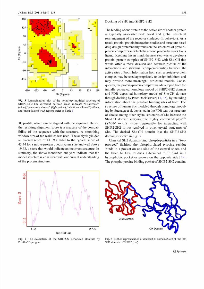
Another important indicator of the stereochemicalquality of the model is the distribution of the main chaintorsion angles phi and psi which may be examined in a Ramachandran plot. The Ramachandran plot of the phi – psi
plots is shown in Fig. 3 while the detailed results are listedin Table 1. The plot clearly shows that all the residues areeither in most favored or additional allowed regions andnone in generously allowed or disallowed regions, suggest-
ing high model quality. Finally, the 3D homology modelwas verified using the Profile-3D program in InsightIIsoftware, shown in Fig. 4. Profile-3D is a program based onalgorithms that measure the compatibility of an amino acidsequence with a three-dimensional structure by reducing thestructure to a one-dimensional representation, known as the
Fig. 2 Superimposed structures of SHIP2-SH2 (magenta), SHIP1-SH2 (blue), and SAP-SH2 (red )
Table 1 Results of protein structure check by PROCHECK andProstat
Residues in most favored regions 72 (86.7%)
Residues in additional allowed regions 11 (13.3%)
Residues in generously allowed regions 0 (0.0%)
Residues in disallowed regions 0 (0.0%) Number of non-glycine and non-proline residues 83
Number of end-residues (excl. Gly and Pro) 2
Number of glycine residues (shown as triangles) 8
Number of proline residues 4
Total number of residues 97
Number of bond distances with significant deviationsb (by Prostat)
0
Number of bond angles with significant deviations (by Prostat)
0
Fig. 1 Multiple sequence alignment using ClustalW. Residues highlighted in red correspond to identical/conserved residues, while residues in red
text are similar in the three proteins. Secondary structural elements are shown for SHIP1 (2YSX)
152 J Chem Biol (2011) 4:149–158
8/3/2019 Imran Siddiqui
http://slidepdf.com/reader/full/imran-siddiqui 5/10
3D profile, which can be aligned with the sequence. Hence,the resulting alignment score is a measure of the compat-ibility of the sequence with the structure. A smoothingwindow size of ten residues was used. The analysis yieldedan overall score of 41.19 similar to the typical score of 43.74 for a native protein of equivalent size and well above19.68, a score that would indicate an incorrect structure. In
summary, the above mentioned analyses indicate that themodel structure is consistent with our current understandingof the protein structure.
Docking of SHC into SHIP2-SH2
The binding of one protein to the active site of another proteinis typically associated with local and global structuralrearrangement of the receptor (induced-fit behavior). As a result, protein – protein interaction studies and structure-baseddrug design preferentially relies on the structures of protein –
protein complexes in which the second protein behaves like a ligand. Keeping this in mind, the next step was to develop a
protein – protein complex of SHIP2-SH2 with Shc-CH that would offer a more detailed and accurate picture of theinteractions and structural complementarities between theactive sites of both. Information from such a protein – proteincomplex may be used appropriately to design inhibitors andmay provide more meaningful structural models. Conse-quently, the protein – protein complex was developed from theinitially generated homology model of SHIP2-SH2 domainand PDB deposited homology model of Shc-CH domainthrough docking by PatchDock server [11, 35], by including
information about the putative binding sites of both. Thestructure of human Shc modeled through homology model-ing by Suenaga et al. deposited in the PDB was our structureof choice among other crystal structures of Shc because theShc-CH domain carrying the highly conserved pTyr 317
(YVNV motif) residue responsible for interacting withSHIP2-SH2 is not resolved in other crystal structures of Shc. The docked Shc-CH domain into the SHIP2-SH2domain is shown in Fig. 5.
Classical SH2 domains bind phosphopeptides in a “two- pronged” fashion; the phosphorylated tyrosine residue binds in a pocket on one side of the central sheet, and
the three to five residues C-terminal to it bind in a hydrophobic pocket or groove on the opposite side [19].The phosphotyrosine-binding pocket of SHIP2-SH2 contains
Fig. 3 Ramachandran plot of the homology-modeled structure of SHIP2-SH2.The different colored areas indicate “disallowed”(white),“generously allowed” (light yellow), “additional allowed”( yellow),and “most favored”(red ) regions (refer to Table 1)
Fig. 4 The evaluation of the SHIP2-SH2-modeled structure byProfile-3D program
Fig. 5 Ribbon representation of docked CH domain (blue) of Shc intoSH2 domain of SHIP2 (red )
J Chem Biol (2011) 4:149–158 153
8/3/2019 Imran Siddiqui
http://slidepdf.com/reader/full/imran-siddiqui 6/10
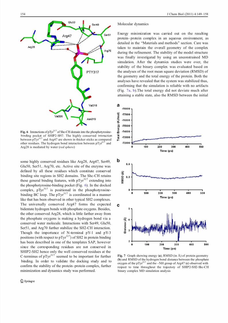
some highly conserved residues like Arg28, Arg47, Ser49,Glu50, Ser51, Arg70, etc. Active site of the enzyme was
defined by all these residues which constitute conserved binding site regions in SH2 domains. The Shc-CH retainsthese general binding features, with pTyr 317 extending intothe phosphotyrosine-binding pocket (Fig. 6). In the dockedcomplex, pTyr 317 is positioned in the phosphotyrosine-
binding BC loop. The pTyr 317 is coordinated in a manner like that has been observed in other typical SH2 complexes.The universally conserved Arg47 forms the expected
bidentate hydrogen bonds with phosphate oxygens. Besides,the other conserved Arg28, which is little farther away fromthe phosphate oxygens is making a hydrogen bond via a conserved water molecule. Interactions with Ser49, Glu50,
Ser51, and Arg70 further stabilize the SH2-CH interaction.Though the importance of N-terminal pY-1 and pY-3
positions (with respect to pTyr 317) of SH2 in protein bindinghas been described in one of the templates SAP, however since the corresponding residues are not conserved inSHIP2-SH2 hence only the well conserved residues at theC-terminus of pTyr 317 seemed to be important for further
binding. In order to validate the docking study and toconfirm the stability of the protein – protein complex, further minimization and dynamics study was performed.
Molecular dynamics
Energy minimization was carried out on the resulting protein – protein complex in an aqueous environment, asdetailed in the “Materials and methods” section. Care wastaken to maintain the overall geometry of the complexduring the refinement. The stability of the model structure
was finally investigated by using an unconstrained MDsimulation. After the dynamics studies were over, thestability of the binary complex was evaluated based onthe analyses of the root mean square deviation (RMSD) of the geometry and the total energy of the protein. Both theanalyses have revealed that the system was stabilized thus,confirming that the simulation is reliable with no artifacts(Fig. 7a, b).The total energy did not deviate much after attaining a stable state, also the RMSD between the initial
Fig. 6 Interactions of pTyr 317 of Shc-CH domain into the phosphotyrosine- binding pocket of SHIP2-SH2. The highly conserved interaction between pTyr 317 and Arg47 are shown in thicker sticks as comparedother residues. The hydrogen bond interaction between pTyr 317 andArg28 is mediated by water (red sphere)
Fig. 7 Graph showing energy (a), RMSD (in Å) of protein geometry(b) and RMSD of the hydrogen bond distance between the phosphateoxygen of the pTyr 317 and the – NH group of Arg47 (c) observed withrespect to time throughout the trajectory of SHIP2-SH2-Shc-CH
binary complex MD simulation analysis
154 J Chem Biol (2011) 4:149–158

8/3/2019 Imran Siddiqui
http://slidepdf.com/reader/full/imran-siddiqui 7/10
and final structures was also quite low, suggesting that thetwo proteins were reasonable in their energy as well asgeometry and their relative movements from their presumed
position was not too large. The highly conserved pTyr 317
and Arg47 interaction, which has been well documented inthe literature as the most important interaction, has also
been studied during the entire dynamic simulation. Asshown in Fig. 7c, the RMSD of the hydrogen bond distance
formed between the phosphate oxygen of the pTyr 317 andthe – NH group of Arg47 residue with respect to the averagestructure, oscillates very narrowly at a fixed position duringthe entire simulation confirming that this is one of the most stable and uniform interaction between the two proteins.Other important interactions have also been noticed duringthe simulation and they also follow the same pattern as of Arg47 (data not shown). Hence, all these studies confirmthat the system remained in equilibrium during the entiresimulation. Figure 8 shows the position of pTyr 317VNVmotif of the Shc-CH into the phosphotyrosine binding siteand the hydrophobic pocket (Val + 3 site) of SH2 domain.
The pTyr 317 retains all the conserved interactions asobserved during the docking studies. The pY + 1 to pY + 3
positions following pTyr 317 belonging to the conserved motif YVNV are coordinated by interactions with the residues inthe hydrophobic cleft of the Val + 3 site. Comparison of theinitial unbound SHIP2-SH2 domain structure with theShc-CH bound form after molecular dynamics shows smalloverall deviations. Importantly, in the unbound form, the EFand BG loops fold inward to close the hydrophobic +3
binding groove, however due to the insertion of Val-320
(pTyr 317 +3 position) the hydrophobic cleft can be observedin its open conformation (Fig. 9).
Docking and molecular dynamic studies of AP22650with SHIP2-SH2
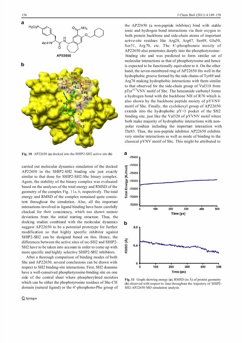
In order to further gain insight in the SHIP2-SH2
interactions with interacting partners, we performed dock-ing studies of an inhibitor, AP22650 (Fig. 10a ). Theinhibitor though initially designed for targeting osteoclastscarrying the src SH2 domain was non-selective. Hence,docking studies were done in order to find out whether thesame could bind to SHIP2-SH2, too. Though shc-SH2 is
just 28.8% identical to SHIP2-SH2 in overall sequencelength, however, there is considerable identity/similarity intheir binding site residues with the exception of few, yet important residues. Interestingly, AP22650 docked quitewell with a FlexX docking energy of −18.067 kcal/mol anda reasonable conformation into the SHIP2-SH2 active site
as shown in Fig. 10b. It has been observed that dockedAP22650 acquire the same position and displayed the sameinteractions in the SHIP2-SH2 as observed for pTyr 317VNVmotif of Shc-CH. The phosphate group of the inhibitor snugly fits into the phosphotyrosine binding site, while the
preceeding hydrophobic groups mimic the +1 and +3 positions of the pTyr 317VNV motif of Shc-CH, therebyentering into the hydrophobic pocket between the BG andEF loop around the hydrophobically conserved Thr 83. Inorder to further affirm this binding conformation along withstability of various important binding interactions, we
Fig. 8 pTyr 317VNV motif of the SHC-CH into the phosphotyrosine binding site and the hydrophobic pocket (Val + 3 site) of SH2 domain
Fig. 9 The open (cyan) and closed (magenta) conformation of SHIP2-SH2 domain
J Chem Biol (2011) 4:149–158 155
8/3/2019 Imran Siddiqui
http://slidepdf.com/reader/full/imran-siddiqui 8/10
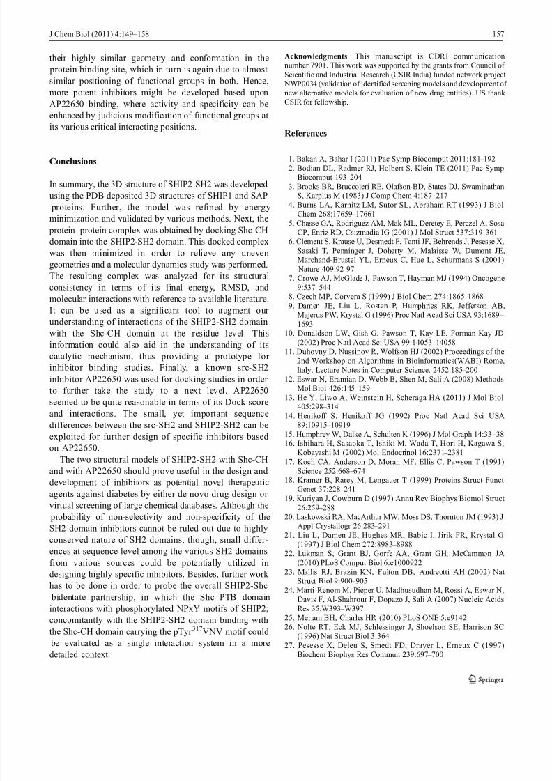
carried out molecular dynamics simulation of the dockedAP22650 in the SHIP2-SH2 binding site just exactlysimilar to that done for SHIP2-SH2-Shc binary complex.
Again, the stability of the binary complex was evaluated based on the analyses of the total energy and RMSD of thegeometry of the complex Fig. 11a, b, respectively. The totalenergy and RMSD of the complex remained quite consis-tent throughout the simulation. Also, all the important interactions involved in ligand binding have been carefullychecked for their consistency, which too shown minutedeviations from the initial starting structure. Thus, thedocking studies combined with the molecular dynamicssuggest AP22650 to be a potential prototype for further modification so that highly specific inhibitor against SHIP2-SH2 can be designed based on this. Hence, the
differences between the active sites of src-SH2 and SHIP2-SH2 have to be taken into account in order to come up withmore specific and highly selective SHIP2-SH2 inhibitors.
After a thorough comparison of binding modes of bothShc and AP22650, several conclusions can be drawn withrespect to SH2 binding-site interactions. First, SH2 domainshave a well-conserved phosphotyrosine-binding site on oneside of the central sheet where phosphorylated moietieswhich can be either the phophotyrosine residues of Shc-CHdomain (natural ligand) or the 4′-phosphono-Phe group of
the AP22650 (a non-peptide inhibitor) bind with stableionic and hydrogen bond interactions via their oxygen to
both protein backbone and side-chain atoms of important active-site residues like Arg28, Arg47, Ser49, Glu50,Ser51, Arg70, etc. The 4′-phosphonate moiety of AP22650 also penetrates deeply into the phosphotyrosine-
binding site and was predicted to form similar set of
molecular interactions as that of phosphotyrosine and henceis expected to be functionally equivalent to it. On the other hand, the seven-membered ring of AP22650 fits well in thehydrophobic groove formed by the side chains of Tyr69 andArg70 making hydrophobic interactions with them similar to that observed for the side-chain group of Val318 from
pTyr 317VNV motif of Shc. The benzamide carbonyl formsa hydrogen bond with the backbone NH of R70 which isalso shown by the backbone peptide moiety of pYVNVmotif of Shc. Finally, the cyclohexyl group of AP22650extends into the hydrophobic pY+3 pocket of the SH2
binding site, just like the Val320 of pYVNV motif where
both make majority of hydrophobic interactions with non- polar residues including the important interaction withThr83. Thus, the non-peptide inhibitor AP22650 exhibitsvery similar interactions as well as mode of binding to theclassical pYNV motif of Shc. This might be attributed to
Fig. 10 AP22650 (a) docked into the SHIP2-SH2 active site (b)
Fig. 11 Graph showing energy (a), RMSD (in Å) of protein geometry(b) observed with respect to time throughout the trajectory of SHIP2-SH2-AP22650 MD simulation analysis
156 J Chem Biol (2011) 4:149–158
8/3/2019 Imran Siddiqui
http://slidepdf.com/reader/full/imran-siddiqui 9/10
their highly similar geometry and conformation in the protein binding site, which in turn is again due to almost similar positioning of functional groups in both. Hence,more potent inhibitors might be developed based uponAP22650 binding, where activity and specificity can beenhanced by judicious modification of functional groups at its various critical interacting positions.
Conclusions
In summary, the 3D structure of SHIP2-SH2 was developedusing the PDB deposited 3D structures of SHIP1 and SAP
proteins. Further, the model was refined by energyminimization and validated by various methods. Next, the
protein – protein complex was obtained by docking Shc-CHdomain into the SHIP2-SH2 domain. This docked complexwas then minimized in order to relieve any unevengeometries and a molecular dynamics study was performed.
The resulting complex was analyzed for its structuralconsistency in terms of its final energy, RMSD, andmolecular interactions with reference to available literature.It can be used as a significant tool to augment our understanding of interactions of the SHIP2-SH2 domainwith the Shc-CH domain at the residue level. Thisinformation could also aid in the understanding of itscatalytic mechanism, thus providing a prototype for inhibitor binding studies. Finally, a known src-SH2inhibitor AP22650 was used for docking studies in order to further take the study to a next level. AP22650seemed to be quite reasonable in terms of its Dock score
and interactions. The small, yet important sequencedifferences between the src-SH2 and SHIP2-SH2 can beexploited for further design of specific inhibitors basedon AP22650.
The two structural models of SHIP2-SH2 with Shc-CHand with AP22650 should prove useful in the design anddevelopment of inhibitors as potential novel therapeuticagents against diabetes by either de novo drug design or virtual screening of large chemical databases. Although the
probability of non-selectivity and non-specificity of theSH2 domain inhibitors cannot be ruled out due to highlyconserved nature of SH2 domains, though, small differ-
ences at sequence level among the various SH2 domainsfrom various sources could be potentially utilized indesigning highly specific inhibitors. Besides, further work has to be done in order to probe the overall SHIP2-Shc
bidentate partnership, in which the Shc PTB domaininteractions with phosphorylated NPxY motifs of SHIP2;concomitantly with the SHIP2-SH2 domain binding withthe Shc-CH domain carrying the pTyr 317VNV motif could
be evaluated as a single interaction system in a moredetailed context.
Acknowledgments This manuscript is CDRI communicationnumber 7901. This work was supported by the grants from Council of Scientific and Industrial Research (CSIR India) funded network project
NWP0034 (validation of identified screening models and development of new alternative models for evaluation of new drug entities). US thank CSIR for fellowship.
References
1. Bakan A, Bahar I (2011) Pac Symp Biocomput 2011:181 – 1922. Bodian DL, Radmer RJ, Holbert S, Klein TE (2011) Pac Symp
Biocomput 193 – 2043. Brooks BR, Bruccoleri RE, Olafson BD, States DJ, Swaminathan
S, Karplus M (1983) J Comp Chem 4:187 – 2174. Burns LA, Karnitz LM, Sutor SL, Abraham RT (1993) J Biol
Chem 268:17659 – 176615. Chasse GA, Rodriguez AM, Mak ML, Deretey E, Perczel A, Sosa
CP, Enriz RD, Csizmadia IG (2001) J Mol Struct 537:319 – 3616. Clement S, Krause U, Desmedt F, Tanti JF, Behrends J, Pesesse X,
Sasaki T, Penninger J, Doherty M, Malaisse W, Dumont JE,Marchand-Brustel YL, Erneux C, Hue L, Schurmans S (2001)
Nature 409:92 – 977. Crowe AJ, McGlade J, Pawson T, Hayman MJ (1994) Oncogene
9:537 – 5448. Czech MP, Corvera S (1999) J Biol Chem 274:1865 – 18689. Damen JE, Liu L, Rosten P, Humphries RK, Jefferson AB,
Majerus PW, Krystal G (1996) Proc Natl Acad Sci USA 93:1689 –
169310. Donaldson LW, Gish G, Pawson T, Kay LE, Forman-Kay JD
(2002) Proc Natl Acad Sci USA 99:14053 – 1405811. Duhovny D, Nussinov R, Wolfson HJ (2002) Proceedings of the
2nd Workshop on Algorithms in Bioinformatics(WABI) Rome,Italy, Lecture Notes in Computer Science. 2452:185 – 200
12. Eswar N, Eramian D, Webb B, Shen M, Sali A (2008) MethodsMol Biol 426:145 – 159
13. He Y, Liwo A, Weinstein H, Scheraga HA (2011) J Mol Biol
405:298 –
31414. Henikoff S, Henikoff JG (1992) Proc Natl Acad Sci USA89:10915 – 10919
15. Humphrey W, Dalke A, Schulten K (1996) J Mol Graph 14:33 – 3816. Ishihara H, Sasaoka T, Ishiki M, Wada T, Hori H, Kagawa S,
Kobayashi M (2002) Mol Endocrinol 16:2371 – 238117. Koch CA, Anderson D, Moran MF, Ellis C, Pawson T (1991)
Science 252:668 – 67418. Kramer B, Rarey M, Lengauer T (1999) Proteins Struct Funct
Genet 37:228 – 24119. Kuriyan J, Cowburn D (1997) Annu Rev Biophys Biomol Struct
26:259 – 28820. Laskowski RA, MacArthur MW, Moss DS, Thornton JM (1993) J
Appl Crystallogr 26:283 – 29121. Liu L, Damen JE, Hughes MR, Babic I, Jirik FR, Krystal G
(1997) J Biol Chem 272:8983 –
898822. Lukman S, Grant BJ, Gorfe AA, Grant GH, McCammon JA(2010) PLoS Comput Biol 6:e1000922
23. Mallis RJ, Brazin KN, Fulton DB, Andreotti AH (2002) Nat Struct Biol 9:900 – 905
24. Marti-Renom M, Pieper U, Madhusudhan M, Rossi A, Eswar N,Davis F, Al-Shahrour F, Dopazo J, Sali A (2007) Nucleic AcidsRes 35:W393 – W397
25. Meriam BH, Charles HR (2010) PLoS ONE 5:e914226. Nolte RT, Eck MJ, Schlessinger J, Shoelson SE, Harrison SC
(1996) Nat Struct Biol 3:36427. Pesesse X, Deleu S, Smedt FD, Drayer L, Erneux C (1997)
Biochem Biophys Res Commun 239:697 – 700
J Chem Biol (2011) 4:149–158 157
8/3/2019 Imran Siddiqui
http://slidepdf.com/reader/full/imran-siddiqui 10/10
28. Phee H, Jacob A, Coggeshall KM (2000) J Biol Chem275:19090 – 19097
29. Pieper U, Eswar N, Webb B, Eramian D, Kelly L, Barkan D,Carter H, Mankoo P, Karchin R, Marti-Renom M, Davis F, Sali A(2009) Nucleic Acids Res 37:D347 – D354
30. Pletneva EV, Sundd M, Fulton DB, Andreotti AH (2006) J MolBiol 357:550 – 561
31. Poy F, Yaffe MB, Sayos J, Saxena K, Morra M, Sumegi J, CantleyLC, Terhorst C, Eck MJ (1999) Mol Cell 4:555 – 561
32. Rarey M, Kramer B, Lengauer T, Klebe G (1996) J Mol Biol261:470 – 489
33. Ryckaert JP, Ciccitti G, Berendsen HJC (1977) J Comp Phys23:327 – 341
34. Saltiel AR (1996) Am J Physiol 270:375 – 38535. Schneidman-Duhovny D, Inbar Y, Nussinov R, Wolfson HJ
(2005) Nucl Acids Res 33:363 – 36736. Shakespeare W, Yang M, Bohacek R, Cerasoli F, Stebbins K,
Sundaramoorthi R, Azimioara M, Vu C, Pradeepan S, Metcalf C,Haraldson C, Merry T, Dalgarno D, Narula S, Hatada M, Lu X,
Schravendijk MR, Adams S, Violette S, Smith J, Guan W, Bartlett C, Herson J, Iuliucci J, Weigele M, Sawyer T (2000) Proc NatlAcad Sci USA 97:9373 – 9378
37. Sleeman MW, Wortley KE, Lai KM, Gowen LC, Kintner J, KlineWO, Garcia K, Stitt TN, Yancopoulos GD, Wiegand SJ, Glass DJ(2005) Nat Med 11:199 – 205
38. Songyang Z, Shoelson SE, McGlade J, Olivier P, Pawson T,Bustelo XR, Barbacid M, Sabe H, Hanafusa H, Yi T, Ren R,Baltimore D, Ratnofsky S, Feldman RA, Cantley LC (1994) Mol
Cell Biol 14:2777 – 278539. Thompson JD, Higgins DG, Gibson TJ (1994) Nucleic Acids Res
22:4673 – 468040. van der Geer P, Wiley S, Lai VKM, Olivier JP, Gish G, Stephens
R, Kaplan D, Shoelson S, Pawson T (1995) Curr Biol 5:404 – 41241. Virkamäki A, Ueki K, Kahn CR (1999) J Clin Invest 103:931 – 94342. Wisniewski D, Strife A, Swendeman S, Erdjument-Bromage H,
Geromanos S, Kavanaugh WM, Tempst P, Clarkson B (1999)Blood 93:2707 – 2720
43. Zhou H, Skolnick J (2007) Biophys J 93:1510 – 1518
158 J Chem Biol (2011) 4:149–158