improving hydrophobicity of n-terminal increased …improving hydrophobicity of n-terminal increased...
TRANSCRIPT
International Journal of Engineering Research & Science (IJOER) ISSN: [2395-6992] [Vol-2, Issue-8, August- 2016]
Page | 1
Improving hydrophobicity of N-terminal increased molecular
stability of mannanase Man1312 Haiyan Zhou
1, Wenjiao Yang
2, Hanhui Peng
3, Yun Tian
4, Yongyao Wu
5*
1,2,3,5College of Bioscience and Biotechnology, Hunan Agricultural University, Changsha 410128, China;
4Key lab of agricultural biochemistry and biotransformation, Hunan Agricultural University, Changsha 410128, China
*Corresponding Author: E-mail: [email protected]; postal address: College of Bioscience and Biotechnology, Hunan
Agricultural University, Changsha, China (postal code: 410128)
Abstract— Protein terminals have important roles in molecular structural stability and in some occasions they have
regulatory roles in catalytic reaction. To expand our understanding on the influences of distal residues mutation, we
explored the molecular stability and kinetics of mannanase Man1312 mutants. For Man1312, the N-terminal loop was more
disordered and changeable; therefore the mutations on N-terminal should have significant effects on enzymic properties. The
experiment was investigated by spectrophotometer, circular dichroism and differential scanning calorimetry assays. As a
result, positive mutations were found from sites of T2 and Q9 and double-site mutant ManY9G2 showed increased catalytic
activity by 7.7% higher than Man1312. Meanwhile, ManY9G2 had significantly increased thermostability with promoted Topt
by 6oC and elongated t1⁄2 by 7 min, which was resulted from the optimized intermolecular forces and a newly-built hydrogen
bond in ManY9G2. In our experiment, the specific residues were mutated from hydrophilic to hydrophobic and the improved
hydrophobicity on N-terminal had a positive impact on the properties of mannanase Man1312.
Highlights
We construct N-terminal multi-site mutants of mannanase Man1312.
We determine the enzyme activity and thermal dynamics of mannanase and its mutants.
Best mutations increase the hydrophobilicity of N-terminal.
N-terminal mutation significantly improves the thermo-stability and slightly increases activity of mannanase.
Keywords— Mannanase, N-terminal, multi-site mutation, structural stability, activity.
I. INTRODUCTION
Hemicelluloses are a series of polysaccharides consisting of linear chain and branches in the plants cell walls which are
associated to the cellulose and lignin forming lignocelluloses biomass (1). Mannan is the most abundant of polysaccharide
present in softwood hemicelluloses and targeted by mannanase to degrade into oligosaccharides. β-Mannanases catalyze
random hydrolysis of beta-1,4-mannosidic bonds by releasing a single β-D-mannose unit from the nonreducing end of
manno-oligo saccharides (2).
In the past few decades, various mannanases were from isolated from plants (3), marine mollusk (4), and a body of bacteria
and fungi (5, 6), but in general, production of β-mannanase by microorganisms is more promising because of its low cost,
high production rate and readily controlled conditions (7).
Mannanases play important roles in fundamental biological processes and also have potential applications in various
industries. They are widely used by industries including food processing, feed, oil mining, paper making, pharmaceutical,
and second generation biofuel (8, 9) as well as manufacture of oligosaccharide (7). Meanwhile, mannanases could be used in
prebiotic preparation which is expected to improve the growth performance of animal.
Mannanase Man1312 screened from Bacillus subtilis (B. subtilis) B23 has the mechanism of Glycoside Hydrolase Family 26
(GH26). Although the members of GH26 family display great diversity in their sequence and function, all of them have a
canonical α/β hydrolase fold, which consists of eight β-sheets flanked by α-helices. This core architecture is the structural
foundation as a stable scaffold to accommodate the catalytic residues and tolerate mutations at some degree without losing its
molecular stability or catalytic machinery. The loop which connects α-helices and β-strand is important to molecular
structural stability and activity as well. Pleiss team studied the sequence and structure of epoxide hydrolases and proposed
that NC-loop might interact with the substrate by defining the substrate-binding pocket and regulating the accessibility of
active site (10, 11). The residue Tyr on the NC-loop of epoxide hydrolases is involved in substrate binding, stabilization of
the transition state, and possibly protonation of the epoxide oxygen (12, 13). Moreover, loops participate in the formation of
the binding sites for metallic cations (14) and ligands (15), mediates protein-protein interactions (16), and modulates protein
International Journal of Engineering Research & Science (IJOER) ISSN: [2395-6992] [Vol-2, Issue-8, August- 2016]
Page | 2
stability (17). All evidences suggest that exploring the functional and evolutionary role of the loop may be important for
understanding the adaptation and evolution of homologous enzymes.
In our previous work, the active sites of mannanase Man1312 was detected and site-directed mutagenesis were performed on
the active sites. H129, E159, H190, E191, W196, F197, W198, and W199 are the residues relevant to mannanase Man1312
activity and substrate binding; Moreover, mutants on H129, H190, and W198 had increased activity by 3.5-, 2.2-, and 3.8-
fold, respectively. In this work, the disordered residues on distal loops were taken into accounts because they are relative to
the molecular structural stability and in some occasions they have regulatory roles in catalytic reaction. In order to explore
the relationship of the residues in distal loop and molecular stability and activity, the mutations on N-terminal loop were
determined. Our work could provide insight into the integration of structure and function and had led to the design and
construction of more stable and active mannanase.
II. MATERIALS AND METHODS
2.1 Computer-aided prediction for the structure of mannanase Man1312
The secondary structure, 3D structure modeling, Van der Waals' force (VDW) and hydrogen bond compute were performed
by the Swiss Model server (http://swissmodel.expasy.org/) (18, 19, 20) using the template 2qha.1.A(the sequence identity
76.19). Structural modeling quality was evaluated with Protein Structure Validation Software (21) (PSVS, http://psvs-1_4-
dev.nesg.org/). The visualization and analysis of structures were performed by PyMOL (22) and Swiss-PDB Viewer
software.
2.2 Cloning, mutagenesis, expression and purification of enzymes
The decoded gene of mannanase Man1312 was transferred in E. coli TOP10 cells and can be cloned by the primers P1 (5’-
ATGCCTACTAAGT-3’) and P2 (5’-TGATTCAGCTATCTGT G-3’). Deletion mutations were constructed with the
QuikChange Mutagenesis protocol (23) by pairs of reverse primers on the recombinant plasmid plasmid pBS-T-man23 which
carried the decoded gene of mannanase Man1312. Conditions of mutagenic PCR were 94oC for 4 min; 20 cycles of 90
oC for
1 min, 55oC for 1 min and 72
oC for 5 min; and finally extension at 72
oC for 20 min. Successful introduction of deletion
mutation was confirmed by DNA sequencing.
The PCR products were digested with BamH I and EcoR I and linked with vector pHY-p43 and then transformed into
Bacillus subtilis(B. subtilis) WB600. The details about expression and purification were described as previously (24).
2.3 Enzyme assay
Protein concentration was measured using the Bradford assay (25). The Mannanase activity assay was improved from the
method of monitoring the release of reducing sugar (26, 27). One unit of enzyme activity was defined as the amount of
enzyme liberating 1 μmol mannose per minute at 50oC and pH 6.8. The activity formula was as follows:
Mannanase activity (U/ml) = 5.56CeVde/VjeVst
5.56 the mole value of 1.0 mg mannose, μmol
Ce amount of mannose produced from hydrolysis, mg
Vde metered volume of enzyme solution, ml
Vje volume of enzyme solution added into the reaction mixture, ml
Vs volume of substrate solution, ml
t time, min
Specific activity of mannanase (units per milligram) =5.56CeVde2 /CpVjeVst
Cp amount of total proteins, mg
2.4 Kinetic parameters of enzymes
Topt was determined at the temperature range of 30~90oC under pH 5.8. T50 was determined at the temperature range of
30~90oC for 20 min and t1/2 was determined at the optimum temperature for 1~40 min. The buffer was 50 mmol/l phosphate
buffer.
Vmax and Km were acquired by fitting enzymatic activities as a function of substrate concentrations to the Michaelis-Menten
equation using non-linear regression of the software GraphPad Prism 5.0. The parameter kcat was obtained by using the
equation kcat=Vmax/[E], where [E] was the molar concentration of the enzymes.

International Journal of Engineering Research & Science (IJOER) ISSN: [2395-6992] [Vol-2, Issue-8, August- 2016]
Page | 3
2.5 Thermal dynamics of enzymes
The secondary structure of mannanase Man1312 and its mutants were monitored by Circular dichroism (CD)
spectropolarimeter (JASCO J810, Japan) from 190 to 260 nm with the parameters as protein concentration 0.1 mg/ml, 0.1 cm
path-length cuvette at 50 nm/min scan speed, response time 2 s, bandwidth 2 nm, and pitch 0.1 nm. For thermodynamic
analysis, the spectra were recorded at 220 nm with the following parameters: protein concentration 0.2 mg/ml, and
temperature interval 0.5oC /min from 40~90
oC. Tm was determined through the change in the CD value.
Differential scanning calorimetry (DSC) was performed on DSC200F3 Differential Scanning Calorimeter (NETZSCH,
German) with protein concentration 1.0 mg/ml. Denaturizing curves were recorded from 40 oC to 90
oC at a rate of 1℃/min.
2.6 Statistical Analysis
Data were presented as the mean±standard error of the mean. Results were compared with the analysis of variance and
Fisher's protected least-significant difference tests, with a significance of P<0.05.
III. RESULTS
3.1 A mutant library on N-terminal loop was developed to discover key positions relevant to enzyme properties
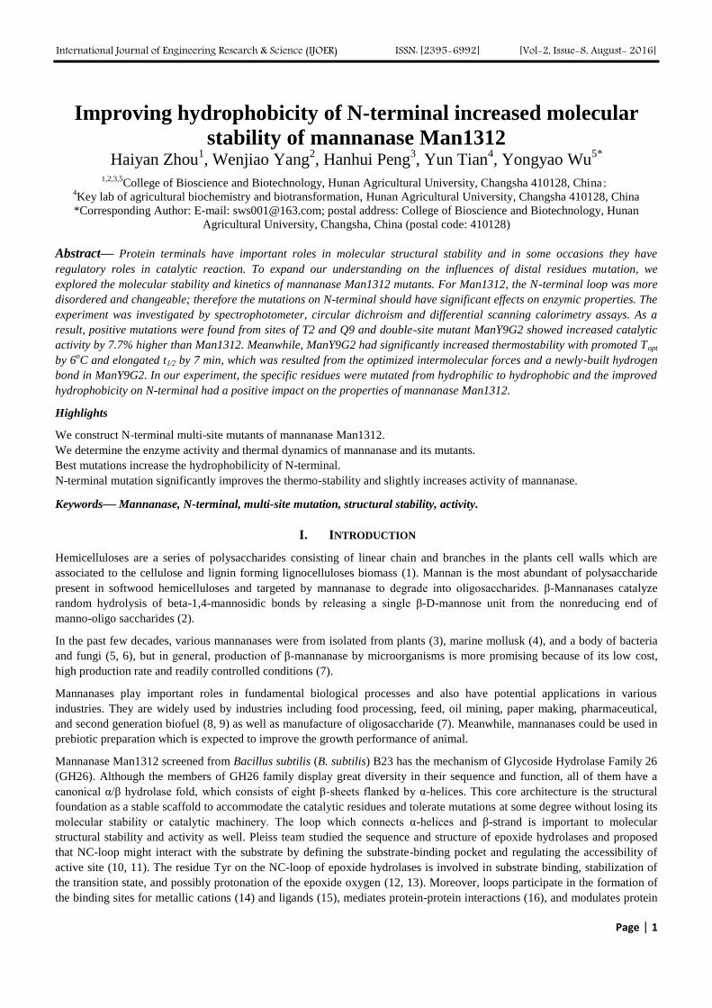
Mannanase Man1312 has a typical (β/α)8 barrel structure of GH26 according to its amino acid sequence similarities. The N-
terminal loop of Man1312 includes the first 11 residues from H1 to Q11 (Fig. 1). Saturation mutagenesis was applied on each
site of N-terminal loop to identify catalytic effects of the substitutions by other amino acid residues at the each position. The
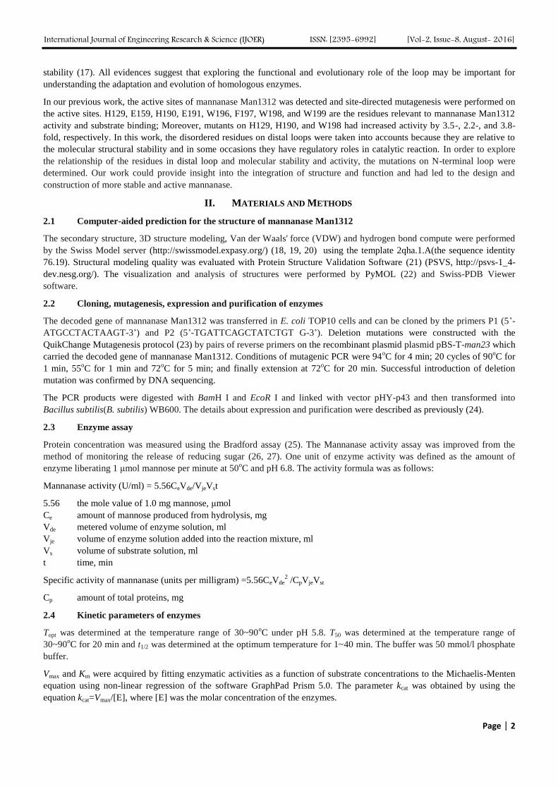
saturation mutant library contained 220 clones, but significant mutations were observed on site T2 and N9 (Fig. 2). Three
mutants showed an enhanced thermo-stability activity but without activity loss (Table 1).
FIG. 1 THE TERTIARY STRUCTURE OF MANNANASE MAN1312 A structure of mannanase Man1312; b residues on N-terminal of mannanase Man1312. The black represented N-
terminal region which is the research target in this paper and its sequence is H-T-V-Y-P-V-N-P-N-A-Q.
FIG. 2 THE ACTIVITY INCREASING RATIO IN THE SATURATION MUTANT LIBRARY
The saturation mutant library contained 220 clones, among which significant mutations were observed on site T2 and N9
with the substitution of alanine and glycine on T2 and tyrosine on N9.

International Journal of Engineering Research & Science (IJOER) ISSN: [2395-6992] [Vol-2, Issue-8, August- 2016]
Page | 4
TABLE 1
ENZYME ACTIVITY AND KINETICS PARAMETERS OF MANNANASE MAN1312 AND ITS MUTANTS T2A, T2G AND
N9Y
3.2 The double mutant was constructed to build top variants
From the single-site saturation mutations, the substitution of alanine and glycine on T2 and tyrosine on N9 showed
significant improvement on enzyme properties. The first top substitution was N9Y, therefore tyrosine was the best and
exclusive mutation on position 9. The second top substitution was T2G and the third was T2A, both of which mutated on
position 2. Two double-site mutants were constructed, each of which combined the N9Y substitution with T2G and T2A
respectively. The assessment of two double-site mutants illuminated the combination of N9Y and T2G was the best variants
with 7.7% higher activity and 28% longer half-life (Table 2).
TABLE 2
ENZYME ACTIVITY AND KINETICS PARAMETERS OF MANNANASE MAN1312 AND ITS MUTANTS
As for double mutant ManY9G2, the residues were mutated from Thr of site 2 and Asn of site 9 to Gly and Tyr. Threonine
and asparagine are both hydrophilic amino acids, but glycine and tyrosine are hydrophobic, therefore the double mutant
improved hydrophobicity of N-terminal which could be a reason of optimized properties of ManY9G2.
3.3 The double-site mutant ManY9G2 exhibited more stable on thermal dynamics
According to the thermal denaturation spectra (Fig. 3a), the Tm values of double-site mutant ManY9G2 was 69oC, which was
5oC higher than that of Man1312. Taking into account that the mutant Tm value was basically consistent with t1⁄2 value, it
could be concluded that the enhanced enzyme thermo-stability might be caused by the increase of Tm. Meanwhile, from the
△G0 profiles between the native and denatured states, the slopes of ManY9G2 decreased more slowly than that of Man1312
(Fig. 3b). Correspondingly, the Man1312 unfold more quickly than ManY9G2. Because of the higher Tm and the slower
decreasing △G0, ManY9G2 was more thermostable. To obtain further information on the thermodynamic stability, thermal
unfolding experiments were carried out by DSC (Fig. 4). As the above results, Tm value of mutant ManY9G2 was ~5oC
higher than Man1312.
Mannanase amount (mg/ml) Mannanase activity (U/mg) Topt T50 t1/2
Man1312 8.5 ± 0.1 245 ± 0.5 60oC 65
oC 25 min
ManT2A 8.65 ± 0.1 253 ± 0.5 62oC 66
oC 26 min
ManT2G 8.6 ± 0.1 260 ± 0.5 62oC 66
oC 26 min
ManN9Y 8.7 ± 0.1 254 ± 0.5 65oC 68
oC 30 min
Mannanase amount
(mg/ml)
Mannanase
activity (U/mg) Km (mg/L) Kcat (s
−1) Topt T50
t1/2
(min)
Man1312 8.5±0.1 245±0.5 9.5±0.5 8.2±0.2 60oC 65
oC 25
ManY9A2 8.65±0.1 256±0.5 8.1±0.3
9.2±0.2 65oC 67
oC 28
ManY9G2 8.7±0.2 264±0.5 7.2±0.2 10.4±0.2
66oC 69
oC 32

International Journal of Engineering Research & Science (IJOER) ISSN: [2395-6992] [Vol-2, Issue-8, August- 2016]
Page | 5
(A) (B)
FIG. 3 FRACTION UNFOLDED (A) AND MOLAR GIBBS FREE ENERGY CHANGES (△G0) (B) DURING THE THERMAL
DENATURATION PROCESS OF MANNANASE The CD data were measured at 220 nm from 310 to 360 K at a heating rate of 0.5℃/min and were converted to an
unfolded fraction through the equation (fu = (y-yn)/(yd-yn)), where y is the ellipticity observed at the given temperature
and yn and yd are the characteristic ellipticities of the folded and unfolded protein. △G0 (dG, J/mol) was calculated by the
standard van’t Hoff equation (△G0 = -RT(fu/(1-fu)).
FIG. 4 CD SPECTRA PROFILES OF MAN1312 AND DOUBLE-SITE MUTANT MANY9G2
CD spectra were recorded from 190 to 260 nm. Protein concentration of 0.1 mg/ml was used for the analysis.
To compare the secondary structural between Man1312 and its mutant ManY9G2, the CD spectra were presented to evaluate
secondary structural elements of helix, sheet, and coil which characterized negative CD spectrum bands at 222 and 210 nm,
215 nm, and 195 nm, respectively.
Both of CD profiles had two clear minima at 210 and 222 nm (Fig. 5), in responding to GH16 hydrolase having both α-helix
and β-sheet features. The CD spectra of Man1312 and ManY9G2 exhibited similar minima at 222 nm, however the
amplitude decreased on ManY9G2 comparing with Man1312. The irregular coil had a characterized negative band at ~195
nm, the spectra minima of ManY9G2 at 210 nm shifted toward higher wavelengths, indicating that the contents of helix and
strand on ManY9G2 increased relatively. Thus, the results stated mutant ManY9G2 increased enzyme stability.

International Journal of Engineering Research & Science (IJOER) ISSN: [2395-6992] [Vol-2, Issue-8, August- 2016]
Page | 6
FIG. 5 THERMAL UNFOLDING OF MAN1312 AND DOUBLE-SITE MUTANT MANY9G2 MONITORED BY DSC
SCANNING Protein concentration of 1.0 mg/ml was used for the analysis. Denaturizing curves were recorded from 40
oC to 90
oC
with a rate of 1oC/min.
3.4 Molecular analysis around mutated sites on N-terminal was performed by computer-aided software
From the VDW prediction (Fig. 6), ManY9G2 formed stronger van der Waals force around position 9, but became weak
around position 2. ManY9G2 showed more stable comparing to Man1312, indicating that substitution on position 9 had more
important effects on thermal stability than position 2. On the other side, around position 9, one more hydrogen bond was built
after being mutated from Asn to Tyr (Fig. 7), but there is no newly-built hydrogen bonds appear around position 2. It can be
perceived that the changes around position 9 should be main reasons for thermos-stability of mannanase ManY9G2.
(6A) (6B)
(6C) (6D)

International Journal of Engineering Research & Science (IJOER) ISSN: [2395-6992] [Vol-2, Issue-8, August- 2016]
Page | 7
(6E) (6F)
FIG. 6 THE VDW ANALYSIS ON N-TERMINAL OF MAN1312 AND MANY9G2 A N-terminus of Man1312; b N-terminus of ManY9G2; c position 2 surrounding of Man1312; d position 2 surrounding
of ManY9G2; e position 9 surrounding of Man1312; f position 9 surrounding of ManY9G2.
(7A) (7B) FIG. 7 HYDROGEN BONDS ANALYSIS AROUND POSITION 9 OF MAN1312 AND MANY9G2
Dash lines represented hydrogen bonds formed by inter-molecules. a hydrogen bonds around position 9 of Man1312
(before mutation); b hydrogen bonds around position 9 of ManY9G2 (after mutation). Before being mutated, three
hydrogen bonds were formed around position 9 between Val6-Pro8, Asn7-Pro8, and Asn7-Ala10; after being mutated,
besides three bonds formed before, one more hydrogen was newly built between Asn7- Tyr9.
IV. CONCLUSION
According to the hydrophobic cluster analysis, Mannanase Man1312 has been classified into GH26 family. The (α/β)8 barrel
is one of GH26 hydrolase features and enzymes in this family display a broad variety of activity, including endo-β-1,4-
mannanases, exo-acting β-mannanase (28), β-1,3:1,4-glucanase (29) and β-1,3-xylanase activities (30). For this reason, they
are one of the most widely used groups of biocatalysts in industry.
In our previous work, the active sites were clarified and the expression system of mannanase Man1312 decoded gene was
optimized. Furthermore, rational evolution strategy was performed on α/β fold of Man1312 to improve the molecular
stability. The present study focused on the relationship of mutations on the N-terminal loop and molecular stability and
activity basing on the structural analysis.
Our results suggest that the N-terminal loop could modulate both catalytic activity and stability of Man1312 and the
mutations increased Man1312 thermostability and activity. The double mutant ManY9G2 increased catalytic activity by
7.7% higher than Man1312 and meanwhile, it increased Topt by 6oC and t1⁄2 elongated by 7 min in compare with Man1312.
International Journal of Engineering Research & Science (IJOER) ISSN: [2395-6992] [Vol-2, Issue-8, August- 2016]
Page | 8
From the spectra and modeling, ManY9G2 increased contents of the helix and strand and thus, led to a slow decreased △G0
tendency in the thermal denaturation process, which indicated ManY9G2 had more mechanical stabilities and higher
stiffness. Moreover, around the mutated positions 9, ManY9G2 formed stronger van der waals force due to its greater
electron cloud overlap and formed a new hydrogen bond between Tyr9 and Asn7, indicating that substitution on position 9
had more important effects on thermal stability.
Similar to our work, Zhai constructed a loop-replacement mutant (L6RM) and solution nuclear magnetic resonance data
show that the L6RM results in significant chemical shift changes in the loop and surrounding regions. The interactions with
the L6RM loop stabilize the enediolate intermediate toward the elimination reaction catalyzed by the LDM (31). Results
from Pertusa suggested the N-terminal domain of TRPM8 had a critical contribution to thermal and chemical sensitivity.
Single point mutations caused a comparable increase in the responses to cold and menthol (32). The five N-terminal
mutations might confer structural stability, and hence, prevent the overall thermal unfolding of the mesophilic Streptomyces
olivaceovirdis xylanase (33). Dumon researched on the 15 most thermostable xylanases, the results showed that the N-
terminal region was more susceptible to thermal unfolding (34).
Our results demonstrate that the specific mutations on N-terminus can have a significant impact on chemical properties of
mannanase Man1312. With the better understanding of sequence-structure- function relationship of the second structure in
different α/β hydrolases, the more benefit for industrial applications would be achieved through engineering this considerable
group of enzymes.
FUNDING
This study was supported by the Natural Science Foundation of China (31401637). The funders had no role in study design,
data collection and analysis, decision to publish, or preparation of the manuscript.
COMPETING INTERESTS
The authors have declared that no competing interests exist.
REFERENCES
[1] L. R. S. Moreira and E. X. F. Filho, “An overview of mannan structure and mannan-degrading enzyme systems.,” Appl. Microbiol.
Biotechnol., vol. 79, no. 2, pp. 165–78, May 2008.
[2] C. Songsiriritthigul, B. Buranabanyat, D. Haltrich, and M. Yamabhai, “Efficient recombinant expression and secretion of a
thermostable GH26 mannan endo-1,4-beta-mannosidase from Bacillus licheniformis in Escherichia coli.,” Microb. Cell Fact., vol. 9,
p. 20, Jan. 2010.
[3] R. Bourgault and J. D. Bewley, “Variation in its C-terminal amino acids determines whether endo-beta-mannanase is active or
inactive in ripening tomato fruits of different cultivars.,” Plant Physiol., vol. 130, no. 3, pp. 1254–62, Nov. 2002.
[4] A. M. Larsson, L. Anderson, B. Xu, I. G. Muñoz, I. Usón, J.-C. Janson, H. Stålbrand, and J. Ståhlberg, “Three-dimensional crystal
structure and enzymic characterization of beta-mannanase Man5A from blue mussel Mytilus edulis.,” J. Mol. Biol., vol. 357, no. 5,
pp. 1500–10, Apr. 2006.
[5] H. Stålbrand, A. Saloheimo, J. Vehmaanperä, B. Henrissat, and M. Penttilä, “Cloning and expression in Saccharomyces cerevisiae of
a Trichoderma reesei beta-mannanase gene containing a cellulose binding domain.,” Appl. Environ. Microbiol., vol. 61, no. 3, pp.
1090–7, Mar. 1995.
[6] T. N. Nazina, T. P. Tourova, A. B. Poltaraus, E. V Novikova, A. A. Grigoryan, A. E. Ivanova, A. M. Lysenko, V. V Petrunyaka, G.
A. Osipov, S. S. Belyaev, and M. V Ivanov, “Taxonomic study of aerobic thermophilic bacilli: descriptions of Geobacillus
subterraneus gen. nov., sp. nov. and Geobacillus uzenensis sp. nov. from petroleum reservoirs and transfer of Bacillus
stearothermophilus, Bacillus thermocatenulatus, Bacillus th,” Int. J. Syst. Evol. Microbiol., vol. 51, no. Pt 2, pp. 433–46, Mar. 2001.
[7] Meenakshi, G. Singh, A. Bhalla, and G. S. Hoondal, “SOLID STATE FERMENTATION AND CHARACTERIZATION OF
PARTIALLY PURIFIED THERMOSTABLE MANNANASE FROM Bacillus sp. MG-33,” BioResources, vol. 5, no. 3. pp. 1689–
1701, 20-Jun-2010.
[8] S. Yoshida, Y. Sako, and A. Uchida, “Cloning, sequence analysis, and expression in Escherichia coli of a gene coding for an enzyme
from Bacillus circulans K-1 that degrades guar gum.,” Biosci. Biotechnol. Biochem., vol. 62, no. 3, pp. 514–20, Mar. 1998.
[9] Y. Tamaku, T. Akaki, T. Morishita, T. Kimura, K. Sakka, and K. Ohmiya, “Cloning, DNA sequencing, and expression of the β-1,4-
mannanase gene from a marine bacterium, Vibrio sp. strain MA-138,” J. Ferment. Bioeng., vol. 83, no. 2, pp. 201–205, Jan. 1997.
[10] S. Barth, M. Fischer, R. D. Schmid, and J. Pleiss, “The database of epoxide hydrolases and haloalkane dehalogenases: one structure,
many functions.,” Bioinformatics, vol. 20, no. 16, pp. 2845–7, Nov. 2004.
[11] S. Barth, M. Fischer, R. D. Schmid, and J. Pleiss, “Sequence and structure of epoxide hydrolases: a systematic analysis.,” Proteins,
vol. 55, no. 4, pp. 846–55, Jun. 2004.

International Journal of Engineering Research & Science (IJOER) ISSN: [2395-6992] [Vol-2, Issue-8, August- 2016]
Page | 9
[12] M. Nardini, I. S. Ridder, H. J. Rozeboom, K. H. Kalk, R. Rink, D. B. Janssen, and B. W. Dijkstra, “The x-ray structure of epoxide
hydrolase from Agrobacterium radiobacter AD1. An enzyme to detoxify harmful epoxides.,” J. Biol. Chem., vol. 274, no. 21, pp.
14579–86, May 1999.
[13] L. T. Elfström and M. Widersten, “Implications for an ionized alkyl-enzyme intermediate during StEH1-catalyzed trans-stilbene
oxide hydrolysis.,” Biochemistry, vol. 45, no. 1, pp. 205–12, Jan. 2006.
[14] Y. Lu and J. S. Valentine, “Engineering metal-binding sites in proteins.,” Curr. Opin. Struct. Biol., vol. 7, no. 4, pp. 495–500, Aug.
1997.
[15] E. M. Maes, A. Weichsel, J. F. Andersen, D. Shepley, and W. R. Montfort, “Role of binding site loops in controlling nitric oxide
release: structure and kinetics of mutant forms of nitrophorin 4.,” Biochemistry, vol. 43, no. 21, pp. 6679–90, Jun. 2004.
[16] C. A. McPhalen, I. Svendsen, I. Jonassen, and M. N. James, “Crystal and molecular structure of chymotrypsin inhibitor 2 from barley
seeds in complex with subtilisin Novo.,” Proc. Natl. Acad. Sci. U. S. A., vol. 82, no. 21, pp. 7242–6, Nov. 1985.
[17] A. D. Nagi and L. Regan, “An inverse correlation between loop length and stability in a four-helix-bundle protein.,” Fold. Des., vol.
2, no. 1, pp. 67–75, Jan. 1997.
[18] M. Biasini, S. Bienert, A. Waterhouse, K. Arnold, G. Studer, T. Schmidt, F. Kiefer, T. G. Cassarino, M. Bertoni, L. Bordoli, and T.
Schwede, “SWISS-MODEL: modelling protein tertiary and quaternary structure using evolutionary information.,” Nucleic Acids
Res., vol. 42, no. Web Server issue, pp. W252–8, Jul. 2014.
[19] K. Arnold, L. Bordoli, J. Kopp, and T. Schwede, “The SWISS-MODEL workspace: a web-based environment for protein structure
homology modelling.,” Bioinformatics, vol. 22, no. 2, pp. 195–201, Jan. 2006.
[20] P. Benkert, M. Biasini, and T. Schwede, “Toward the estimation of the absolute quality of individual protein structure models.,”
Bioinformatics, vol. 27, no. 3, pp. 343–50, Feb. 2011.
[21] A. Bhattacharya, R. Tejero, and G. T. Montelione, “Evaluating protein structures determined by structural genomics consortia.,”
Proteins, vol. 66, no. 4, pp. 778–95, Mar. 2007.
[22] W. L. DeLano, “The case for open-source software in drug discovery.,” Drug Discov. Today, vol. 10, no. 3, pp. 213–7, Feb. 2005.
[23] W. Wang and B. A. Malcolm, “Two-stage PCR protocol allowing introduction of multiple mutations, deletions and insertions using
QuikChange Site-Directed Mutagenesis.,” Biotechniques, vol. 26, no. 4, pp. 680–2, Apr. 1999.
[24] H. Zhou, Y. Yang, X. Nie, W. Yang, and Y. Wu, “Comparison of expression systems for the extracellular production of mannanase
Man23 originated from Bacillus subtilis B23.,” Microb. Cell Fact., vol. 12, p. 78, Jan. 2013.
[25] M. M. Bradford, “A rapid and sensitive method for the quantitation of microgram quantities of protein utilizing the principle of
protein-dye binding.,” Anal. Biochem., vol. 72, pp. 248–54, May 1976.
[26] B. V McCleary, “A simple assay procedure for beta-D-mannanase.,” Carbohydr. Res., vol. 67, no. 1, pp. 213–21, Nov. 1978.
[27] B. V McCleary, Biomass Part A: Cellulose and Hemicellulose, vol. 160. Elsevier, 1988.
[28] A. Cartmell, E. Topakas, V. M.-A. Ducros, M. D. L. Suits, G. J. Davies, and H. J. Gilbert, “The Cellvibrio japonicus mannanase
CjMan26C displays a unique exo-mode of action that is conferred by subtle changes to the distal region of the active site.,” J. Biol.
Chem., vol. 283, no. 49, pp. 34403–13, Dec. 2008.
[29] E. J. Taylor, A. Goyal, C. I. P. D. Guerreiro, J. A. M. Prates, V. A. Money, N. Ferry, C. Morland, A. Planas, J. A. Macdonald, R. V
Stick, H. J. Gilbert, C. M. G. A. Fontes, and G. J. Davies, “How family 26 glycoside hydrolases orchestrate catalysis on different
polysaccharides: structure and activity of a Clostridium thermocellum lichenase, CtLic26A.,” J. Biol. Chem., vol. 280, no. 38, pp.
32761–7, Sep. 2005.
[30] T. Araki, S. Hashikawa, and T. Morishita, “Cloning, sequencing, and expression in Escherichia coli of the new gene encoding beta-
1,3-xylanase from a marine bacterium, Vibrio sp. strain XY-214.,” Appl. Environ. Microbiol., vol. 66, no. 4, pp. 1741–3, Apr. 2000.
[31] X. Zhai, M. K. Go, A. C. O’Donoghue, T. L. Amyes, S. D. Pegan, Y. Wang, J. P. Loria, A. D. Mesecar, and J. P. Richard, “Enzyme
architecture: the effect of replacement and deletion mutations of loop 6 on catalysis by triosephosphate isomerase.,” Biochemistry,
vol. 53, no. 21, pp. 3486–501, Jun. 2014.
[32] M. Pertusa, A. González, P. Hardy, R. Madrid, and F. Viana, “Bidirectional modulation of thermal and chemical sensitivity of
TRPM8 channels by the initial region of the N-terminal domain.,” J. Biol. Chem., vol. 289, no. 32, pp. 21828–43, Aug. 2014.
[33] S. Zhang, K. Zhang, X. Chen, X. Chu, F. Sun, and Z. Dong, “Five mutations in N-terminus confer thermostability on mesophilic
xylanase.,” Biochem. Biophys. Res. Commun., vol. 395, no. 2, pp. 200–6, Apr. 2010.
[34] C. Dumon, A. Varvak, M. A. Wall, J. E. Flint, R. J. Lewis, J. H. Lakey, C. Morland, P. Luginbühl, S. Healey, T. Todaro, G.
DeSantis, M. Sun, L. Parra-Gessert, X. Tan, D. P. Weiner, and H. J. Gilbert, “Engineering hyperthermostability into a GH11
xylanase is mediated by subtle changes to protein structure.,” J. Biol. Chem., vol. 283, no. 33, pp. 22557–64, Aug. 2008.