improvement in the acidithiobacillus ferrooxidans protein
TRANSCRIPT

Page 1/22
Mutation Effect Investigation on Cytochrome C552Protein Instability and Electron TransferImprovement in the Acidithiobacillus FerrooxidansBacteria Respiratory ChainMahnaz Shojapour ( [email protected] )
Payame Noor University Faculty of Basic SciencesFaezeh Fatemi
Materials and Nuclear Fuel Research School, Nuclear Science and Technology Research Institute,Tehran, IranMarzieh Dehghan Shasaltaneh
University of Zanjan Faculty of SciencesSomayeh Farahmand
Payame Noor University Faculty of Basic Sciences
Research Article
Keywords: Acidithiobacillus ferrooxidans, E121D mutation, MD simulation, RCY/Cytochrome c552complex, Respiratory chain
Posted Date: June 4th, 2021
DOI: https://doi.org/10.21203/rs.3.rs-441104/v1
License: This work is licensed under a Creative Commons Attribution 4.0 International License. Read Full License

Page 2/22
AbstractCytochrome c552 (Cyc1) is a protein in the electron transport chain of the Acidithiobacillus ferrooxidans
(Af) bacteria which obtain their energy from oxidation Fe2+ to Fe+3. The electrons are directed throughCyc2, RCY (rusticyanin), Cytochrome c552, and Cox aa3 proteins to O2. Cytochrome c552 protein consistsof two chains, A and B. In the present study, a new mutation (E121D) in the A chain of cytochrome c552
protein was selected due to electron receiving from Histidine 143 of RCY. Then, the changes performed inthe E121D mutant were evaluated by MD simulations analyzes. Cytochrome c552 and RCY proteins weredocked by a Patchdock server. By E121D mutation, the connection between the two chains in Cytochromec552 was enhanced by an additional hydrogen bond between Zn1388 and aspartate 121. Asp 121 inchain A gets farther from Zn 1388 in chain B. Therefore, the aspartate gets closer to Cu 1156 of the RCYleading to the higher stability of the RCY/Cytochrome c552 complex. Further, an acidic residue (Glu121)becomes a more acidic residue (Asp121) and improving the electron transfer to Cytochrome c552 protein.The results of RMSF analysis showed further ligand �exibility in mutation. This leads to �uctuation of theactive site and increases redox potential at the mutation point and the speed of electron transfer. Thisstudy also predicts that in all respiratory chain proteins, electrons probably enter the �rst active site viaglutamate and exit through the second active site of each respiratory chain protein and through histidine.
IntroductionCytochrome c552 protein is an essential protein in the electron transport chain on the Af bacteriummembrane. This bacterium is considered one of the main bacteria involved in the metals bioleachingprocess. Bioleaching is the process of extracting metals from their minerals by using microorganisms.The electrons are transmitted through several protein carriers including the Cyc2, RCY, Cytochrome c552,and Cox aa3. Cyc2 is a protein that is available in the external membrane of Af. This protein was
proposed as the �rst electron receptor on the respiratory chain between iron (Fe2+) and oxygen [1]. Theterminal electron receptor is a cytochrome oxidase (Cox aa3) in the cytoplasmic membrane. Apart frommany other anaerobic respiratory chains, the bioenergetics metabolism of this organism involves severalproteins with the highest redox potential and is encoded by rus and petI operons for the downward andupward pathways, respectively. Redox potential is considered as a measure of the tendency of thechemical species (e.g. aqueous solutions) to either gains or loses electrons. A solution with a higherreduction potential (more positive) has more tendency to gain electrons and vice versa. The path of therespiratory chain (downward pathway) could be Cyc2 → RCY → Cytochrome c552 → Cox aa3 → O2 [2].
Biochemical studies have shown that Fe2+ is oxidized by Cyc2, which is present in the outer membrane. Inaddition, the electrons are directed to O2 through RCY and Cytochrome c552 periplasmic proteins, and�nally, Cox aa3 in the internal membrane [3–5]. These enzymes with molecular oxygen reductioneventually led to the production of water molecules [6, 7]. Each of these enzymes has two active sites,one associated with the previous protein to receive electrons (�rst active site) and the other associatedwith the next protein to release electrons (second active site). The electron transfer reason in the

Page 3/22
respiratory chain is the redox potential difference at chain components, and there is a high correlationbetween the amount of redox potential and electron reception. The standard redox potential (E0) isconsidered as a numerical measure of convenience which can be reductions to the structure or make iteasy to accept the electrons. If E0 is more positive, it means more readiness for electron acceptance, andif E0 is more negative, it means more readiness for releasing the electron. Therefore, electrons can befreely transferred from a set with a lower E0 to a higher E0. In the electron transport chain, each receiverhas a larger E0 than the electron donator. The electrons are transferred from Cu 1156 of RCY to the hemeA in Cytochrome c552 via charged residue interactions RCY and Cytochrome c552. His143 of RCY wasorganized to create a stable interaction with Glu121 of Cytochrome c552 at RCY/Cytochrome c552
complex [8, 9].
Cytochrome c552 protein consists of two chains, A and B. E121 of Cytochrome c552 interacts with H143 ofRCY via zinc-binding at RCY/Cytochrome c552 complex. Based on studies conducted in the third andfourth structures of Cytochrome c552 protein by PyMOL software, there are three zinc ions associatedwith histidine in each chain. In Chain A, we have H 39 to Zn (1187), H 97 to Zn (1188), and H 152 to Zn(1189). In Chain B, we have H 239 to Zn (1388), H 297 to Zn (1390), and H 352 to Zn (1389). There are sixzinc ions in Cytochrome c552, but only Zn 1388, which binds to histidine 239 in the chain B, is involved inconnecting to both Cytochrome c552 chains (Fig. 1).
In addition, Zinc (1388) is involved in binding glutamate 121 of chain A in Cytochrome c552 to histidine143 of RCY. In fact, Zn 1388 is involved in both the connection of two-chain A, B in Cytochrome c552 andthe formation of the RCY/Cytochrome c552 complex. In other words, histidine 143 in RCY may act ashistidine 239 in the Cytochrome c552 crystal dimer [8] (Fig. 2A& B).
Jafarpour et al. (2020) studied the effect of the mutation on the RCY second active site [10]. In thepresent study, the effect of the novel mutation (E121D) on the same type of bacterium at the Cytochromec552 �rst active site was investigated to speed up the bioleaching process by MD simulation methods.
Materials And MethodsHomology-Based Modeling of Wild Type and Mutant Structures
A. ferrooxidans ATCC 23270 (Id code: B7JAQ6) was selected as a model using the UniProt database [11].The Cytochrome c552 protein crystallographic structure with a resolution of 2.13 and 98% identity wasproposed as a template structure by the I-TASSER server (PDB ID: 1h1o). 3D structures were generatedusing modeler software (version 9.12). And, 100 pdb �les were designed with different angles by usingModeler software. Finally, the �rst 25 pdb �les sorted from low to high according to DOPE were selected.
Docking

Page 4/22
Molecular docking is a computational procedure that aims to predict the favored orientation of a ligandto its macromolecular target (receptor) when these are bound to each other to form a stable complex [12].The protein ability (enzyme) to interact with small molecules to form a supramolecular complex plays animportant role in protein dynamics, enhancing or inhibiting its biological function [13].
Setting up parameters for docking
The wild and mutated protein Cytochrome c552 required Zn 1388 as a ligand involved in the proteinstructure and action. Thus, the assembly calculations were done with the AutoDock software version 4.2based on the Lamarck genetic algorithm and experimental free energy function. The box center was seton the 121 glutamate residue, and the size of the box was adjusted for the free turning of the ligand [14-16]. Finally, the Cytochrome c552 and RCY proteins were docked by the Patchdock server to check thestatus of the two proteins relative to each other [17].
Mutations
Initially, we used MUpro and I-stable servers to predict the stability of mutated protein. According to bothservers, this mutation reduced protein stability (Table 1). The Cytochrome c552 mutant model wasgenerated using PyMOL software at position 121 from glutamate to aspartate in wild protein and themutant structure was saved as a PDB structure [18]. Some servers such as ProSA-web, Q Mean, verify 3D,and RAMPAGE support structural wild and mutant [19].
Simulation System Setup
To explore the structural effect on Cytochrome c552 protein due to E121D point mutation, comparativeMD simulation studies were performed in GROMACS 5.1.4 package on an Ubuntu Linux system andGROMOS96 43a1 force-�eld [20-23]. The modeled protein was solvated in a cubic box by the SPC216water model [24]. Then the system was neutralized by adding, 4 Na+ and 12 Cl- ions to replace the �rstSPC water molecule in all directions [25]. After 5000 steps of energy minimization, MD simulation wasrun in NVT and NPT groups. The �rst phase of the NVT was run for a period of 100 ps and the secondphase of the NPT was done for 100 ps under the position restraints condition for heavy atoms [26]. FinalMD was run for 100 ns, and the atomic coordinates were saved every two ps to identify the structuralchange in the conformation of the Cytochrome c552 protein [19]. Then, the results of the generated �leswere visualized with VMD (Visual molecular dynamics) software [27,28] and analyzed using standardsoftware presented by GROMACS 5.1.4 [28].
pathway Analysis
MD simulation results were evaluated by using trajectory �les obtained, and the structural behavior ofwild and mutant structures was compared. Then, the comparative analysis was performed for wild andmutant types. In the next method, root mean square deviation (RMSD), Radius of gyration (Rg), RootMean Square Fluctuation (RMSF), Solvent Accessible Surface Area (SASA), and Database of secondary

Page 5/22
structure assignments for all Protein (DSSP) were analyzed. Finally, hydrogen bonds (H Bonds) shapedby special residues of the protein to the solvent at the time of simulation were calculated [23].
ResultsChecking the quality of the model produced
Employing four servers including ProSA-web, QMEAN, RAMPAGE, and Verify 3D, the best model wasselected before and after mutation. The overall model quality was additionally examined using ProSA-web along with QMEAN servers. Then, the standardized score was calculated, which is a total modelquality index. The results of PorSA-web and QMEAN Z-score indicated that the models have desirablequalities. In addition, 81.52% of amino acids having a score of 3D-1D> = 0.2 in verify 3D and RAMPAGEwith 98.9% residues in the allowed region. The wild-type and E121D mutant were visualized using PyMOLas shown in Figure 2A-B.
The hydrogen bonding results analysis in Glu, Asp 121 in wild-type and mutant protein using PyMOLsoftware
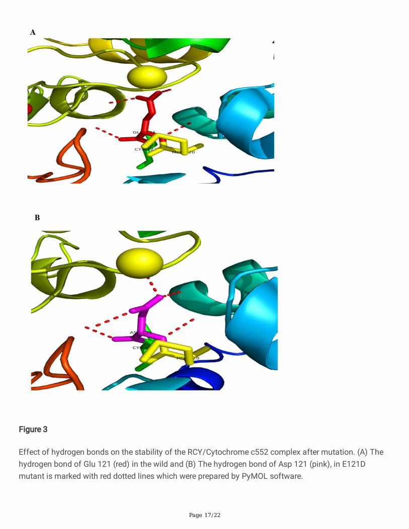
The results showed that glutamate has four hydrogen bonds in the side chain with the surroundingmolecules in wild-type. However, there is an extra hydrogen bond that binds the aspartate to Zn1388 ofchain B in E121D mutation (Fig.3A-B).
Analysis of the results of the distance between Zn 1388 of chain B and Cu1156 of RCY to glutamate 121and aspartate 121 (after mutation) chain A using PyMOL software.
In E121D mutation, an increase occurred in the distance between Zn1388 of chain B and aspartate 121 ofchain A compared to glutamate 121 (2.2 angstroms in E121 and 4.4 in D121). Also, a decrease occurredin the distance between aspartate 121 of chain A and Cu1156 of RCY at RCY/Cytochrome c552 complex(16.9 angstroms in E121 and 15.3 in D121) (Fig. 4A-B).
The results of MD simulation analysis and effects mutation on stability and protein secondary structure
MD simulation was performed to study the con�guration, and stability in the wild-type protein and thechanges of these parameters in the mutant protein, for both types of proteins. In addition, MD simulationis considered a useful method to support the experimental results in protein mutations through structuralspeci�cations at the atomic level [29]. RMSD, RMSF, SASA, H-bonds, Rg, and DSSP were analyzedthroughout the simulation time (Table 2, 3).
To �nd out the effect of the mutation on conformational changes and the stability of the proteinstructure, the RMSD of Cα atoms was calculated for both the wild-type and mutant compared to theoriginal structure [19]. The RMSD plot of the wild and mutant structures indicated the convergencepattern during 100 ns simulations (Fig. 5A). Convergence obtained for wild and mutant structurescon�rmed that the mutant protein has less sustainability than the wild-type.

Page 6/22
The RMSF of each residue was measured to assign the mutation effect on the residues' dynamicbehavior in wild and mutant proteins [19]. Overall, the mutant protein had more �exibility (RMSF) than thewild-type (+0.0002 increase) [30]. The RMSF plan is plotted for wild and mutant structures, as shown inFigure 5B.
Intermolecular hydrogen bonding is the electrostatic force between two polar bonds. In this bonding,hydrogen is bonded covalently to the highly electronegative atoms like nitrogen and oxygen. H bond wascalculated for wild and mutant types. The average number of H bonds for the mutated structure was125.78 ± 4.52 and -2 number decreased compared to wild protein (Table 2). The difference in the mutantH bonds con�rmed the �exibility of the mutant structure (Fig. 5C).
Rg determines the compression of the protein. As the radius of gyration is higher, the protein tends to bemore unstable, unfolding, and low compact [31]. The values of Rg for mutated variants were 1.62 ± 0.001nm, with a slight change (+0.01 increase) in protein folding and its compactness (Table 2) (Fig. 5D).
SASA indicates the level of protein access to the solvent [32]. The change in SASA measured theaccessibility of the protein to the solvent in the wild and mutant structure during simulations (Fig. 5E).The average of SASA for the mutated structure was 103.10 ± 0.59 nm2 and +1.23 increased compared towild protein (Table 2).
Figure 6 shows the alterations in the secondary structure of the protein during the simulations. The DSSPscheme has been used to calculate the secondary structure of wild-type and mutant protein. The analysisof wild and mutant structures shows the number of amino acids involved in different structures and theincrease or decrease in the number of amino acids in the structures. The most changes are related to theturn structure with an increase of 2.27 amino acids, and the α helix structure with a decrease of 1.27amino acids. The bar graph of the secondary structure for wild-type and E121D mutant systems is shownin Figure 6A. The amount of Structure, coil, Turn, and beta bridge in the mutant protein has increased but,beta-sheet, alpha-helix, have diminished. The highest value is shown in the secondary structure of the twosystems for coil and alpha-helix. And the largest change is related to the turn structure (1.24% increase),which is shown in Table 3.
Results of φ and ψ angle analysis of amino acids in wild and mutant proteins by Ramachandran method
The results were reported as diagrams of changes in angles according to time, to investigate the state ofthe protein at the mutation point. According to the angles φ and ψ before and after mutation using theRamachandran method, it was found that the amino acid studied did not change signi�cantly in terms ofangles φ and ψ (Fig. 7, Table 4).
The Wild and mutant proteins Ramachandran scheme produced by PROCHECK
To compare the Ramachandran results of simulated wild and mutant protein (E121D), the pdb �les wereuploaded and the results are shown in Figure 8 and Table 5. Before the mutation, 95.5% of the amino

Page 7/22
acids are present in most favored regions, which after the mutation, its amount has decreased to 84%.There were no amino acids in the allowed and disallowed regions before the mutation, but after theE121D mutation, two of the amino acids (alanine 9, histidine 54) were in the allowed regions.
DiscussionFollowing previous studies, E121D mutation at the �rst active site and the electron entry point from theRCY to Cytochrome c552 were conducted on Af bacterium. Molecular dynamics simulation studies onwild-type and mutant identi�ed the in�uence of mutations at the active site. The trajectory analysis wasused to calculate protein changes and showed the changes in protein structure associated with increased�exibility at the mutation point (+ 0.0017 increase).
DSSP is an algorithm that deals with information on the secondary structure of proteins [33]. DSSPanalysis aims to measure the content of the secondary structure of a protein as a function of time. Theanalysis of the wild and mutated structures shows the number of amino acids involved in differentstructures, as well as increasing or decreasing the number of amino acids in the structure. In the DSSPscheme, with the conversion of glutamate to aspartate, structure, coil, β Bridge, and Turn increased by0.62%, 0.08%, 0.09% and 1.24%, respectively, and β Sheet, α helix, 5-helix, and 3-helix decreased by0.006%, 0.69%, 0.48%, and 0.11%, respectively. A decrease of 0.69% in the alpha-helix in the DSSP plan isrelated to the fact that aspartate tends to disrupt a helix because their side chains contain hydrogen-bonddonors or acceptors in proximity to the main chain, where they compete for main-chain NH and COgroups [34]. The number of amino acids which participate in protein structure eventually increased by0.62% (Table 3).
As mentioned, the Af bacterium is considered as one of the main bacteria involved in the metalbioleaching process, and bioleaching is the extraction of metals from their minerals through using thesebacteria [11]. RCY and Cytochrome c552 are considered as consecutive proteins in the electron transportchain Af bacterium. In this way, the electron is transmitted from RCY to Cytochrome c552 [9].
Patra et al. indicated that His143 of RCY has a stable interaction with Glu121 of periplasmic Cytochromec552. In addition, glutamate 121 is associated with iron and causes electron transfer to heme A [9].
Malarte et al. evaluated glutamate 121, which played a key role in the formation of the RCY/Cytochromec552 complex. They found that the complex was destroyed by the E121A mutation and the conversion ofan electrically charged glutamate to a hydrophobic alanine [35]. The formation of the complexsigni�cantly modi�ed the pK value of the exposed histidine ligand to the Cu ion and regulated themidpoint potential of the redox in the site of Cu [36]. Abergel et al. reported the role of the divalent Zncation, which prevents RCY/Cytochrome c552 interaction by making E121 unavailable [8]. The results ofMD analysis demonstrate the effect of E121D mutation on the unfolding of the cytochrome c552 proteinand the increased distance between Zn 1388 and aspartate 121 (4.4 angstroms instead of 2.2 angstromsfor glutamate). Moreover, there is a decrease in the distance between Cu 1156 in RCY and the

Page 8/22
Cytochrome c552 aspartate 121, which creates a more stable RCY/Cytochrome c552 complex (15.3angstroms instead of 16.9 angstroms for glutamate) (Fig. 4A-B). The kd value of glutamate for zinc ionsand copper ions is 10− 5.5 and 10− 8 at pH 4.5, respectively (the lower kd value leads to the higher bindinga�nity of the ligand to its target) [8]. The glutamate interacts with zinc inhibiting the interaction withRusticyanin. Further, with E121D mutation, the role of zinc inhibiting on the Asp 121 Cytochrome c552
decreases and one could expect shorter distances with the Rustycianin copper. Therefore, its tendency tocopper 1156 of RCY increases, and the RCY/Cytochrome c552 complex becomes more stable after themutation.
In this study, we proposed the novel E121D mutation in which glutamate has four hydrogen bonds in theside chain with the surrounding molecules. However, in aspartate, there is an extra hydrogen bond thatbinds this amino acid to Zn1388 in the B chain and creates a stronger bond between the two chains(Fig. 3A-B).
Abergel et al. reported that histidine is deprotonated due to the 3.2 Å distance between Nε H143 and OγE12, and their formation of hydrogen bonds between. The RCY redox potential decreases and,consequently, the transfer of electrons from the RCY to Cytochrome c552 is more e�cient [8]. Thismutation converts an acidic residue into a more acidic residue Therefore, His143 becomes moredeprotonated, reduces the redox potential at the RCY midpoint, and improves electron transfer toCytochrome c552 protein at RCY/Cytochrome c552 complex.
Also, the results of RMSF analysis show more �exibility in the mutation point compared to the wild type.The E121D mutation reduces stability at the active site. Finally, instability of the active site leads toincreased ligand �exibility (ΔS) and ΔG decrease and increase in the value of E0 which is proved by twoformulas Gibbs free energy (ΔG = ΔH – TΔS) and Nernst (E0 = −ΔG/nF) [37]. Finally, by the increase inredox potential, electron transfer from RCY to Cytochrome c552 at RCY/Cytochrome c552 complex isaccelerated. In this groundbreaking study, we found that in all respiratory chain proteins, electronsprobably enter through the glutamate in the �rst active site and exit through histidine at the second activesite of each respiratory chain protein. Therefore, by converting glutamate to aspartate at the electronentry point and converting histidine to arginine at the electron exit point, and destabilizing the active siteof each protein in the respiratory chain, the electron transfer rate in the chain, followed by the bioleachingprocess, can be improved.
ConclusionBased on the results, the mutated protein (E121D) is unfolded more than wild protein. The analysis of thehydrogen bond indicates that the mutant protein is more �exible. However, in aspartate, there is an extrahydrogen bond that binds this amino acid to Zn1388 in the B chain and creates a stronger bond betweenthe two chains. By converting glutamate 121 to aspartate, a decrease occurs in the role of zinc inhibitingon the Asp 121 Cytochrome c552. Therefore, the aspartate gets closer to Cu 1156 of the RCY and results
Page 9/22
in higher stability of the RCY/Cytochrome c552 complex. On the other hand, an acidic residue (Glu121)becomes a more acidic residue (Asp121), and His143 is more deprotonated inducing a reduction of theredox potential at the rusticyanin midpoint improving the electron transfer to Cytochrome c552 protein.Also, by the increase in �exibility and decreasing ΔG in the active site, the redox potential at the mutationpoint increases, and the electron transfer to Cytochrome c552 improves. For future studies, we proposeextensive research on electron transfer improvement by selecting the best mutations obtained bybioinformatics methods at all active sites (electron entry and exit points) in all respiratory chain proteins.In this way, The amino acids in the �rst active site of the proteins, namely glutamate, are converted toaspartate with more acidic properties. And the amino acid histidine converted to the more alkaline aminoacid arginine in the second active site position. As a result, the active site becomes more unstable and thespeed of electron transfer in the chain increases. Finally, biotechnological methods can clone bacteriathat have the above mutations to improve the bioleaching process.
DeclarationsFunding
No funding was received for conducting this study.
Acknowledgments
We also thank Ms. Saba Miri for her valuable technical advice.
Author contribution
The protocol designed, conceptualized by F.F., Manuscript preparation, and analysis, and supervision isdone by M.Sh. General consultation of the project and editing of the manuscript was done by M.D.Sh.Part of the text editing was done by S.F. The manuscript was reviewed and approved by all authors.
Data Availability Statement
The analyses during the current study are available from the corresponding author on reasonable request.
Con�ict of Interest
The authors declare that they have no con�icts of interest to this work.
References1. Fatemi F, Miri S, Jahani, S (2017) Effect of metal sul�de pulp density on gene expression of electron
transporters in Acidithiobacillus sp. FJ2. Arch Microbiol, 199(4): 521-530. DOI: 10.1007/s00203-016-1318-1

Page 10/22
2. Rawlings D. E (2005) Characteristics and adaptability of iron-and sulfur-oxidizing microorganismsused for the recovery of metals from minerals and their concentrates. MICROB CELL FACT. 4, 13.https://doi.org/10.1186/1475-2859-4-13
3. Ingledew W. J (1982) Thiobacillus ferrooxidans the bioenergetics of an acidophilic chemolithotroph.BBA-BIOENERGETICS. 683, 89-117. https://doi.org/10.1016/0304-4173(82)90007-6
4. Holmes D. S, Bonnefoy V (2007) Genetic and bioinformatic insights into iron and sulfur oxidationmechanisms of bioleaching organisms. Biomining, Springe. 281-307. https://doi.org/10.1007/978-3-540-34911-2_14
5. Yarzabal A, Appia-Ayme C, Ratouchniak J, & Bonnefoy V (2004) Regulation of the expression of theAcidithiobacillus ferrooxidans rus operon encoding two cytochromes c, a cytochrome oxidase andrusticyanin. Microbiology +, 150(7), 2113-2123. DOI:10.1099/mic.0.26966-0
�. Bruscella P, Appia-Ayme C, Levican G, Ratouchniak J, Jedlicki E, Holmes D. S, et al (2007) Differentialexpression of two bc1 complexes in the strict acidophilic chemolithoautotrophic bacteriumAcidithiobacillus ferrooxidans suggests a model for their respective roles in iron or sulfur oxidation.Microbiology +, 153(1), 102-110. DOI 10.1099/mic.0.2006/000067-0
7. Lyons J. A, Aragão D, Slattery O, Pisliakov A. V, Soulimane, T, Caffrey, M (2012) Structural insightsinto electron transfer in caa 3-type cytochrome oxidase. Nature, 487(7408), 514.https://doi.org/10.1038/nature11182
�. Abergel C, Nitschke W, Malarte G, Bruschi M, Claverie, J.-M, Giudici-Orticoni, M.-T (2003) The structureof Acidithiobacillus ferrooxidans c4-cytochrome: a model for complex-induced electron transfertuning. Structure, 11(5), 547-555. DOI:10.1016/S0969-2126(03)00072-8
9. Patra M. C, Pradhan S K, Rath S N, Maharana J (2013) Structural Analysis of Respirasomes inElectron Transfer Pathway of Acidithiobacillus ferrooxidans: A Computer-Aided Molecular DesigningStudy. ISRN Biophysics, 2013. http://dx.doi.org/10.1155/2013/295718
10. Jafarpour R, Fatemi F, Eidi A, & Mehrnejad F (2020) Effect of the Met148Leu Mutation on theStructure and Dynamics of the Rusticyanin Protein from Acidithiobacillus sp. FJ2. Biomol Struct Dyn(just-accepted), 1-19. https://doi.org/10.1080/07391102.2020.1775119
11. Jahani S, Fatemi F, Firoz-e-zare M, Zolfaghari, M (2015) Isolation and characterization ofAcidithiobacillus ferrooxidans strain FJS from Ramsar, Iran. Electronic J Biol, 11(4), 138-146.
12. Lengauer T, & Rarey, M (1996) Computational methods for biomolecular docking. Curr. Opin. Struct.Biol, 6(3), 402-406. https://doi.org/10.1016/S0959-440X(96)80061-3
13. Hernández-Santoyo A, Tenorio-Barajas A. Y, Altuzar V, Vivanco-Cid H, Mendoza-Barrera C (2013)Protein-protein and protein-ligand docking. Protein engineering-technology and application.http://dx.doi.org/10.5772/56376
14. Abdel-Hamid M. K, McCluskey A (2014) In Silico docking, molecular dynamics and binding energyinsights into the bolinaquinone-clathrin terminal domain binding site. Molecules, 19(5), 6609-6622.https://doi.org/10.3390/molecules19056609

Page 11/22
15. Dehghan-Shasaltaneh M, Lanjanian H, Riazi G. H, & Masoudi-Nejad, A (2018) The importance of α-CT and Salt bridges in the Formation of Insulin and its Receptor Complex by ComputationalSimulation. Iran J Pharm Res: IJPR, 17(1), 63. available online at http://www.ijpr.ir
1�. Ling B, Zhang R, Wang Z, Liu Y, & Liu C (2010) Study on the interactions of SMAC mimetics withXIAP-BIR3 domain by docking and molecular dynamics simulations. J THEOR COMPUT CHEM,9(04), 797-812. DOI: 10.1142/S0219633610005980
17. DeLano W. L (2002) Pymol: An open-source molecular graphics tool. CCP4 Newsletter on proteincrystallography, 40(1), 82-92.
1�. Ghasemi F, Zomorodipour A, Karkhane A. A, Khorramizadeh M. R (2016) In silico designing of hyper-glycosylated analogs for the human coagulation factor IX. Journal of J Mol Graph Model, 68, 39-47.DOI: 10.1016/j.jmgm.2016.05.011
19. van Gunsteren W. F, Billeter S. R, Eising A. A, Hünenberger P. H, Krüger P, Mark A. E, et al. (1996)Biomolecular simulation: the {GROMOS96} manual and user guide.
20. Imani S, Cheng J, Shasaltaneh M. D, Wei C, Yang L, Fu S, et al. (2017) Genetic identi�cation andmolecular modeling characterization reveal a novel PROM1 mutation in Stargardt4-like MacularDystrophy. ONCOTARGET. doi: 10.18632/oncotarget.22343. eCollection 2018 Jan 2
21. Castelle C, Guiral M, Malarte G, Ledgham F, Leroy G, Brugna M, et al. (2008) A new iron-oxidizing/O2-reducing supercomplex spanning both inner and outer membranes, isolated from the extremeacidophile Acidithiobacillus ferrooxidans. J BIOL CHEM, 283(38), 25803-25811.DOI:10.1074/jbc.M802496200
22. Srikumar P, & Rohini K (2013) Exploring the structural insights on human laforin mutation K87A inLafora disease—a molecular dynamics study. Appl Biochem Biotechnol, 171(4), 874-882.
23. Berendsen H, Grigera J, & Straatsma T (1987) The missing term in effective pair potentials. Journalof Physical Chemistry, 91(24), 6269-6271.
24. Linder T, Wang S, Zangerl-Plessl E.-M, Nichols C. G, & Stary-Weinzinger A (2015) Molecular dynamicssimulations of KirBac1. 1 mutants reveal global gating changes of Kir channels. Journal of chemicalinformation and modeling, 55(4), 814-822. DOI: 10.1021/acs.jcim.5b00010
25. Berendsen H. J, Postma J. v, van Gunsteren W. F, DiNola A, & Haak J (1984) Molecular dynamics withcoupling to an external bath. J PHYS CHEM-US, 81(8), 3684-3690. DOI: 10.1007/s12010-013-0393-x
2�. Humphrey W, Dalke A, Schulten K (1996) VMD: visual molecular dynamics. J. Mol. Graph, 14(1), 33-38. https://doi.org/10.1016/0263-7855(96)00018-5
27. Hess B, Kutzner C, Van Der Spoel D, & Lindahl E (2008) GROMACS 4: algorithms for highly e�cient,load-balanced, and scalable molecular simulation. J CHEM THEORY COMPUT, 4(3), 435-447. DOI:10.1021/ct700301q
2�. Dodson G. G, Lane D. P, Verma C. S (2008) Molecular simulations of protein dynamics: new windowson mechanisms in biology. EMBO reports, 9(2), 144-150. DOI: 10.1021/ct700301q
29. Zhao Y, Zeng C, & Massiah M. A (2015) Molecular dynamics simulation reveals insights into themechanism of unfolding by the A130T/V mutations within the MID1 zinc-binding Bbox1 domain.

Page 12/22
PLoS One, 10(4).
30. Lobanov M. Y, Bogatyreva N. Galzitskaya O (2008) Radius of gyration as an indicator of proteinstructure compactness. Mol Biol+, 42(4), 623-628. DOI: 10.1134/S0026893308040195
31. Satpati S, Manohar K, Acharya N, Dixit A (2017) Comparative molecular dynamics studies ofheterozygous open reading frames of DNA polymerase eta (η) in pathogenic yeast Candida albicans.SCI REP-UK, 7, 41087. https://doi.org/10.1038/srep41087
32. Kabsch W, Sander C (1983) Dictionary of protein secondary structure: pattern recognition ofhydrogen‐bonded and geometrical features. BIOPOLYMERS, 22(12), 2577-2637.https://doi.org/10.1002/bip.360221211
33. Berg J. M, Tymoczko J. L, & Stryer L (2013) Das Immunsystem. In Stryer Biochemie, pp. 993-1024.
34. Malarte G, Leroy G, Lojou E, Abergel C, Bruschi M, Giudici-Orticoni, M. T. (2005). Insight into molecularstability and physiological properties of the diheme cytochrome CYC41 from the acidophilicbacterium Acidithiobacillus ferrooxidans. Biochemistry, 44(17), 6471-6481. DOI: 10.1021/bi048425b
35. Marie-Thérese Giudici-Orticoni F, Guerlesquin M. B, & Nitschke W (1999) Interaction-induced RedoxSwitch in the Electron Transfer Complex Rusticyanin-Cytochrome c4. J BIOL CHEM DOI:10.1074/jbc.274.43.30365
TablesTable. 1 Protein stability upon mutation at 27℃ and pH = 2, using two servers MUpro and I-Mutant 2.0.
MUpro I-Mutant 2.0
Stability DDG ( Kcal/mol) Stability DDG ( Kcal/mol)
Decrease -1.25 Decrease -0.18
DDG<0: Decrease of Stability, DDG>0: Increase of Stability.
Table. 2 The average of RMSD, RMSF, SASA, H-Bonds, and Rg values of Wild and E121D Mutant.
Parameters Wild E121D Mutant
RMSD 0.0241±0.001 0.0243±0.001
RMSF 0.0202±0.002 0.0204±0.002
Rg 1.61±0.001 1.62±0.001
SASA 101.87±0.52 103.10±0.59
NH bonds 127.54±4.03 125.78±4.52

Page 13/22
The values of RMSD, RMSF and Rg are given in nm, values of SASA given in nm2, and values of NHbonds given in numbers.
Table. 3 the secondary structure average for Wild and E121D Mutant during the 100 ns MD simulation.
SecondaryStructure
Wild type Wild type(%)
E121DMutant
E121D Mutant(%)
Differenceamount
Structure 109.52 ±3.37
59.52 110.66 ±2.71
60.14 1.14 (0.62%) ↑
Coil 50.52 ±1.14
27.45 50.68 ±1.02
27.54 0.16(0.08%) ↑
B-Sheet 0.10 ± 0.69 0.054 0.09 ± 0.64 0.048 0.01(0.006%) ↓
B-Bridge 4.33 ±1.045
2.35 4.5 ± 1.26 2.44 0.17(0.09%) ↑
Turn 22.59 ±2.66
12.27 24.86 ±2.22
13.51 2.27(1.24%) ↑
A-Helix 82.49 ±2.41
44.83 81.22 ±1.21
44.14 1.27(0.69%) ↓
5-Helix 5.85 ± 2.06 3.18 4.98 ± 1.53 2.70 0.87(0.48%) ↓
3-Helix 0.79 ± 1.35 0.43 0.59 ± 1.21 0.32 0.20(0.11%) ↓
Table. 4 Average, minimum and maximum values of dihedral angles φ and ψ during simulation in Wildand E121D protein.
Wild E121D
GLU121 Average
min max ASP121 Average
min max
φ
φ
-55.31±7.80 -80.41 -25.52 -55.69±8.35 -84.14 -26.18
ψ ψ
-50.42±9.84 -84.71 -23.38 -59.34±11.22 -89.07 -26.23

Page 14/22
Table. 5 Comparison of statistical parameters of Ramachandran plot for Wild and E121D protein.
Status of the residues in theRamachandran plot
amino acidsNumber
(Wild)
amino acidsNumber
E121D
aminoacids
Percentage
Wild
aminoacids
Percentage
E121D
Residues in most favored regions[A,B,L]
149 131 95.5 84
Residues in additional allowedregions [a,b,l,p]
7 23 4.5 14.7
Residues in generously allowedregions [~a,~b,~l,~p]
0 2 0 1.3
Residues in disallowed regions 0 0 0 0
Number of non-glycine and non-proline residues
156 156 100 100
Number of end-residues (excl. Glyand Pro)
2 2
Number of glycine residues (shownas triangles)
16 16
Number of proline residues 10 10
Total number of residues 184 184
Figures

Page 15/22
Figure 1
There are six zinc ions in Cytochrome c552. (A) Three zinc ions are associated with histidine in eachchain. In Chain A, H 39 to Zn (1187), H 97 to Zn (1188), and H 152 to Zn (1189) and shown in Purple. InChain B, H 239 to Zn (1388), H 297 to Zn (1390), and H 352 to Zn (1389) and shown in dark blue. Zn1388, which binds to histidine 239 in chain B, is involved in connecting to both Cytochrome c552 chainsand shown in yellow and H2O 2068 in blue, and SO4 1387 in green.

Page 16/22
Figure 2
Structure of the RCY/Cytochrome c552 complex (chain A of Cytochrome c552: Light and dark blue, chainB of Cytochrome c552: green and yellow: RCY: red and brown). (A) Wild: Glu121 of chain A, red and His143 of RCY, dark blue, Zn 1388 of chain B, yellow and Cu 1156 of RCY, orange. (B) E121D mutant: Asp121of chain A, pink and His 143 of RCY, dark blue, Zn 1388 of chain B, yellow and Cu 1156 of RCY, orange.
Page 17/22
Figure 3
Effect of hydrogen bonds on the stability of the RCY/Cytochrome c552 complex after mutation. (A) Thehydrogen bond of Glu 121 (red) in the wild and (B) The hydrogen bond of Asp 121 (pink), in E121Dmutant is marked with red dotted lines which were prepared by PyMOL software.

Page 18/22
Figure 4
The effect of mutation on reducing the distance and stability of RCY/Cytochrome c552 complex (A) Thedistances of the glutamate 121 in chain A to Zn 1388 in chain B and the distances of the glutamate 121in chain A to Cu1156 of RCY. (B) The distances of the aspartate 121 in chain A to Zn 1388 in chain B andthe distances of the aspartate 121 in chain A to Cu1156 of RCY are marked with blue dotted lines whichwere prepared by PyMOL software.

Page 19/22
Figure 5
Functional effects of the E121D mutation on Cytochrome c552 protein. (A) RMSD plots of backboneatoms of the wild-type and E121D complex systems for 100 ns of simulation, where time step is plottedon X-axis and RMSD (nm) is plotted on Y-axis. (B) Cα-RMSF plots of the wild-type and E121D complexsystem for 100 ns of simulation, where residue number is plotted on the X-axis while RMSF (nm) isplotted on the Y-axis. (C)The number of H bonds is plotted where there is a time step on X-axis while NH

Page 20/22
bonds are plotted on Y-axis. (D) The Radius of gyration (Rg) is plotted where there is a time step on X-axiswhile Rg is plotted on Y-axis. (E) SASA is plotted where there is a time step on X-axis while SASA (nm) isplotted on Y-axis. The mutation diagrams (E121D) have been shown in red and the wild-type in blue.
Figure 6
Secondary structure analysis of wild-type in comparison with the mutant form (E121D). (A & B) TheDSSP plots (100 ns) of wild-type and E121D mutant systems at 300 K. (C) The bar graph of the residue

Page 21/22
number of secondary structure for wild-type and the mutant.
Figure 7
Dihedral Angles φ and ψ of Amino Acid Glu121, Asp121 in Wild and Mutant E121D Protein.
Figure 8
Ramachandran scheme to investigate the status of amino acids in Wild and Mutant E121D proteinsgenerated by PROCHECK. Red indicates the most favored regions, yellow indicates allowed regions, beigeindicates tolerated regions and white indicates unfavorable regions.
Supplementary Files

Page 22/22
This is a list of supplementary �les associated with this preprint. Click to download.
Graphicalabstract.docx