improved poly -l-lactic acid mini-plate and screws …...screws were used in cranio maxillofacial...
TRANSCRIPT

Improved Poly -L-Lactic Acid Mini-Plate and Screws
f.or Cranio-Maxillofacial Surgery -
Experimental and clinical studies
Yuji Nnr<eNISHI, M.D. , Tatsuo NareJtnae, M.D. , Yohko YosnIuuRa, M'D. '
Kaoru TereuI, M'D' and Jun Uxet' M'D'
日本形成外科学会会誌
舅曽2 0曜案 舅害1 21計 (pp. 7 2 3へ ′7 3 0)男 llll
2000年 12月 発行

日形会誌 (J」pn.PRS),20:723~ 730,2000
<Original>
―-723-―
Improved Poly-L-Lactic Acid Mini-Plate and Screwsfor CraniO―NIaxlllofacial Surgery:
Experilnental and clinical studies
Yuji NAKANISHI,M.D.*,Tatsuo NAKAJIMA,M.D.**,Yohko YosHIMURA,M.D.***,
Kaoru TAKAMI,h/1.D.***and Jun UKAI,M.D.***
*Dのα〃πιπ′グ Pι鑢″ ακ″Rιθθ2s′協ι″υι Sπ ttι鶴 おι κιあ助紗′″ι κθあ ιrttυθκグ机 Is‐ι,5ヱδ`θ
ζ′
(ffθα″:4ssなタタ笈υ41ζ多″グNttαηたカグ)**Dのαγttι″ ぼ Pιαsιたαπ″Rιθθπs′ /7rθ″υι
`賜
箸ιり'力
θθノび フИιググιれι,κιあ しrηグυι冬グク,動ゎθ,Iこリー8582
(Jθα″:P%の4動なzθ A物力″グタ2Zα )
***五の αγ′πι″ げ P:偲″ απ′ Rιιθηs′協θ″υι Sπttιり,■力θθノグ ′lイι″′ε物の Fググ″ ル α′ル しr″υθバグ机 Tρノθ滋らイη ″ 92
(ルαグ:Pグ 拘 カカθ ttS力物 %z)
AbstractPoly-L-lactic acid has become a popular bioabsorbable material in the field of plastic surgery. Though
PLLA plates have previously been reported, they generally had a high molecular weight and were difficult to
absorb. Therefore, we developed a system with a low molecular weight that is both easy to absorb and strong.
Improved mini-plates and screws were used to treat experimental skull fractures in rabbits. Fracture healing
and plate and screw absorption were investigated using gross inspection and microscopy. Male Japanese white
rabbits (n:15) weighing apbrox 3 kg were used. After the outer table of the skull was elevated, it was
repositioned and fixed with a miniplate and screws. At 3, 6, 9, 12, and 24 months after fixation, the plate was
exposed. Tissue specimens were removed and examined using light and electron microscopy. The plates and
screws were used in cranio maxillofacial surgery from January 1995, these mini-plates and screws were used
in 45 cases. A11 patients had an uneventful postoperative course. There were no compiications. Polylactic acid
mini plates may eventually replace metallic hardware in these applications.
Key Words ! biodegradable material, fracture fixation, fracture healing, Poly L lactic acid mini-plate and
screw, facial fracture
INTRODUCTION
Bioabsorbable materials were introduced in the
1960's when sutures made of polyglycolic acid came
into use. Since then, bioabsorbable materials in a
variety of shapes have been developed and applied
clinically, including mesh used to reinforce the
chest and abdominal walls and pins used to buttress
the ribs. The strength of the material was directlyproportional, while the absorption time was
inversely proportional, to the molecular weight. Weprepared a strong readily absorbable mini plate
with a low molecular weight by extrudingpolylactic acid. The effects of this mini-plate on
fracture healing were examined in animals. The
device was then applied clinically in the field of
cranio maxillofacial surgery.
MATERIALS AND METHODS
Poly L lactic acid (PLLA) rods were manu-
factured with an extruder using a PLLA polymer(Mw:3.0-5.0 x 10s) . The rods were initiallydrawn using several deformation ratios and tem-
peratures. High strength PLLA rods were shaped
into mini-plates and screws (Mw:2.0x 10⁵) using
a deformation ratio of 2.5. The length of the plate
was 20 mm, the height was 2 mm, and the width was
5 mm. The screws had a thread diameter of 2.7 mm

Improved Poly I- I-actic Acid Mini Plate and Scrervs for Cranio Maxillofacial Surgery
cut and repositioned outer table
,rr rrlrrr.
\\\\\r::--fixed with a PLLA plate and screws
Fig. 1 Schematic showing the repositioned outer tablefixed with a poly L lactic acid plate and screws.
Fig. 3 Photograph at 6 months after fixation showingthe opaque plate r,l.ith evidence of degradation. Thereare few findings suggestive of absorption.
and a height of 7 mm. Both plates and screws were
made from the same clinical materials.Male Japanese white rabbits (n:15) weighing
apbrox 3 kg were used. After the outer table of theskull was incised and elevated, it was repositioned
and fixed with a PLLA mini-plate and screws(Fis. 1). At 3 (n-3),6 (n:3),9 (n:3), 12 (n:3), and 24 (n:3) months after fixation, the plate
was exposed and the surrounding tissue was in-
spected macroscopically. Tissue specimens were
then removed, cut vertically into sections, and
stained with hematoxylin-eosin (light micros-copy) or uranyl acetate and lead (electron micros-
copy).
RESULTS
Macroscopic examinationAt 3 months, the PLLA mini plates were densely
adherent to the bone and good union of the frac-tures was noted. The plate surface was more
Fig. 2 Photograph at 3 months after fixation showingthe opaque plate that exhibited no change in hardness.
Favorable osteosynthesis is noted at the fracture site.The plate is firmly adherent to the bone.
Fig. 4 Photograph at 9 months after fixation showingprogressive degradation and the begining of absorp-tion.
opaque than before operation. There were no
changes in shape (Fig. 2). At 6 months, the surfaceof the plate was opaque and degradation was
apparent, but there were few findings suggestive ofabsorption (Fig. 3). At 9 months, the changes inthe plate shape suggested that degradation was
progressing and that absorption had begun (Fig.
4). At 12 months, the plate was transparent and
gelatinous. The plates and screws broke easily(Fig. 5) . At 24 months, there was almost completedegradation, although a small amount of materialremained and absorption was not judged to be
complete (FiS. 6).
Microscopic examinationHistologic examination at 3 months showed good
union at the fracture site without inflammatory cellinfiltration (Fig. 7). A small amount of degrada-
tion and absorption were apparent at 6 months.

日形会誌 (2000)
Fig.5 Photographing the transparent
at 12 months after fixation show-gelatinous plate.
Fig. 7 Photomicrograph at 3 months after fixationshowing a good union at the fracture site withoutinflammatory cel1 infiltration (Hematoxylin-eosinstain, x 8).
The plates and screws were in direct contact withthe bone without evidence of inflammation (Fig.
8). At 9 months, fibrous tissue was seen invadingthe plates. Degradation and absorption were evi-
dent at the upper and lower ends of the screws.
Bone filled the spaces left by the absorbed material(Fig. 9) . At 12 months, the plates were not recog-
nizable on histologic examination. The degradationand absorption of the screws was more advanced
and a greater amount of bone formation was noted(Fig. 10) . At 24 months, the plates had undergone
complete degradation. There was considerableprogress in the degradation of the screws. At highmagnifications, foam cells were seen occupying the
spaces left by the dissolving screws, in preparation
for phagocytosis (Fig. 11a-c).Scanning electron microscopy at 3 months
revealed the formation of small pores within the
―-725-―
Fig. 6 Photograph at 24 months after fixation showingalmost complete degradation. Absorption is not com-plete.
Fig. 8 Photomicrograph at 6 months after fixationshowing a small amount of degradation and absorp-tion (Hematoxylin-eosin, x8).The plate is in direct contact with the bone, withoutevidence of inflammation.
plate due to the hydrolysis (Fig. 12) . At 12 months,
electron microscopy revealed degradation and
small residual fragments of the PLLA plates (Fig.
13).
CLINICAL APPLICATION
Prior to clinical application, the optimum shape
and specifications of the PLLA plates were inves-
tigated.At first, a simple, flat plate was designed. Use of
the screws required tapping with a drill. To encour-
陪
躙
陽
鳳

-726-
Fig. 9 Photomicrograph at 9 months after fixationshowing fibrous tissue invading rifts in the plate(Hematoxylin eosin, x8) . Degradation and absorp-tion are noted at both ends of the screw. Bone fills thespaces left by the absorbed material.
Improved I'oly L Lactic Acid Mini Plate and Screws for Cranio Maxillofacial Surgery
Fig. 10 Photomicrograph at 12 months after fixationshowing no evidence of the plate (Hematoxy lineosin, X 8). Degradation and absorption were noted.
ヽ耐
ム”
抒
(a)
(b) (c)
Fig. 11 Photomicrograph at 24 months after fixation(Hematoxylin eosin,). (a) The plate has undergonecomplete degradation. There is considerable degrada-tion of the screw ( x 8). (b) ( x 50). (c) The spaces
in the dissolved screw are occupied by foam cells ( x200) .
age widespread use, a screw with standard threads
was subsequently constructed. Based on the
requirements for bending strength, the extension
rate (deformation rate) for both the plates and
screws was set at 2 .5 times. Since it was difficult tobend the original plate laterally, the central portion
was made thinner. Further refinements then were
made to allow the plates to conform to the three

日形会誌 (2000) ―-727-―
Fig. 12 Scanning electron micrograph at 3 monthsafter fixation showing the formation of small pores
within the plate due to hydrolysis.
Fig. 13 Scanning electron micrograph at 12 monthsafter fixation showing degradation and small frag-ments of the plate.
dimensional structure of the face (Fig 14). These
plates and screws were used clinically in 45 patients
from January 1995. The longest follow up was 5
years. All patients gave written, informed consent
and this study was performed in accordance withthe Helsinki Declaration revised in 1989. There
were no major complications and the postoperative
recovery was uneventful in all patients.
CASE REPORTS
Case 1 i A 21-year-old woman who sustained a
Le-Fort I fracture in a traffic accident underwent
open reduction. The maxillary buttress was fixedwith four PLLA mini-plates and screws. Three
dimensional computed tomography showed good
results and the results at 4 years were satisfactory
' ,., r-, ' ,r1r, t n'r', i'Fig. 14 Photographs showing the innovative changes
in the shape of the Poly-L lactic acid mini-plate(above). The original plate was difficult to bend toconform to the three-dimensional structure of theface. Then the central portion was made thinner toallow for bending (middle). The final design of plate
and screw. The plate was made to allow it to conformto the three-dimensional facial skeleton (below) .
(Fis. 15).
Case 2 I A 28-year-old man underwent treatmentfor a post-traumatic deformity of the zygoma.
Osteotomy of the zygoma and augmentation of the
orbital floor with apatite ceramics was performed.
Fixation of the osteotomy site was accomplished
with one PLLA mini-plate along the lateral and
one mini-plate along the inferior orbital rim. At 3
years, the results were satisfactory (Fis. 16).
DISCUSSION
After bioabsorbable fibers were introduced for

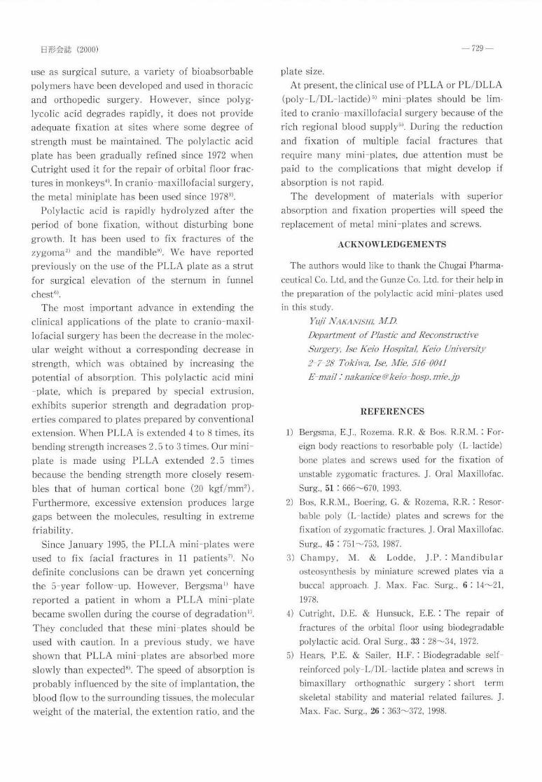
日形会誌 (2000)
use as surgical suture, a variety of bioabsorbable
polymers have been developed and used in thoracic
and orthopedic surgery However, since polyg―
lycolic acid degrades rapidly, it does not provide
adequate fixation at sites 、vhere some degree of
strength must be maintained. The polylactic acid
plate has been gradually refined since 1972 、vhen
Cutright used it for the repair of orbital floor frac―
tures in rllonkeys4).In Cranio― maxillofacial surgery,
the metal rlliniplate has been used since 19783)
Polylactic acid is rapidly hydrolyzed after the
period of bone fixation, without disturbing bone
gro、vth. It has been used to fix fractures of the
zygoma2)and the mandible9). ヽヽre have reported
previously on the use of the PLLpL plate as a strut
for surgical elevation of the sternum in funnel
chest6).
The most important advance in extending the
clinical applications of the plate to cranio― maxil―
lofacial surgery has been the decrease in the molec―
ular 、veight without a corresponding decrease in
strength, 、vhich was obtained by increasing the
potential of absorption. This polylactic acid nlini
―plate, which is prepared by special extrusion,
exhibits superior strength and degradation prop―
erties compared to plates prepared by conventional
extension.1ヽ rhen PLLA is extended 4 to 8 tirnes,its
bending strength increases 2.5 to 3 tilnes Our rnini―
plate is made using PLL2ヽ extended 2.5 tirlles
because the bending strength more closely resenl‐
bles that of human cortical bone(20 kgf/mm2).
Furthermore, excessive extension produces large
gaps between the molecules, resulting in extreme
friability.
Since January 1995,the PLLメ に lnini―plates were
used to fix facial fractures in ll patients7). No
definite conclusions can be dra、 vn yet concerning
the 5-year foHow― up. However, Bergsmal)have
reported a patient in whorll a PLLA mini― plate
became s、vollen during the course of degradationl).
They concluded that these rnini― plates should be
used with caution. In a previous study, 、ve have
sho、vn that PLLA nlini― plates are absorbed more
slowly than expected8).The speed of absorption is
probably influenced by the site of implantation,the
blood flo、v to the surrounding tissues,the rnolecular
、veight of the rllaterial,the extention ratio,and the
-729-plate size.
At present, the clinical use of PLLA or PL/DLLA(poly L/Dl-lactide)5) mini plates should be lim-
ited to cranio maxillofacial surgery because of the
rich regional blood supplys). During the reduction
and fixation of multiple facial fractures thatrequire many mini plates, due attention must be
paid to the complications that might develop ifabsorption is not rapid.
The development of materials with superior
absorption and fixation properties will speed the
replacement of metal mini-plates and screws.
ACKNOWLEDGEMENTS
The authors would like to thank the Chugai Pharma-
ceutical Co. Ltd, and the Gunze Co. Ltd. for their help in
the preparation of the polylactic acid mini-plates used
in this study.
Yuji N,atr,tNtsnt, M.D.
Department of Plastic and Reconstructive
Surgery, Ise Keio Hospital, Keio [Iniversity2 7 28 Tokiwa, Ise, Mie, 516 0041
E mail I nakanice@keio hosp.mie.jp
REFERENCES
1) Bergsma, E.J., Rozema. R.R. & Bos. R.R.M.: For-
eign body reactions to resorbable poly (L-lactide)
bone plates and screws used for the fixation of
unstable zygomatic fractures. J. Oral Maxillofac.
Surg., 51 : 666-670, 1993.
2) Bos, R.R.M., Boering, G. & Rozema, R.R. I Resor-
bable poly (L-lactide) plates and screws for the
fixation of zygomatic fractures. J. Oral Maxillofac.
Surg., 45 : 751-753, 1987.
3) Champy, M. & Lodde, J.P. : Mandibularosteosynthesis by miniature screwed plates via abuccal approach. J. Max. Fac. Surg., 6 : l4-2I,1978.
4) Cutright, D.E. & Hunsuck, E.E. : The repair of
fractures of the orbital floor using biodegradable
polylactic acid. Oral Surg., 33 '. 28-34, 1972.
5) Hears, P.E. & Sailer, H.F. : Biodegradable self-
reinforced poly-L/DL lactide platea and screws in
bimaxillary orthognathic surgery I short term
skeletal stability and material related failures. J.
Max. Fac. Surg., 26 : 363-372, 1998.

-730- Improved Poly-L-Lactic Acid
Nakajima, T., Yoshimura, Y. & Nakanishi, Y. :
Modified procedures for pectus excavation repair I
Use of the vascularized rib strut and bioabsorbable
poly-L lactide plate. Plast. Surg. Tech., l, 209
-216, 1995.
Nakanishi, Y. & Nakajima, T. I Use of the bioab-
sorbable poly-L lactide mini-plate for cranio-max-illofacial surgery (First report). Progress in Medi-
cine (Japan), 16 : 450-452,1996.
Mini-Plate and Screws for Cranio Maxillofacial Surgery
8) Nakanishi, Y. & Nakajima, T. : Use of the bioab-
sorbable poly-L-lactide mini-plate for cranio-max-illofacial surgery (second report). Progress inMedicine (Japan) ,17 : 277-278, 1997.
9) Suuronen, R I Comparison of absorbable self-rein-
forced poly-L-lactide screws and metallic screws in
the fixation of mandibular condyle osteotomies l
An experimental study in sheep. J. Oral Maxillofac.
Sure., 49 r 989-995, 1991.