important role of autophagy in regulation of metabolic ... · important role of autophagy in...
TRANSCRIPT
PHYSIOLOGICAL RESEARCH • ISSN 0862-8408 (print) • ISSN 1802-9973 (online) 2014 Institute of Physiology v.v.i., Academy of Sciences of the Czech Republic, Prague, Czech Republic
Fax +420 241 062 164, e-mail: [email protected], www.biomed.cas.cz/physiolres
Physiol. Res. 63: 409-420, 2014
REVIEW
Important Role of Autophagy in Regulation of Metabolic Processes in Health, Disease and Aging Z. PAPÁČKOVÁ1, M. CAHOVÁ1 1Department of Metabolism and Diabetes, Institute for Clinical and Experimental Medicine, Prague, Czech Republic
Received October 15, 2013
Accepted January 31, 2014
On-line April 3, 2014
Summary
Autophagy is the basic catabolic mechanism that involves
degradation of dysfunctional cellular components through the
action of lysosome as well as supplying energy and compounds
for the synthesis of essential biomacromolecules. This process
enables cells to survive stress from the external environment like
nutrient deprivation. Autophagy is important in the breakdown of
proteins, carbohydrates and lipids as well. Furthermore, recent
studies have shown that autophagy is critical in wide range of
normal human physiological processes, and defective autophagy
is associated with diverse diseases, including lysosomal storage
disease, myopathies, neurodegeneration and various metabolic
disorders. This review summarizes the most up-to-date findings
on what role autophagy plays in metabolism.
Key words
Autophagy Lysosome Metabolism Autophagosome Protein
Lipid Carbohydrate
Corresponding author
M. Cahová, Institute for Clinical and Experimental Medicine,
Department of Metabolism and Diabetes, Videnska 1958/9,
Prague 4, 14021, Czech Republic. Fax: +420261363027. E-mail:
Introduction
Autophagy, or the process of degradation of
intracellular components in lysosomes, has been
traditionally linked to cellular energy balance and
nutritional status (Mizushima et al. 2004). Lysosomes are
cellular organelles responsible for an important portion of
the degradative activity that is necessary for maintaining
cellular homeostasis and defense (Dell'Angelica et al.
2000). Plasma membrane proteins and their ligands are
targeted into lysosomes by the endosomal pathway when
destined for degradation. Extracellular pathogens reach
lysosomes for destruction as part of cellular defense via
fusion of phagosomes with lysosomes. Intracellular
components that need to be degraded, either because they
are dysfunctional or to meet cellular energetic or
metabolic demands, reach lysosomes by several pathways
collectively termed “autophagy” (Eskelinen and Saftig
2009). The discovery of the autophagic molecular
machinery has been rapidly followed by numerous
studies supporting the occurrence of autophagic alteration
in different common human disorders such as cancer,
neurodegenerative and muscular diseases, metabolic
syndrome, and infectious disorders, among others. Most
of these connections of autophagy with cellular
physiology and disease have emphasized the important
function of autophagy in quality control and clearance of
altered and damaged intracellular proteins and organelles,
its contribution to cellular remodeling through
degradation of structural components, or its role in
cellular defense as part of both innate and acquired
immunity (Mizushima et al. 2008).
Autophagic pathways
The process of autophagy is conserved from
yeast to mammals. During the past decade researchers

410 Papáčková and Cahová Vol. 63
have uncovered the existence of yeast autophagy genes
and the molecular mechanisms of autophagy have been
studied extensively in Saccharomyces cerevisiae. These
discoveries were followed by the identification of
mammalian orthologs with similar roles and provide
a series of reagents for characterizing the molecular
machinery of the autophagy system (Klionsky 2007). The
process of autophagy is similar in yeast and mammalian
cells, but there are some distinct differences.
Autophagy, the definition of which is “self-
eating”, can be induced by starvation or other forms of
nutrient deprivation to supply a variety of substrates for
cellular energy generation (Finn and Dice 2006). Three
forms of autophagy have been defined: microautophagy,
chaperone-mediated autophagy (CMA) and
macroautophagy. In microautophagy, organelles or
protein are taken up within an invagination of the
lysosomal membrane for breakdown (Yorimitsu and
Klionsky 2005). CMA is a specific process that removes
individual proteins that contain a specific peptide motif
recognized by the chaperone protein Hsp70 (70 kDa heat
shock cognate protein). The chaperone-protein complex
translocates to the lysosome where it binds to lysosome-
associated membrane protein 2A (LAMP-2A) for protein
internalization and degradation (Orenstein and Cuervo
2010).
Macroautophagy is a non-specific process
occurring when a portion of cytosol is engulfed by
a double-membrane structure, termed an autophagosome,
that fuses with a lysosome whose enzymes degrade the
cellular constituents sequestered in the autophagosome
(Mehrpour et al. 2010). The regulation of this process is
complex and controlled by the coordinated actions of
autophagy-regulated genes (Atgs), over 30 of which have
been identified both in yeast and humans (Mizushima and
Levine 2010). Studies in yeast indicate that an initial
structure called an isolation membrane, or phagophore,
becomes a nascent autophagosome whose ends elongate
until they form the completely enclosed autophagosome.
The source of the double membrane is controversial, but
it might be derived from the endoplasmatic reticulum
(ER), mitochondria or plasma membrane (Hamasaki and
Yoshimori 2010). The exact mechanism for the formation
and elongation of the autophagosomal double membrane
is unclear. However, a number of multi-protein
complexes are known to be involved in these processes,
as discussed below.
There are three major pathways that regulate the
process of macroautophagy (Fig. 1). The first one is
dependent on the target of rapamycin complex 1
(TORC1) pathway. In direct response to nutrients supply,
or nutrient-induced insulin secretion, class I
phosphatidylinositol 3-kinase (PI3K) activates protein
kinase B (Akt) and TORC1. This signaling pathway
blocks macroautophagy through the ability of TORC1
(mammalian homologue of mammalian target of
rapamycin mTOR) to inhibit Atg1 (mammalian
homologue ULK1/2) from recruiting its partner Atg13
(mammalian homologue mAtg13) and Atg17
(mammalian homologue FIP200) (Neufeld 2010). The
Atg1-Atg13-Atg17 (ULK1/2-mAtg13-FIP200) complex
recruits and organizes other proteins for the developing
autophagosome.
Fig. 1. The three major pathways that regulate process of macroautophagy. The first is an inhibitory pathway in which nutrient or insulin stimulation of the mTOR signaling pathway blocks autophagosome formation (red line). Two other pathways are stimulatory. The first, phosphorylation of Bcl-2 dissociates it from beclin-1, which allows beclin-1 to form complex with Vps34 and Atg14, which is required for induction of autophagy (blue line). The other pathway involves a series of conjugation steps that generate LC3-II and the Atg5-Atg12-Atg16 protein complex, which are both necessary for autophagosome formation (green line). IR: insulin receptor, PE: phosphatidylethanolamine, 3-MA: 3-methyladenine.
2014 Autophagy and Metabolism 411
The second pathway that regulates autophagy is
mediated by Atg6 (mammalian homologue beclin1),
which forms a complex with the class III PI3K Vps34.
Activation of the Atg1-Atg13-Atg17 (ULK1/2-mAtg13-
FIP200) complex leads to organization of the Atg6
(beclin1)-Vps34 complex on the lipid membrane. Vps34
produces phosphatidylinositol 3-phosphate, which is
involved in recruiting other proteins to the autophagy
complex (Cheong et al. 2005). It is important to
distinguish this PI3K from the insulin-activated, class I
PI3K, which activates mTOR. Vps34 is the target of
the widely used pharmacologic inhibitor of autophagy
3-methyladenine (Seglen and Gordon 1982). Beclin 1 is
an important interface between the autophagic and cell
death pathway because the anti-apoptotic protein Bcl-2
and Bcl-XL bind beclin 1 to inhibit autophagy (Levine et
al. 2008).
The third pathway that mediates autophagosome
formation and elongation involves 2 ubiquitin-like
conjugation processes that generate membrane-bound
protein complexes. In the first, Atg7 and Atg10 mediate
the conjugation of Atg12 to Atg5, which subsequently
interact with Atg16. The Atg12-Atg5 complex associates
with the membrane and then dissociates when the
autophagosome is fully formed. Another critical
conjugation reaction involves Atg8 (mammalian analog
microtubule-associated protein 1 light chain 3, LC3). In
nutrient-rich condition Atg8 labels the PAS to the
vacuole. Moreover, upon starvation, the Atg8 translokate
the PAS to autophagosomes (Cheong and Klionsky
2008). LC3 is constitutively cleaved by Atg4 to produce
LC3-I. When autophagic signal is induced, Atg7 and
Atg3 mediate the conjugation of LC3-I to the membrane
lipid phosphatidylethanolamine, to form LC3-II
(Ichimura et al. 2000). LC3-II associates with the
autophagosomal membrane, where the lipidated protein
can mediate membrane elongation and closure. LC3-II is
degraded late in the autophagic pathway after
autophagosome fusion with the lysosome (Tanida et al.
2005).
Taken together, the maturation and degradation
of autophagosome and autolysosome is a complicated
process regulated by many autophagy-associated
proteins. Physiologically, autophagy occurs at a low basal
level, which is enhanced during starvation as well as in
the response to the accumulation of non-required cellular
components in order to accomplish its homeostatic
mission. Pathologically, however, dysfunctions of
autophagy are the cause of several diseases.
Autophagy in carbohydrate homeostasis
The regulatory function of autophagy has a role
in glucose metabolism, particularly in glycogenolysis.
Glycogen degradation by phosphorylase and debranching
enzymes occurs primarily in cytosol, but lysosomal acid
glycosidases also contribute to glycogen breakdown.
There is an important role for autophagy in regulating
cellular glycogen stores, and indeed, glucagon, one of the
most important hormones controlling glycogen
metabolism, was the first hormone known to activate
autophagy (Schworer et al. 1979). Autophagy is acutely
required during childbirth in which the neonatal liver
induces autophagy to mobilize glycogen stores to
increase availability of glucose (Kalamidas and Kotoulas
2000). At birth the cardiac muscle of mice has a very
high glycogen content, but glycogen levels fall coinciding
with the appearance of glycogen-containing autophagic
vacuoles. Similarly, glucagon and adrenalin enhance the
breakdown of cardiac muscle glycogen by increasing the
number, size, and total volume of glycogen-containing
autophagic vacuoles, and the activity of acid glucosidase
(lysosomal glycogen-hydrolyzing enzyme) (Kotoulas
et al. 2004). Lysosomal mannose 6- and glucose
6-phosphatase modulate the phosphorylation of glucose
favoring its exit from the lysosome.
Autophagy can indirectly impact glucose
metabolism by modulating pancreatic β-cell mass and
function. Mice with β-cell-specific inhibition of
macroautophagy reveal progressive β-cell degradation
and decreased insulin secretion. These models
demonstrated that the absence of autophagy within
β-cells resulted in the impaired insulin secretion, as well
as overall impaired glucose homeostasis in the animal
(Ebato et al. 2008). β-cells are characterized by active
mitochondrial respiration. Several independent lines of
evidence indicate that autophagy, particularly mitophagy
(autophagy of mitochondria), is of essential importance
for β-cell homeostasis as it removes damaged
mitochondria. Accordingly, any disruption of this quality
control mechanism is expected to result in accumulation
of damaged mitochondria and increases in the
accompanying negative consequences, such as, elevated
ROS production, severe oxidative stress and β-cell
apoptosis. Another essential role of autophagy lies in the
regulation of intracellular insulin stores and, more
generally, in whole protein turnover (Marsh et al. 2007).
Rab3A knockout mouse exhibits a dysfunction in insulin
secretion due to a defect in β-cell granule transport

412 Papáčková and Cahová Vol. 63
concomitantly with a normal rate of proinsulin synthesis
and processing. Despite this disconnection between
insulin production and secretion, insulin content in
isolated islets from Rab3A knockout mice is normal due
to a marked up-regulation of autophagy that keeps the
granule number constant.
Studies in db/db mice with diet-induced obesity
and insulin resistance have revealed increased
autophagosome formation and β-cell expansion. It is
possible that autophagy protects against chronic lipid
stress in the pancreas in these settings, particularly since
β-cell autophagy deficient rodents failed to display
similar increases in β-cells mass (Jung et al. 2008).
Closer examination of the β-cells revealed these cells had
accumulated aggregates of polyubiquinated proteins and
structurally abnormal mitochondria. These results are
consistent with the housekeeping and known recycling
function of autophagy. As such, these models suggest that
a functional autophagic system is necessary to maintain
β-cell health and that distruption of autophagy can lead to
profound metabolic impairment.
Autophagy in protein catabolism
Although lysosomes contain a broad array of
hydrolases (lipases, proteases, glycosidases and
nucleotidases) that allows them to degrade many different
molecules, most of the functional studies on autophagy
have focused on protein breakdown. In fact, for a long
time, changes in the rate of degradation of long-lived
proteins were used to monitor autophagy. In the liver,
autophagy was estimated to degrade from 1.5 to 5 % of
total proteome per hour under fed or starved conditions
(Deter et al. 1967). Autophagy was thus responsible for
up to 70 % of intracellular protein breakdown in this
organ during prolonged (24 h) fasting, which was later
confirmed in mice with hepatic knockouts of essential
autophagy genes (Komatsu et al. 2005). There are two
purposes for protein breakdown: to utilize amino acids
for cellular fueling and to replenish the intracellular pool
of amino acids required to maintain protein synthesis.
Amino acids can be utilized in fasting to provide
substrates for gluconeogenesis and ketogenesis (in liver)
and to replenish the intracellular pool of amino acids. The
contribution of different proteolytic systems to these
processes seems to be timed with the duration of
starvation. Studies in cultured cells have shown that the
proteasome system contributes most of the amino acids to
this pool during first hours of starvation (Vabulas and
Hartl 2005), whereas macroautophagy starts soon after
and reaches a peak at about 6-8 h later (Deter et al. 1967).
In fact, although autophagosomes are still visible up to
24 h of starvation, the maximal rate of autophagosome
formation is reached approximately at the sixth hour of
starvation and decline progressively after that. The exact
mechanism by which amino acids signal through mTOR
to downregulate macroautophagy is still unclear, but the
contribution of Vps34, Ras-related small GTPases (that
relocate mTOR to the lysosomal compartment) and
a bidirectional transporter that exchanges L-glutamine for
essential amino acids have all been involved in this
signaling process (Nicklin et al. 2009).
Autophagy in lipid metabolism
Lipid droplets (LDs) are intracellular deposits of
lipid esters surrounded by a monolayer of phospholipids
and separated from hydrophilic cytosolic environment by
a coat of structural proteins, known as perilipins
(Fujimoto and Parton 2011). As with many other
organelles, LDs have been shown to adapt to changes in
the cellular environment and to interact with other
intracellular compartments in a regulated manner, with
different outcomes. First, the interaction between LDs
and other organelles may be a source of membrane lipids.
Second, LD interaction with mitochondria or
peroxisomes may enhance the provision of lipid for
β-oxidation. Finally, lipid droplets originating from the
ER and maintaining a close connection with this
organelle may facilitate exchange of lipids and proteins
between both compartments to meet metabolic
requirements of the cell (Ohsaki et al. 2009).
Mobilization of the lipids inside the LD occurs through
lipolysis that is activated in response to the increased
energy demand but also in response to a large affluence
of lipids to prevent stores from becoming
compromisingly enlarged for cells (Lass et al. 2011). The
rate of lipolysis is modulated by the interaction of lipases
present at the surface of the LD with the structural
proteins that surround LD and with inhibitory proteins in
the cytosol.
Although mobilization of LD by lipolysis has
been attributed to the LD-associated cytosolic lipases like
adipose triacylglycerol lipase (ATGL) or hormone
sensitive lipase (HSL), recent studies have revealed a role
for autophagy in LD breakdown. Lipolysis and autophagy
share striking similarities. Both are essential catabolic
pathways activated in response to nutrient deprivation.
2014 Autophagy and Metabolism 413
They are under identical hormonal control, being
inhibited by insulin or activated by glucagon (Finn and
Dice 2006). Intracellular lipids were not previously
considered autophagic substrates, but the similarities
between lipolysis and autophagy, together with the
existence of lysosomal lipases, suggested a possible link
between these two pathways. An interrelationship
between the two processes has been demonstrated by the
finding that autophagy mobilizes lipids from lipid
droplets for metabolism, through a process termed,
“lipophagy” (Singh et al. 2009) (Fig. 2). We have
previously shown that manipulation of either autophagy
or lysosomal activity independently and comparably
affects intracellular lipid degradation and lipid-
degradation product formation in primary hepatocytes
and liver slices incubated in vitro. At least part of TAG in
the liver is degraded in lysosomes and normal autophagic
flux is necessary for this process (Skop et al. 2012).
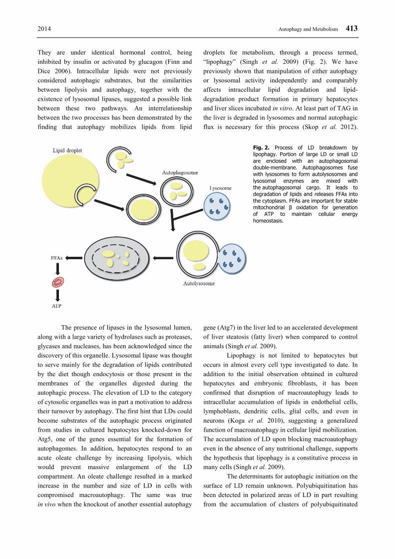
Fig. 2. Process of LD breakdowm by lipophagy. Portion of large LD or small LD are enclosed with an autophagosomal double-membrane. Autophagosomes fuse with lysosomes to form autolysosomes and lysosomal enzymes are mixed with the autophagosomal cargo. It leads to degradation of lipids and releases FFAs into the cytoplasm. FFAs are important for stable mitochondrial β oxidation for generation of ATP to maintain cellular energy homeostasis.
The presence of lipases in the lysosomal lumen,
along with a large variety of hydrolases such as proteases,
glycases and nucleases, has been acknowledged since the
discovery of this organelle. Lysosomal lipase was thought
to serve mainly for the degradation of lipids contributed
by the diet though endocytosis or those present in the
membranes of the organelles digested during the
autophagic process. The elevation of LD to the category
of cytosolic organelles was in part a motivation to address
their turnover by autophagy. The first hint that LDs could
become substrates of the autophagic process originated
from studies in cultured hepatocytes knocked-down for
Atg5, one of the genes essential for the formation of
autophagomes. In addition, hepatocytes respond to an
acute oleate challenge by increasing lipolysis, which
would prevent massive enlargement of the LD
compartment. An oleate challenge resulted in a marked
increase in the number and size of LD in cells with
compromised macroautophagy. The same was true
in vivo when the knockout of another essential autophagy
gene (Atg7) in the liver led to an accelerated development
of liver steatosis (fatty liver) when compared to control
animals (Singh et al. 2009).
Lipophagy is not limited to hepatocytes but
occurs in almost every cell type investigated to date. In
addition to the initial observation obtained in cultured
hepatocytes and embryonic fibroblasts, it has been
confirmed that disruption of macroautophagy leads to
intracellular accumulation of lipids in endothelial cells,
lymphoblasts, dendritic cells, glial cells, and even in
neurons (Koga et al. 2010), suggesting a generalized
function of macroautophagy in cellular lipid mobilization.
The accumulation of LD upon blocking macroautophagy
even in the absence of any nutritional challenge, supports
the hypothesis that lipophagy is a constitutive process in
many cells (Singh et al. 2009).
The determinants for autophagic initiation on the
surface of LD remain unknown. Polyubiquitination has
been detected in polarized areas of LD in part resulting
from the accumulation of clusters of polyubiquitinated
414 Papáčková and Cahová Vol. 63
apolipoprotein B (ApoB) on their surface (Ohsaki et al.
2006). It seems that ApoB can undergo both proteasomal
and lysosomal degradation as proteasome inhibition
caused an increase of autophagic vacuoles abundance and
ApoB content in lysosomes. Whether or not the
lysosomal degradation of ApoB occurs as a result of the
activation of lipophagy and how the accumulation of this
protein contributes to the initiation of the process require
future investigation. Of particular interest is the fact that
LDs have been shown to dynamically interact with two of
the organelles – the ER and mitochondria – that have
been proposed as sites of formation of the limiting
membrane of the autophagosomes. Interaction with the
ER may be related to LD biogenesis, as this is the
compartment from where these organelles originate, but
may also favor the distribution of lipids from the LD
towards other organelles through the endosecretory
pathway. In the case of mitochondria, the proximity of
LDs to the outer-membrane of this organelle could
facilitate delivery of the FFA for mitochondrial
β-oxidation. However, considering the described
association of the autophagic initiation complex to
precise area in the membrane of ER and the
mitochondria, and the formation of cup-like precursors of
the limiting membrane of the autophagosome from these
regions, it is tempting to at least propose that the
previously described interactions of LD with these
organelles could contribute to the initiation of their
autophagic degradation.
Mobilization of LD by autophagy was first
observed both in cultured hepatocytes in response to fatty
acid exposure and in the liver of mice maintained on
a diet enriched in fat for prolonged period of time (Singh
et al. 2009). The liver responds to the massive influx of
lipids from the blood by up-regulating LD biogenesis as
a defense mechanism against the toxicity of FFAs, which
upon esterification get converted into TAG that are stored
in LDs (Lass et al. 2011). However, in order to prevent
uncontrolled expansion of LD, activation of lipolysis also
occurs under these conditions and contributes to
maintaining of LD size. Failure to regulate lipid
accumulation in hepatocytes may be the basis of
pathologic conditions such as liver steatosis and
steatohepatitis (Christian et al. 2013). Autophagy has
now been added to the mechanism that controls the
growth of the hepatic LD under these conditions
(Greenberg et al. 2011).
After the first observations demonstrating the
existence of lipophagy and the upregulation of this
process in response to lipid challenge, numerous studies
have confirmed the stimulatory effect of dietary lipids on
the autophagic process. In contrast, an equal number of
studies have reported inhibition of autophagy in response
to high concentrations of particular lipids. In animals
exposed to a high-fat diet for prolonged periods of time, it
is possible to detect an increase in autophagic activity
during the first weeks of treatment that is followed by
a gradual decrease in autophagy. This decrease in
autophagy further contributes to the expansion of the LD
compartment, eventually leading to hepatotoxicity and
steatosis (Singh et al. 2009). Other results showed that in
the liver, the autophagic response to the increased fat
supply in the diet is biphasic. At the beginning of high-fat
diet feeding autophagy flux is stimulated but over time
autophagy flux is nearly completely impaired (Papackova
et al. 2012).
Recently, a significant role for autophagy in
lipid metabolism has been revealed in human enterocytes
(Khaldoun et al. 2014). Enterocytes have to deal with
massive alimentary lipids upon food consummation.
They orchestrate complex lipid trafficking events that
lead to secretion of TAG-rich lipoproteins and the
transient storage of lipids in LDs. The authors showed
that delivering alimentary lipid micelles to polarized
human enterocytes induced an immediate autophagic
response. This was accompanied by rapid capture of
newly synthesized LDs by nascent autophagosomal
structures at the ER membrane and hence targeting them
to the lysosomes. They proposed an interesting
hypothesis according to which the autophagosomes,
despite their primary lysosomal-delivery function, could
also be used as a “hiding” compartment in the cell in
order to avoid excessive TAG accumulation from the
membranes where lipid biosynthesis occurs. Such a local
program could act as a global protection and adaptation
response to the arrival of neutral lipids, as is the situation
for enterocytes during the postprandial phase (Khaldoun
et al. 2014).
Autophagy in other disease
A basal and constitutive level of autophagy is
indispensable for intracellular homeostasis and quality
control for healthy individuals. The molecular dissection
of autophagy and the growing number of physiological
function attributed to this process are leading to a better
understanding of the role of autophagy in disease.
Mounting evidence has demonstrated that many of its

2014 Autophagy and Metabolism 415
physiological function is strongly related to human
diseases such as cancer, myopahies, bacterial and viral
infections and neurodegenerative, liver and heart diseases
(Fig. 3).
Direct evidence for impaired autophagy in
a myopathy was first obtained when the gene encoding
a lysosomal membrane protein (lamp2) was knocked out
in mice (Saftig et al. 2001). The predominant phenotype
of these mice is a massive accumulation of autophagic
vacuoles in cells of the liver, muscle and heart. Despite
the increased number of autophagic vesicles, the rate of
lysosomal degradation of proteins is reduced because the
clearance of autophagosomes through lysosomal fusion is
impaired. Autophagy of glycogen is not limited to the
liver. In fact, altered autophagic degradation of glycogen
stores may underlie the basis of some muscle disorders
that are now classified as autophagic vacuolar myopathies
such as Danon disease, X-linked vacuolar myopathy with
excessive autophagy and Pompe disease (Fukuda et al.
2007). Glycogen granules accumulate in muscles of
Danon disease patients resulting in cardiomyopathy,
proximal muscle weakness and mental retardation
(Nishino et al. 2000). The histological resemblance of the
skeletal and heart muscles from the lamp-2 knockout
mice to those from patients with Danon disease led to the
identification of mutations in LAMP2 as the primary
defect in this lysosomal storage disease (Yamamoto et al.
2001).
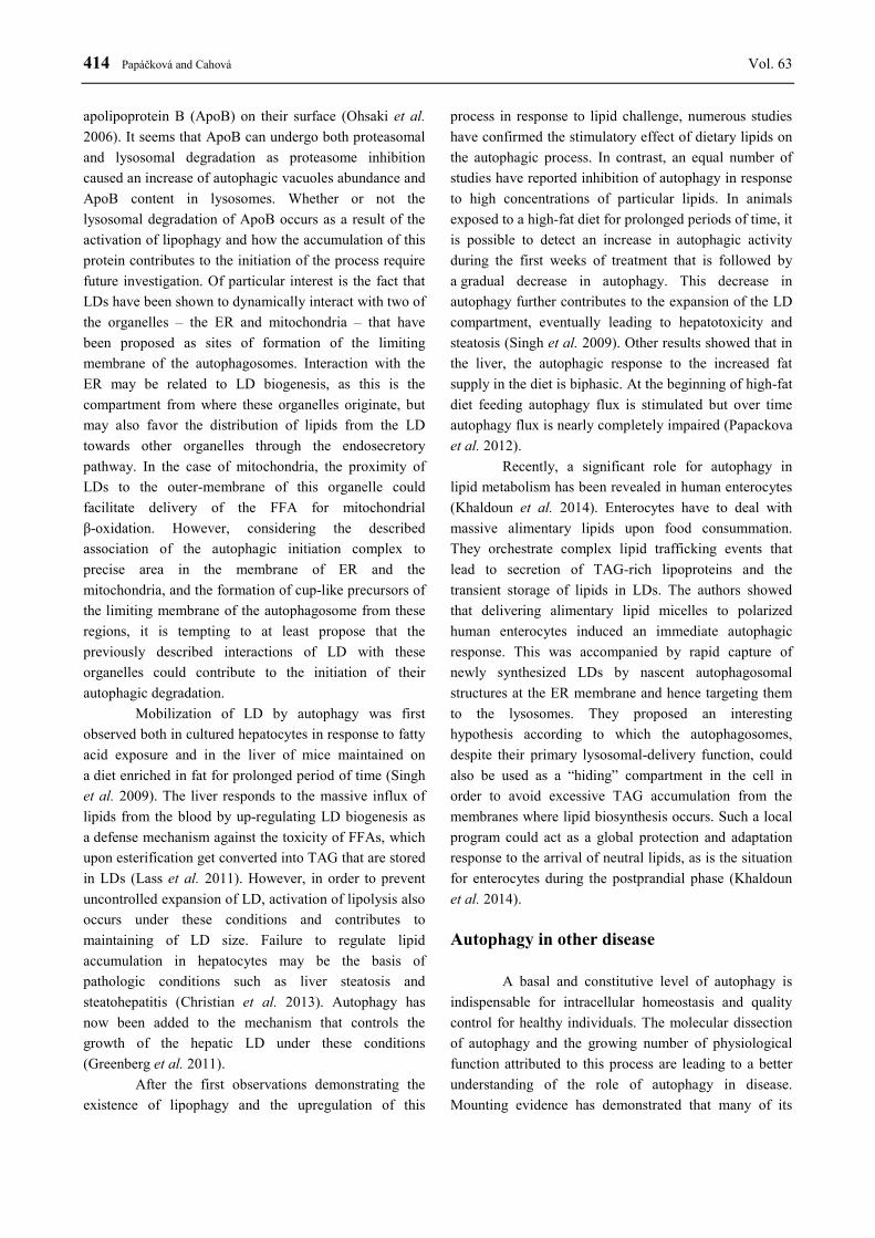
Fig. 3. Autophagy in disease. Intensity of autophagy is important for the homeostasis in all organisms. Decreased or excessively increased autophagic activity could progress various disorders.
Degradation of proteins by autophagy
contributes to quality control and prevents proteotoxicity
associated with accumulation of abnormal proteins. In
fact, defective autophagy often associates with formation
of proteins aggregates and it is likely the basis for protein
conformational disorders such as Alzheimer´s,
Huntington´s and Parkinson´s disease (Komatsu et al.
2006). Although the mutated protein is different in each
of these disorders, the sequence of events leading to
protein aggregation is apparently exactly the same and
proceeds as follows: 1) the abnormal conformation of
affected protein exposes normally hidden hydrophobic
residues; 2) the cell responds to these abnormal proteins
by activating chaperone system and cytosolic proteases;
and 3) in the initial stages of the disorder, chaperones and
proteases can sometimes revert, or at least slow down,
protein aggregation. However, as the levels of the
pathogenic protein increase, the process becomes
irreversible and even the “helpful” proteins become
trapped in the aggregates. Although this might have
originally been a defensive mechanism against
hydrophobic patches, the hydrophobic nature of these
proteins makes them resistant to attack by cytosolic
proteases, leaving removal by macroautophagy as the

416 Papáčková and Cahová Vol. 63
only viable possibility (Michalik and Van Broeckhoven
2003). Recent studies show that, at least in experimental
systems, the activation of macroautophagy facilitates the
removal of newly formed aggregates (Webb et al. 2003).
Insulin signaling is critical for the regulation of
glucose homeostasis in the adipose and muscle. Eating an
unhealthy diet and obesity contribute to the development
of insulin resistance where hepatic, adipose and muscle
tissues no longer respond to insulin signaling, thus
resulting in hyperglycemia (Martyn et al. 2008).
Recently, a role for autophagy in insulin signaling has
been discovered. Intriguingly, it seems that the role of
autophagy in the regulation of insulin signaling in
different tissues may be opposite. Hyperinsulinemic high-
fat diet fed mice and ob/ob mice both display impaired
hepatic autophagy activity indicated by decreased
expression of autophagy markers and increased levels of
p62, a protein that is normally degraded by autophagy.
Decreased autophagy in ob/ob mice led to a decreased
insulin signaling and induction of ER stress, both of
which were rescued by atg7 overexpression (Yang et al.
2010). This suggests that insulin signaling down-
regulates autophagy activity in the liver during
a hyperinsulinemic state. In contrast, autophagy was
shown to be up-regulated in adipose tissue of obese and
T2D patients. Kovsan et al. (2011), in a human study,
reveals direct correlation between different types and
degrees of obesity and autophagic activity and fat
deposits size. Surprisingly, autophagy was extreme high
in omental fat tissue extract from obese individuals and
was also increased in insulin-resistant obese subjects
(Kovsan et al. 2011). It has been suggested that diabetic
state is associated with pseudo starved state, leading to
increased adipocyte autophagy, elevated TAG hydrolysis
and enhanced plasma fatty acid concentration (Ost et al.
2010).
One of the prominent features of T2D clinical
manifestation is the defective angiogenesis and
consequent microvascular complications. Some
conditions that are tightly associated with T2D, like
inflammation or chronic hyperglycemia, are connected
also with alterations in autophagy regulation, but the
relationship between altered autophagy and the
development of these complications is still not fully
understood. It has been reported that inflammatory
molecules as MCP-1, TNF-α, IL-1β and IL-8 are known
to promote angiogenesis. Roy et al. (2012) demonstrated
that all these pathways converge at the MCP-induced
protein (MCPIP), that is able to switch on the cascade of
oxidative stress, ER stress, autophagy and angiogenic
differentiation in HUVEC cells. Interestingly, inhibition
of each particular step caused inhibition of each
subsequent step that was postulated. These data suggest
that cellular stress evoked by the diabetic milieu may be
translated into de novo angiogenesis (Roy and
Kolattukudy 2012). Somewhat contradictory results were
reported by Liu et al. (2012), who showed that
methylglyoxal (MGO) (a small carbohydrate compound
that is elevated in T2D) stimulated autophagic
degradation of VEGFR2 in endothelial cells. Suppression
of autophagy either by inhibitors or siRNA, but not of the
proteasome and caspase, normalized both the VEGFR2
protein levels and angiogenesis. Conversely, induction of
autophagy either by rapamycin or overexpression of LC3
and Beclin-1 reduced VEGFR2 and angiogenesis (Liu et
al. 2012).
Retinopathy belongs to the serious pathologies
associated with T2D progression. Most retinal cells are
fully or terminally differentiated cells; therefore, a steady
supply of nutrients, cell growth and cell cycle control are
important for long-term survival (Dyer and Cepko 2000,
Lee et al. 2006) and make these cells extremely sensitive
to metabolic disturbance. Recently, it has been published
that the crucial role in the process of retinopathy plays
a pro-oxidant and pro-apoptotic thioredoxin interacting
protein (TXNIP) (Singh, 2013). TXNIP contributes to the
oxidative and nitrosative stress by significant attenuation
of antioxidant cell capacity, induction of mitochondrial
damage and consequent stimulation of mitophagy. In the
end, this cascade of events may program autophagic cell
death involving caspase-3 or cellular energy collapse.
Nonetheless, in the broader context the autophagy could
serve as an adaptive and even protective mechanism
preventing excessive bursts of reactive oxygen species
production and necrotic cell death.
Aging
Morphological alterations in the lysosomal
system and changes in its enzymatic content are common
in almost all tissues from older mammals (Ward 2002).
Considering the physiological functions of autophagy, the
cellular consequences of diminished autophagy flux are
easily inferred and include inefficient removal of
damaged intracellular structures, alterations in cellular
homeostasis, inability to adapt to extracellular changes
and poor defensive response against damaging agents.
Decreased protein degradation plays an important role in
2014 Autophagy and Metabolism 417
aging with regard to prolonging protein lifespan, which
increases the probability of their undesired alteration. The
defects in the autophagic/lysosomal proteolytic system
may be the main cause of reduced protein degradation,
since the proteasomal system cannot digest larger
proteins or impaired organelles. Indeed, there is evidence
of gradual reduction in autophagy with age (Cuervo et al.
2005). Autophagy activation may protect an organism
from aging due to the increased ability to get rid of
damaged proteins and organelles. Caloric restriction, the
only intervention that delays aging and increases lifespan,
reverses the decline in autophagy that occurs with age
and may come about through reduced insulin/IGF-1
signaling (Kenyon 2005). Fasting can promote longevity,
but it may cause potentially adverse effects of caloric
restriction on human health and hence alternative
approaches are currently being studied that mimic the
beneficial effects if caloric restriction. The application of
antilipolytic drugs that increase autophagy and extend
longevity is a good example (Bergamini 2005).
Conclusion
Autophagy has been shown to complement
“classical pathways” in the catabolism of carbohydrates,
proteins and mitochondria in fasting and nutrient
deficiency. It is now proposed that autophagy also
participates in the regulation of lipid metabolism. This
finding necessitates a re-evaluation of much of the
knowledge and assumptions about LD metabolism in
light of this new alternative pathway of lipolysis.
Lipophagy is likely to be an important metabolic pathway
in the supply of energy for specific cellular function. The
findings to date indicate that further investigations of
lipophagy might increase our understanding of the role of
LD breakdown in cell physiology and provide new
avenues to treat diseases resulting from defects in lipid
metabolism or storage. Autophagy is necessary for
regulation of metabolic processes. In addition,
malfunctions of autophagy have been implicated in the
development of several diseases and so manipulation of
this critical cellular process might be a promising
therapeutic target.
Conflict of Interest There is no conflict of interest.
Acknowledgements This study was supported by MH CZ – DRO (“Institute
for Clinical and Experimental Medicine – IKEM,
IN 00023001”).
References
BERGAMINI E: Targets for antiageing drugs. Expert Opin Ther Targets 9: 77-82, 2005.
CHEONG H, KLIONSKY DJ: Biochemical methods to monitor autophagy-related processes in yeast. Methods
Enzymol 451: 1-26, 2008.
CHEONG H, YORIMITSU T, REGGIORI F, LEGAKIS JE, WANG CW, KLIONSKY DJ: Atg17 regulates the
magnitude of the autophagic response. Mol Biol Cell 16: 3438-3453, 2005.
CHRISTIAN P, SACCO J, ADELI K: Autophagy: Emerging roles in lipid homeostasis and metabolic control. Biochim
Biophys Acta 1831: 819-824, 2013.
CUERVO AM, BERGAMINI E, BRUNK UT, DROGE W, FFRENCH M, TERMAN A: Autophagy and aging: the
importance of maintaining "clean" cells. Autophagy 1: 131-140, 2005.
DELL'ANGELICA EC, MULLINS C, CAPLAN S, BONIFACINO JS: Lysosome-related organelles. FASEB J 14:
1265-1278, 2000.
DETER RL, BAUDHUIN P, DE DUVE C: Participation of lysosomes in cellular autophagy induced in rat liver by
glucagon. J Cell Biol 35: C11-C16, 1967.
DYER MA, CEPKO CL: Control of Müller glial cell proliferation and activation following retinal injury. Nat Neurosci
3: 873-880, 2000.
EBATO C, UCHIDA T, ARAKAWA M, KOMATSU M, UENO T, KOMIYA K, AZUMA K, HIROSE T, TANAKA
K, KOMINAMI E, KAWAMORI R, FUJITANI Y, WATADA H: Autophagy is important in islet homeostasis
and compensatory increase of beta cell mass in response to high-fat diet. Cell Metab 8: 325-332, 2008.
ESKELINEN EL, SAFTIG P: Autophagy: a lysosomal degradation pathway with a central role in health and disease.
Biochim Biophys Acta 1793: 664-673, 2009.
418 Papáčková and Cahová Vol. 63
FINN PF, DICE JF: Proteolytic and lipolytic responses to starvation. Nutrition 22: 830-844, 2006.
FUJIMOTO T, PARTON RG: Not just fat: the structure and function of the lipid droplet. Cold Spring Harb Perspect
Biol 3: a004838, 2011.
FUKUDA T, ROBERTS A, PLOTZ PH, RABEN N: Acid alpha-glucosidase deficiency (Pompe disease). Curr Neurol
Neurosci Rep 7: 71-77, 2007.
GREENBERG AS, COLEMAN RA, KRAEMER FB, MCMANAMAN JL, OBIN MS, PURI V, YAN QW, MIYOSHI
H, MASHEK DG: The role of lipid droplets in metabolic disease in rodents and humans. J Clin Invest 121:
2102-2110, 2011.
HAMASAKI M, YOSHIMORI T: Where do they come from? Insights into autophagosome formation. FEBS Lett 584:
1296-1301, 2010.
ICHIMURA Y, KIRISAKO T, TAKAO T, SATOMI Y, SHIMONISHI Y, ISHIHARA N, MIZUSHIMA N, TANIDA
I, KOMINAMI E, OHSUMI M, NODA T, OHSUMI Y: A ubiquitin-like system mediates protein lipidation.
Nature 408: 488-492, 2000.
JUNG HS, CHUNG KW, WON KIM J, KIM J, KOMATSU M, TANAKA K, NGUYEN YH, KANG TM, YOON KH,
KIM JW, JEONG YT, HAN MS, LEE MK, KIM KW, SHIN J, LEE MS: Loss of autophagy diminishes
pancreatic beta cell mass and function with resultant hyperglycemia. Cell Metab 8: 318-324, 2008.
KALAMIDAS SA, KOTOULAS OB: Glycogen autophagy in newborn rat hepatocytes. Histol Histopathol 15: 1011-
1018, 2000.
KENYON C: The plasticity of aging: insights from long-lived mutants. Cell 120: 449-460, 2005.
KHALDOUN SA, EMOND-BOISJOLY MA, CHATEAU D, CARRIÈRE V, LACASA M, ROUSSET M,
DEMIGNOT S, MOREL E: Autophagosomes contribute to intracellular lipid distribution in enterocytes. Mol
Biol Cell 25: 118-132, 2014.
KLIONSKY DJ: Autophagy: from phenomenology to molecular understanding in less than a decade. Nat Rev Mol Cell
Biol 8: 931-937, 2007.
KOGA H, KAUSHIK S, CUERVO AM: Altered lipid content inhibits autophagic vesicular fusion. FASEB J 24: 3052-
3065, 2010.
KOMATSU M, WAGURI S, CHIBA T, MURATA S, IWATA J, TANIDA I, UENO T, KOIKE M, UCHIYAMA Y,
KOMINAMI E, TANAKA K: Loss of autophagy in the central nervous system causes neurodegeneration in
mice. Nature 441: 880-884, 2006.
KOMATSU M, WAGURI S, UENO T, IWATA J, MURATA S, TANIDA I, EZAKI J, MIZUSHIMA N, OHSUMI Y,
UCHIYAMA Y, KOMINAMI E, TANAKA K, CHIBA T: Impairment of starvation-induced and constitutive
autophagy in Atg7-deficient mice. J Cell Biol 169: 425-434, 2005.
KOTOULAS OB, KALAMIDAS SA, KONDOMERKOS DJ: Glycogen autophagy. Microsc Res Tech 64: 10-20, 2004.
KOVSAN J, BLUHER M, TARNOVSCKI T, KLOTING N, KIRSHTEIN B, MADAR L, SHAI I, GOLAN R,
HARMAN-BOEHM I, SCHON MR, GREENBERG AS, ELAZAR Z, BASHAN N, RUDICH A: Altered
autophagy in human adipose tissues in obesity. J Clin Endocrinol Metab 96: E268-E277, 2011.
LASS A, ZIMMERMANN R, OBERER M, ZECHNER R: Lipolysis – a highly regulated multi-enzyme complex
mediates the catabolism of cellular fat stores. Prog Lipid Res 50: 14-27, 2011.
LEE TC, ALMEIDA D, CLAROS N, ABRAMSON DH, COBRINIK D: Cell cycle-specific and cell type-specific
expression of Rb in the developing human retina. Invest Ophthalmol Vis Sci 47: 5590-5598, 2006.
LEVINE B, SINHA S, KROEMER G: Bcl-2 family members: dual regulators of apoptosis and autophagy. Autophagy
4: 600-606, 2008.
LIU H, YU S, ZHANG H, XU J: Angiogenesis impairment in diabetes: role of methylglyoxal-induced receptor for
advanced glycation endproducts, autophagy and vascular endothelial growth factor receptor 2. PLoS One 7:
e46720, 2012.
MARSH BJ, SODEN C, ALARCON C, WICKSTEED BL, YAEKURA K, COSTIN AJ, MORGAN GP, RHODES CJ:
Regulated autophagy controls hormone content in secretory-deficient pancreatic endocrine beta-cells. Mol
Endocrinol 21: 2255-2269, 2007.
MARTYN JA, KANEKI M, YASUHARA S: Obesity-induced insulin resistance and hyperglycemia: etiologic factors
and molecular mechanisms. Anesthesiology 109: 137-148, 2008.

2014 Autophagy and Metabolism 419
MEHRPOUR M, ESCLATINE A, BEAU I, CODOGNO P: Autophagy in health and disease. 1. Regulation and
significance of autophagy: an overview. Am J Physiol Cell Physiol 298: C776-C785, 2010.
MICHALIK A, VAN BROECKHOVEN C: Pathogenesis of polyglutamine disorders: aggregation revisited. Hum Mol
Genet 12 (Spec No 2): R173-R186, 2003.
MIZUSHIMA N, LEVINE B, CUERVO AM, KLIONSKY DJ: Autophagy fights disease through cellular self-
digestion. Nature 451: 1069-1075, 2008.
MIZUSHIMA N, LEVINE B: Autophagy in mammalian development and differentiation. Nat Cell Biol 12: 823-830,
2010.
MIZUSHIMA N, YAMAMOTO A, MATSUI M, YOSHIMORI T, OHSUMI Y: In vivo analysis of autophagy in
response to nutrient starvation using transgenic mice expressing a fluorescent autophagosome marker. Mol Biol
Cell 15: 1101-1111, 2004.
NEUFELD TP: TOR-dependent control of autophagy: biting the hand that feeds. Curr Opin Cell Biol 22: 157-168,
2010.
NICKLIN P, BERGMAN P, ZHANG B, TRIANTAFELLOW E, WANG H, NYFELER B, YANG H, HILD M,
KUNG C, WILSON C, MYER VE, MACKEIGAN JP, PORTER JA, WANG YK, CANTLEY LC, FINAN
PM, MURPHY LO: Bidirectional transport of amino acids regulates mTOR and autophagy. Cell 136: 521-534,
2009.
NISHINO I, FU J, TANJI K, YAMADA T, SHIMOJO S, KOORI T, MORA M, RIGGS JE, OH SJ, KOGA Y, SUE
CM, YAMAMOTO A, MURAKAMI N, SHANSKE S, BYRNE E, BONILLA E, NONAKA I, DIMAURO S,
HIRANO M: Primary LAMP-2 deficiency causes X-linked vacuolar cardiomyopathy and myopathy (Danon
disease). Nature 406: 906-910, 2000.
OHSAKI Y, CHENG J, FUJITA A, TOKUMOTO T, FUJIMOTO T: Cytoplasmic lipid droplets are sites of
convergence of proteasomal and autophagic degradation of apolipoprotein B. Mol Biol Cell 17: 2674-2683,
2006.
OHSAKI Y, CHENG J, SUZUKI M, SHINOHARA Y, FUJITA A, FUJIMOTO T: Biogenesis of cytoplasmic lipid
droplets: from the lipid ester globule in the membrane to the visible structure. Biochim Biophys Acta 1791:
399-407, 2009.
ORENSTEIN SJ, CUERVO AM: Chaperone-mediated autophagy: molecular mechanisms and physiological relevance.
Semin Cell Dev Biol 21: 719-726, 2010.
OST A, SVENSSON K, RUISHALME I, BRANNMARK C, FRANCK N, KROOK H, SANDSTROM P, KJOLHEDE
P, STRALFORS P: Attenuated mTOR signaling and enhanced autophagy in adipocytes from obese patients
with type 2 diabetes. Mol Med 16: 235-246, 2010.
PAPACKOVA Z, DANKOVA H, PALENICKOVA E, KAZDOVA L, CAHOVA M: Effect of short- and long-term
high-fat feeding on autophagy flux and lysosomal activity in rat liver. Physiol Res 61 (Suppl 2): S67-S76,
2012.
ROY A, KOLATTUKUDY PE: Monocyte chemotactic protein-induced protein (MCPIP) promotes inflammatory
angiogenesis via sequential induction of oxidative stress, endoplasmic reticulum stress and autophagy. Cell
Signal 24: 2123-2131, 2012.
SAFTIG P, TANAKA Y, LULLMANN-RAUCH R, VON FIGURA K: Disease model: LAMP-2 enlightens Danon
disease. Trends Mol Med 7: 37-39, 2001.
SEGLEN PO, GORDON PB: 3-Methyladenine: specific inhibitor of autophagic/lysosomal protein degradation in
isolated rat hepatocytes. Proc Natl Acad Sci USA 79: 1889-1892, 1982.
SCHWORER CM, COX JR, MORTIMORE GE: Alteration of lysosomal density by sequestered glycogen during
deprivation-induced autophagy in rat liver. Biochem Biophys Res Commun 87: 163-170, 1979.
SINGH LP: Thioredoxin interacting protein (TXNIP) and pathogenesis of diabetic retinopathy. J Clin Exp Ophthalmol
4: doi: 10.4172/2155-9570.1000287, 2013.
SINGH R, KAUSHIK S, WANG Y, XIANG Y, NOVAK I, KOMATSU M, TANAKA K, CUERVO AM, CZAJA MJ:
Autophagy regulates lipid metabolism. Nature 458: 1131-1135, 2009.
420 Papáčková and Cahová Vol. 63
SKOP V, CAHOVA M, PAPACKOVA Z, PALENICKOVA E, DANKOVA H, BARANOWSKI M, ZABIELSKI P,
ZDYCHOVA J, ZIDKOVA J, KAZDOVA L: Autophagy-lysosomal pathway is involved in lipid degradation
in rat liver. Physiol Res 61: 287-297, 2012.
TANIDA I, MINEMATSU-IKEGUCHI N, UENO T, KOMINAMI E: Lysosomal turnover, but not a cellular level, of
endogenous LC3 is a marker for autophagy. Autophagy 1: 84-91, 2005.
VABULAS RM, HARTL FU: Protein synthesis upon acute nutrient restriction relies on proteasome function. Science
310: 1960-1963, 2005.
WARD WF: Protein degradation in the aging organism. Prog Mol Subcell Biol 29: 35-42, 2002.
WEBB JL, RAVIKUMAR B, ATKINS J, SKEPPER JN, RUBINSZTEIN DC: Alpha-Synuclein is degraded by both
autophagy and the proteasome. J Biol Chem 278: 25009-25013, 2003.
YAMAMOTO A, MORISAWA Y, VERLOES A, MURAKAMI N, HIRANO M, NONAKA I, NISHINO I: Infantile
autophagic vacuolar myopathy is distinct from Danon disease. Neurology 57: 903-905, 2001.
YANG L, LI P, FU S, CALAY ES, HOTAMISLIGIL GS: Defective hepatic autophagy in obesity promotes ER stress
and causes insulin resistance. Cell Metab 11: 467-478, 2010.
YORIMITSU T, KLIONSKY DJ: Autophagy: molecular machinery for self-eating. Cell Death Differ 12: 1542-1552,
2005.