impact of the host on toxoplasm a stage...
TRANSCRIPT
OPEN ACCESS | www.microbialcell.com 1 Microbial Cell | in press
www.microbialcell.com
Review
ABSTRACT The unicellular parasite Toxoplasma gondii infects warm-blooded
animals and humans, and it is highly prevalent throughout the world. Infec-
tion of immunocompetent hosts is usually asymptomatic or benign but leads
to long-term parasite persistence mainly within neural and muscular tissues.
The transition from acute primary infection towards chronic toxoplasmosis is
accompanied by a developmental switch from fast replicating and metaboli-
cally highly active tachyzoites to slow replicating and largely dormant brady-
zoites within tissue cysts. Such developmental differentiation is critical for T.
gondii in order to complete its life cycle and for pathogenesis. Herein, we
summarize accumulating evidence indicating a major impact of the host cell
physiology on stage conversion between the tachyzoite and the bradyzoite
stage of the parasite. Withdrawal from cell cycle progression, proinflammato-
ry responses, reduced availability of nutrients and extracellular adenosine can
indeed induce tachyzoite-to-bradyzoite differentiation and tissue cyst for-
mation. In contrast, high glycolytic activity as indicated by increased lactate
secretion can inhibit bradyzoite formation. These examples argue for the in-
triguing possibility that after dissemination within its host, T. gondii can sense
its cellular microenvironment to initiate the developmental program towards
the bradyzoite stage in distinct cells. This may also explain the predominant
localization of T. gondii in neural and muscular tissues during chronic toxo-
plasmosis.
Impact of the host on Toxoplasma stage differentiation
Carsten G.K. Lüder1,*
and Taibur Rahman1
1 Institute for Medical Microbiology, University Medical Center Goettingen, Goettingen, Germany.
* Corresponding Author:
Carsten G.K. Lüder, Institute for Medical Microbiology, Kreuzbergring 57, 37075 Goettingen, Germany; Tel: +49 551 395869;
Fax: +49 551 395861; E-mail: [email protected]
INTRODUCTION
Developmental switching between life cycle stages is criti-
cal for the biology of a large number of unicellular micro-
organisms. It is also important for the course of infectious
diseases caused by bacteria, fungi and protozoa. Examples
include differentiation between planktonic and sessile bac-
teria during biofilm formation [1], between yeast and hy-
phae and vice versa during infection with distinct Candida
spp. [2], or between Anopheles-transmitted sporozoites
and liver stage merozoites of Plasmodium spp. during ma-
laria [3]. Characterization of those factors that regulate
these developmental processes is of fundamental interest
to understand the biology of microbial cells and the evolu-
tion of multicellular organisms. It can also lead to the iden-
tification of novel targets for efficient intervening with
onset or progression of infectious diseases.
Toxoplasma gondii is an obligate intracellular parasite
of the phylum Apicomplexa which comprises a variety of
pathogens of utmost importance for human and animal
health. T. gondii is widespread throughout the world and
threatens the health particularly of immunocompromised
individuals or that of fetuses from recently infected preg-
nant women [4]. In otherwise healthy adults, primary in-
fection with T. gondii is regularly controlled by the ensuing
Th1-type cell-mediated immune response but nevertheless
gives rise to chronic persistence of the parasite, possibly
for the host’s life. The parasite’s ability to persist for ex-
tended periods of time is critical for transmission between
hosts. Its appropriate regulation likely was and still is sub-
ject of selective pressure during evolution.
The life cycle of T. gondii is complex with members of
the Felidae and warm-blooded vertebrates, i.e., mammals
and birds, as final or intermediate hosts, respectively, and
different phases of sexual and asexual reproduction. Not
surprisingly, it also involves different parasite stages with
sexual gametocytes, transmissible sporozoites, rapidly pro-
liferating tachyzoites and dormant bradyzoites being the
most prominent ones. Transition between the different
stages requires extensive morphological remodeling, ex-
pression of distinct transcriptomes and proteomes, as well
Received originally: 20.04.2017;
in revised form: 07.06.2017,
Accepted 07.06.2017,
Published 22.06.2017.
Keywords: Toxoplasma gondii, stage
conversion, bradyzoite, parasite host-
interaction, host cell, metabolism, cell
cycle, immune response.
Abbreviations:
HFF - human foreskin fibroblasts,
LDL - low-density lipoproteins,
LPS – lipopolysaccharide,
MyHC - myosin heavy chain,
NO – nitric oxide,
PV - parasitophorous vacuole,
SkMCs - skeletal muscle cells,
SNP - sodium nitroprusside.

C.G.K. Lüder and T. Rahman (2017) Toxoplasma stage differentiation
OPEN ACCESS | www.microbialcell.com 2 Microbial Cell | in press
as several physiological adaptations [5, 6]. The differentia-
tion of tachyzoites to bradyzoites within intermediate
hosts is experimentally amenable after in vitro infection of
different host cells and after in vivo infection of laboratory
or farm animals [5, 7-10]. It is also of major biological and
medical interest because the transition between tachyzo-
ites and bradyzoites correlates with the acute or chronic
phase of toxoplasmosis, respectively.
Tachyzoites of T. gondii develop after oral transmission
of infective sporozoites or bradyzoites to intermediate
hosts, presumably within the intestinal epithelium (Fig. 1).
They rapidly divide within a membrane-bound intracellular
compartment, i.e. the parasitophorous vacuole (PV) by a
process called endodyogeny [11]. The doubling time of this
stage depends on the parasite strain and can be as fast as
~5 hours in type I strains but takes 6 - 12 hours in type II
and III strains [12]. Tachyzoites are metabolically highly
active indicating a high demand for nutrients from the en-
vironment. After 5 to 6 cell divisions, tachyzoites egress
from the host cell in a Ca2+
-dependent manner and can
then infect neighboring host cells [13, 14]. T. gondii invades
its host cell by a parasite-driven process that depends on
the parasites’ actin-myosin machinery and the discharge of
microneme and rhoptry proteins from the apical complex
[15]. Importantly, the active host cell infection enables T.
gondii to invade any nucleated cell of its host. Infection of
monocytes and dendritic cells particularly contributes to
the dissemination of the parasite throughout the hosts’
body [16, 17]. A sustained parasite replication with tissue
damage due to host cell lysis and/or the ensuing cell-
mediated immune response correlates with the acute
phase of infection and possibly overt disease under certain
circumstances. In most cases, however, vigorous cell-
mediated immunity is able to restrict tachyzoite replication
and to even kill tachyzoites in a timely fashion thereby
preventing overt symptoms.
Bradyzoites of T. gondii differentiate from tachyzoites
within 6 - 9 days after oral infection of mice with oocysts or
bradyzoites (Fig. 1) [11], but they can develop as early as 3
days after infection with tachyzoites [18]. Bradyzoites are
also located inside a PV which, however, over time ma-
tures to a so-called intracellular tissue cyst. The most
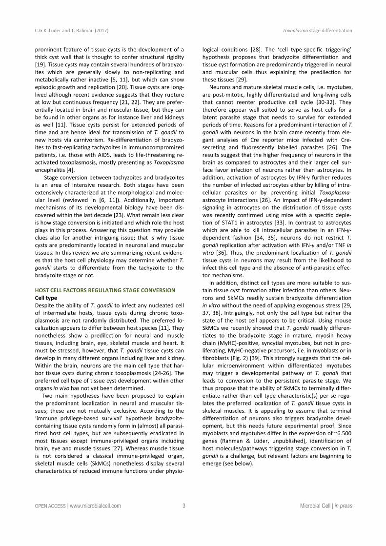
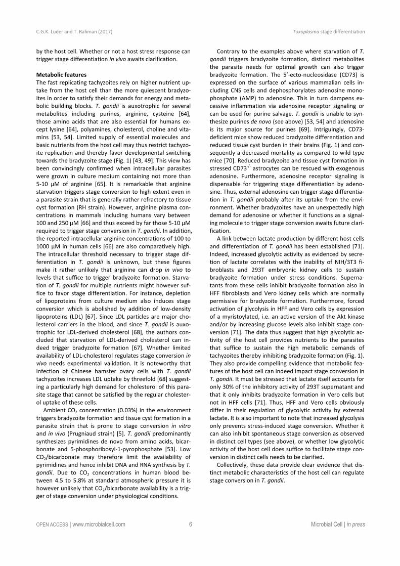
FIGURE 1: T. gondii stage conversion and its regulation by the host (cell) microenvironment. Within one to two weeks of infection, highly
active tachyzoites (two parasitophorous vacuoles within human foreskin fibroblasts are depicted; left micrograph) convert to relatively
dormant bradyzoites (a tissue cyst from mouse brain containing hundreds of individual bradyzoites is shown; right micrograph). Formation
of long-lived bradyzoite-containing tissue cysts is crucial for parasite transmission to new hosts. Reconversion of bradyzoites to tachyzoites
can occur in immunocompromised individuals (e.g. those with AIDS or transplant recipients (TPX)) leading to life-threatening disease. Dis-
tinct characteristics of the infected host cell or of bystander cells can trigger or inhibit differentiation towards the bradyzoite stage as indi-
cated in the upper part of the figure. Noteworthy, parasite-intrinsic triggers may also govern stage differentiation (not depicted). See main
text for further details.
C.G.K. Lüder and T. Rahman (2017) Toxoplasma stage differentiation
OPEN ACCESS | www.microbialcell.com 3 Microbial Cell | in press
prominent feature of tissue cysts is the development of a
thick cyst wall that is thought to confer structural rigidity
[19]. Tissue cysts may contain several hundreds of bradyzo-
ites which are generally slowly to non-replicating and
metabolically rather inactive [5, 11], but which can show
episodic growth and replication [20]. Tissue cysts are long-
lived although recent evidence suggests that they rupture
at low but continuous frequency [21, 22]. They are prefer-
entially located in brain and muscular tissue, but they can
be found in other organs as for instance liver and kidneys
as well [11]. Tissue cysts persist for extended periods of
time and are hence ideal for transmission of T. gondii to
new hosts via carnivorism. Re-differentiation of bradyzo-
ites to fast-replicating tachyzoites in immunocompromized
patients, i.e. those with AIDS, leads to life-threatening re-
activated toxoplasmosis, mostly presenting as Toxoplasma
encephalitis [4].
Stage conversion between tachyzoites and bradyzoites
is an area of intensive research. Both stages have been
extensively characterized at the morphological and molec-
ular level (reviewed in [6, 11]). Additionally, important
mechanisms of its developmental biology have been dis-
covered within the last decade [23]. What remain less clear
is how stage conversion is initiated and which role the host
plays in this process. Answering this question may provide
clues also for another intriguing issue; that is why tissue
cysts are predominantly located in neuronal and muscular
tissues. In this review we are summarizing recent evidenc-
es that the host cell physiology may determine whether T.
gondii starts to differentiate from the tachyzoite to the
bradyzoite stage or not.
HOST CELL FACTORS REGULATING STAGE CONVERSION
Cell type
Despite the ability of T. gondii to infect any nucleated cell
of intermediate hosts, tissue cysts during chronic toxo-
plasmosis are not randomly distributed. The preferred lo-
calization appears to differ between host species [11]. They
nonetheless show a predilection for neural and muscle
tissues, including brain, eye, skeletal muscle and heart. It
must be stressed, however, that T. gondii tissue cysts can
develop in many different organs including liver and kidney.
Within the brain, neurons are the main cell type that har-
bor tissue cysts during chronic toxoplasmosis [24-26]. The
preferred cell type of tissue cyst development within other
organs in vivo has not yet been determined.
Two main hypotheses have been proposed to explain
the predominant localization in neural and muscular tis-
sues; these are not mutually exclusive. According to the
‘immune privilege-based survival’ hypothesis bradyzoite-
containing tissue cysts randomly form in (almost) all parasi-
tized host cell types, but are subsequently eradicated in
most tissues except immune-privileged organs including
brain, eye and muscle tissues [27]. Whereas muscle tissue
is not considered a classical immune-privileged organ,
skeletal muscle cells (SkMCs) nonetheless display several
characteristics of reduced immune functions under physio-
logical conditions [28]. The ‘cell type-specific triggering’
hypothesis proposes that bradyzoite differentiation and
tissue cyst formation are predominantly triggered in neural
and muscular cells thus explaining the predilection for
these tissues [29].
Neurons and mature skeletal muscle cells, i.e. myotubes,
are post-mitotic, highly differentiated and long-living cells
that cannot reenter productive cell cycle [30-32]. They
therefore appear well suited to serve as host cells for a
latent parasite stage that needs to survive for extended
periods of time. Reasons for a predominant interaction of T.
gondii with neurons in the brain came recently from ele-
gant analyses of Cre reporter mice infected with Cre-
secreting and fluorescently labelled parasites [26]. The
results suggest that the higher frequency of neurons in the
brain as compared to astrocytes and their larger cell sur-
face favor infection of neurons rather than astrocytes. In
addition, activation of astrocytes by IFN-γ further reduces
the number of infected astrocytes either by killing of intra-
cellular parasites or by preventing initial Toxoplasma-
astrocyte interactions [26]. An impact of IFN-γ-dependent
signaling in astrocytes on the distribution of tissue cysts
was recently confirmed using mice with a specific deple-
tion of STAT1 in astrocytes [33]. In contrast to astrocytes
which are able to kill intracellular parasites in an IFN-γ-
dependent fashion [34, 35], neurons do not restrict T.
gondii replication after activation with IFN-γ and/or TNF in
vitro [36]. Thus, the predominant localization of T. gondii
tissue cysts in neurons may result from the likelihood to
infect this cell type and the absence of anti-parasitic effec-
tor mechanisms.
In addition, distinct cell types are more suitable to sus-
tain tissue cyst formation after infection than others. Neu-
rons and SkMCs readily sustain bradyzoite differentiation
in vitro without the need of applying exogenous stress [29,
37, 38]. Intriguingly, not only the cell type but rather the
state of the host cell appears to be critical. Using mouse
SkMCs we recently showed that T. gondii readily differen-
tiates to the bradyzoite stage in mature, myosin heavy
chain (MyHC)-positive, syncytial myotubes, but not in pro-
liferating, MyHC-negative precursors, i.e. in myoblasts or in
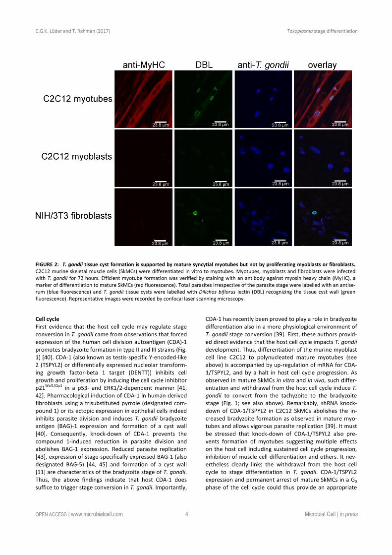
fibroblasts (Fig. 2) [39]. This strongly suggests that the cel-
lular microenvironment within differentiated myotubes
may trigger a developmental pathway of T. gondii that
leads to conversion to the persistent parasite stage. We
thus propose that the ability of SkMCs to terminally differ-
entiate rather than cell type characteristic(s) per se regu-
lates the preferred localization of T. gondii tissue cysts in
skeletal muscles. It is appealing to assume that terminal
differentiation of neurons also triggers bradyzoite devel-
opment, but this needs future experimental proof. Since
myoblasts and myotubes differ in the expression of ~6.500
genes (Rahman & Lüder, unpublished), identification of
host molecules/pathways triggering stage conversion in T.
gondii is a challenge, but relevant factors are beginning to
emerge (see below).
C.G.K. Lüder and T. Rahman (2017) Toxoplasma stage differentiation
OPEN ACCESS | www.microbialcell.com 4 Microbial Cell | in press
Cell cycle
First evidence that the host cell cycle may regulate stage
conversion in T. gondii came from observations that forced
expression of the human cell division autoantigen (CDA)-1
promotes bradyzoite formation in type II and III strains (Fig.
1) [40]. CDA-1 (also known as testis-specific Y-encoded-like
2 (TSPYL2) or differentially expressed nucleolar transform-
ing growth factor-beta 1 target (DENTT)) inhibits cell
growth and proliferation by inducing the cell cycle inhibitor
p21Waf1/Cip1
in a p53- and ERK1/2-dependent manner [41,
42]. Pharmacological induction of CDA-1 in human-derived
fibroblasts using a trisubstituted pyrrole (designated com-
pound 1) or its ectopic expression in epithelial cells indeed
inhibits parasite division and induces T. gondii bradyzoite
antigen (BAG)-1 expression and formation of a cyst wall
[40]. Consequently, knock-down of CDA-1 prevents the
compound 1-induced reduction in parasite division and
abolishes BAG-1 expression. Reduced parasite replication
[43], expression of stage-specifically expressed BAG-1 (also
designated BAG-5) [44, 45] and formation of a cyst wall
[11] are characteristics of the bradyzoite stage of T. gondii.
Thus, the above findings indicate that host CDA-1 does
suffice to trigger stage conversion in T. gondii. Importantly,
CDA-1 has recently been proved to play a role in bradyzoite
differentiation also in a more physiological environment of
T. gondii stage conversion [39]. First, these authors provid-
ed direct evidence that the host cell cycle impacts T. gondii
development. Thus, differentiation of the murine myoblast
cell line C2C12 to polynucleated mature myotubes (see
above) is accompanied by up-regulation of mRNA for CDA-
1/TSPYL2, and by a halt in host cell cycle progression. As
observed in mature SkMCs in vitro and in vivo, such differ-
entiation and withdrawal from the host cell cycle induce T.
gondii to convert from the tachyzoite to the bradyzoite
stage (Fig. 1; see also above). Remarkably, shRNA knock-
down of CDA-1/TSPYL2 in C2C12 SkMCs abolishes the in-
creased bradyzoite formation as observed in mature myo-
tubes and allows vigorous parasite replication [39]. It must
be stressed that knock-down of CDA-1/TSPYL2 also pre-
vents formation of myotubes suggesting multiple effects
on the host cell including sustained cell cycle progression,
inhibition of muscle cell differentiation and others. It nev-
ertheless clearly links the withdrawal from the host cell
cycle to stage differentiation in T. gondii. CDA-1/TSPYL2
expression and permanent arrest of mature SkMCs in a G0
phase of the cell cycle could thus provide an appropriate
FIGURE 2: T. gondii tissue cyst formation is supported by mature syncytial myotubes but not by proliferating myoblasts or fibroblasts.
C2C12 murine skeletal muscle cells (SkMCs) were differentiated in vitro to myotubes. Myotubes, myoblasts and fibroblasts were infected
with T. gondii for 72 hours. Efficient myotube formation was verified by staining with an antibody against myosin heavy chain (MyHC), a
marker of differentiation to mature SkMCs (red fluorescence). Total parasites irrespective of the parasite stage were labelled with an antise-
rum (blue fluorescence) and T. gondii tissue cysts were labelled with Dilichos biflorus lectin (DBL) recognizing the tissue cyst wall (green
fluorescence). Representative images were recorded by confocal laser scanning microscopy.
C.G.K. Lüder and T. Rahman (2017) Toxoplasma stage differentiation
OPEN ACCESS | www.microbialcell.com 5 Microbial Cell | in press
signal in muscle tissue in vivo to trigger T. gondii stage con-
version. Intriguingly, in mice, CDA-1/TSPYL2 is highly ex-
pressed in different regions of the brain, including cerebral
cortex and hippocampus and it is expressed to a lower
extent also in the gonads [46, 47]. It will thus be interesting
to see whether CDA-1/TSPYL2 and cell cycle inhibition also
triggers T. gondii stage conversion in the brain. The mech-
anism of how CDA-1 regulates bradyzoite differentiation is
currently unknown.
Pro-inflammatory immune responses
Pro-inflammatory molecules were among the first host-
derived signals for which pro-bradyzoite activities have
been confirmed. Activation of murine macrophages with
IFN-γ and/or LPS efficiently induces differentiation of T.
gondii to the bradyzoite stage (Fig. 1) [48, 49]. The level of
macrophage activation is critical, since only intermediate
levels of activation induce expression of bradyzoite-specific
antigens [49]. This was explained by findings that only par-
asites that show limited replication but not a complete halt
of replication are able to differentiate towards the brady-
zoite stage [49]. Later on it was confirmed that transition
through a G2-related cell cycle phase prior to mitosis and
cytokinesis is required for T. gondii to enter the differentia-
tion program to the bradyzoite stage [43]. Thus, non-
activated host macrophages favor direct S phase-to-mitosis
progression indicative for the tachyzoite cell cycle [12],
while too strongly activated macrophages prevent T. gondii
reaching a putative G2-associated checkpoint that may be
required for initiating the developmental program towards
bradyzoite formation. Bradyzoite formation in activated
macrophages correlates with nitric oxide (NO) production
by the inducible NO synthase (iNOS, also designated NOS2),
and the NO donor sodium nitroprusside (SNP) partially
mimics activation by IFN-γ/LPS [49]. The underlying mech-
anism of NO-mediated stage conversion in T. gondii seems
to partially involve NO reacting with iron-sulfur centers of
proteins of the host cells mitochondrial electron transport
chain [49]. It is interesting to note that this links macro-
phage activation and its impact on bradyzoite formation in
T. gondii with host cell metabolism (see also below). Since
SNP even at high concentrations, however, only partially
mimics the effect of macrophage activation, mechanisms
others than the direct toxicity of NO appear to also impact
bradyzoite formation. Induction of reactive oxygen species
and starvation of T. gondii for tryptophan by activation of
the oxidative burst [50] or the indoleamine 2,3-
dioxygenase (IDO) [51, 52], respectively, are possible can-
didates, but this needs to be validated. Furthermore, since
iNOS catalyzes the production of NO by converting arginine
to citrulline, and since T. gondii is auxotrophic for arginine
[53, 54], arginine limitation can also contribute to inducing
T. gondii stage conversion in activated macrophages.
Other cell types including human foreskin fibroblasts
(HFF), murine astrocytes or rat neurons, astrocytes and
microglia do not increasingly support the differentiation
from tachyzoites to bradyzoites after activation with IFN-γ
[37, 40, 55-57]. This might be due to inappropriate activa-
tion of iNOS in these cells since SNP-derived exogenous NO
does induce bradyzoite formation in human fibroblasts or
rat brain cells to various extents [37, 58, 59]. In human
fibroblasts, IL-6 also induces differentiation towards brady-
zoites and formation of cyst-like structures [55]. Together,
these data indicate that pro-inflammatory signals from the
host cell can trigger stage conversion in T. gondii in a cell
type- and context-dependent fashion. The significance of
these findings for the course of infection in vivo is less clear.
Detection of bradyzoites in vivo coincides with develop-
ment of a vigorous cell-mediated immune response and
this has supported the view that pro-inflammatory signals
may trigger stage conversion in the infected host. Addi-
tionally, immunological competence is clearly required to
prevent reactivation of latent tissue cysts during chronic
toxoplasmosis in humans [4, 60] and mice [61-63]. Howev-
er, those cell types supporting bradyzoite formation after
activation with pro-inflammatory cytokines are not pre-
ferred host cells for parasite persistence in vivo, and those
which harbor latent tissue cysts during chronic infection do
obviously not support bradyzoite formation in response to
pro-inflammatory signals in vitro. Direct evidence for acti-
vation of host cells by pro-inflammatory molecules trigger-
ing stage conversion in vivo is thus lacking.
A possible scenario is, however, a bystander effect of ac-
tivated inflammatory macrophages or other immune cells
triggering stage conversion in adjacent Toxoplasma-
infected non-hematopoietic cells including neurons and
muscle cells via diffusion of NO (Fig. 1). Alternatively, acti-
vated macrophages could also trigger formation of brady-
zoite-containing tissue cysts which can then infect neigh-
boring cells via migration of free bradyzoites or even of
tissue cysts [5]. Both scenarios await nevertheless future
verification. It is nevertheless generally accepted that cell-
mediated immunity is necessary to stabilize tissue cysts
and to prevent reactivation [4, 60, 61]. This is supported by
the finding that murine astrocytes support long-term culti-
vation of tissue cysts in vitro [57].
Host stress response
Applying extracellular stress including alkaline pH, heat
shock or treatment of infected host cells with toxic sub-
stances as for instance sodium arsenite or mitochondrial
inhibitors can induce bradyzoite differentiation and/or
tissue cyst formation in vitro (reviewed in [29]). Whereas
these stressors are artificial they have nonetheless helped
to study T. gondii stage differentiation in vitro. In the con-
text of this review their ability to induce stage differentia-
tion raises the question whether they directly act on the
parasite or indirectly via the host stress response or both.
Exposure of extracellular parasites to alkaline pH or to SNP
prior to infection of host cells in the absence of exogenous
stress triggers bradyzoite development [58]. However, the
extent to which bradyzoite formation is induced under
these conditions is lower as compared to treatment of
infected host cells with these stressors [58]. Whereas it is
difficult to draw robust conclusions from these data due to
different length of treatments, they nevertheless might
suggest that stage conversion in T. gondii is both a direct
parasite response to stress and an indirect effect mediated
C.G.K. Lüder and T. Rahman (2017) Toxoplasma stage differentiation
OPEN ACCESS | www.microbialcell.com 6 Microbial Cell | in press
by the host cell. Whether or not a host stress response can
trigger stage differentiation in vivo awaits clarification.
Metabolic features
The fast replicating tachyzoites rely on higher nutrient up-
take from the host cell than the more quiescent bradyzo-
ites in order to satisfy their demands for energy and meta-
bolic building blocks. T. gondii is auxotrophic for several
metabolites including purines, arginine, cysteine [64],
those amino acids that are also essential for humans ex-
cept lysine [64], polyamines, cholesterol, choline and vita-
mins [53, 54]. Limited supply of essential molecules and
basic nutrients from the host cell may thus restrict tachyzo-
ite replication and thereby favor developmental switching
towards the bradyzoite stage (Fig. 1) [43, 49]. This view has
been convincingly confirmed when intracellular parasites
were grown in culture medium containing not more than
5-10 µM of arginine [65]. It is remarkable that arginine
starvation triggers stage conversion to high extent even in
a parasite strain that is generally rather refractory to tissue
cyst formation (RH strain). However, arginine plasma con-
centrations in mammals including humans vary between
100 and 250 µM [66] and thus exceed by far those 5-10 µM
required to trigger stage conversion in T. gondii. In addition,
the reported intracellular arginine concentrations of 100 to
1000 µM in human cells [66] are also comparatively high.
The intracellular threshold necessary to trigger stage dif-
ferentiation in T. gondii is unknown, but these figures
make it rather unlikely that arginine can drop in vivo to
levels that suffice to trigger bradyzoite formation. Starva-
tion of T. gondii for multiple nutrients might however suf-
fice to favor stage differentiation. For instance, depletion
of lipoproteins from culture medium also induces stage
conversion which is abolished by addition of low-density
lipoproteins (LDL) [67]. Since LDL particles are major cho-
lesterol carriers in the blood, and since T. gondii is auxo-
trophic for LDL-derived cholesterol [68], the authors con-
cluded that starvation of LDL-derived cholesterol can in-
deed trigger bradyzoite formation [67]. Whether limited
availability of LDL-cholesterol regulates stage conversion in
vivo needs experimental validation. It is noteworthy that
infection of Chinese hamster ovary cells with T. gondii
tachyzoites increases LDL uptake by threefold [68] suggest-
ing a particularly high demand for cholesterol of this para-
site stage that cannot be satisfied by the regular cholester-
ol uptake of these cells.
Ambient CO2 concentration (0.03%) in the environment
triggers bradyzoite formation and tissue cyst formation in a
parasite strain that is prone to stage conversion in vitro
and in vivo (Prugniaud strain) [5]. T. gondii predominantly
synthesizes pyrimidines de novo from amino acids, bicar-
bonate and 5-phosphoribosyl-1-pyrophosphate [53]. Low
CO2/bicarbonate may therefore limit the availability of
pyrimidines and hence inhibit DNA and RNA synthesis by T.
gondii. Due to CO2 concentrations in human blood be-
tween 4.5 to 5.8% at standard atmospheric pressure it is
however unlikely that CO2/bicarbonate availability is a trig-
ger of stage conversion under physiological conditions.
Contrary to the examples above where starvation of T.
gondii triggers bradyzoite formation, distinct metabolites
the parasite needs for optimal growth can also trigger
bradyzoite formation. The 5’-ecto-nucleosidase (CD73) is
expressed on the surface of various mammalian cells in-
cluding CNS cells and dephosphorylates adenosine mono-
phosphate (AMP) to adenosine. This in turn dampens ex-
cessive inflammation via adenosine receptor signaling or
can be used for purine salvage. T. gondii is unable to syn-
thesize purines de novo (see above) [53, 54] and adenosine
is its major source for purines [69]. Intriguingly, CD73-
deficient mice show reduced bradyzoite differentiation and
reduced tissue cyst burden in their brains (Fig. 1) and con-
sequently a decreased mortality as compared to wild type
mice [70]. Reduced bradyzoite and tissue cyst formation in
stressed CD73-/-
astrocytes can be rescued with exogenous
adenosine. Furthermore, adenosine receptor signaling is
dispensable for triggering stage differentiation by adeno-
sine. Thus, external adenosine can trigger stage differentia-
tion in T. gondii probably after its uptake from the envi-
ronment. Whether bradyzoites have an unexpectedly high
demand for adenosine or whether it functions as a signal-
ing molecule to trigger stage conversion awaits future clari-
fication.
A link between lactate production by different host cells
and differentiation of T. gondii has been established [71].
Indeed, increased glycolytic activity as evidenced by secre-
tion of lactate correlates with the inability of NIH/3T3 fi-
broblasts and 293T embryonic kidney cells to sustain
bradyzoite formation under stress conditions. Superna-
tants from these cells inhibit bradyzoite formation also in
HFF fibroblasts and Vero kidney cells which are normally
permissive for bradyzoite formation. Furthermore, forced
activation of glycolysis in HFF and Vero cells by expression
of a myristoylated, i.e. an active version of the Akt kinase
and/or by increasing glucose levels also inhibit stage con-
version [71]. The data thus suggest that high glycolytic ac-
tivity of the host cell provides nutrients to the parasites
that suffice to sustain the high metabolic demands of
tachyzoites thereby inhibiting bradyzoite formation (Fig. 1).
They also provide compelling evidence that metabolic fea-
tures of the host cell can indeed impact stage conversion in
T. gondii. It must be stressed that lactate itself accounts for
only 30% of the inhibitory activity of 293T supernatant and
that it only inhibits bradyzoite formation in Vero cells but
not in HFF cells [71]. Thus, HFF and Vero cells obviously
differ in their regulation of glycolytic activity by external
lactate. It is also important to note that increased glycolysis
only prevents stress-induced stage conversion. Whether it
can also inhibit spontaneous stage conversion as observed
in distinct cell types (see above), or whether low glycolytic
activity of the host cell does suffice to facilitate stage con-
version in distinct cells needs to be clarified.
Collectively, these data provide clear evidence that dis-
tinct metabolic characteristics of the host cell can regulate
stage conversion in T. gondii.
C.G.K. Lüder and T. Rahman (2017) Toxoplasma stage differentiation
OPEN ACCESS | www.microbialcell.com 7 Microbial Cell | in press
CONCLUSIONS
The data discussed above provide strong evidence that the
host cell microenvironment regulates stage differentiation
in T. gondii. Host cell cycle, host cell metabolism and in-
flammatory responses, the latter possibly by acting on in-
fected non-immune bystander cells, clearly affect
bradyzoite formation in vitro. Distinct features as for in-
stance the CD73-mediated adenosine formation have been
proved to regulate bradyzoite and tissue cyst formation
also in vivo (see above). We thus propose that differentia-
tion towards the bradyzoite stage can occur spontaneously
when the parasite encounters an appropriate host cell en-
vironment. Terminally differentiated SkMCs and neurons
may represent such a suitable cellular niche and this may
explain the predominant localization of tissue cysts in mus-
cular and neural tissues during chronic toxoplasmosis. In-
flammatory responses including NO production may addi-
tionally limit parasite replication and hence promote stage
differentiation. Following infection with sporozoites or
bradyzoites, T. gondii can also spontaneously slow down its
cell cycle and differentiate to the bradyzoite stage after
~20 divisions of rapid tachyzoite growth, i.e. via a pro-
grammed developmental pathway [43, 72]. The factors
regulating tachyzoite-to-bradyzoite differentiation are thus
likely diverse and complex.
From the findings discussed in this review several im-
portant questions emerge. Firstly, do host cell cycle, host
cell metabolism and inflammatory responses act inde-
pendently on T. gondii to initiate bradyzoite differentia-
tion? Secondly, what are the host cell signals that are per-
ceived by the parasite and how are these transduced into
the program that ultimately leads to stage conversion? And
finally, do the same signals trigger stage conversion within
different host cell types (and/or different host species) and
does this indeed explain the predilection of T. gondii to
persist in certain tissues, i.e. brain and muscle? Elucidating
these issues is critical to better understand the develop-
mental processes in T. gondii and possibly also in other
Apicomplexa.
ACKNOWLEDGMENTS
We are grateful to Izabela Swierzy for providing micrograph
images presented in Figure 2. Taibur Rahman is supported by
a PhD scholarship from Interweave Erasmus Mundus Action 2
Partnership. We also acknowledge support by the German
Research Foundation (DFG) and the Open Access Publication
Funds of the University of Goettingen.
CONFLICT OF INTEREST
The authors declare no conflict of interest.
COPYRIGHT
© 2017 Lüder and Rahman. This is an open-access article
released under the terms of the Creative Commons Attrib-
ution (CC BY) license, which allows the unrestricted use,
distribution, and reproduction in any medium, provided
the original author and source are acknowledged.
Please cite this article as: Carsten G.K. Lüder and Taibur Rahman
(2017). Impact of the host on Toxoplasma stage differentiation.
Microbial Cell: in press.
REFERENCES 1. Claessen D, Rozen DE, Kuipers OP, Sogaard-Andersen L, van Wezel
GP (2014). Bacterial solutions to multicellularity: a tale of biofilms,
filaments and fruiting bodies. Nat Rev Microbiol 12(2): 115-124.
2. Hnisz D, Bardet AF, Nobile CJ, Petryshyn A, Glaser W, Schock U,
Stark A, Kuchler K (2012). A histone deacetylase adjusts transcription
kinetics at coding sequences during Candida albicans morphogenesis.
PLoS Genet 8(12): e1003118.
3. Coppens I (2011). Metamorphoses of malaria: the role of autophagy
in parasite differentiation. Essays Biochem 51:127-136.
4. Montoya JG, Liesenfeld O (2004). Toxoplasmosis. Lancet 363(9425):
1965-1976.
5. Dzierszinski F, Nishi M, Ouko L, Roos DS (2004). Dynamics of Toxo-
plasma gondii differentiation. Eukaryot Cell 3(4): 992-1003.
6. Knoll LJ, Tomita, T., Weiss, L. M. (2014). Bradyzoite Development.
In: Weiss LM, Kim, K., editors. Toxoplasma gondii. The Model Apicom-
plexan: Perspectives and Methods. Academic Press, London, Wal-
tham, San Digo; pp 521 - 549.
7. Lindsay DS, Dubey JP, Blagburn BL, Toivio-Kinnucan M (1991). Ex-
amination of tissue cyst formation by Toxoplasma gondii in cell cul-
tures using bradyzoites, tachyzoites, and sporozoites. J Parasitol
77(1): 126-132.
8. McHugh TD, Holliman RE, Butcher PD (1994). The in vitro model of
tissue cyst formation in Toxoplasma gondii. Parasitol Today 10(7):
281-285.
9. Ferguson DJ (2004). Use of molecular and ultrastructural markers to
evaluate stage conversion of Toxoplasma gondii in both the interme-
diate and definitive host. Int J Parasitol 34(3): 347-360.
10. Pittman KJ, Aliota MT, Knoll LJ (2014). Dual transcriptional profiling
of mice and Toxoplasma gondii during acute and chronic infection.
BMC Genomics 15:806.
11. Dubey JP, Lindsay DS, Speer CA (1998). Structures of Toxoplasma
gondii tachyzoites, bradyzoites, and sporozoites and biology and de-
velopment of tissue cysts. Clin Microbiol Rev 11(2): 267-299.
12. Radke JR, Striepen B, Guerini MN, Jerome ME, Roos DS, White MW
(2001). Defining the cell cycle for the tachyzoite stage of Toxoplasma
gondii. Mol Biochem Parasitol 115(2): 165-175.
13. Blader IJ, Coleman BI, Chen CT, Gubbels MJ (2015). Lytic Cycle of
Toxoplasma gondii: 15 Years Later. Annu Rev Microbiol 69:463-485.
14. Nagamune K, Hicks LM, Fux B, Brossier F, Chini EN, Sibley LD
(2008). Abscisic acid controls calcium-dependent egress and develop-
ment in Toxoplasma gondii. Nature 451(7175): 207-210.
15. Frenal K, Polonais V, Marq JB, Stratmann R, Limenitakis J, Soldati-
Favre D (2010). Functional dissection of the apicomplexan glideosome
molecular architecture. Cell Host Microbe 8(4): 343-357.
16. Lambert H, Hitziger N, Dellacasa I, Svensson M, Barragan A (2006).
Induction of dendritic cell migration upon Toxoplasma gondii infection
potentiates parasite dissemination. Cell Microbiol 8(10): 1611-1623.
C.G.K. Lüder and T. Rahman (2017) Toxoplasma stage differentiation
OPEN ACCESS | www.microbialcell.com 8 Microbial Cell | in press
17. Courret N, Darche S, Sonigo P, Milon G, Buzoni-Gatel D, Tardieux I
(2006). CD11c- and CD11b-expressing mouse leukocytes transport
single Toxoplasma gondii tachyzoites to the brain. Blood 107(1): 309-
316.
18. Dubey JP, Frenkel JK (1976). Feline toxoplasmosis from acutely
infected mice and the development of Toxoplasma cysts. J Protozool
23(4): 537-546.
19. Tomita T, Bzik DJ, Ma YF, Fox BA, Markillie LM, Taylor RC, Kim K,
Weiss LM (2013). The Toxoplasma gondii Cyst Wall Protein CST1 Is
Critical for Cyst Wall Integrity and Promotes Bradyzoite Persistence.
PLoS Pathog 9(12): e1003823.
20. Watts E, Zhao Y, Dhara A, Eller B, Patwardhan A, Sinai AP (2015).
Novel Approaches Reveal that Toxoplasma gondii Bradyzoites within
Tissue Cysts Are Dynamic and Replicating Entities In Vivo. MBio 6(5):
e01155-01115.
21. Ferguson DJ, Hutchison WM, Pettersen E (1989). Tissue cyst rup-
ture in mice chronically infected with Toxoplasma gondii. An immuno-
cytochemical and ultrastructural study. Parasitol Res 75(8): 599-603.
22. Dubey JP, Ferreira LR, Alsaad M, Verma SK, Alves DA, Holland GN,
McConkey GA (2016). Experimental Toxoplasmosis in Rats Induced
Orally with Eleven Strains of Toxoplasma gondii of Seven Genotypes:
Tissue Tropism, Tissue Cyst Size, Neural Lesions, Tissue Cyst Rupture
without Reactivation, and Ocular Lesions. PLoS One 11(5): e0156255.
23. White MW, Radke JR, Radke JB (2014). Toxoplasma development -
turn the switch on or off? Cell Microbiol 16(4): 466-472.
24. Ferguson DJ, Hutchison WM (1987). An ultrastructural study of the
early development and tissue cyst formation of Toxoplasma gondii in
the brains of mice. Parasitol Res 73(6): 483-491.
25. Ferguson DJ, Hutchison WM (1987). The host-parasite relationship
of Toxoplasma gondii in the brains of chronically infected mice. Vir-
chows Arch A Pathol Anat Histopathol 411(1): 39-43.
26. Cabral CM, Tuladhar S, Dietrich HK, Nguyen E, MacDonald WR,
Trivedi T, Devineni A, Koshy AA (2016). Neurons are the Primary Tar-
get Cell for the Brain-Tropic Intracellular Parasite Toxoplasma gondii.
PLoS Pathog 12(2): e1005447.
27. Kristensson K, Mhlanga JD, Bentivoglio M (2002). Parasites and the
brain: neuroinvasion, immunopathogenesis and neuronal dysfunc-
tions. Curr Top Microbiol Immunol 265:227-257.
28. Wiendl H, Hohlfeld R, Kieseier BC (2005). Immunobiology of mus-
cle: advances in understanding an immunological microenvironment.
Trends Immunol 26(7): 373-380.
29. Ferreira-da-Silva Mda F, Barbosa HS, Gross U, Lüder CG (2008).
Stress-related and spontaneous stage differentiation of Toxoplasma
gondii. Mol Biosyst 4(8): 824-834.
30. Walsh K, Perlman H (1997). Cell cycle exit upon myogenic differen-
tiation. Curr Opin Genet Dev 7(5): 597-602.
31. Deneris ES, Hobert O (2014). Maintenance of postmitotic neuronal
cell identity. Nat Neurosci 17(7): 899-907.
32. Frade JM, Ovejero-Benito MC (2015). Neuronal cell cycle: the
neuron itself and its circumstances. Cell Cycle 14(5): 712-720.
33. Hidano S, Randall LM, Dawson L, Dietrich HK, Konradt C, Klover PJ,
John B, Harris TH, Fang Q, Turek B, Kobayashi T, Hennighausen L,
Beiting DP, Koshy AA, Hunter CA (2016). STAT1 Signaling in Astrocytes
Is Essential for Control of Infection in the Central Nervous System.
MBio 7(6): e01881-16.
34. Peterson PK, Gekker G, Hu S, Chao CC (1995). Human astrocytes
inhibit intracellular multiplication of Toxoplasma gondii by a nitric
oxide-mediated mechanism. J Infect Dis 171(2): 516-518.
35. Halonen SK, Taylor GA, Weiss LM (2001). Gamma interferon-
induced inhibition of Toxoplasma gondii in astrocytes is mediated by
IGTP. Infect Immun 69(9): 5573-5576.
36. Schluter D, Deckert M, Hof H, Frei K (2001). Toxoplasma gondii
infection of neurons induces neuronal cytokine and chemokine pro-
duction, but gamma interferon- and tumor necrosis factor-stimulated
neurons fail to inhibit the invasion and growth of T. gondii. Infect
Immun 69(12): 7889-7893.
37. Lüder CG, Giraldo-Velasquez M, Sendtner M, Gross U (1999). Tox-
oplasma gondii in primary rat CNS cells: differential contribution of
neurons, astrocytes, and microglial cells for the intracerebral devel-
opment and stage differentiation. Exp Parasitol 93(1): 23-32.
38. Ferreira-da-Silva Mda F, Takacs AC, Barbosa HS, Gross U, Lüder CG
(2009). Primary skeletal muscle cells trigger spontaneous Toxoplasma
gondii tachyzoite-to-bradyzoite conversion at higher rates than fibro-
blasts. Int J Med Microbiol 299(5): 381-388.
39. Swierzy IJ, Luder CG (2015). Withdrawal of skeletal muscle cells
from cell cycle progression triggers differentiation of Toxoplasma
gondii towards the bradyzoite stage. Cell Microbiol 17(1): 2-17.
40. Radke JR, Donald RG, Eibs A, Jerome ME, Behnke MS, Liberator P,
White MW (2006). Changes in the expression of human cell division
autoantigen-1 influence Toxoplasma gondii growth and development.
PLoS Pathog 2(10): e105.
41. Chai Z, Sarcevic B, Mawson A, Toh BH (2001). SET-related cell
division autoantigen-1 (CDA1) arrests cell growth. J Biol Chem
276(36): 33665-33674.
42. Tu Y, Wu W, Wu T, Cao Z, Wilkins R, Toh BH, Cooper ME, Chai Z
(2007). Antiproliferative autoantigen CDA1 transcriptionally up-
regulates p21(Waf1/Cip1) by activating p53 and MEK/ERK1/2 MAPK
pathways. J Biol Chem 282(16): 11722-11731.
43. Radke JR, Guerini MN, Jerome M, White MW (2003). A change in
the premitotic period of the cell cycle is associated with bradyzoite
differentiation in Toxoplasma gondii. Mol Biochem Parasitol 131(2):
119-127.
44. Bohne W, Gross U, Ferguson DJ, Heesemann J (1995). Cloning and
characterization of a bradyzoite-specifically expressed gene
(hsp30/bag1) of Toxoplasma gondii, related to genes encoding small
heat-shock proteins of plants. Mol Microbiol 16(6): 1221-1230.
45. Parmley SF, Weiss LM, Yang S (1995). Cloning of a bradyzoite-
specific gene of Toxoplasma gondii encoding a cytoplasmic antigen.
Mol Biochem Parasitol 73(1-2): 253-257.
46. Lin CW, Huang TN, Wang GS, Kuo TY, Yen TY, Hsueh YP (2006).
Neural activity- and development-dependent expression and distribu-
tion of CASK interacting nucleosome assembly protein in mouse brain.
J Comp Neurol 494(4): 606-619.
47. Tao KP, Fong SW, Lu Z, Ching YP, Chan KW, Chan SY (2011). TSPYL2
is important for G1 checkpoint maintenance upon DNA damage. PLoS
One 6(6): e21602.
48. Bohne W, Heesemann J, Gross U (1993). Induction of bradyzoite-
specific Toxoplasma gondii antigens in gamma interferon-treated
mouse macrophages. Infect Immun 61(3): 1141-1145.
49. Bohne W, Heesemann J, Gross U (1994). Reduced replication of
Toxoplasma gondii is necessary for induction of bradyzoite-specific
antigens: a possible role for nitric oxide in triggering stage conversion.
Infect Immun 62(5): 1761-1767.
50. Murray HW, Rubin BY, Carriero SM, Harris AM, Jaffee EA (1985).
Human mononuclear phagocyte antiprotozoal mechanisms: oxygen-
dependent vs oxygen-independent activity against intracellular Toxo-
plasma gondii. J Immunol 134(3): 1982-1988.
C.G.K. Lüder and T. Rahman (2017) Toxoplasma stage differentiation
OPEN ACCESS | www.microbialcell.com 9 Microbial Cell | in press
51. Habara-Ohkubo A, Shirahata T, Takikawa O, Yoshida R (1993).
Establishment of an antitoxoplasma state by stable expression of
mouse indoleamine 2,3-dioxygenase. Infect Immun 61(5): 1810-1813.
52. Daubener W, MacKenzie CR (1999). IFN-gamma activated in-
doleamine 2,3-dioxygenase activity in human cells is an antiparasitic
and an antibacterial effector mechanism. Adv Exp Med Biol 467:517-
524.
53. Fox BA, Chaudhary, K., Bzik, D. J. (2007). Nucleotides and Amino
Acids. In: Ajioka JWS, D., editors. Toxoplasma Molecular and Cellular
Biology. Horizon Bioscience, Norfolk, UK.; pp 365-387. ISBN: 978-1-
904933-34-2.
54. Lüder CGK, Seeber, F. (2016). Toxoplasma. In: Walochnik J, Duche-
ne, M., editors. Molecular Parasitology Protozoan Parasites and their
Molecules. Springer Nature, Wien; pp 217-239.
55. Weiss LM, Laplace D, Takvorian PM, Tanowitz HB, Cali A, Wittner
M (1995). A cell culture system for study of the development of Toxo-
plasma gondii bradyzoites. J Eukaryot Microbiol 42(2): 150-157.
56. Soete M, Camus D, Dubremetz JF (1994). Experimental induction
of bradyzoite-specific antigen expression and cyst formation by the RH
strain of Toxoplasma gondii in vitro. Exp Parasitol 78(4): 361-370.
57. Jones TC, Bienz KA, Erb P (1986). In vitro cultivation of Toxoplasma
gondii cysts in astrocytes in the presence of gamma interferon. Infect
Immun 51(1): 147-156.
58. Weiss LM, Ma YF, Takvorian PM, Tanowitz HB, Wittner M (1998).
Bradyzoite development in Toxoplasma gondii and the hsp70 stress
response. Infect Immun 66(7): 3295-3302.
59. Kirkman LA, Weiss LM, Kim K (2001). Cyclic nucleotide signaling in
Toxoplasma gondii bradyzoite differentiation. Infect Immun 69(1):
148-153.
60. Mamidi A, DeSimone JA, Pomerantz RJ (2002). Central nervous
system infections in individuals with HIV-1 infection. J Neurovirol 8(3):
158-167.
61. Gazzinelli R, Xu Y, Hieny S, Cheever A, Sher A (1992). Simultaneous
depletion of CD4+ and CD8+ T lymphocytes is required to reactivate
chronic infection with Toxoplasma gondii. J Immunol 149(1): 175-180.
62. Beaman MH, Araujo FG, Remington JS (1994). Protective reconsti-
tution of the SCID mouse against reactivation of toxoplasmic encepha-
litis. J Infect Dis 169(2): 375-383.
63. Scharton-Kersten TM, Yap G, Magram J, Sher A (1997). Inducible
nitric oxide is essential for host control of persistent but not acute
infection with the intracellular pathogen Toxoplasma gondii. J Exp
Med 185(7): 1261-1273.
64. Chaudhary K, Roos DS (2005). Protozoan genomics for drug dis-
covery. Nat Biotechnol 23(9): 1089-1091.
65. Fox BA, Gigley JP, Bzik DJ (2004). Toxoplasma gondii lacks the
enzymes required for de novo arginine biosynthesis and arginine
starvation triggers cyst formation. Int J Parasitol 34(3): 323-331.
66. Wu G, Morris SM, Jr. (1998). Arginine metabolism: nitric oxide and
beyond. Biochem J 336 (Pt 1):1-17.
67. Ihara F, Nishikawa, Y. (2014). Starvation of low-density lipoprotein-
derived cholesterol induces bradyzoite conversion in Toxoplasma
gondii. Parasites & Vectors 7: 248-252.
68. Coppens I, Sinai AP, Joiner KA (2000). Toxoplasma gondii exploits
host low-density lipoprotein receptor-mediated endocytosis for cho-
lesterol acquisition. J Cell Biol 149(1): 167-180.
69. Krug EC, Marr JJ, Berens RL (1989). Purine metabolism in Toxo-
plasma gondii. J Biol Chem 264(18): 10601-10607.
70. Mahamed DA, Mills JH, Egan CE, Denkers EY, Bynoe MS (2012).
CD73-generated adenosine facilitates Toxoplasma gondii differentia-
tion to long-lived tissue cysts in the central nervous system. Proc Natl
Acad Sci U S A 109(40): 16312-16317.
71. Weilhammer DR, Iavarone AT, Villegas EN, Brooks GA, Sinai AP,
Sha WC (2012). Host metabolism regulates growth and differentiation
of Toxoplasma gondii. Int J Parasitol 42(10): 947-59.
72. Jerome ME, Radke JR, Bohne W, Roos DS, White MW (1998). Tox-
oplasma gondii bradyzoites form spontaneously during sporozoite-
initiated development. Infect Immun 66(10): 4838-4844.