impact of sox2 and cd44 as cancer stem cell markers in
TRANSCRIPT

Impact of SOX2 and CD44 as Cancer Stem Cell Markers in Urinary Bladder Carcinoma Basma Saed Amer and Alaa Ibrahim Amer

INTERNATIONAL JOURNAL OF CANCER AND BIOMEDICAL RESEARCH (IJCBR)
R E S E A R C H A R T I C L E
IJCBR (jcbr.journals.ekb.eg) is published by Egyptian Society of Cancer Research (eacr.tanta.edu.eg) and sponsored by the Egyptian Knowledge Bank (www.ekb.eg)
Impact of SOX2 and CD44 as Cancer Stem Cell Markers in Urinary Bladder Carcinoma Basma Saed Amer and Alaa Ibrahim Amer Pathology Department, Faculty of Medicine, Tanta University, Egypt
Background: In Egypt, bladder cancer (BC) is the third common malignant tumor. The most important items of CSCs regulatory core are transcription factors like SOX2. The cell adhesion molecule CD44 has also been found as a cell surface marker with CSCs in multiple types of tumors, like BC. Aim: This study was conducted to detect the expression of SOX2 and CD44 and correlate their expression with the available pathological parameters. Materials and Methods: The study was done on 80 cases of BC (60 cases of transitional cell carcinoma, 17 cases of squamous cell carcinoma and three cases of adenocarcinoma), 20 specimens were collected by radical cystectomy and 60 specimens were collected by transurethral resection. The specimens were immunostained with SOX2 and CD44. Results: SOX2 was positive in 46 cases of urothelial carcinoma (76.7%), 11 cases of SCC (64.7%) and all adenocarcinoma cases. SOX2 immunostaining was significantly increased with muscular invasion, and high stage in urinary bladder carcinomas. CD44 was positive in 46/60 cases of urothelial carcinoma (76.7%) and all cases of squamous cell carcinoma. The basal cell layer of adjacent, apparently normal urothelium, was also expressed a positive reaction for CD44. There was significant inverse relation in statistics between CD44 and tumor grade. CD44 was also inversely correlated with muscle invasion. Conclusion: SOX2 overexpression could be used as a marker of poor progression in bladder carcinoma cases. It could be a target for an efficient therapeutic strategy of BC treatment, high grades and more liability for infiltration. BC is associated with low expression or complete loss of CD44 immune reactivity.
Keywords: Bladder cancer; cancer stem cells; SOX2, CD44; immunohistochemical markers
Editor-in-Chief: Prof. M.L. Salem, PhD - Article DOI: 10.21608/jcbr.2021.63456.1178
Article history
Received: February 16, 2021 Revised: May 2, 2021 Accepted: May 7, 2021 Correspondence to:
Basma Saed Amer, MD Pathology Department, Faculty of Medicine, Tanta University, Egypt Tel.: 040 3274447 Email: [email protected]
INTRODUCTION
In Egypt, bladder cancer (BC) is the third malignant common tumors. Its incidence is 8.7% of all malignant tumors in both sexes with more predominance in males as reported by National Cancer Registry with more expected cases in the future (12,762 by 2025 and 28, 337 by 2050) (Al-Sharaky et al. 2020). Previous studies indicated that HER2/neu, SPINK1, PD-L1 and p53 may be considered as predicting biomarkers for progression in bladder carcinoma (Atef and Bedeer, 2021; Heabah and Bedeer, 2021).
Tumor recurrence and multiple-focality are important signs of these tumors. Multiple studies suggested that bladder tumors
originated from a primary transformed progenitor cell (Zhu et al., 2017). Carcinoma of the bladder is considered a heterogeneous stem cell tumor with increasing morbidity and death rates if it is not treated properly. The presence of cancer stem cells (CSCs) is associated with tumor progression, reappearance, metastasis, and resistance to conventional chemotherapy and makes complete elimination of the tumor difficult (Abugomaa et al., 2020; Zhang et al., 2012; Bellmunt, 2018).
The normal stem cells found in the basal cell layer of the urothelium allow regeneration and healing of the urothelium after injury (Garg, 2015). Multiple immune markers can be used for detecting stem cells, for example, CD44, CK5, CK17, and laminin receptors (Hatina and

Amer and Amer, 2021
IJCBR Vol. 5(3): 27-34 28
Schulz, 2012). CSCs may originate from mutated normal stem cells (Jordan, 2009). The normal urothelium consists of several layers of transitional cells, divided into basal cells, intermediate cells, and umbrella cells, which can undergo malignant potentials and change to CSCs (Garg, 2015; Ohishi et al., 2016). Excellence in treatment plans needs more understanding of the CSC population and their molecular biology (Abugomaa et al., 2020). The most important items of CSCs regulatory core are transcription factors such as OCT4, SOX-2, and Nanog. They play an important role in the regulatory network for maintaining the ‘stemness’ state of stem cells (Atlasi et al., 2007).
SOX2, one of the SOX family (SRY-related high mobility group box), is an important transcription factor involved in the pluripotency, self-reappearance and differentiation of embryonic stem cell (Tung et al., 2010). It is important to detect cell and regulate cell proliferation (Sarkar and Hochedlinger, 2013). SOX2 immunostaining is expressed in many cancers and is correlated with CSCs (Ferone et al. 2016; Lundberg et al. 2016). SOX2 is involved in cancer progression as it has been found in tumors with lower degrees of differentiation (Hussenet and du Manoir, 2010). As for carcinoma of the bladder, SOX2 immunoexpression is linked to tumor progression (Kitamura et al. 2013; Ruan et al., 2013). SOX2-immunoexpression cells were found in the normal urothelium, but in urothelial malignancies remains elusive (Zhu et al., 2017).
CD44, a cell surface adhesion molecule, is expressed in several tumors and involved in cancer cell proliferation, differentiation, angiogenesis, and disease prognosis (Usui et al., 2017). Expression of CD44 cells is found in the basal layer of the normal urothelium. All CD44 glycoproteins play the main role in the progression of multiple malignant tumors, for example, urothelial tumor (Brandt et al., 2009; Elbadawy et al., 2019). Repeated failure of cancer therapies for bladder carcinoma may be due to their low effect on CSCs. The development of new CSC-targeted therapies is currently hindered by the lack of reliable markers for the identification of CSCs and the
poor understanding of their behavior and fate (Zhu et al., 2017).
The main objective of this work was to study the expression of CSCs markers SOX2 and CD44 in urinary bladder carcinoma and to correlate their expression with the available pathological parameters.
MATERIALS AND METHODS Tissue specimens
The present study was carried out on 80 cases of bladder carcinomas selected during the period of the study from January 2018 to January 2020. Inclusion criteria included all available cases of primary bladder carcinoma with a full clinicopathological sheet and the presence of tissue blocks in good condition. Paraffin blocks of the patients were retrieved from the archives of the Pathology Department, Faculty of Medicine, Tanta University. All procedures performed in the current study were accepted by the Local Research Ethics Committee of Faculty of Medicine, Tanta University (approval code number 34252). Tissue specimens were in the form of radical cystectomy (20 specimens) and transurethral resection (TUR, 60 specimens). Cases were categorized according to the histopathological types into 60 cases of transitional cell carcinoma, 17 cases of squamous cell carcinoma and 3 cases of adenocarcinoma. Patient’s data on age, sex, clinical presentation, and staging were obtained from files of surgery and oncology reports. Paraffin-embedded sections of 4–5 micron in thickness were stained with ordinary H&E to reassess the histologic diagnosis, tumor grade as well as the extent of invasion (in TUR specimens), and T-stage (in cystectomy specimens) of bladder carcinomas.
Histopathological assessment
Histopathologic types for bladder carcinoma cases were according to WHO classification, 2016. Grading was performed according to WHO/ISUP grading criteria (Moch et al., 2016). Staging of the tumor and muscle invasion were done according to TNM American Joint Committee on Cancer Union International Center Cancer staging system (AJCC-UICC), classifies the tumor into non-muscle invasive bladder cancer (NMIBC) (pTa and pT1) or
SOX2 and CD44 as Cancer Stem Cell Markers in Bladder Carcinoma
IJCBR Vol. 5(3): 27-34 29
muscle-invasive bladder cancer MIBC (pT2, pT3 and pT4) (Moch et al., 2016).
Immunohistochemical staining
Paraffin-embedded sections of 4 mm thickness were dewaxed and rehydrated. They were incubated for 10 min in 3% hydrogen peroxide to block the endogenous peroxidase. The slides were immersed in acetic acid and put in an oven for antigen retrieval, and then washed with PBS. The slides were immersed with primary antibodies at room temperature. SOX2 for rabbit polyclonal antibody (Cat no. E18601, Spring Bioscience, USA, 1: 100) and CD44 mouse monoclonal antibody (MS-668-P0; Labvision, 1: 100 dilution) were used. Thereafter, the slides were incubated with a secondary antibody (Dako EnVision K4007 detection system) for 30 min and 3,30 diaminobenzidine as chromogen. Positive controls were normal skin tissue for SOX2 and normal human tonsil sections for CD44. Negative controls were prepared by replacing the primary antibody with PBS. Which counterstain was used? Sections were counter-stained with hematoxylin.
Interpretation of immunohistochemical sections
Immunoreactivity was quantified by counting the number of positively stained tumor cells per 10 high-power fieldsx400; the results were expressed as the percentage of positively stained tumor cells. SOX2 staining reactions were detected as brownish staining, mainly in the nucleus, scoring the intensity was as follows: (0) negative, (+1) weak, (+2) moderate, and (+3) strong staining. Scoring the percentage of positive cells was as follows: (0) negative, (+1) less than 25%, (+2) 25–50%, (+3) 51–75%, and (+4) more than 75%. IHC scoring was done by multiplying the intensity score by the percentage score as follows: (0) no expression, (1–4) weak, (5–8) moderate, and (9–12) strong expression (Chiu et al., 2020). The CD44 staining reaction was predominantly membranous. The scoring was according to the percentage of positive cells as follows: negative (0) positive cells were up to 1%; (+1) positive cells were more than1% and up to 10%; (+2) positive cells were more than 10and up to 60%; and (+3), positive cells were more than60% (Lu et al., 2011).
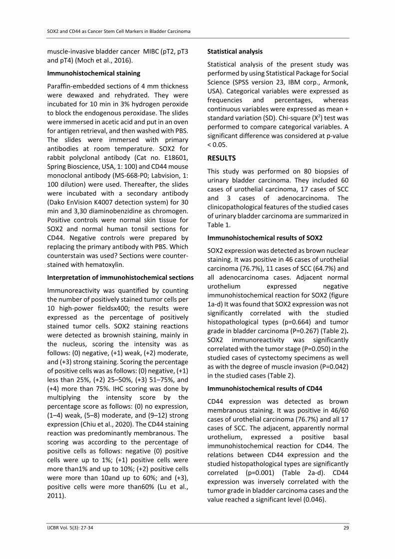
Statistical analysis
Statistical analysis of the present study was performed by using Statistical Package for Social Science (SPSS version 23, IBM corp., Armonk, USA). Categorical variables were expressed as frequencies and percentages, whereas continuous variables were expressed as mean + standard variation (SD). Chi-square (X2) test was performed to compare categorical variables. A significant difference was considered at p-value < 0.05.
RESULTS
This study was performed on 80 biopsies of urinary bladder carcinoma. They included 60 cases of urothelial carcinoma, 17 cases of SCC and 3 cases of adenocarcinoma. The clinicopathological features of the studied cases of urinary bladder carcinoma are summarized in Table 1.
Immunohistochemical results of SOX2
SOX2 expression was detected as brown nuclear staining. It was positive in 46 cases of urothelial carcinoma (76.7%), 11 cases of SCC (64.7%) and all adenocarcinoma cases. Adjacent normal urothelium expressed negative immunohistochemical reaction for SOX2 (figure 1a-d) It was found that SOX2 expression was not significantly correlated with the studied histopathological types (p=0.664) and tumor grade in bladder carcinoma (P=0.267) (Table 2). SOX2 immunoreactivity was significantly correlated with the tumor stage (P=0.050) in the studied cases of cystectomy specimens as well as with the degree of muscle invasion (P=0.042) in the studied cases (Table 2).
Immunohistochemical results of CD44
CD44 expression was detected as brown membranous staining. It was positive in 46/60 cases of urothelial carcinoma (76.7%) and all 17 cases of SCC. The adjacent, apparently normal urothelium, expressed a positive basal immunohistochemical reaction for CD44. The relations between CD44 expression and the studied histopathological types are significantly correlated (p=0.001) (Table 2a-d). CD44 expression was inversely correlated with the tumor grade in bladder carcinoma cases and the value reached a significant level (0.046).

Amer and Amer, 2021
IJCBR Vol. 5(3): 27-34 30
Table 1. Summary of clinicopathological features of studied cases of urinary bladder carcinoma
Clinicopathological features Total (8) (%)
Age (Mean ± SD.) 62.86 ± 8.89 <40 years 2 2.5% >40 years 78 97.5% Gender Male 64 80% Female 16 20% Histopathological type Urothelial 60 75% SCC 17 21.2% Adenocarcinoma 3 3.8% Grade High grade 54 67.5% Low grade 26 32.5%
Degree of muscle invasion (80) Absent 28 (35) 35% Present 52 (65) 65%
Table 2. The relation of SOX2 and CD44 expression with different histopathological features
Variables SOX2 -ve SOX2 +ve p-value CD44 -ve CD44 +ve p-value
Histopathological type Urothelial (60) 14 (23.3%) 46 (76.7%) p=MC
0.664 14 (23.3%) 46 (76.7%) p=MC
*0.001 SCC (17) 6 (35.3%) 11 (64.7%) 0 17 (100%) Adenocarcinoma (2) 0 2 (100%) 2 (100%) 0 Histopathological Grade Low grade (26) 9 (34.6%) 17 (65.4%) 0.168 2 (7.7%) 24 (92.3%) *0.040 High grade (54) 11 (20.4%) 43 (79.6%) 15 (27.8%) 39 (72.2%) Muscle invasion (80) Ta, T1 (28) 11 (39.3%) 17 (60.7%) *0.030 0 28 (100%) *0.001 T2, T3, T4 (52) 9 (17.3%) 43 (82.7%) 17 (32.7%) 35 (67.3%) Stage (n=20) cystectomy
T2 (7) 5 (71.4%) 2 (28.6%) p=MC *0.050
3 (42.8%) 4 (57.2%) p=MC 0.374 T3 (11) 2 (18.2%) 9 (81.8%) 2 (18.2%) 9 (81.8%)
T4 (2) 0 2 (100%) 1 (50%) 1 (50%)
square test, *: Statistically significant at p < 0.05-square test, FE: Fisher Exact for Chi-square test, MC: Monte Carlo for Chi-: Chi2c
As regards the tumor stage, it was noticed that CD44 expression decreased in tumors with high stage, but the values did not reach statistically significant levels (p=0.374). Moreover, CD44 expression significantly decreased with muscle invasion in the studied cases (p=0.001) (Table 2 & Figure 2a-d)
DISCUSSION
Cancer stem cells are a subtype of cancer cells in tumors that have stem cell-like features. CSCs were identified in various types of cancer (Lu et al., 2010). CSCs in bladder cancer have been studied frequently, but the roles of bladder CSCs are still controversial Bass et al, 2009. CSCs are supposed for carcinogenesis initiation with higher apoptosis resistance properties than
differentiated cancer cells. Chemoresistance is a serious problem for high-grade bladder cancer patients during prolonged therapy. Recent researches detect that cancer stem-like cells may play role in recurrence and chemoresistance. (Namekawa et al., 2020).
SOX2, MYC, KLF4and OCT4 are transcription factors involved in embryonic stem cell (ESC) and induced pluripotent stem cell (iPSC) signals. They have been associated with tumor progression in different carcinoma (Ben-Porath et al., 2008). SOX2 plays important role in the self-renewal and differentiation of progenitor cells in different adult tissue (Hu et al, 2010). Over-expression in CSCs plays an essential role in tumor genesis and the progression of various

SOX2 and CD44 as Cancer Stem Cell Markers in Bladder Carcinoma
IJCBR Vol. 5(3): 27-34 31
Figure 1. (a) Non-infiltrating papillary urothelial carcinoma showing negative SOX2 immunoreactivity (x200), (b) Low grade Infiltrating urothelial carcinoma showing positive nuclear expression of SOX2 score (+1) (x200), (c) High grade Infiltrating urothelial carcinoma showing positive nuclear expression of SOX2 score (+2) (x400) and (d) Well-differentiated squamous cell carcinoma showing positive nuclear expression of SOX2 score (+3) (x100).
types of human carcinomas (Chen et al., 2012). The results of the present study revealed that positive SOX2 expression was significant with advanced tumor stage, and with evident muscle invasion. Intense SOX2 expression by carcinoma cells is associated to a great extent, with poor differentiation states, as SOX2 is found to make cells in a state of undifferentiating by regulating the transcription factor called Nanog (Papapetrou et al., 2009). These results are in agreement with those of Gayyed and Twfiek (2015) who reported the expression of SOX2 and the staging of the tumor is significant, suggesting that SOX2 could be used as a molecular marker for the prognosis of bladder tumors. In previous researches, SOX2 was highly expressed in high-grade urothelial carcinoma (Ruan et al., 2013). Chiu et al. (2020) also found that positive SOX2 expression was significantly contributed to bladder tumor recurrence after surgical resection. They recommended that a high percentage of SOX2
positive cells should support more radical therapy. They concluded that SOX2 develops bladder cancer cell by induction of the IGF2/IGF1R pathway; pharmacological inhibition of this pathway inhibits bladder cancer cell survival.
These results highlight the role of SOX2 in oncogenesis and therapeutic targets in bladder carcinoma. CD44 is a cell surface marker linked with CSCs in various human tumors, including urinary bladder carcinoma (Klatte et al., 2010). In our study, CD44 was expressed in 78.8% of the studied cases of urinary bladder carcinoma. Between cases of urothelial carcinoma, CD44 expression was inversely correlated with tumor grade. Similarly, Omran and Ata (2012), Abd El-Fattah et al. (2014) and Yikilmaz et al. (2016) found a statistically significant decrease in CD44 expression with the increase in the tumor grade in cases of urothelial carcinoma. This could be explained by the more aggressive behavior usually seen with high-grade tumors with loss of

Amer and Amer, 2021
IJCBR Vol. 5(3): 27-34 32
Figure 2. (a) Normal urothelium adjacent to urothelial carcinoma showing basal positive CD44 membranous immunoreactivity score (+1) (x200), (b) Papillary urothelial neoplasm of low malignant potential showing positive CD44 membranous immunoreactivity score (+2) (x400), (c) Low grade infiltrating urothelial carcinoma showing negative CD44 immunoreactivity (x200) and (d) High grade infiltrating urothelial carcinoma showing negative CD44 immunoreactivity (x200). adhesion molecules such as CD44. Regarding, the relationship between CD44 immunoreactivity and the stage of urinary bladder carcinoma, it was noticed that negative CD44 expression within tumors with high stage, but did not reach statistically significant values.
This observation might be attributed to the small number of specimens of cystectomy in the study. However, among TUR specimens, CD44 expression was inversely correlated with muscle invasion. These results coincided with those reported by Omran and Ata (2012), Abd El-Fattah et al. (2014) and Yikilmaz et al. (2016) who revealed that CD44 expression inversely correlated with the tumor stage, which may enable prediction of the tumor progression, as loss or decrease in the adhesion molecule (CD44) would stimulate invasiveness of tumor cells. On the other hand, Keymoosi et al. (2014) reported that CD44 positivity was detected in 43% of urothelial carcinoma cases, which was correlated with high tumor grades and recurrence with no significant association
lamina propria involvement and muscular invasion. Wu et al. (2018) reported that the CD44 positive staining was significantly correlated with high tumor stage, lymph node metastasis and recurrence in muscle-invasive bladder cancer patients with curative treatment, these results were in agreement with Siddiqui et al. (2020) who reported that bladder carcinoma cases with CD44 positive expression had poor prognosis with a high rate of recurrence.
CONCLUSIONS
SOX2 overexpression could be utilized as a marker of poor progression in bladder carcinoma cases. It could be a target for an efficient therapeutic strategy of BC treatment, high grades and more liability for infiltration. BC is associated with low expression or complete loss of CD44 immunoreactivity.
CONFLICTS OF INTEREST
All authors declared no conflicts of interest.

SOX2 and CD44 as Cancer Stem Cell Markers in Bladder Carcinoma
IJCBR Vol. 5(3): 27-34 33
FUND
No fund was received for this work.
REFERENCES Abd El-Fattah GA, El-Sawi RM, Abd-Rabuh RM
(2014). Prognostic value of CD44 and HER2/Neu in invasive transitional cell carcinoma of the urinary bladder. Medical Journal of Cairo University, 82:81–87.
Abugomaa A, Elbadawy M, Yamawaki H, Usui T, Sasaki K (2020). Emerging roles of cancer stem cells in bladder cancer progression, tumorigenesis, and resistance to chemotherapy: A potential therapeutic target for bladder cancer. Cells, 9(1): 235.
Al-Sharaky DR, Kandil MA EH, Aiad HAS, El-Hosary EM, Alagizy HA, Elshenawy MAS, El-Rebey HS (2020). ROC-1, P21 and CAIX as markers of tumor aggressiveness in bladder carcinoma in Egyptian patients. Diagnostic Pathology, 15: 1-11.
Atef A, Bedeer A. (2021). Evaluation of HER2/neu and SPINK1 as an adjunct to the prognosis of urothelial dysplasia and urothelial carcinoma of the urinary bladder. International Journal of Cancer and Biomedical Research, 5(2):111-122.
Atlasi Y, Mowla SJ, Ziaee SAM, Bahrami AR (2007). OCT-4, an embryonic stem cell marker, is highly expressed in bladder cancer. International Journal of Cancer,120, 1598–1602.
Bass A J, Watanabe H, Mermel C H, Yu S, Perner S, Verhaak R G, Kim S Y, Wardwell L , Ramos AH (2009). SOX2 is an amplified lineage-survival oncogene in lung and esophageal squamous cell carcinomas. Nature Genetics, 41(11): 1238-1242.
Bellmunt, J (2018). Stem-like signature predicting disease progression in early stage bladder cancer. The Role of E2F3 and SOX4. Biomedicines, 6(3): 85.
Ben-Porath I, Thomson MW, Carey V J, Ge R, Bell G W, Regev A, Weinberg R A (2008). An embryonic stem cell–like gene expression signature in poorly differentiated aggressive human tumors. Nature Genetics, 40(5): 499.
Brandt WD, Matsui W, Rosenberg JE, He X, Ling S, Schaeffer EM, ,Berman DM (2009). Urothelial carcinoma: stem cells on the edge. Cancer and Metastasis Reviews, 28(3-4): 291-304.
Chen S, Xu Y, ChenY, LiX, Mou W, Wang L, Liu Y, Reisfeld RA, Xiang R ,LiN (2012). SOX2 gene regulates the transcriptional network of oncogenes and affects tumorigenesis of human lung cancer cells. PloS One, 7(5): e36326.
Chiu YF, Wu CC, Kuo MH, Miao CC, Zheng MY, Chen PY, Lin SC, Chang JL, WangYH, Chou YT (2020). Critical role of SOX2–IGF2 signaling in aggressiveness of bladder cancer. Scientific Reports, 10(1): 1-13.
Elbadawy M, UsuiT, MoriT, Tsunedomi R, Hazama S, Nabeta R, Uchide T, Fukushima R, Yoshida T, Tanaka T (2019). Establishment of a novel experimental model for muscle-invasive bladder cancer using a dog bladder cancer organoid culture. Cancer Science, 110(9): 2806-2821.
Ferone G, Song JY, Sutherland KD, Bhaskaran R, MonkhorstK, Lambooij JP, Proost N, Gargiulo G, Berns A (2016). SOX2 is the determining oncogenic switch in promoting lung squamous cell carcinoma from different cells of origin. Cancer Cell 30: 519–532.
Garg M (2015). Urothelial cancer stem cells and epithelial plasticity: current concepts and therapeutic implications in bladder cancer. Cancer and Metastasis Reviews, 34(4): 691-701.
Gayyed MF, Tawfiek ER (2015). Utility of SOX2 and livin co-expression in the prognosis of bladder cancer with bilharzial and non-bilharzial bladder status. World Journal of Oncology, 6(5): 446-455.
Heabah NA, Bedeer AE. (2021). Assessment of PD-L1 and p53 expression in urinary bladder carcinoma: Association with different clinicopathologic characteristics. International Journal of Cancer and Biomedical Research, 5(1):15-26.
Hatina J, Schulz WA (2012). Stem cells in the biology of normal urothelium and urothelial carcinoma. Neoplasma, 59(6): 728-736.
Hu Q, Zhang L, Wen J, Wang S, Li M, Feng R, Yang X, Li L (2010). The EGF receptor-sox2-EGF receptor feedback loop positively regulates the self-renewal of neural precursor cells. Stem Cells, 28(2): 279-286.
Hussenet T, du Manoir S (2010). SOX2 in squamous cell carcinoma: amplifying a pleiotropic oncogene along carcinogenesis. Cell Cycle, 9(8): 1480-1486.
Jordan C T (2009). Cancer stem cells: controversial or just misunderstood? Cell Stem Cell, 4(3): 203-205.
Keymoosi H, Gheytanchi E, Asgari M, Shariftabrizi A, Madjd, Z. (2014). ALDH1 in combination with CD44 as putative cancer stem cell markers are correlated with poor prognosis in urothelial carcinoma of the urinary bladder. Asian Pacific Journal of Cancer Prevention, 15(5): 2013-20.
Kitamura H, Torigoe T, Hirohashi Y, Asanuma H, Inoue R, Nishida S, Tanaka T, Fukuta F,

Amer and Amer, 2021
IJCBR Vol. 5(3): 27-34 34
Masumori N, Sato N, Tsukamoto T (2013). Prognostic impact of the expression of ALDH1 and SOX2 in urothelial cancer of the upper urinary tract. Modern Pathology. 26: 117–124.
Klatte T, Seligson DB, Rao JY, Yu H, de Martino M, Garraway I, et al (2010). Absent CD44v6 expression is an independent predictor of poor urothelialbladder cancer outcome. The Journal of Urology,183: 2403–2408.
Kuncova´ J, Urban M, Mandys V (2007). Expression of CD44s and CD44v6 in transitional cell carcinomas of the urinary bladder: comparison with tumour grade, proliferative activity and p53 immunoreactivity of tumour cells. APMIS 115:1194–1205.
Lu Y, Futtner, C, Rock JR, Xu X, Whitworth W, Hogan BL, Onaitis MW (2010). Evidence that SOX2 overexpression is oncogenic in the lung. PloS One, 5(6): e11022.
Lu¨ X, Xu K, Lu¨ H, Yin Y, Ma C, Liu Y, et al (2011). CD44 +/CD24 – cells are transit progenitors and do not determine the molecular subtypes and clinical parameters in breast carcinomas. Ultrastructural Pathology,35: 72–78.
Lundberg IV, Edin S, Eklof V, Oberg A, Palmqvist R, Wikberg ML (2016). SOX2 expression is associated with a cancer stem cell state and down-regulation of CDX2 in colorectal cancer. BMC Cancer 16: 471.
Moch H, Humphrey PA, Ulbright TM, Reuter VE (2016). WHO classification of tumours of the urinary system and male genital organs (Vol. 8). International Agency for Research on Cancer (IARC). European Urology:1-14.
Namekawa T, Ikeda K, Horie-Inoue K, Suzuki T, Okamoto K, Ichikawa T, Yano A, Kawakami S, Inoue S (2020). ALDH1A1 in patient-derived bladder cancer spheroids activates retinoic acid signaling leading to TUBB3 overexpression and tumor progression. International Journal of Cancer, 146(4): 1099-1113.
Ohishi T, Koga F, Migita T (2016). Bladder cancer stem-like cells: their origin and therapeutic perspectives. International Journal of Molecular Sciences, 17(1): 43.
Omran OM, Ata HS (2012). CD44s and CD44v6 in diagnosis and prognosis of human bladder cancer. Ultrastructural Pathology, 36:145–152.
Papapetrou EP, Tomishima MJ, Chambers SM, Mica Y, Reed E, Menon J, Tabar V, Mo Q, Studer L, Sadelain M (2009). Stoichiometric and
temporal requirements of Oct4, Sox2, Klf4, and c-Myc expression for efficient human iPSC induction and differentiation. Proceedings of the National Academy of Sciences of the United States of America, 106:12759–12764.
Ruan J, Wei B, Xu Z, Yang S, Zhou Y, Yu M, Liang J, Jin K, Huang X, Lu P, Cheng H (2013). Predictive value of Sox2 expression in transurethral resection specimens in patients with T1 bladder cancer. Medical Oncology, 30: 445.
Sarkar A, Hochedlinger K (2013). The sox family of transcription factors: versatile regulators of stem and progenitor cell fate. Cell Stem Cell 12: 15–30.
Siddiqui Z, Srivastava AN, Sankhwar SN, Dalela D, Singh V, Zaidi N, Fatima N, Bno I, Anjum S (2020). Synergic effects of cancer stem cells markers, CD44 and embryonic stem cell transcription factor Nanog, on bladder cancer prognosis. British Journal of Biomedical Science, 77(2): 69-75.
Tung CL, Hou PH, Kao YL, Huang YW, Shen CC, Cheng YH, Wu SF, Lee MS, Li C (2010) SOX2 modulates alternative splicing in transitional cell carcinoma. Biochem Biophys Res Commun Biochemical and Biophysical Research Communication 393(3): 420-425.
Usui T, Sakurai M, Nishikawa S, Umata K, Nemoto Y, Haraguchi T, Itamoto K, Mizuno T, Noguchi S, Iwai S (2017). Establishment of a dog primary prostate cancer organoid using the urine cancer stem cells. Cancer Science, 108(12): 2383-2392.
Wu CT, Lin WY, Chen WC, Chen MF (2018). Predictive value of CD44 in muscle-invasive bladder cancer and its relationship with IL-6 signaling. Annals of Surgical Oncology, 25(12): 3518-3526.
Yikilmaz TN, Dirim A, Ayva ES, Ozdemir H, Ozkardes H (2016). Clinical use of tumor markers for the detection and prognosis of bladder carcinoma: A comparison of CD44, cytokeratin 20 and survivin. Urology Journal, 13(3): 2677-2683.
Zhang Y, Wang Z, Yu J, zhong Shi J, Wang C, hua Fu W, Chen ZW, Yang J (2012). Cancer stem-like cells contribute to cisplatin resistance and progression in bladder cancer. Cancer Letters, 322(1): 70-77.
Zhu F, Qian W, Zhang H, Liang Y, Wu M, Zhang Y, Zhang X, Gao Q, Li, Y (2017). SOX2 is a marker for stem-like tumor cells in bladder cancer. Stem Cell Reports, 9(2): 429-437.