impact of detergents on the protein histochemistry of various cell types of the gill epithelium of...
TRANSCRIPT

ECOTOXICOLOGY AND ENVIRONMENTAL SAFETY 1$206-2 11 ( 1988)
Impact of Detergents on the Protein Histochemistry of Various Cell Types of the Gill Epithelium of Rita rita
DEBASISH ROY’
Department of Zoology, Banaras Hindu University, Varanasi-221005, India and Department of Zoology, Utkal University, Bhubaneswar, India
Received September 23, I98 7
Fish, Rita rita, were exposed to an anionic detergent, dodecylbenzene sodium sulfonate, 6.9 mg per litre of tap water (96~hr LCso of the detergent). A gradual decrease in the protein constitu- ents of the major cell types, viz, the epithelial cells and the goblet mucous cells in the epithelium lining the gill arch, gill filament, and club cells present only in the gill arch epithelium has been observed by using a series of h&chemical techniques. B 1988 Academic FWZ, IDC.
INTRODUCTION
The widespread use of synthetic detergents, due to their lower cost and better wash- ing capability, has considerably added to the pollution of cultivable waters and has posed a great threat to the whole of the aquatic environment. Abel ( 1974) studied in detail earlier reports of the effects induced by synthetic detergents on various systems of aquatic organisms, including fish and microinvertebrates. This review infers that a considerable amount of literature exists on the histomorphological changes influ- enced by detergents in fish gill. Subsequently, Abel and Skidmore (1975), Abel (1976), Fukuda (1983), and Misra et al. (1985) shed light on the histopathological changes in gills caused by detergents at both light and electron microscopic levels. However, a detailed examination of histochemical alterations in the nature of differ- ent cell types, present in fish gills, has altogether been neglected. In this communica- tion, the changes effected by an anionic detergent, dodecylbenzene sodium sulfonate, in the protein constituents of various cell types of the gills of a freshwater teleost, Rita rita, have been incorporated.
MATERIALS AND METHODS
Live specimens of R. rita ( 12- 16 cm in length) were collected from the riversides of the Ganges at Varanasi, U.P., and were acclimated to optimum laboratory condi- tions for about 30 days before experimentation. Fish during acclimatization were fed with minced goat liver on alternate days. Fecal material and uneaten food were siphoned out of the aquaria every day. Static bioassay tests were performed as de- scribed in the Standard methods (APHA, AWWA, & WPCF, 15th edition) for evalu- ating the 96-hr LC5,, of an anionic detergent, dodecylbenzene sodium sulphonate. All the subsequent experiments were set at 6.9 mg litre-’ (96~hr LCX, of the detergent). Calculated amounts of detergent for 30 litres of water in aquaria of 2 X 1 X 1.5 ft were dissolved in a small amount of water and then thoroughly mixed into the aquaria water. Fish from both control and experimental aquaria were removed at
’ Present address, 464; Saheednagar, Bhubaneswar, Orissa, India
0147-6513/88 $3.00 Copyright 8 1988 by Academic F’res& Inc. AS rights of reproduction in any form msetved.
206
DETERGENT-INDUCED CHANGES IN GILL EPITHELIUM 207
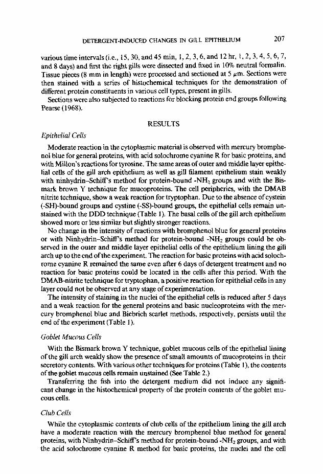
various time intervals (i.e., 15, 30, and 45 min, 1,2,3,6, and 12 hr, 1,2, 3,4, 5,6,7, and 8 days) and first the right gills were dissected and fixed in 10% neutral formalin. Tissue pieces (8 mm in length) were processed and sectioned at 5 pm. Sections were then stained with a series of histochemical techniques for the demonstration of different protein constituents in various cell types, present in gills.
Sections were also subjected to reactions for blocking protein end groups following Pearse (1968).
RESULTS
Epithelial Cells
Moderate reaction in the cytoplasmic material is observed with mercury bromphe- no1 blue for general proteins, with acid solochrome cyanine R for basic proteins, and with Millon’s reactions for tyrosine. The same areas of outer and middle layer epithe- lial cells of the gill arch epithelium as well as gill filament epithelium stain weakly with ninhydrin-SchifFs method for protein-bound -NH2 groups and with the Bis- mark brown Y technique for mucoproteins. The cell peripheries, with the DMAB nitrite technique, show a weak reaction for tryptophan. Due to the absence of cystein (-SH)-bound groups and cystine (-SS)-bound groups, the epithelial cells remain un- stained with the DDD technique (Table 1). The basal cells of the gill arch epithelium showed more or less similar but slightly stronger reactions.
No change in the intensity of reactions with bromphenol blue for general proteins or with Ninhydrin-Schiffs method for protein-bound -NH2 groups could be ob- served in the outer and middle layer epithelial cells of the epithelium lining the gill arch up to the end of the experiment. The reaction for basic proteins with acid soloch- rome cyanine R remained the same even after 6 days of detergent treatment and no reaction for basic proteins could be located in the cells after this period. With the DMAB-nitrite technique for tryptophan, a positive reaction for epithelial cells in any layer could not be observed at any stage of experimentation.
The intensity of staining in the nuclei of the epithelial cells is reduced after 5 days and a weak reaction for the general proteins and basic nucleoproteins with the mer- cury bromphenol blue and Biebrich scarlet methods, respectively, persists until the end of the experiment (Table 1).
Goblet Mucous Cells
With the Bismark brown Y technique, goblet mucous cells of the epithelial lining of the gill arch weakly show the presence of small amounts of mucoproteins in their secretory contents. With various other techniques for proteins (Table I), the contents of the goblet mucous cells remain unstained (See Table 2.)
Transferring the fish into the detergent medium did not induce any signifi- cant change in the histoehemical property of the protein contents of the goblet mu- cous cells.
Club Cells
While the cytoplasmic contents of club cells of the epithelium lining the gill arch have a moderate reaction with the mercury bromphenol blue method for genera1 proteins, with Ninhydrin-Schiff s method for protein-bound -NH* groups, and with the acid solochrome cyanine R method for basic proteins, the nuclei and the cell

TABL
E 1
A SU
MM
ARY
OF
HIS
T~C
HEM
ICAL
C
HAN
GES
IN
F~O
TEIN
CO
NSTI
TUEN
TS
OF
EPIT
HEL
IAL
CELL
S IN
THE
GIL
L EP
ITH
ELIIJ
M
OF
Ritu
rit
a AT
VAR
IOU
S IN
TER
VALS
O
F DE
TERG
ENT
EX
PO
SU
RE
Epith
elial
ceUs
Outer
lay
er Mi
ddle
layer
Basa
l lay
er
Chem
ical
ccm
stitu
ents
Histo
chem
ical
techn
ique
Color
C
I 2
3 4
5 6
1 8
C 1
2 3
4 5
6 7
8 C
1 2
3 4
5 6
7 8
Gene
ral
pmtei
n
Basic
pro
teins
Ba
sic
0ucIe
opm
tein.s
Ty
msin
e
Tlypto
phan
Qstei
n bo
und
~ulry
dryl
(-SH)
gr
oups
Cysti
ne
boun
d dis
ulfide
(-S
S)
grou
ps
Muc
apm
teins
Hg-b
mm
phen
ol blu
e BI
Ninh
ydrin
/Sch
iff P
Deam
inatio
n/ninh
ydrin
/ -
Schil
T Ac
id so
lochr
ome
cyan
ine
R BI
Bie
brich
sc
arlet
S
Millio
n rea
ction
P
kdina
tion/M
illion
reacti
on
- DM
AEnit
rite
BI
Perfo
rmic
acid/
oxida
tion/
- DM
AB
nitrite
an
d iod
inatio
n/DM
AB
nitrite
Di
bydm
xydin
aphth
yl-
- dis
ultide
(D
DD)
Male
imide
ox
idatio
n/DDD
-
and
icdina
tion/D
DD
Petfo
rmic
acid/
AkGn
blu
e -
Perfo
rmic
acid/
oxida
tion/
- pe
rform
ic ac
id/Al&
n blu
e an
d thi
c&co
late
redU
CtiO
~lpn
form
iC
acid/
Alcian
blu
e Bi
smar
k br
own
Y
Br
r
__-
- -
- -
- --
:r +.
+*
f.
+.
- -
+. p
;:
+b +
b fb
*b
_+b
p +’
+b
+*
+a
+. +
. +.
+.
- -
+. +
. __
_-
- -
----
- -
- -
- -
- -
+*
- -
- _
- -
- -
- -
-
1:
;I +.
-
- 1:
;.I
*t
J *b
*b
+.
+.
+.
+
* +.
-
- -
- -
- -
- -
_ _
- -
- -
- -
- -
- -
- -
___-
_-
- -
---
----
--
- *.
-
- -
- -
- -
-
t+’ +a
t+’ +b
t+’ f=
*’
- -
- -
- _
- _
1:
;: +*
+.
-
- 1:
+b
+b
;I
*b
+b
!z
g p
+a
+.
+a
+.
+.
- 1
E -
- -
- -
- -
_ iz
-
- -
- -
_ -
_ -
- -
- -
- -
- 5

TABL
E 2
A SU
MM
ARYO
FHIS
TOC
HEM
ICAL
CH
ANG
ESIN
PRO
TEIN
CO
NST
ITU
ENTS
OFG
LAN
DC
ELLS
INTH
EGIL
LEPI
THEL
IUM
OF
Ritu
rita
ATV
AR
IOU
SIN
TER
VA
LSO
FDE
TER
GE
NTE
XP
~SU
RE
Club
cells
Goble
t HU
MUS
cells
outer
lay
er M
iddl
elay
eran
dbas
alla
yn
CO
kX
Chem
ical
cons
tituen
t Hi
stach
emica
itech
nique
sy
mbo
l C
1 2
3 4
5 67
8C
1234
5678
C I23
4567
8
Gene
ral
protei
ns
Hg-br
omph
enol
blue
F’m
tein
boun
d-NH1
Ni
nhyd
rin/S
cbiff
grou
ps
Deam
inatio
n/ninh
ydrin
/ sC
hi5
Basic
pro
teins
Ac
id so
lochr
ome
cyan
ine
R
Basic
nu
cleop
rotein
s Bie
brich
sc
arlet
Ty
msin
e
Tryp
topha
n
Cyste
in bo
und
sWdry
1 (-S
H)
groJ
Jps
Cysti
ne
boun
d dis
ulphid
e (-S
S)
grou
ps
Mien
re
actio
n Io
dina
tion/
Mill
onre
atiio
n D
MA
B-n
itrite
P
erfo
rmic
ac
id ox
idatio
n/ DM
AB
nitrite
an
d iad
inatio
n/DM
A&oit
rite
Diby
droxy
dinap
hthyl-
dis
ulphid
e (D
DD)
Male
imide
ox
idatio
n/DDD
an
d lod
inatio
n/DDD
Pe
rform
ic ac
id/Al&
n blu
e
Per
form
icac
idox
idal
ion/
pe
rfotic
ac
id/Alc
ian
blue
and
thiqt
lywlat
e re
ducti
on/
perfo
rmic
acid/
Al&n
blue
Bi.&
ark
brow
n Y
Bl
P - BI
s P - BI
- - - - BI
- -
-__
----
----
- -
- _
__
- -
--_
- P
tbd
;b - - - - - - -
- -
- -
- i’
9 +’

210 DEBASISH ROY
boundaries have a strong reaction with all these techniques. With Millon’s reaction for tyrosine, with Bismark brown Y for mucoproteins, and with DMAB-nitrite for tryptophan, the cytoplasmic contents of these cells show a weak reaction. However, only the nuclei of these cells exhibit a moderate reaction with the Biebrich scarlet method for basic nucleoproteins.
The contents and the nuclei of club cells in the deeper layer of the epithelium lining the gill arch stain comparatively less darkly with the mercury bromphenol blue and acid solochrome cyanine R methods.
Histochemical analysis does not indicate any substantial change in the staining behavior of the club cells of the epithelium lining the gill arch up to 2 days of detergent treatment. Subsequently, the intensity of the staining reaction decreases and a weak reaction for general proteins, basic proteins, and protein-bound -NH2 groups in the club cells of all the strata is left, after the third day onward, until the end of the experi- ment. Positive reaction for tyrosine with Millon’s reaction, for mucoproteins with Bismark brown Y, and for tryptophan with the DMAB-nitrite technique disappears after the fourth day.
The staining intensity with Biebrich scarlet for basic nucleoproteins becomes weak alter 2 days and continues to remain weak until the end of the experiment.
DISCUSSION
The present investigation observes a gradual decrease in the intensity ofthe staining reaction in different protein constituents of various cell types, present in the gill arch and gill filament epithelium, indicating the occurrence of certain changes in the chemistry of the protein moieties induced by the detergent.
The series of events and the mechanism of detergent poisoning leading to death of the fish are not yet known. The immediate cause ofdeath for acute detergent intoxica- tion where extensive gill damage occurs is likely to be either asphyxiation or loss of osmotic or ionic stability. But asphyxiation alone is never the only cause of fish death, rather, it is difficult to say whether asphyxiation is the primary cause associated with various other secondary factors or whether these secondary factors together give rise to the primary one.
Much research has been carried out in the past on the interaction of proteins and synthetic detergents Putnam (1948). It is established that detergents in low concentra- tion can alter proteins reversibly or irreversibly. At higher concentrations, when the detergent is largely in the form of micelles, detergents have the property of solubilizing organic material. Hotchkiss (1946), while discussing the bactericidal effects of syn- thetic detergents, suggested that a succession of events takes place, the first of which involves combination with oppositely charged ions on the bacterial sites. He believes this to be succeeded by membrane injury causing cytolysis, autolysis, possibly selec- tive inactivation of metabolic systems and eventual death. The possibility of a similar mechanism in fish gill epithelium may not be ignored. Helenius and Simon (1975) explained that the presence of highly charged proteins or glycoproteins, or a dense protein network on the surface of the biological membrane, may affect the penetra- tion, thereby facilitating the effects of detergents on the membrane. Few authors in the 1940s considered that synthetic detergents could produce their diverse effects by a combination with proteins primarily through the action of electrostatic forces.
DETERGENT-INDUCED CHANGES IN GILL EPITHELIUM 211
CONCLUSIONS
Synthetic detergents are known to exert disruptive effect on the gills of fish. This report, for the first time, aims at the study of change in the different protein moieties in various cell types of gill arch epithelium. The typical manner of decrease of staining reaction of protein constituents indicate the detergent action on the gill epithelium to be instant and the detergents may not only be exerting their effects through contact, but also may be penetrating through the membrane.
ACKNOWLEDGMENTS
Thanks are due to the University Grants Commission, New Delhi, and the concerned Principal Investi- gator for necessary facilities and financial assistance.
REFERENCES
ABEL, P. D. (1974). Toxicity of synthetic detergents to fish and aquatic invertebrates. J. Fish Biol. 6,279- 298.
ABEL, P. D. ( 1976). Toxic action of several lethal concentrations of an anionic detergent on the gills of the brown trout (Salmo trutta. L) J. Fish Biol. 9,441~446.
ABEL, P. D., AND SKIDMORE, J. F. (1975). Toxic effects of an anionic detergent on the gills of rainbow trout. Water Res. 9,759-765.
APHA, AWWA, AND WPCF (1980). Standard methods for the examination of water and waste water, 15th ed., Washington.
FUKUDA, Y. (1983). Specific reactions of gold fish gills exposed to linear alkylbenzene sulfonate. Japan. .I. Ichthyol. 30(3), 268-274.
HELENIUS, A., AND SIMON, K. ( 1975). Solubilization of membranes by detergents. B&him. Biophys. Acta 41529-79.
HOTCHKISS, R. D. (1946). The nature of the bacteriocidal action of surface active agents. Ann. N. Y. Acad. Sci. 46,479-493.
MISRA, V., LAL, H., CHAWLA, G., AND VISWANATHAN, P. N. (1985). Pathomorphological changes in the gills of fish fingerlings (Cirrhina mrigala) by linear alkylbenzene sulfonate. Ecotoxicol. Environ. Sat 10, 302-308.
PEAR%, A. G. E. (1968). Histochemistry: Theoretical and Applied, Vol. 1,3rd ed. Churchill, London. PUTNAM, F. W. (1948). The interactions of proteins and synthetic detergents. Adv. Protein Chem. 4, 79-
122.