immunosuppressive effect of litsea cubeba l. essential oil
TRANSCRIPT

International Journal of
Molecular Sciences
Article
Immunosuppressive Effect of Litsea cubebaL. Essential Oil on Dendritic Cell and ContactHypersensitivity Responses
Hsin-Chun Chen 1, Wen-Te Chang 2, You-Cheng Hseu 1, Hsing-Yu Chen 2,Cheng Hsuan Chuang 2, Chi-Chen Lin 3, Meng-Shiou Lee 2,* and Ming-Kuem Lin 2,*
1 Department of Cosmeceutics, College of Biopharmaceutical and Food Sciences, China Medical University,No. 91, Hsueh-Shih Road, Taichung 40402, Taiwan; [email protected] (H.-C.C.);[email protected] (Y.-C.H.)
2 Department of Chinese Pharmaceutical Sciences and Chinese Medicine Resources,College of Biopharmaceutical and Food Sciences, China Medical University, No. 91, Hsueh-Shih Road,Taichung 40402, Taiwan; [email protected] (W.-T.C.); [email protected] (H.-Y.C.);[email protected] (C.H.C.)
3 Institute of Medical Technology, College of Life Science, National Chung Hsing University, Taichung 402,Taiwan; [email protected]
* Correspondence: [email protected] (M.-S.L.); [email protected] (M.-K.L.);Tel.: +886-4-2205-3366 (ext. 5208) (M.-S.L.); +886-4-2205-3366 (ext. 5212) (M.-K.L.)
Academic Editor: Paula AndradeReceived: 27 May 2016; Accepted: 8 August 2016; Published: 12 August 2016
Abstract: Litsea cubeba L., also named as Makauy, is a traditional herb and has been used ascooking condiment or tea brewing to treat diseases for aborigines. The present study wasundertaken to explore the chemical compositions of the fruit essential oil of L. cubeba (LCEO) andthe immunomodulatory effect of LCEO on dendritic cells and mice. The LCEO was analyzedusing gas chromatography (GC) and gas chromatography/mass spectrometry (GC/MS) with directinjection (DI/GC) or headspace-solid phase microextraction (HS-SPME/GC). In total, 56 componentswere identified, of which 48 were detected by DI/GC and 49 were detected by HS-SPME/GC.The principal compounds were citral (neral and geranial). An immunosuppressive activity of LCEOwas investigated with bone marrow-derived dendritic cells (DCs) which have a critical role totrigger the adaptive immunity. Additionally, the inhibitory effect of LCEO on immune responsewas elucidated by performing the contact hypersensitivity (CHS) responses in mice. Our resultsclearly showed that LCEO decreases the production of TNF-α and cytokine IL-12 in a dose-dependentmanner in lipopolysaccharide (LPS)-stimulated DCs. CHS response and the infiltrative T cells wereinhibited in the tested ears of the mice co-treated with LCEO. We demonstrate, for the first time,that the LCEO mainly containing citral exhibits an immunosuppressive effect on DCs and mice,indicating that LCEO can potentially be applied in the treatment of CHS, inflammatory diseases, andautoimmune diseases.
Keywords: Litsea cubeba; essential oil; dendritic cell; immunosuppressive; citral
1. Introduction
In the immune system, various immune cells are highly communicative with each other byvarious cytokines and are in charge of the defense against foreign pathogen infection and maintaininghealth. Inflammation is a major component of our immune response. Although inflammation is anatural defense, the persistence of the process for abnormally long periods can be harmful and hasbeen recognized as a major risk factor for various human diseases, including cardiovascular disease,
Int. J. Mol. Sci. 2016, 17, 1319; doi:10.3390/ijms17081319 www.mdpi.com/journal/ijms

Int. J. Mol. Sci. 2016, 17, 1319 2 of 11
metabolic disorder, neurological disease, and cancer [1,2]. Thus, reduction of chronic inflammationwould be beneficial to prevent the pathological progression of these human diseases. In such cases, theinflammatory response should be suppressed. Among the immune cells, dendritic cells (DCs) are thebest antigen-presenting cells and are in charge of the induction of adaptive immunity [3–5]. To initiateadaptive immunity, DCs present a specified antigen on the surface to be recognized by naïve T cells.This recognition triggers the differentiation of the specified antigen-specific T cells. Next, a strongand specific T cell-based immune response is built up to attack the “pathogens” which present thespecified antigen. With such a critical role, DCs are thought to be an ideal target when attempting toevaluate potential immune modulators [6–8].
Litsea cubeba L. belongs to the family Lauraceae, which is widely distributed in Japan, Taiwan,Southern China, and Southeastern Asia [9]. All parts of this plant emanate a pungent gingery odor [10].Fruits of L. cubeba are spicy condiments, frequently used in the aboriginal cuisine of Taiwan [11]. Theessential oil of its fruit has been used as a flavor enhancer in cigarettes, cosmetics, and foods, and asraw material to produce citral (neral and geranial) [12]. Its pharmacological effects have been reportedto have an antimicrobial [9,13], antioxidative [14], anticancer [10,15], anti-inflammatory [11], andinsecticidal activities [12,16]. However, there is no report on immunosuppressive effects of L. cubebaon DCs and its relative immune response in vivo. In this study, L. cubeba essential oil (LCEO) wasextracted from the fresh fruits. The inhibitory effect of the LCEO on dendritic cell activation wasexamined. In addition, the contact hypersensitive response was conducted to examine the in vivoimmunosuppressive effects in mice.
2. Results and Discussion
2.1. Constituents of the Essential Oils
Punyarajun and Nandhasri (1981) extracted essential oils from unripe L. cubeba berries of Thaiorigin and the yield was ca. 3.0% [17]. Ho et al. (2010) used hydrodistillation to extract the leaf andfruit essential oils of L. cubeba from Taiwan, and the yields were 13.9% ± 0.09% and 4.0% ± 0.03%,v/w, respectively [10]. Liu and Yang (2012) extracted the fruit essential oils of L. cubeba from Taiwan,and the yields were 4.5% ± 0.2% [9]. Jiang et al. (2009) reported that the fruit of L. cubeba contains3%–5% of essential oils which are rich in citral [12]. In the present study, the yield of the essential oilsobtained from the fresh fruit of L. cubeba by steam distillation was 3.7% ± 0.4%. The yield is similar tothat reported in these published studies.
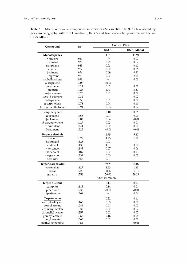
As shown in Table 1, a total of 48 components were identified by gas chromatography usingdirect injection (DI/GC). These components include 12 monoterpenes, five sesquiterpenes, seventerpene alcohols, three terpene aldehydes, two terpene ketones, six terpene esters, five terpene oxides,four aliphatic aldehydes, one aliphatic ketone, and three aliphatic ester. The principal compoundswere citral (neral and geranial) accounting for 88.02%. Other constituents identified in significantproportions were 6-methyl-5-hepten-2-one, β-myrcene, limonene, linalool, citronellal, and verbenol.Terpene aldehydes (89.25%) were the most abundant compounds in the essential oil (Table 1). In linewith the studies from Seo et al. [16], Ho et al. [10], Kejlová et al. [18], Liu and Yang [9], this studyshowed that terpene aldehydes were the most abundant volatile compounds and neral and geranialwere the most major components in the oil.
Int. J. Mol. Sci. 2016, 17, 1319 3 of 11
Table 1. Means of volatile compounds in Litsea cubeba essential oils (LCEO) analyzed bygas chromatography with direct injection (DI/GC) and headspace-solid phase microextraction(HS-SPME/GC).
Compound RI z Content (%) y
DI/GC HS-SPME/GC
Monoterpenes 4.01 11.91α-thujene 921 – w 0.02α-pinene 931 0.22 0.72
camphene 945 0.03 0.10sabinene 972 0.07 0.06β-pinene 976 0.09 0.20β-myrcene 980 0.77 2.11
α-phellandrene 998 – 0.01α-terpinene 1007 <0.01 –ρ-cymene 1014 0.01 0.01limonene 1026 2.73 8.50
cis-β-ocimene 1026 0.01 0.02trans-β-ocimene 1032 – 0.02γ-terpinene 1050 0.01 0.01α-terpinolene 1078 0.04 0.11
1,3,8-ρ-menthatriene 1094 0.03 0.02
Sesquiterpenes 0.10 0.06α-copaene 1366 0.01 0.01β-elemene 1382 0.04 <0.01
β-caryophyllene 1429 0.03 0.04α-humulene 1441 0.02 0.01δ-cadinene 1525 <0.01 <0.01
Terpene alcohols 2.75 5.22linalool 1079 1.23 1.11
isopulegol 1128 0.03 –verbenol 1130 1.31 3.81
α-terpineol 1183 0.07 0.06cis-carveol 1189 0.07 0.19cis-geraniol 1237 0.03 0.05
nerolidol 1558 0.01 –
Terpene aldehydes 89.25 75.09citronellal 1127 1.23 1.63
neral 1226 38.02 34.17geranial 1256 50.00 39.29
(2854.05 mmol/L)
Terpene ketone 0.14 0.10camphor 1113 0.14 0.04
piperitone 1230 <0.01 <0.01piperitenone 1308 – 0.06
Terpene ester 0.32 0.14methyl salicylate 1163 0.05 0.01
bornyl acetate 1286 0.01 0.02terpinenyl acetate 1335 0.07 0.02citronellyl acetate 1357 0.02 0.02
geranyl acetate 1362 0.16 0.06neryl acetate 1366 0.01 0.01
methyl cinnamate 1384 – <0.01

Int. J. Mol. Sci. 2016, 17, 1319 4 of 11
Table 1. Cont.
Compound RI z Content (%) y
DI/GC HS-SPME/GC
Terpene oxide 0.16 0.171,8-cineole 1019 0.12 0.14
trans-linalool oxide 1055 <0.01 0.02cis-rose oxide 1086 <0.01 <0.01
trans-rose oxide 1089 – <0.01limonene oxide 1128 0.01 –
caryophyllene oxide 1571 0.03 0.01
Aliphatic aldehydes 0.01 0.033-methyl butanal 631 <0.01 –2-methyl butanal 636 <0.01 –
pentanal 697 – <0.01hexanal 776 <0.01 0.01
2,6-dimethylhept-5-enal 1047 0.01 0.02
Aliphatic ketone 1.19 2.236-methyl-5-hepten-2-one 962 1.19 2.23
Aliphatic alcohol – <0.012-methyl-3-buten-2-ol 600 – <0.01
Aliphatic esters 0.01 0.01ethyl isovalerate 825 <0.01 –isoamyl acetate 864 0.01 0.01
ethyl tiglate 915 <0.01 <0.01Z Retention indices, using paraffin (C5-C25) as references; y Values are means of triplicates; w undetectable.
The headspace-solid phase microextraction (HS-SPME) method has been reported to be anexcellent tool for the analysis of herbs because it is simple, fast, and does not leave any residues [19].In this study, a total of 49 components were identified by GC and GC/MS with HS-SPME method.These components include 15 monoterpenes, five sesquiterpenes, seven terpene alcohols, three terpenealdehydes, three terpene ketones, seven terpene esters, six terpene oxides, four aliphatic aldehydes,one aliphatic ketone, one aliphatic alcohol, and three aliphatic esters. Terpene aldehydes (75.09%) werethe most abundant compounds in the oils (Table 1).
Comparative analysis of these compounds identified by these two methods, showed that56 compounds were detected in total, of which, 48 were identified by DI/GC and 49 by HS-SPME/GC.As shown in Table 1, HS-SPME/GC analysis revealed higher percentages of monoterpenes, terpenealcohols, and aliphatic ketone, but lower percentages of terpene aldehydes and terpene esters thanDI/GC analysis. Some monoterpenes such as α-thujene, α-phellandrene, and trans-β-ocimene couldbe identified only by HS-SPME/GC. This indicates that the more complete constituents of essentialoils can be identified with the combination of DI/GC and HS-SPME/GC.
2.2. L. cubeba Essential Oils (LCEO) Inhibited the Activation of Dendritic Cells (DCs)
To test that the cytotoxic effect of L. cubeba essential oils (LCEO) on dendritic cells (DCs), theviability of mouse bone marrow-derived DCs treated with the different concentrations of LCEO wasexamined. The result showed no significant effect of 5 × 10⁴-, 1 × 10⁵-, 2 × 10⁵- and 4 × 10⁵-folddiluted LCEO on DCs, although 5 × 10⁴-fold diluted LCEO exhibited a little cytotoxic effect (Figure 1).Thus, the concentrations of 1 × 10⁵-, 2 × 10⁵-, and 4 × 10⁵-fold diluted LCEO was used for thefollowing inhibition experiment. TNF-α and IL-12 are hallmarks of DC activation [3,6–8]. To elucidatethe immunomodulatory activity of the LCEO, the effect of LCEO on TNF-α and IL-12 production byDCs stimulated by lipopolysaccharides (LPS) was examined. The results showed that the amounts

Int. J. Mol. Sci. 2016, 17, 1319 5 of 11
of TNF-α and IL-12 produced by LPS-induced DCs were inhibited by the presence of LCEO in adose-dependent manner (Figure 2). This indicated that LCEO possess an inhibitory activity to DCactivation. The IC50 of LCEO for TNF-α and IL-12 was approximately 1 × 10⁵- and 2 × 10⁵-folddilution, respectively.Int. J. Mol. Sci. 2016, 17, 1319 5 of 11
Figure 1. L. cubeba essential oils (LCEO) did not impair cell viability of dendritic cells (DCs). DCs were treated with LCEO at different concentrations (5 × 10⁴-, 1 × 10⁵-, 2 × 10⁵- and 4 × 10⁵-fold dilutions) at 37 °C in 5% CO2/air for 24 h. The cytotoxicity of LCEO was examined by Cell Counting Kit-8 (CCK-8) assay (Sigma-Aldrich, St. Louis, MO, USA). NS p > 0.05 (Mann–Whitney U-test) for the comparison between LCEO-treated and untreated DCs.
(A) (B)
Figure 2. The release of TNF-α (A) and IL-12 (B) by lipopolysaccharide (LPS)-induced DCs were inhibited by LCEO. The DCs were treated with LPS or LPS + LCEO at different concentrations (1 × 10⁵-, 2 × 10⁵- and 4 × 10⁵-fold dilutions). Supernatants were collected after 6 h to detect TNF-α and 24 h to detect IL-12. The amounts of TNF-α and IL-12 were determined by enzyme-linked immuno sorbent assay (ELISA). Each value represents the mean ± SD (standard deviation) of the data obtained from three wells for each treatment. * p < 0.05, ** p < 0.01 and *** p < 0.001 (Mann–Whitney U-test) for the comparison between the LCEO treated LPS-activated DC groups and the untreated LPS-activated DC group. All data are representative of three independent experiments.
2.3. The Contact Hypersensitivity (CHS) Response Is Attenuated in Mice Co-Treated with LCEO
The above findings indicate that LCEO is able to inhibit the activation of DCs and, thus, we are able to postulate logically that LCEO is able to prevent DC-mediated immune response. Therefore, DNFB-induced CHS was performed to examine the inhibition as the immune response stimulated by DNFB is a type of cell-mediated response. Mice were sensitized by painting their abdomens with DNFB in the absence or presence of LCEO. The hypersensitivity response to DNFB at the ears was then examined. The results of the histological analyses (Figure 3A) and the increase of thickness of the tested ears (Figure 3B), showed that the tested ears were significantly swollen in DNFB-sensitized
Figure 1. L. cubeba essential oils (LCEO) did not impair cell viability of dendritic cells (DCs). DCs weretreated with LCEO at different concentrations (5 × 10⁴-, 1 × 10⁵-, 2 × 10⁵- and 4 × 10⁵-fold dilutions) at37 ◦C in 5% CO2/air for 24 h. The cytotoxicity of LCEO was examined by Cell Counting Kit-8 (CCK-8)assay (Sigma-Aldrich, St. Louis, MO, USA). NS p > 0.05 (Mann–Whitney U-test) for the comparisonbetween LCEO-treated and untreated DCs.
Int. J. Mol. Sci. 2016, 17, 1319 5 of 11
Figure 1. L. cubeba essential oils (LCEO) did not impair cell viability of dendritic cells (DCs). DCs were treated with LCEO at different concentrations (5 × 10⁴-, 1 × 10⁵-, 2 × 10⁵- and 4 × 10⁵-fold dilutions) at 37 °C in 5% CO2/air for 24 h. The cytotoxicity of LCEO was examined by Cell Counting Kit-8 (CCK-8) assay (Sigma-Aldrich, St. Louis, MO, USA). NS p > 0.05 (Mann–Whitney U-test) for the comparison between LCEO-treated and untreated DCs.
(A) (B)
Figure 2. The release of TNF-α (A) and IL-12 (B) by lipopolysaccharide (LPS)-induced DCs were inhibited by LCEO. The DCs were treated with LPS or LPS + LCEO at different concentrations (1 × 10⁵-, 2 × 10⁵- and 4 × 10⁵-fold dilutions). Supernatants were collected after 6 h to detect TNF-α and 24 h to detect IL-12. The amounts of TNF-α and IL-12 were determined by enzyme-linked immuno sorbent assay (ELISA). Each value represents the mean ± SD (standard deviation) of the data obtained from three wells for each treatment. * p < 0.05, ** p < 0.01 and *** p < 0.001 (Mann–Whitney U-test) for the comparison between the LCEO treated LPS-activated DC groups and the untreated LPS-activated DC group. All data are representative of three independent experiments.
2.3. The Contact Hypersensitivity (CHS) Response Is Attenuated in Mice Co-Treated with LCEO
The above findings indicate that LCEO is able to inhibit the activation of DCs and, thus, we are able to postulate logically that LCEO is able to prevent DC-mediated immune response. Therefore, DNFB-induced CHS was performed to examine the inhibition as the immune response stimulated by DNFB is a type of cell-mediated response. Mice were sensitized by painting their abdomens with DNFB in the absence or presence of LCEO. The hypersensitivity response to DNFB at the ears was then examined. The results of the histological analyses (Figure 3A) and the increase of thickness of the tested ears (Figure 3B), showed that the tested ears were significantly swollen in DNFB-sensitized
Figure 2. The release of TNF-α (A) and IL-12 (B) by lipopolysaccharide (LPS)-induced DCs wereinhibited by LCEO. The DCs were treated with LPS or LPS + LCEO at different concentrations (1 × 10⁵-,2 × 105- and 4 × 10⁵-fold dilutions). Supernatants were collected after 6 h to detect TNF-α and 24 h todetect IL-12. The amounts of TNF-α and IL-12 were determined by enzyme-linked immuno sorbentassay (ELISA). Each value represents the mean ± SD (standard deviation) of the data obtained fromthree wells for each treatment. * p < 0.05, ** p < 0.01 and *** p < 0.001 (Mann–Whitney U-test) for thecomparison between the LCEO treated LPS-activated DC groups and the untreated LPS-activated DCgroup. All data are representative of three independent experiments.
2.3. The Contact Hypersensitivity (CHS) Response Is Attenuated in Mice Co-Treated with LCEO
The above findings indicate that LCEO is able to inhibit the activation of DCs and, thus, we areable to postulate logically that LCEO is able to prevent DC-mediated immune response. Therefore,DNFB-induced CHS was performed to examine the inhibition as the immune response stimulated

Int. J. Mol. Sci. 2016, 17, 1319 6 of 11
by DNFB is a type of cell-mediated response. Mice were sensitized by painting their abdomens withDNFB in the absence or presence of LCEO. The hypersensitivity response to DNFB at the ears was thenexamined. The results of the histological analyses (Figure 3A) and the increase of thickness of the testedears (Figure 3B), showed that the tested ears were significantly swollen in DNFB-sensitized mice butnot in DNFB plus LCEO-treated mice (by painting), indicating that LCEO significantly inhibits the CHSin the DNFB-sensitized mice. Moreover, CD3+ T cells, which are activated by DC cells, were examinedby immunostaining analysis in the tissue of the tested ears. The results showed that infiltrative T cellsare significantly reduced (Figure 4A). To quantify the inhibitory effect, the number of the infiltrativeT (CD3+) cells in the tested ears was counted. The results showed that infiltrative CD3+ cells weresignificantly reduced by the presence of LCEO (Figure 4B). Collectively, these results provided evidencethat LCEO have the potential to prevent or treat delayed-type hypersensitivity/type-4 hypersensitivity;for example, allergic contact dermatitis.
Int. J. Mol. Sci. 2016, 17, 1319 6 of 11
mice but not in DNFB plus LCEO-treated mice (by painting), indicating that LCEO significantly inhibits the CHS in the DNFB-sensitized mice. Moreover, CD3+ T cells, which are activated by DC cells, were examined by immunostaining analysis in the tissue of the tested ears. The results showed that infiltrative T cells are significantly reduced (Figure 4A). To quantify the inhibitory effect, the number of the infiltrative T (CD3+) cells in the tested ears was counted. The results showed that infiltrative CD3+ cells were significantly reduced by the presence of LCEO (Figure 4B). Collectively, these results provided evidence that LCEO have the potential to prevent or treat delayed-type hypersensitivity/type-4 hypersensitivity; for example, allergic contact dermatitis.
Figure 3. The contact hypersensitivity (CHS) response was attenuated in mice that had been treated with LCEO. 2,4-Dinitro-1-fluorobenzene (DNFB)-induced hypersensitivity response was carried out as described in the “Materials and Methods”. Mice were sensitized with vehicle (blue), 0.5% DNFB (purple), 100-fold diluted LCEO (red), 50-fold diluted LCEO (green), 0.5% DNFB + 100-fold diluted LCEO (light blue), or 0.5% DNFB + 50-fold diluted LCEO (orange) by painting their abdomens. The hypersensitivity response was examined by histological analysis using hematoxylin and eosin staining (A), and by measuring the thickness of the tested ear at 4, 8, 12, and 24 h (B). The scale bar represents 0.2 mm. Each value represents as mean ± SD from data of each group. ** p < 0.01 (Mann–Whitney U-test) for the comparison between the LCEO-treated DNFB-sensitized mouse group and the untreated DNFB-sensitized mouse group.
Figure 3. The contact hypersensitivity (CHS) response was attenuated in mice that had been treatedwith LCEO. 2,4-Dinitro-1-fluorobenzene (DNFB)-induced hypersensitivity response was carried outas described in the “Materials and Methods”. Mice were sensitized with vehicle (blue), 0.5% DNFB(purple), 100-fold diluted LCEO (red), 50-fold diluted LCEO (green), 0.5% DNFB + 100-fold dilutedLCEO (light blue), or 0.5% DNFB + 50-fold diluted LCEO (orange) by painting their abdomens.The hypersensitivity response was examined by histological analysis using hematoxylin and eosinstaining (A), and by measuring the thickness of the tested ear at 4, 8, 12, and 24 h (B). The scalebar represents 0.2 mm. Each value represents as mean ± SD from data of each group. ** p < 0.01(Mann–Whitney U-test) for the comparison between the LCEO-treated DNFB-sensitized mouse groupand the untreated DNFB-sensitized mouse group.

Int. J. Mol. Sci. 2016, 17, 1319 7 of 11Int. J. Mol. Sci. 2016, 17, 1319 7 of 11
Figure 4. Infiltrative CD3+ cells were significantly reduced in the tissue of the tested ears treated with LCEO. (A) CD3+ cells were detected by CD3 immunohistochemistry in the tissue of the tested ears. The scale bar represents 0.2 mm; (B) The number of CD3+ cells in ten immunostained tissue samples from the tested ears of each group was determined by manually counting the number of red cells (CD3+) under a light microscope. The counts were summarized and then used to make the plot. Each value represent as the mean ± SD. *** p < 0.001 (Student’s t-test) for comparison with the untreated DNFB-sensitized mouse group.
By chemical analysis, we found that neral and geranial were the most common components. Liao et al. separated citral into neral and geranial in pure forms and demonstrated their anti-inflammatory activity, and neral showed a greater anti-inflammatory activity, including significant inhibition of cytokine secretion and inflammatory molecule expression of LPS-stimulated macrophages [11]. Therefore, it is likely that neral and geranial can be the major active constituents in LCEO which contribute to the immunosuppressive effects exhibited in the present study.
Increasingly, recent research has focused on identifying immune modulators in native resources, particularly in edible material. The reason is that the compounds in such materials are relatively safe to humans and, thus, may be regarded as safe immune modulators. L. cubeba has long been used to treat various diseases and as a functional food for aborigines, thus can be taken as a good native resource candidate. In this study, LCEO extracted from L. cubeba fruits was shown to possess immunomodulatory activity, as seen by the immunosuppressive activity to DCs in DNFB-sensitized mice. Therefore, the in vitro and in vivo results revealed that LCEO has the ability to inhibit hypersensitivity responses by affecting DC functioning. Moreover, DCs play a role to develop chronic inflammation and autoimmunity [20,21]. Thus, we have provided, for the first time, evidence that LCEO may be a promising agent for the treatment of inflammation and autoimmune diseases.
Figure 4. Infiltrative CD3+ cells were significantly reduced in the tissue of the tested ears treated withLCEO. (A) CD3+ cells were detected by CD3 immunohistochemistry in the tissue of the tested ears.The scale bar represents 0.2 mm; (B) The number of CD3+ cells in ten immunostained tissue samplesfrom the tested ears of each group was determined by manually counting the number of red cells(CD3+) under a light microscope. The counts were summarized and then used to make the plot. Eachvalue represent as the mean ± SD. *** p < 0.001 (Student’s t-test) for comparison with the untreatedDNFB-sensitized mouse group.
By chemical analysis, we found that neral and geranial were the most common components.Liao et al. separated citral into neral and geranial in pure forms and demonstrated theiranti-inflammatory activity, and neral showed a greater anti-inflammatory activity, includingsignificant inhibition of cytokine secretion and inflammatory molecule expression of LPS-stimulatedmacrophages [11]. Therefore, it is likely that neral and geranial can be the major active constituents inLCEO which contribute to the immunosuppressive effects exhibited in the present study.
Increasingly, recent research has focused on identifying immune modulators in native resources,particularly in edible material. The reason is that the compounds in such materials are relativelysafe to humans and, thus, may be regarded as safe immune modulators. L. cubeba has long beenused to treat various diseases and as a functional food for aborigines, thus can be taken as a goodnative resource candidate. In this study, LCEO extracted from L. cubeba fruits was shown to possessimmunomodulatory activity, as seen by the immunosuppressive activity to DCs in DNFB-sensitizedmice. Therefore, the in vitro and in vivo results revealed that LCEO has the ability to inhibithypersensitivity responses by affecting DC functioning. Moreover, DCs play a role to develop chronicinflammation and autoimmunity [20,21]. Thus, we have provided, for the first time, evidence thatLCEO may be a promising agent for the treatment of inflammation and autoimmune diseases.

Int. J. Mol. Sci. 2016, 17, 1319 8 of 11
3. Materials and Methods
3.1. Plant Material
Fresh Litsea cubeba fruits were collected from a spicebush farm Wanrong Township,Hualien, Taiwan. These fruits were washed using running water and then air-dried at roomtemperature overnight.
3.2. Methods
3.2.1. Preparation of L. cubeba Essential Oil
Fresh fruits of L. cubeba (400 g) were homogenized for 2 min with 1200 mL of deionized water.The homogenate was put into a 5 L round-bottom flask and steam-distilled for 4 h to extract theessential oils. The oil was dried over anhydrous sodium sulfate. The prepared samples wereimmediately stored in brown flasks at −20 ◦C (freezer) prior to analyses by gas chromatography(GC) and bioassays.
3.2.2. Analysis of the Volatile Constituents
(1) Direct injection analytic method (DI): 1 µL of essential oil was injected into the gaschromatograph injection unit. All experiments in the present study were performed in triplicate.
(2) Headspace-solid phase microextraction (HS-SPME) analysis: A 50/30 µm divinylbenzene/carboxen/polydimethylsiloxane fiber (Supelco, Inc., Bellefonte, PA, USA) was exposed to each sample(1 mL) as placed in a 22 mL vial (precleaned # 27343 clear screw cap vials; Supelco, Bellefonte, PA,USA) for 20 min at 25 ◦C; the fiber was then injected into the gas chromatograph injection unit.
(3) Analysis of GC: quantitative analyses of the volatile compounds were performed using anAgilent 7890A GC (Santa Clara, CA, USA) equipped with a DB-1 (60 m × 0.25 mm i.d., 0.25 µmfilm thickness) fused-silica capillary column with a flame ionization detector. The oven temperaturewas held at 40 ◦C for 1 min and then raised to 150 ◦C at 2 ◦C/min and held for 1 min, and thenincreased from 150 to 200 ◦C at 10 ◦C/min and held for 3 min. Injector and detector temperatureswere maintained at 250 ◦C and 300 ◦C, respectively. The nitrogen gas flow rate was 1 mL/min. Kovatsindices were calculated for the separated components relative to a C5-C25 n-alkanes mixture [22]. Themethod used was modified as previously described [23].
(4) Analysis of GC-MS: qualitative analyses of volatile compounds were identified using anAgilent 7890B GC (Santa Clara, CA, USA) equipped with a DB-1 (60 m × 0.25 mm i.d., 0.25 µm filmthickness) fused-silica capillary column coupled to an Agilent model 5977 N MSD mass spectrometer(MS) (Agilent model 5977 N MSD mass spectrometer). The GC conditions in the GC-MS analysis werethe same as in the GC analysis. The injector temperature was maintained at 250 ◦C. The helium gasflow rate was 1 mL/min. The electron energy was 70 eV at 230 ◦C. The constituents were identifiedby matching their spectra with those recorded in an MS library (Wiley 7n). The constituents wereconfirmed by comparing the Kovats indices or GC retention time data with data published in theliterature or those of authentic standards. The method used was modified as previously described [23].
3.3. Preparation of Mouse Bone Marrow-Derived Dendritic Cells
C57BL/6 mice, which were purchased from Taiwan, were used in this study. All animals werehoused in a specific pathogen-free facility in the Division of Laboratory Animals, China MedicalUniversity. All mice were maintained and handled according to standard protocols and the protocolswas approved (103-156-N, 27 December 2012) by the Institutional Animal Care and Use Committee,China Medical University. The bones of mice were collected and bone marrow-derived dendritic cells(DCs) were prepared as previously described [6–8].

Int. J. Mol. Sci. 2016, 17, 1319 9 of 11
3.4. Cytotoxicity Assay of LCEO
The cytotoxicity of LCEO was examined by Cell Counting Kit-8 (CCK-8) assay (Sigma-Aldrich,St. Louis, MO, USA). The LCEO was diluted into 50-fold diluted stock with dimethyl sulfoxide.The DCs were treated with LCEO at different concentrations (5 × 10⁴-, 105-, 2 × 10⁵-, and 4 × 10⁵-folddilution in final) at 37 ◦C in 5% CO2/air for 24 h. The cells were then harvested and the viabilitymeasured according to manufacturer’s instruction.
3.5. Measurement of Cytokines Production by DCs
Cytokine production was measured by enzyme-linked immuno sorbent assay (ELISA) asdescribed previously [6–8]. The DCs were treated with lipopolysaccharide (LPS, 100 ng/mL) fromEscherichia coli 055:B5 (Sigma) or LPS + LCEO (5 × 10⁴-, 1 × 10⁵-, and 2 × 10⁵-fold dilution in final)for 6 h for TNF-α and 24 h for IL-12. The production of TNF-α and IL-12p70 was measured using theELISA kit (eBioscience, San Diego, CA, USA).
3.6. The Assay of Contact Hypersensitivity (CHS) Response
2,4-Dinitro-1-fluorobenzene (DNFB; Sigma-Aldrich, St. Louis, MO, USA)-stimulatedhypersensitivity was conducted as previously described [8,24]. Briefly, 12 mice were used and groupedinto four groups. To bring about sensitization, their abdomens were painted with vehicle, DNFB,50-fold diluted LCEO, DNFB + 50-fold diluted LCEO, or DNFB + 100-fold diluted LCEO every dayfor 5 days. Then, both ears of all mice were painted with DNFB on the sixth day. The phenotype ofthe CHS were determined histologically in 24 h using hematoxylin and eosin staining. The thicknessof the tested ear were measured. The increase of the thickness was calculated by the thickness ofthe challenged ear minus the thickness of the unchallenged ear. By immunostaining analysis usinganti-CD3 antibody, the number of infiltrating T cells in the tested ear was detected and calculated aspreviously described [8].
3.7. Data Analysis
In order to assess the significance of the differences in the levels of the cytokines and the increaseof thickness of ear, the Mann–Whitney U-test was used. In order to assess the significance of thedifferences in the numbers of CD3+ T cells, the Student’s t-test with a two-tailed distribution andtwo-sample equal variance was used. Values of ** p < 0.01 and *** p < 0.001 were considered highlysignificant. A value of * p < 0.05 was considered significant.
4. Conclusions
A total of 56 components were identified in LCEO. Forty-eight were detected by DI/GC, and 49were detected by HS-SPME/GC. The principal compounds were neral and geranial (citral). LCEOinhibits DC functioning. Thus, LCEO may be useful in the treatment of inflammatory diseases.
Acknowledgments: This work was supported by research grants from the Council of Agriculture, ExecutiveYuan (Taiwan) (104AS-3.2.2-FD-Z1), Ministry of Education (Taiwan) (1038142*), and China Medical University(CMU104-TC-02).
Author Contributions: Conceived and designed the experiments: Hsin-Chun Chen, Meng-Shiou Lee,Ming-Kuem Lin. Performed the experiments: Hsin-Chun Chen, Hsing-Yu Chen, Cheng Hsuan Chuang. Analyzedthe data: Meng-Shiou Lee, Wen-Te Chang, Ming-Kuem Lin. Contributed reagents/materials/analysis tools:Wen-Te Chang, You-Cheng Hseu, Chi-Chen Lin. Wrote the paper: Hsin-Chun Chen, Meng-Shiou Lee,Ming-Kuem Lin.
Conflicts of Interest: The authors declare no conflict of interest.

Int. J. Mol. Sci. 2016, 17, 1319 10 of 11
References
1. Libby, P. Inflammatory mechanisms: The molecular basis of inflammation and disease. Nutr. Rev. 2007, 65,S140–S146. [CrossRef] [PubMed]
2. Pan, M.H.; Lai, C.S.; Ho, C.T. Anti-inflammatory activity of natural dietary flavonoid. Food Funct. 2010, 1,15–31. [CrossRef] [PubMed]
3. Banchereau, J.; Steinman, R.M. Dendritic cells and the control of immunity. Nature 1998, 392, 245–252.[CrossRef] [PubMed]
4. Guermonprez, P.; Valladeau, J.; Zitvogel, L.; Thery, C.; Amigorena, S. Antigen presentation and T cellstimulation by dendritic cells. Annu. Rev. Immunol. 2002, 20, 621–667. [CrossRef] [PubMed]
5. Rudulier, C.D.; Kroeger, D.R.; Bretscher, P.A. Distinct roles of dendritic and B cells in the activation of naiveCD4(+) T cells. Immunotherapy 2012, 4, 355–357. [CrossRef] [PubMed]
6. Lin, M.K.; Yu, Y.L.; Chen, K.C.; Chang, W.T.; Lee, M.S.; Yang, M.J.; Cheng, H.C.; Liu, C.H.; Chen, D.C.;Chu, C.L. Kaempferol from Semen Cuscutae attenuates the immune function of dendritic cells. Immunobiology2011, 216, 1103–1109. [CrossRef] [PubMed]
7. Lin, C.C.; Pan, I.H.; Li, Y.R.; Pan, Y.G.; Lin, M.K.; Lu, Y.H.; Wu, H.C.; Chu, C.L. The adjuvant effects ofhigh-molecule-weight polysaccharides purified from Antrodia cinnamomea on dendritic cell function andDNA vaccines. PLoS ONE 2015, 10, e0116191. [CrossRef] [PubMed]
8. Lin, M.K.; Lee, M.S.; Chang, W.T.; Chen, H.Y.; Chen, J.F.; Li, Y.R.; Lin, C.C.; Wu, T.S. Immunosuppressiveeffect of zhankuic acid C from Taiwanofungus camphoratus on dendritic cell activation and the contacthypersensitivity response. Bioorg. Med. Chem. Lett. 2015, 25, 4637–4641. [CrossRef] [PubMed]
9. Liu, T.T.; Yang, T.S. Antimicrobial impact of the components of essential oil of Litsea cubeba from Taiwan andantimicrobial activity of the oil in food systems. Int. J. Food Microbiol. 2012, 156, 68–75. [CrossRef] [PubMed]
10. Ho, C.L.; Ou, J.P.; Liu, Y.C.; Hung, C.P.; Tsai, M.C.; Liao, P.C.; Wang, E.I.; Chen, Y.L.; Su, Y.C. Compositionsand in vitro anticancer activities of the leaf and fruit oils of Litsea cubeba from Taiwan. Nat. Prod. Commun.2010, 5, 617–620. [PubMed]
11. Liao, P.C.; Yang, T.S.; Chou, J.C.; Chen, J.; Lee, S.C.; Kuo, Y.H.; Ho, C.L.; Chao, L.K.P. Anti-inflammatoryactivity of neral and geranial isolated from fruits of Litsea cubeba Lour. J. Funct. Foods 2015, 19, 248–258.[CrossRef]
12. Jiang, Z.; Akhtar, Y.; Bradbury, R.; Zhang, X.; Isman, M.B. Comparative toxicity of essential oils of Litseapungens and Litsea cubeba and blends of their major constituents against the cabbage looper, Trichoplusia ni.J. Agric. Food Chem. 2009, 57, 4833–4837. [CrossRef] [PubMed]
13. Wang, H.; Liu, Y. Chemical composition and antibacterial activity of essential oils from different parts ofLitsea cubeba. Chem. Biodivers. 2010, 7, 229–235. [CrossRef] [PubMed]
14. Hwang, J.K.; Choi, E.M.; Lee, J.H. Antioxidant activity of Litsea cubeba. Fitoterapia 2005, 76, 684–686.[CrossRef] [PubMed]
15. Seal, S.; Priyajit Chatterjee, P.; Bhattacharya1, S.; Pal, D.; Dasgupta, S.; Kundu, R.; Mukherjee, S.;Bhattacharya, S.; Bhuyan, M.; Bhattacharyya, P.R.; et al. Vapor of volatile oils from Litsea cubeba seedinduces apoptosis and causes cell cycle arrest in lung cancer cells. PLoS ONE 2012, 7, e47014. [CrossRef][PubMed]
16. Seo, S.M.; Kim, J.; Lee, S.G.; Shin, C.H.; Sang-Chul Shin, S.C.; Park, I.K. Fumigant antitermitic activity ofplant essential oils and components from ajowan (Trachyspermum ammi), allspice (Pimenta dioica), caraway(Carum carvi), dill (Anethum graveolens), geranium (Pelargonium graveolens), and litsea (Litsea cubeba) oilsagainst Japanese termite (Reticulitermes speratus Kolbe). J. Agric. Food Chem. 2009, 57, 6596–6602. [PubMed]
17. Punyarajun, S.; Nandhasri, P. Volatile oil from Litsea cubeba in Thailand. Mahidol Univ. J. Pharm. Sci. 1981, 8,65–71.
18. Kejlová, K.; Jírová, D.; Bendová, H.; Gajdoš, P.; Kolárová, H. Phototoxicity of essential oils intended forcosmetic use. Toxicol. In Vitro 2010, 24, 2084–2089. [CrossRef] [PubMed]
19. Yang, Y.; Xiao, Y.; Liu, B.; Fang, X.; Yang, W.; Xu, J. Comparison of headspace solid-phase microextractionwith conventional extraction for the analysis of the volatile components in Melia azedarach. Talanta 2011, 86,356–361. [CrossRef] [PubMed]
20. Galkina, E.; Ley, K. Immune and inflammatory mechanisms of atherosclerosis. Annu. Rev. Immunol. 2009, 27,165–197. [CrossRef] [PubMed]

Int. J. Mol. Sci. 2016, 17, 1319 11 of 11
21. Oyoshi, M.K.; He, R.; Kumar, L.; Yoon, J.; Geha, R.S. Cellular and molecular mechanisms in atopic dermatitis.Adv. Immunol. 2009, 102, 135–226. [PubMed]
22. Schomburg, G.; Dielmann, G. Identification by means of retention parameters. J. Chromatogr. Sci. 1973, 11,151–159. [CrossRef]
23. Yeh, C.H.; Tsai, W.Y.; Chiang, H.M.; Wu, C.S.; Lee, Y.I.; Lin, L.Y.; Chen, H.C. Headspace solid-phasemicroextraction analysis of volatile components in Phalaenopsis Nobby’s Pacific Sunset. Molecules 2014, 19,14080–14093. [CrossRef] [PubMed]
24. Chen, J.P.; Liao, N.S.; Lai, S.L.; Hsu, L.; Mao, W.Y.; Ku, M.C.; Liao, F. Reduced2,4-dinitro-1-fluorobenzene–induced contact hypersensitivity response in IL-15 receptor a-deficientmice correlates with diminished CCL5/RANTES and CXCL10/IP-10 expression. Eur. J. Immunol. 2005, 35,690–698. [CrossRef] [PubMed]
© 2016 by the authors; licensee MDPI, Basel, Switzerland. This article is an open accessarticle distributed under the terms and conditions of the Creative Commons Attribution(CC-BY) license (http://creativecommons.org/licenses/by/4.0/).