immunomodulation and new therapeutic strategies … · immunomodulation and new therapeutic...
TRANSCRIPT

Immunomodulation and new therapeutic strategies in Lyme borreliosis
Dissertation
zur Erlangung des akademischen Grades
des Doktors der Naturwissenschaften
an der Universität Konstanz (Fachbereich Biologie)
vorgelegt von
Isabel Diterich
Datum der mündlichen Prüfung: 3. März 2003
Referent: PD Dr. Dr. T. Hartung
Prof. Dr. A. Wendel

Dedicado a Carlos y a mi familia
List of publications
List of publications
Major parts of this thesis are published or submitted for publication:
• Diterich, I., L. Härter, D. Hassler, A. Wendel, and T. Hartung. 2001.
Modulation of cytokine release in ex vivo stimulated blood from borreliosis
patients. Infect Immun 69 (2):687-694.
• Diterich, I. and T. Hartung. 2001. 2001. Borrelia burgdorferi s.l., the infectious
agent of Lyme borreliosis. Contrib Microbiol 8:72-89.
• Diterich, I., C. Rauter, C.J. Kirschning and T. Hartung. 2003. Borrelia
burgdorferi induced immune anergy as a model of persistence via
immunosuppression. (submitted).
• Diterich, I., C. Rauter, A. Wendel and T. Hartung. 2003 Experimental therapy
of Lyme borreliosis with Granulocyte Colony-Stimulating Factor (Filgrastim).
(submitted).
Contribution to other publications:
• Rauter, C., R. Oehme, I. Diterich, M. Engele, and T. Hartung. 2002.
Distribution of clinically relevant borrelia genospecies in ticks assessed by a
novel, single-run, real-time PCR. J Clin Microbiol 40 (1):36-43.
• Renner, P., I. Diterich, S. Morath, and T. Hartung. 2002. Isolation and
characterization of immunostimulatory components of Borrelia burgdorferi s.s.. (in preparation).
• von Aulock, S., E.M. Boneberg, I. Diterich, and T. Hartung. 2002. G-CSF
(Filgrastim) treatment primes for incresased prostanoid release. (submitted).

Acknowledgment
Acknowledgement The work presented in this thesis was carried out between July 1999 and January
2003 at the chair of Biochemical Pharmacology at the University of Konstanz under
the instructions of PD Dr. Dr. Thomas Hartung.
My special thank goes to my supervisor PD Dr. Dr. Thomas Hartung. He enabled this
study not only by giving me helpful advises and stimulating ideas, but also by
providing excellent working facilities, including the attendance of conferences and the
maintenance of cooperations.
Special thanks go to Prof. Dr. Albrecht Wendel for giving me the opportunity to
perform my PhD thesis in his group. His constant encouragement and interest is
strongly appreciated.
The help of PD Dr. D. Hassler is greatly acknowledged. Clinical data would not have
been possible without his support providing me with patient samples and sharing
important information from his outstanding experience in the practice.
I thank Carolin Rauter and Corinna Hermann for her continuous help and support
and I am grateful to Lars Hareng, Stephanie Traub, Markus Müller and Sigfried
Morath for their constructive criticism and valuable scientific discussions. I am
indebted to Sonja von Aulock for critically reading my manuscripts. Furthermore I am
thankful to Pascal Renner, for maintenance of research on week ends and during
holidays, to Petra Krause for supporting my experimental work as a HiWi and to
Sebastian Hoffmann for help with statistical analysis.
I am grateful to Margarete Kreuer-Ullmann for her tireless commitment, Ulla Gebert,
Gregor Pinski, Ina Seuffert, Annette Haas, Ilona Kindinger and Leonardo Cobianchi
for their excellent technical assistance and Gudrun Kugler for secretarial assistance.
Finally, I thank all members of the “Arbeitsgruppe Wendel” for contributing to the
exceptional working atmosphere and for an unforgettable time in- and outside the
lab.

Abbreviations
Abbreviations Bb Borrelia burgdorferi
BLP bacterial lipoprotein
CHO chinese hamster ovary
CpG synthetical bacterial DNA, oligonucleotides with CG-rich motives
CRASP complement regulator-acquiring surface protein
ELISA enzyme-linked immunosorbent assay
GAP-DH glyceraldehyde-3-phosphate dehydrogenase
G-CSF granulocyte colony-stimulating factor
hLFA-1 human lymphocyte-function-associated antigen-1
i.v. intravenous
IFNγ interferon gamma
IL interleukin
IRAK interleukin-1 receptor-associated kinase
LA Lyme arthritis
LAM arabinose-capped lipoarabinomannan
LB Lyme borreliosis
LPS lipopolysaccharid
LTA lipoteichoic acid
MAP mitogen-activated protein
MALP macrophage-activating lipopeptide
MHC major histocompatibility complex
mu murine
MyD88 myeloid differentiation protein
NF-κB nuclear factor kappa B
PBMC peripheral blood mononuclear cell
PBS phosphate buffered saline
PMN polymorphonuclear neutrophil
r recombinant
RT-PCR reverse transcription polymerase chain reaction
S. aureus Staphylococcus aureus
s.c. subcutaneous
s.l. sensu lato

Abbreviations
s.s. sensu stricto
SEM standard error of the mean
Th T helper
TLR Toll-like receptor
TNF tumor necrosis factor
vlsE variable major protein-like sequence expressed
vs versus
WBC white blood cell

Table of contents
Table of Contents
1 Introduction ..........................................................................................................1
1.1 Lyme borreliosis................................................................................................1
1.2 Toll-like receptors .............................................................................................1
1.3 Tolerance and cross-tolerance ........................................................................2
1.4 Granulocyte colony-stimulating factor (G-CSF) .............................................3
1.5 Aims of the study ..............................................................................................4
2 Borrelia burgdorferi s.l., the infectious agent of Lyme borreliosis ................6
2.1 Introduction .......................................................................................................6
2.2 Transmission vectors .......................................................................................6
2.3 Pathogen: Borrelia burgdorferi s.l. ..................................................................7
2.4 Lyme borreliosis................................................................................................9
2.4.1 Incidence and seroprevalence of Lyme borreliosis ...........................................9
2.4.2 Borrelia: an emerging pathogen? ...................................................................10
2.4.3 Diagnosis........................................................................................................11
2.4.3.1 Microbiological detection method.................................................................11
2.4.3.2 Serology.......................................................................................................12
2.4.3.3 Molecular biological detection method by polymerase chain reaction..........12
2.4.3.4 Species-specific diagnosis ...........................................................................13
2.4.4 Clinical manifestations ....................................................................................13
2.4.4.1 Early, localized stage ...................................................................................14
2.4.4.2 Early, disseminated stage............................................................................14
2.4.4.3 Late, chronic stage.......................................................................................14
2.5 Immunpathogenesis and persistence ...........................................................15
2.5.1 T-cell response ...............................................................................................16
2.5.2 Phagocytosis ..................................................................................................17
2.5.3 Inflammation versus anti-inflammation ...........................................................17
2.6 Therapy ............................................................................................................18
2.6.1 Prophylaxis .....................................................................................................18
2.6.2 Antibiotic treatment .........................................................................................19
2.6.3 Vaccine...........................................................................................................19

Table of contents
2.7 Conclusion, perspectives...............................................................................20
2.7.1 Co-infection and co-transmission ...................................................................20
2.7.2 Immunomodulation versus host-predisposition...............................................20
2.8 New therapy concepts ....................................................................................22
3 Modulation of cytokine release in ex vivo stimulated blood from borreliosis patients ....................................................................................................................24
3.1 Abstract............................................................................................................24
3.2 Introduction .....................................................................................................24
3.3 Material and Methods......................................................................................26
3.3.1 Patients and healthy controls..........................................................................26
3.3.2 Cultivation of Borrelia burgdorferi ...................................................................27
3.3.3 Preparation of Borrelia lysate .........................................................................27
3.3.4 Whole blood incubation ..................................................................................28
3.3.5 Cytokine measurement...................................................................................28
3.3.6 Statistics .........................................................................................................29
3.4 Results .............................................................................................................29
3.4.1 Comparison of ex vivo endotoxin inducible cytokine release in whole blood
from borreliosis patients and healthy controls ...........................................................29
3.4.2 Cytokine release induced by heat-killed or sonified Borrelia in whole blood
from healthy donors...................................................................................................31
3.4.3 Comparison of cytokine release induced by Borrelia lysate and by endotoxin in
blood from healthy donors.........................................................................................32
3.4.4 Comparison of ex vivo cytokine release from borreliosis patients to healthy
controls in response to Borrelia lysate.......................................................................34
3.5 Discussion .......................................................................................................35
4 Borrelia burgdorferi induced immune anergy as a model of persistence via immunosuppression ...............................................................................................38
4.1 Abstract............................................................................................................38
4.2 Introduction .....................................................................................................39
4.3 Material and Methods......................................................................................40
4.3.1 Borrelia cultivation and preparation of Borrelia-specific stimuli .......................40
4.3.2 Isolation of human peripheral blood mononuclear cells ..................................41

Table of contents
4.3.3 Mice ................................................................................................................42
4.3.4 Isolation of primary bone marrow cells from mice...........................................42
4.3.5 In vitro desensitization and re-stimulation experiments ..................................42
4.3.6 MTT-assay......................................................................................................43
4.3.7 Cytokine measurement in culture supernatant by ELISA................................43
4.3.8 RNA-extraction and TLR2-mRNA-quantification.............................................44
4.3.9 Statistics .........................................................................................................45
4.4 Results .............................................................................................................45
4.4.1 Borrelia-induced tolerance in human PBMC...................................................45
4.4.2 Comparison of TNFα-inducing potency of different bacterial stimuli...............46
4.4.3 Borrelia-induced cross-tolerance to LTA and LPS..........................................46
4.4.4 LPS and LTA-induced cross-tolerance to Borrelia-specific stimuli..................47
4.4.5 IL-10 is involved in tolerance induction by Borrelia and LPS ..........................48
4.4.6 TLR2-downregulation by Borrelia-induced tolerance......................................49
4.4.7 TLR2 but not TLR4 is required for tolerance- and cross-tolerance-induction by
Borrelia ......................................................................................................................50
4.5 Discussion .......................................................................................................52
5 Experimental Therapy of Lyme borreliosis with Granulocyte Colony-Stimulating Factor (Filgrastim) ..............................................................................56
5.1 Abstract............................................................................................................56
5.2 Introduction .....................................................................................................57
5.3 Materials and Methods....................................................................................58
5.3.1 Case report.....................................................................................................58
5.3.2 Whole blood incubation ..................................................................................59
5.3.3 Experiments with C3H/HeN mice ...................................................................59
5.3.4 Experiments with SCID mice ..........................................................................60
5.3.5 Murine leukocyte counts .................................................................................60
5.3.6 Determination of cytokine production by peritoneal lavage cells ....................60
5.3.7 Quantitative real-time PCR of Borrelia DNA in murine tissue .........................61
5.3.8 Cytokine ELISA ..............................................................................................61
5.3.9 Statistics .........................................................................................................62
5.4 Results .............................................................................................................62
5.4.1 Patient case report..........................................................................................62

Table of contents
5.4.2 Course of B. garinii-infection in C3H/HeN mice under Filgrastim treatment ...63
5.4.3 Determination of dissemination kinetics of B. garinii-infection in C3H/HeN
mice...........................................................................................................................64
5.4.4 Effect of Filgrastim treatment on B. garinii numbers in tissues of C3H/HeN
mice...........................................................................................................................66
5.4.5 Effect of Filgrastim treatment on B. burgdorferi infection in SCID mice ..........66
5.5 Discussion .......................................................................................................68
6 Discussion..........................................................................................................73
6.1 Borrelia-induced immunogenicity versus host predisposition...................73
6.1.1 Borrelia-induced immunogenicity....................................................................73
6.1.2 Host predisposition .........................................................................................74
6.2 Hypotheses of Borrelia persistence ..............................................................74
6.2.1 Immunomodulation as a possible immune evasion strategy...........................75
6.3 Borrelia lipoproteins and the Toll-like receptor 2.........................................76
6.4 Borrelia-induced tolerance and cross-tolerance ..........................................77
6.4.1 Toll-like receptors and signaltransduction pathways in Borrelia-induced
tolerance ...................................................................................................................78
6.4.2 Regulation of the TLR in Borrelia-induced tolerance ......................................79
6.4.3 Role of soluble mediators in Borrelia-induced tolerance.................................79
6.5 Adjuvant immunotherapy in Lyme borreliosis .............................................80
6.5.1 Case report.....................................................................................................80
6.5.2 Mouse model ..................................................................................................81
7 Summary.............................................................................................................84
8 Zusammenfassung ............................................................................................86
9 References..........................................................................................................88

Chapter 1: Introduction
1
1 Introduction
1.1 Lyme borreliosis
Lyme borreliosis (LB), which was first described in the mid-1970's, represents the
most frequent vector-borne disease in many European countries (1) and the USA (2).
In endemic regions of Southern Germany an incidence of LB between 50 – 600 per
100.000 inhabitants was found (3, 4). According to recent studies up to 52% of highly
exposed individuals in endemic areas of Baden Württemberg are infected with
Borrelia burgdorferi (B. burgdorferi) and up to 24% of ticks are infected with this
pathogen (5). B. burgdorferi, the causative agent of LB, is a corkscrew-shaped
bacterium which belongs to the family of Spirochaetaceae. Infection which occurs via
a tick bite either leads to a subclinical stage or results in a range of clinical symptoms
divided into three stages: the early localized, the disseminated and the late, chronic
stage. Different organs can be affected, including the heart, joints, skin and central
nervous system. Diagnosis of LB still represents a major problem since the
commercially available and in-house tests do not offer the desirable standardized
performance. If infection with B. burgdorferi is not adequately treated, i.e. as early as
possible and with the recommended antibiotics, it may lead to a chronic
multisystemic disorder, which is difficult to cure.
The most puzzling feature of LB is that B. burgdorferi is often not eradicated even in
the presence of an active immune response. Several hypotheses have been put
forward to explain persistence of Borrelia in the human host during months and years
in spite of active immune cells. In the thesis presented here the question how Borrelia
persist in the human host was addressed, providing some evidence that the
pathogen modulates the host’s immune response by inducing anti-inflammatory
responses and rendering monocytes and macrophages anergic.
1.2 Toll-like receptors
Recently the toll family of highly conserved transmembrane receptor proteins has
been identified. The members of this family share a highly homologous cytoplasmic
domain, similar to the IL-1-receptor, a very short transmembrane domain, and an
extracellular portion consisting of a various number of leucine-rich repeats. They are
expressed on immune cells and on tissue cells and represent a critical link between

Chapter 1: Introduction
2
immune stimulants produced by microorganisms and the initiation of host defense.
Activation of these receptors results in the release of antimicrobial peptides,
inflammatory cytokines and costimulatory molecules that initiate adaptive immunity.
Up to now at least 10 different members have been identified, which vary in their
ligand specificity, expression patterns and presumably in signal transduction
pathways, consequently activating different genes. Toll-like receptor (TLR) 4, which
was the first TLR found in humans, was identified as the major LPS recognition
receptor. TLR2 is characterized by associating with TLR1 and TLR6 and having the
broadest spectrum of ligands, including lipoteichoic acid from Gram positive bacteria,
peptidoglycan, zymosan and lipoproteins from Treponema, Mycoplasma and also B.
burgdorferi. However, the role of TLR2 in recognition of other membrane components
of Borrelia is not conclusively clear and will be addressed in this thesis.
1.3 Tolerance and cross-tolerance
In its simplest terms “endotoxin tolerance” refers to a hyporesponsive state following
a second or additional dose of endotoxin in contrast to the responses observed after
an initial exposure to endotoxin. Since its first descriptions in the 1960s extensive
research has been undertaken to understand the molecular and cellular background
of this phenomenon. The "lipopolysaccharide-tolerant" phenotype of macrophages
and monocytes is characterized by reduced TNFα-, IL-1β- and IL-6-release,
enhanced cyclooxygenase-2 activation, inhibition of mitogen-activated protein kinase
activation, and impaired NFκB-translocation upon re-stimulation with LPS. Similarly
as human monocytes and macrophages can become tolerant in vitro, monocytic cells
from patients with systemic inflammatory response syndrome and sepsis have many
characteristics of endotoxin tolerance. It is postulated that the clinical significance of
this desensitization is a natural regulatory mechanism aimed to control an otherwise
autodestructive sytemic inflammation. Recently, increasing evidence is coming up
showing that other stimuli than LPS including lipoteichoic acid, Staphylococcus
aureus, macrophage-activating lipopeptide (MALP) -2, bacterial DNA (CpG), and
arabinose-capped lipoarabinomannan (LAM) render macrophages anergic to each
other as well as to LPS. Based on these findings the term “cross-tolerance” or
“hetero-tolerance” has been coined to describe tolerance induction between different
stimuli. In the present study it was investigated whether Borrelia burgdorferi also has
the capacity to desensitize macrophages. Since Borrelia-induced
Chapter 1: Introduction
3
hyporesponsiveness could represent a mechanism enabling the survival of this
pathogen in the host despite the presence of immune cells.
1.4 Granulocyte colony-stimulating factor (G-CSF)
In line with its name the granulocyte colony-stimulating factor (G-CSF) is a
hematopoietic growth factor which recruites granulocytes from the bone marrow.
However, since its discovery two decades ago, much more functions of this
pleiotropic protein have been described. In addition to controlling production and
maturation of neutrophilic granulocytes in the bone marrow it also primes mature
granulocytes resulting in an increased oxidative burst or phagocytosis. Furthermore,
it exerts pronounced anti-apoptotic effect on these cells. While G-CSF acts on
neutrophils as a pro-inflammatory cytokine by augmenting their bactericidal functions,
it influences monocytes with its anti-inflammatory properties, reducing their release of
pro-inflammatory cytokines such as TNFα, IL-12 and IL-1β. The lymphocytes seem
not to be influenced directly by G-CSF, but lacking monocyte factors attenuate IFNγ-
formation.
Due to its unique anti-infectious and hematopoietic pharmacological properties on the
one hand, and the fact that it is very safe on the other hand, G-CSF has become one
of the most prominent endogenous proteins produced biotechnologically in broad
clinical use. In this work G-CSF was employed in combination with antibiotics as an
immunosupportive treatment to test a novel therapy for late stage LB.

Chapter 1: Introduction
4
1.5 Aims of the study
Lyme borreliosis (LB) is the most common tick borne disease in European countries
and the United states. An incidence of 50 – 600 per 100.000 inhabitants has been
reported and up to 24% Borrelia burgdorferi-infected ticks were found in highly
endemic areas. Since the first description of LB in the mid 1970s, it is still unclear
how Borrelia persist facing the bodies phagocytic and other immune clearance
mechanisms. They sometimes even persist in case of antibiotic therapy. Thus,
understanding the immunopathology of Borrelia infection represents still a major
challenge.
A vaccine, which was only partially effective since it exclusively protected against
infection with Borrelia burgdorferi s.s., but not against the two other strains
pathogenic for humans in Europe, has been available for about three years.
However, since its withdrawal in spring 2002 no vaccine exists to protect population
from this infection. The lack of a vaccine on the one hand and the sometimes
unsatisfactory treatment on the other hand, illustrate the need to find new concepts in
treatment of this disease. Since a better understanding of immunopathology of LB is
important for novel therapeutic interventions, immune avoidance of Borrelia was
examined in the first part of the present thesis: In particular the following issues were
addressed:
Characterization of the immune response of blood leukocytes from late stage
LB patients in comparison to healthy donors
Comparison of Borrelia- and LPS-induced cytokine release in blood from
healthy donors
Characterization of Borrelia-induced immunosuppression as a possible
immune evasion mechanism
Characterization of Borrelia-induced anergy of blood leukocytes in an in vitro
model of immunomodulation and comparison with endotoxin tolerance
Based on the results of the first part, the aim of the second part was to propose a
new concept in LB therapy. The procedure was as follows:

Chapter 1: Introduction
5
Establishing the mouse model of Lyme borreliosis
Testing the effect of immunosupportive treatment with the hematopoietic
growth factor G-CSF (Filgrastim) on ankle swelling and bacterial burden in two
different Borrelia-infected mouse strains
These studies represented the basis for a clinical trial, in which the combination
therapy of antibiotics plus Filgrastim in a late stage LB patient was tested.

Chapter 2: B. burgdorferi, the infectious agent of borreliosis
6
2 Borrelia burgdorferi s.l., the infectious agent of Lyme borreliosis
Isabel Diterich and Thomas Hartung
Biochemical Pharmacology, University of Konstanz,
Published in Emerging Bacterial Pathogens. Contrib Microbiol.
2.1 Introduction
Classical symptoms and manifestations of Lyme borreliosis (LB) were first described
at the beginning of the 20th century in Sweden (6) and Germany (7), and an
association to a tick-borne non-pyogenic bacterium responsive to penicillin was
postulated (8). In the mid-1970's, in Lyme, Connecticut, USA, the rheumatologist
Allen Steere observed a geographic clustering of children with juvenile rheumatoid
arthritis, which was often preceded by a distinctive skin rash, the Erythema migrans
(EM), and linked to antecedent tick bites. The multisystemic nature of the illness was
recognized, with adoption of the term Lyme disease (or Lyme borreliosis) (9).
In 1983, W. Burgdorfer detected spirochetes in the midgut of the tick I. ricinus and
identified this ectoparasite in this way as a vector of the newly described LB (10). A
few years later, Barbour succeeded in culturing the spirochetes in a modified Kelly's
medium (BSK).
2.2 Transmission vectors
Different authors report that transmission of LB bacteria to humans occurs by birds
and insects such as mosquitoes, flies and fleas, however, the most important vectors
for these bacteria are the ticks. Humans do not represent the natural hosts for these
ectoparasites which normally feed on mice, deer, other mammals and birds. The
presence of Borrelia burgdorferi sensu lato (B. burgdorferi), the causative agent of
LB, has been shown in many different tick species, but not all of them transmit the
bacteria effectively. Four species have been identified as the main vectors for LB:

Chapter 2: B. burgdorferi, the infectious agent of borreliosis
7
Ixodes ricinus (I. ricinus) and I. persulcatus in Eurasia, and I. scapularis and I.
pacificus in the United States.
Ticks have a three-stage life cycle (Figure 2.1), which extends over a period of two to
six years and begins in spring when the larvae hatch from a batch of about 2000
eggs deposited by a female tick in the ground in fall. After their first feed
(preferentially on small rodents) they molt into nymphs and are inactive during the
winter. In the following early spring, the nymphs feed, usually on small mammals and
humans, and molt into adults in fall. These adult ticks attach to larger animals,
especially deer, where they feed and mate. Finally, female ticks drop and lay their
eggs on the ground, thus the cycle starts again in the next spring with the hatch of
the larvae.
During each of the three stages, ticks feed only once on an animal host and this is
when infection occurs. After a tick feeds on an infected host, the bacteria remain
confined to the midgut of the tick until the next feed. When the tick, in its next stage,
attaches to another host and blood enters the tick gut, the bacteria migrate to the
tick’s salivary glands and are injected with its saliva into the host.
2.3 Pathogen: Borrelia burgdorferi s.l.
Borrelia burgdorferi, the causative agent of LB, is a Gram-negative, corkscrew-
shaped, microaerophilic bacterium which belongs to the family of Spirochaetaceae.
Like all spirochetes, B. burgdorferi has a protoplasmic cylinder that is surrounded first
Eggs
FALLFALL SUMMERSUMMER
Eggs Larvae Nymphs
Nymphs
dormantNymphs
Adults
dormantAdults
WINTERWINTER SPRINGSPRINGEggs
FALLFALL SUMMERSUMMER
Eggs Larvae Nymphs
Nymphs
dormantNymphs
Adults
dormantAdults
WINTERWINTER SPRINGSPRING
Figure 2.1. Life cycle of Lyme borreliosis ticks

Chapter 2: B. burgdorferi, the infectious agent of borreliosis
8
by a cell membrane, then by 7 to 11 periplasmic flagellae, and finally by an outer
membrane, that is only loosely associated with the underlying structures. The outer
membrane of B. burgdorferi is composed of 51% lipids, 46% proteins and 3%
carbohydrates. The outer membrane of B. burgdorferi is made up of at least 30
different immunogenic proteins, including major outer surface proteins and prominent
antigens such as OspA (30 kDa), OspB (34 kDa), and OspC (23 kDa), OspD
(28kDa), OspE (19.2 kDa), OspF (26.1 kDa), OspG (22 kDa) and a 93 kDa-protein.
Quantitative and qualitative differences in the protein profile have been described
between isolates of B. burgdorferi genospecies from Europe and the United States.
The sequencing of the complete genome of B. burgdorferi s.s., including its various
plasmids was achieved by C.M. Fraser et al. (11), demonstrating that B. burgdorferi
has a linear chromosome (910 725 base-pairs) and 11 plasmids. Unexpectedly, the
genome did not contain high numbers of putative virulence genes, however, it was
full of multi-copy plasmid-encoded genes for the proteins of the outer membrane. The
function of these extrachromasomal genes is still unclear, but it is hypothesized that
they determine the antigenic identity of these organisms and are responsible for
adaptive antigenic variation.
At present, B. burgdorferi sensu lato can be divided taxonomically into at least ten
different species, which are often restricted to different “continents”: B. burgdorferi
sensu stricto is present in Europe and in USA, but absent from Russia and Asia. The
genospecies B. garinii, B. afzelii, B. valaisiana and B. lusitaniae are found in Eurasia
whereas B. japonica, B. tanukii and B. turdae are restricted to Japan and finally B.
andersonii and B. bissettii are only present in the USA. Moreover, a number of
genomic groups, not yet named, increase this diversity.
Only three of the 10 different Borrelia genospecies, are undoubtedly pathogenic for
humans: Borrelia burgdorferi sensu stricto, B. garinii, and B. afzelii. The other
genospecies have until now not been isolated from human cases of LB and are only
known from isolates obtained from ticks or wild animals. Indirect serologic methods
as well as PCR results suggest that B. valaisiana could be associated with pathologic
symptoms. In Europe and Asia, B. valaisiana has been isolated from different tick
species and recently this genospecies has been reported to be most common in ticks
in Ireland. The least information is available for B. bissettii, a species mostly
encountered in California. No strain belonging to this species has been isolated from
a human patient in USA, although rare cases of human disease due to this species

Chapter 2: B. burgdorferi, the infectious agent of borreliosis
9
have been reported in Europe (12). Reports on B. lusitaniae are still rare, and only a
few strains have been isolated from ticks in Portugal, Central Europe, and Tunisia.
2.4 Lyme borreliosis
2.4.1 Incidence and seroprevalence of Lyme borreliosis
At present LB is the most common arthropod-borne infectious disease in temperate
climate zones around the world. Clinically confirmed cases of LB have been reported
all over Eurasia (13), (14), and the USA (15). According to the Center for Disease
Control and Prevention (CDC), LB accounts for more than 95% of all reported vector-
borne illness in the United States and the overall incidence rate of reported cases is
about 5 per 100,000 population and year. It is evident that the prevalence of Lyme
borreliosis varies considerably in different European countries with an overall
increasing prevalence from west to east. The overall incidence rate of reported cases
in Germany is approximately 25 per 100.000 habitants (16), and in some high
endemic areas there is a seroprevalence of 17% (17).
Table 2.1. Estimated annual incidence of Lyme borreliosis in selected European countriesa
Country Incidence per 100,000 Annual number of cases UK* 0.3 200 Ireland 0.6 30 France 16.0 7200 Germany 25.0 20000 Switzerland 30.4 2000 Czech Republic 39.0 3500 Bulgaria 55.0 3500 Sweden (south) 69.0 7120 Slovenia 120.0 2000 Austria 130.0 14000
abased on Report of WHO workshop on Lyme borreliosis Diagnosis and Surveillance, Warsaw, Poland, 20-22 June, 1995, WHO/CDS/VPH/95. (1996) 141-1.
It should be taken into account, that the epidemiological data are mostly based on
heterogeneous studies using either direct methods such as prospective clinical
studies or indirect methods such as the measurement of seroprevalence, the
assessment of the abundance of ticks in general or the prevalence of B. burgdorferi-
infected ticks. Additionally, the data are misrepresented by the difficulty of
serodiagnostic criteria, the under-reporting of EM by the patient and the incoherence

Chapter 2: B. burgdorferi, the infectious agent of borreliosis
10
of seropositivity and disease outcome. Probably the best method to obtain “correct”
epidemiological data is to combine different methods, i.e. to assess the prevalence of
B. burgdorferi-infected ticks and to correlate these data to seroprevalence studies in
the same region.
The transmission occurs through salivation during the feeding process on an animal
host, however, the transmission risk depends on the duration of tick feeding.
Apparently the time of transmission varies between the United States, where it
usually takes place after the tick has been feeding for more than 36 hours (18), and
Europe, where it has been reported that transmission can already occur after 24
hours (19). In general, it is assumed that no infection is transmitted during the first 12
hours after a tick bite.
2.4.2 Borrelia: an emerging pathogen?
Since surveillance of LB was initiated in the mid-1980’s, the annual average
incidence of reported cases has continually increased. This appears even more
surprising as knowledge, attitude and behavior of both clinicians and the public
towards the disease has improved vastly in the last few years. There are different
possible explanations for this increase: First, the development of better serological
methods in diagnosis, the optimization of clinical case definitions, and further the
improved information about the distribution of genospecies, ticks and reservoir hosts,
might have led to less false negative and to more accurate diagnosis. In the United
States and Europe the incidence of new LB cases per population and their
geographical distribution are continually monitored on a national level. Public health
authorities must rely on laboratory and physician reports to evaluate trends, identify
areas of high and low risk and develop new strategies in disease control. However,
there is still considerable underreporting and surveillance methods still vary among
different countries.
Second, some authors believe that human demographics, including reforestation and
suburban migration lead to increased human exposure to ticks and thus to higher risk
of developing LB. According to their opinion, habitats with large numbers of infected
ticks are increasingly frequented as a consequence of the changed outdoor
recreational activities and occupations in the last few years. Thus conflicting views
exist in this respect.

Chapter 2: B. burgdorferi, the infectious agent of borreliosis
11
In addition to the overall trend of increasing LB incidence in already established
endemic areas, there is also a geographic spread of Bb to new areas. This could be
due to the growing mobility of host population including pets and other animals.
Different measures have been explored to eliminate deer and rodents, known to be
the main hosts for ticks, from high endemic areas. However the results of these
experiments were never satisfactory as many other animals are reservoir hosts and
the methods were not practical for large-scale use. Measures to control the tick
population have also been tried, which also did not lead to convincing results. At
present, the most effective preventative measures to stop the increasing incidence of
LB is to inform the public about the disease and increase the awareness of LB.
2.4.3 Diagnosis
At present diagnosis of LB still represents a major problem since the commercially
available and in-house tests still vary considerably in their specificity and sensitivity
and therefore do not meet the desirable standardized performance. False positive
tests, resulting for instance from cross-reactivity, lead to misdiagnosis and
inappropriate treatment. False negative results, originating from the lack of
sensitivity, have more serious effects for the patients since the disease might develop
into a chronic stage, which is more difficult to treat. Although clinical manifestations of
the illness are variable and rarely exclusive for B. burgdorferi infection, diagnosis
must be made in the light of careful evaluation of the patient's clinical history,
physical findings, laboratory evidence and exposure risk evaluation. On the other
hand, infection with B. burgdorferi should not be excluded if awareness or
recollection of a tick bite are not present as this is not always the case.
2.4.3.1 Microbiological detection method The isolation of the causative agent in culture is a direct method to detect the live
pathogen. The disadvantage is that it is expensive, time consuming and difficult,
because of the need for a special bacteriologic medium and laborious observation of
cultures. B. burgdorferi can be cultured from 80% or more of biopsy specimens taken
from early Erythema migrans lesions, but only from about 10% of cerebrospinal fluid
(CSF) samples.

Chapter 2: B. burgdorferi, the infectious agent of borreliosis
12
2.4.3.2 Serology The most commonly used laboratory test is currently the detection of antibodies
against B.b. in patient’s serum or CSF by staining methods. The limitations of this
test are the delay in the development of an antibody response, cross-reactivities with
other organisms, difficulty in distinguishing past from present infections, and lack of
sensitivity and standardization.
A two-step serological approach has been proposed to increase specificity of
diagnosis. It is recommended to examine first with a sensitive first test, either an
enzyme-linked immunosorbent assay (ELISA) or an indirect immunofluorescence
assay (IFA), followed secondly by testing with the more specific Western immunoblot,
to corroborate unclear or positive results obtained with the first test. In this second
test, IgM or IgG antibodies against individual Bb antigens, which have been
separated by gel electrophoresis, can be detected. The Western immunoblot is a
suitable procedure to distinguish differentiated immune responses, but the
interpretation of the number and intensity of bands must still be standardized.
Except for early and late neuroborreliosis, where antibody production should better
be tested in cerebrospinal fluid, serum is the convenient clinical specimen for the
detection of B.b.-specific antibodies. Patients with early, disseminated or late-stage
disease usually have strong serological reactivity and demonstrate specific antibody
binding patterns to B. burgdorferi antigens. Thus, antibiotic treatment in early,
localized disease may blunt or abrogate the antibody response and lead to false-
negative results. Further, it is important to consider that antibodies often persist for
months or years following successfully treated or untreated infection. This reveals
that seroreactivity alone cannot be used as a marker of active disease.
2.4.3.3 Molecular biological detection method by polymerase chain reaction Diagnosis by polymerase chain reaction (PCR), a method which amplifies genomic
DNA of B. burgdorferi, is receiving increasing attention, but so far it has not been
standardized for routine diagnosis of LB, as the results are still not sufficiently reliable
to be used on their own. Skin, blood, urine, cerebrospinal and synovial fluid are the
clinical specimens that can be utilized for the detection of Borrelia DNA. Different
target sequences (e.g. rRNA genes, intergenic spacers, fla and OspA genes) are
currently used. The advantage of PCR is represented by its high sensitivity and the
possibility to sequence the PCR amplificate to determine Borrelia subspecies.

Chapter 2: B. burgdorferi, the infectious agent of borreliosis
13
Interestingly, the PCR result in the synovial fluid rapidly turns negative after
successful antibiotic therapy (20). On the other hand, PCR cannot distinguish
between live and dead organisms and it is very susceptible to false-positive results.
2.4.3.4 Species-specific diagnosis As has been shown in 4.3. up to now no laboratory LB test is definitive nor valid as a
“gold standard”. Consequently, the main objective is to develop new approaches in
LB diagnosis, such as species-specific testing. Increasing data indicate that particular
clinical manifestations are associated with different species of B. burgdorferi (see
chapter 4.5.). Therefore, detection of the given genospecies in the patients’
specimens might allow a more systematic and accurate treatment of clinical
manifestations. A useful method to detect B. burgdorferi genospecies in clinical
samples could be “real-time Polymerase Chain Reaction (PCR)”, offering the
advantage, that different species can be tested simultaneously in one single run by
melting point analysis.
Another important point to address is the considerable polymorphism of antigen
composition among the individual genospecies. Taking this into account, an
interesting approach would be the use of different Borrelia antigens in each endemic
region to detect antibodies in patient serum from the corresponding area.
2.4.4 Clinical manifestations
Infection with Borrelia burgdorferi can be subclinical, or result in a range of clinical
symptoms, depending on the length of time after the infection and the organs
affected. Clinical presentations can generally be divided into three stages (Figure
2.2): the early, localized, the early, disseminated and the late, chronic stage. If left
untreated, the illness progresses from an acute to a chronic stage. The intervals
between these stages can vary considerably in their duration. Furthermore, some
patients present with late manifestations without having experienced, or noticed,
early stage symptoms.

Chapter 2: B. burgdorferi, the infectious agent of borreliosis
14
2.4.4.1 Early, localized stage A typical early symptom of LB, is a slowly expanding red rash (EM), often with central
clearing at the site of the tick bite. The rash only emerges in about 50% of all cases,
it usually appears within a week to a month after the bite and then slowly expands
over several days. The cause for this characteristic skin manifestation is the
infiltration of plasma and immune cells, which are recruited to attack the pathogen.
The EM, which resolves spontaneously within three to four weeks, is sometimes
overlooked by the affected person. General flu-like symptoms such as headache,
myalgia, arthralgia, fever and stiff neck can also appear at this stage.
2.4.4.2 Early, disseminated stage The generalized stage, which starts after about 8 to 10 weeks after the infection, is
marked by a bacteremia, this means a systemic spread of the pathogen in the body.
The spirochetes disseminate via the bloodstream and lymphatic system to multiple
sites, including the heart, the liver, and the nervous system. Manifestations of this
stage may include multiple secondary skin lesions (lymph adenoma), mild hepatitis,
carditis, arthritis with effusion (joint swelling) and a spectrum of neurologic
abnormalities.
2.4.4.3 Late, chronic stage If left untreated, B. burgdorferi infection may progress to the late disseminated stage
weeks to months or even years after infection. The most common presentation at this
stage is chronic Lyme arthritis, which is characterized by intermittent swelling and
pain of one or a few, usually large, weight-bearing joints such as the knees.
Infectionmonthsdays weeks years
Acrodermatitis chronica atrophicans
Arthritis
Erythema chronicum migrans
acute systemic chronic
Borrelia Lymphocytoma
Neuroborreliosis
Infectionmonthsdays weeks years
Acrodermatitis chronica atrophicans
Arthritis
Erythema chronicum migrans
acute systemic chronic
Borrelia Lymphocytoma
Neuroborreliosis
Figure 2.2. Clinical course of Lyme borreliosis

Chapter 2: B. burgdorferi, the infectious agent of borreliosis
15
Another presentation is acrodermatitis chronica atrophicans (ACA), an unusual skin
affection characterized by a lymphocytic infiltration, which is mixed with plasma cells,
in the dermis and often also in the subcutis. The long-standing red discoloration,
usually on extensor surface of extremities, sometimes with dough-like swelling,
ultimately becomes atrophic. It is characterized by a violaceous plaque with
epidermal atrophy, hyperkeratosis, and destruction of the epidermal appendages.
Finally, late stage neurological disorders include chronic Lyme meningoencephalitis,
chronic axonal polyneuropathy, or encephalopathy, the latter usually manifested by
cognitive disorders, sleep disturbance, fatigue, and personality changes. LB is not
fatal, however the damages which occur during an infection are often irreversible and
can be disabling.
There is increasing evidence that each Borrelia genospecies correlates with different
disease manifestations of LB, i.e. B. afzelii is often associated with symptoms of ACA
(21), which is relatively common in central Europe and in Scandinavia, but very rare
in the United States. In western Europe neurological symptoms seem to be the most
common manifestation and they are most frequently associated with B. garinii (22).
Finally, infection with B. burgdorferi sensu stricto appears to lead more frequently to
arthritis, which is the most frequent manifestation in the United States (23). The
pathognomic symptom Erythema migrans occurs after infection with all three species
showing that there also exists overlap between the species in relation to infection
associated symptoms.
2.5 Immunpathogenesis and persistence
Although a Borrelia infection leads to activation of monocytes and granulocytes, and
further induces a prominent antibody and T helper (Th) cell cytokine response in
humans, no protective immunity is conferred, indicating that Borrelia-induced
activation of the immune system alone is not sufficient to eradicate the pathogen and
to protect against ongoing infection. Several hypotheses have been suggested to
explain the persistence of Borrelia in the human host (Figure 2.3): Localization of the
spirochetes in immunepriviledged sites, such as intracellular compartments (24), as
well as in the extracellular matrix (25), has been shown. Others suspect a high
variation of surface antigens in B. burgdorferi (26), similar to Borrelia hermsii which
causes relapsing fever (27). Furthermore, a shift in the T helper cell response is
discussed as the cause of the treatment resistant form of LB (28). A self-propagating

Chapter 2: B. burgdorferi, the infectious agent of borreliosis
16
induction of autoimmunity following infection with Borrelia spec. represents an
alternative explanation for the development of chronic disease. This view is
corroborated by the finding of homology between the Borrelia outer surface protein A
(OspA) and the human LFA-1 antigen (29). Another hypothesis is that the host’s
immune response is modulated by the pathogen (30, in press): based on the ability of
microorganisms to shift or suppress the host’s immune response in a direction
favorable to the survival of the pathogen has been shown for viral, bacterial and
parasitic infections, and has hence led to the concept of microbial modulins. Indeed
there is some evidence that patients with persistent LB show an impaired immune
response not only to Borrelia but also to other bacterial stimuli (13).
2.5.1 T-cell response
The first indications for the involvement of a cellular immune response in LB evolved
from animal models, showing that arthritis-susceptible mouse-strains predominantly
produced T helper 1 (Th1) cytokines, whereas in arthritis-resistant mice mainly T
helper 2 (Th2) cytokines were released (31). Later, further evidence that the cellular
immune response against B. burgdorferi is predominantly characterized by a T helper
cell type 1 pattern was found by Gross et al. demonstrating that in synovial fluid
specimens from patients with arthritis the Th1-like cytokine pattern dominated (32). In
accordance with these results, others could show in a mouse model, that the Th1-
response during a Borrelia infection and the severity of arthritis could be reduced by
treatment with anti-IL-12 (33).
In summary, the preferential Th1-like situation associated with LB reflects that T-cells
seem to have a modulating capacity, leading to an imbalance between Th1/Th2
Figure 2.3. Hypotheses on the cause of persistent Lyme borreliosis
Intracellular Intercellular Antigen-variability
Auto-immunity
Immuno-modulationlocalisation
Intracellular Intercellular Antigen-variability
Auto-immunity
Immuno-modulationlocalisation

Chapter 2: B. burgdorferi, the infectious agent of borreliosis
17
cytokine response. Accordingly, pathology of LB could partially be a consequence of
changes in T-cell-subsets.
2.5.2 Phagocytosis
Electron microscopy studies revealed that professional phagocytes incorporate the
long and thin Borrelia through active uptake processes, either by conventional
phagocytosis (34) or by one of the two preferential processes: „coiling phagocytosis“
(35), which is used by monocytes, and “tube phagocytosis” seen with granulocytes
(36). Coiling phagocytosis seems to be a mixture of macropinocytosis and
conventional phagocytosis. Briefly, spirochetes attach to the phagocyte and are then
wrapped in single folds of the plasma membrane, called pseudopod coils, which
enroll the bacteria in multiple turns until finally engulfing them completely. “Tube
phagocytosis”, which takes approximately 20 minutes, starts with a head-on
attachment of Borrelia to the neutrophils, which induces the latter to form a thin, tube-
like protrusion surrounding increasingly the often intensively moving spirochete and
finally covering them completely. Finally, the bacteria are drawn into the cell and the
tube is retracted.
Lysosomal (34) and non-lysosomal degradation of Borrelia in the phagocytes (37)
have been reported. Moreover NO and oxygen radicals seem to be involved in the
killing of Borrelia in the macrophages (38). Suhonen et al. observed that neutrophil
functions such as oxidative burst, calcium mobilization and phagocytosis are induced
by Borrelia in a complement-dependent manner (39). At present, not very much is
known about the phagocytosis-promoting receptors for Borrelia. There is some
evidence that the integrin CR3 (39) and the Fc-receptor (40) seem to be receptors
involved in the interactions and adherence of the spirochete and the phagocytes.
Others observe that non-Fc-mediated phagocytosis takes place when the
spirochetes are not opsonized (41). The identification of Borrelia receptors and the
regulatory mechanisms involved in phagocytosis still have to be elucidated, which will
probably lead to a better understanding of the pathology of LB in humans.
2.5.3 Inflammation versus anti-inflammation
LB is characterized by a puzzling discrepancy: Borrelia persist quietly during long
periods at lesional sites without obviously activating the immune system, but on the
other hand, they are capable of inducing a strong local inflammatory reaction during

Chapter 2: B. burgdorferi, the infectious agent of borreliosis
18
short lasting clinical manifestations leading to tissue damage, which can be detected
clinically and histologically. Different authors could show that isolated lipoproteins
from the outer membrane of B. burgdorferi, strongly activate monocytes in vitro,
resulting in a strong pro-inflammatory response, characterized by the release of
TNFα, IL-1β and IL-6 (42), (43), (44). In line with these observations is the finding
that treatment with the anti-inflammatory IL-11 reduced arthritis in murine LB (45).
However, the induction of a strong pro-inflammatory response conflicts with the
persistence of the spirochetes in the tissues during months and years in the
presence of active immune cells. Importantly, increasing evidence is coming up
showing that Borrelia also induce anti-inflammatory features such as the release of
anti-inflammatory cytokines i.e. IL-10 in PBMC (peripheral blood mononuclear cells)
from humans and rhesus monkeys (46). Additionally, the cytokine pattern induced by
Borrelia lysate in human whole blood differed significantly from that induced by
bacterial endotoxins, showing a reduced release of TNFα and IFNγ versus an
enhanced secretion of IL-10 and G-CSF (30). The anti-inflammatory influence of
Borrelia is also reflected by the reduced release capacity of pro-inflammatory
cytokines in ex-vivo stimulated blood from patients with persistent LB, in comparison
to the blood from healthy volunteers (30). Further a decrease in expression of MHC
markers on Langerhans cells in skin of ACA patients has been observed. In
summary, the data about the pro- and anti-inflammatory effects of Borrelia on
immune cells are still ambiguous. However, anti-inflammatory changes elicited by
Borrelia might favor the persistence of the spirochete, and in this way contribute to its
pathogenicity.
2.6 Therapy
2.6.1 Prophylaxis
The use of antibiotics following a tick bite is not generally recommended for the
following reasons: The results of different studies, carried out to assess the value of
the prophylactic use of antibiotics immediately after tick removal, were very
contradictory. Further, only a minority of ticks except in few endemic areas are
infected and finally infection can be prevented by prompt removal of the tick. The
immediate, correct (no squeezing, no oil, no glue) removal of the tick appears to be
the most effective prophylactic measure.

Chapter 2: B. burgdorferi, the infectious agent of borreliosis
19
2.6.2 Antibiotic treatment
Important strategies to avoid the difficulties and costs of complicated and late-stage
LB are the early, correct diagnosis and proper antibiotic treatment. Treatment is
recommended for patients showing symptoms with adequate supporting laboratory
evidence for diagnosis to prevent possible progression of the disease. A range of
antibiotics are available (tetracyclines, penicillin, cephalosporins, etc.) and their
selection and use vary in different countries. However, different studies have shown
quinolones, first–generation cephalosporins, rifampicin, and aminoglycosides are not
sufficiently effective in their activity against B. burgdorferi. Furthermore, the
combination, prolongation (>1month), or repetition of antimicrobial therapy in different
studies led to contradictory data. Within 24h of the start of antibiotics, patients may
transiently have intensified signs and symptoms consistent with a Jarisch-Herxheimer
reaction. Prognosis is good for most persons treated early and correctly, however
about 10% of LB patients do not respond sufficiently even to repeated antibiotic
treatment. Different hypotheses are currently discussed to explain the reason for
these treatment resistant cases (20), (26).
2.6.3 Vaccine
The development of safe and effective vaccines is of great importance due to the
difficulties presented by both the diagnosis and treatment of LB. Interest has focused
on several highly immunogenic outer surface proteins (OspA, B, C) and the most
intensively studied of these is OspA. A vaccine for use in humans is now available in
the United States (LYMErixTM, Smithkline Beecham Pharmaceuticals). It is made
from lipidated rOspA of B. burgdorferi sensu stricto. Evidence from several studies in
animals indicates that rOspA vaccine may exert its principal protective effect by
eliciting antibodies that kill LB spirochetes within the tick gut, when the ingested
blood meets the bacteria.
LYMErix™ is administered by intramuscular injection. Three doses are required for
optimal protection: the first dose should be followed by the second dose after one
and the third dose after 12 months. The safety and immunogenicity of alternate
dosing schedules are currently being evaluated and approval of this vaccine was
guarded as there are still doubts about the long-term usage of the product (47).
The OspA vaccine is designed for USA where B. burgdorferi s.s. (OspA serotype 1)
appears to be the only human pathogen of LB. Unfortunately, antibodies generated

Chapter 2: B. burgdorferi, the infectious agent of borreliosis
20
against this OspA serotype are not cross-protective, since this outer surface protein
varies considerably between the different Borrelia species. Furthermore, it is only
expressed in the tick, but not in humans, as a shift from OspA to OspC occurs
following transmission into the host’s blood.
Since European B. burgdorferi s.l. appears to be much more heterogeneous, it will
probably be necessary to produce a more complex mixture of immunogenic proteins
to achieve full protection in Europe. The necessity for vaccination is likely to vary
considerably in different areas. Therefore, the geographic risk as well as a person's
activities and behaviors relating to tick exposure should be taken into account before
the use of the vaccine is recommended.
2.7 Conclusion, perspectives
2.7.1 Co-infection and co-transmission
The tick vector of B. burgdorferi s.l., Ixodes ricinus, also transmits separately or
simultaneously to LB-bacteria other zoonotic organisms, including Babesia, Ehrlichia
and encephalitis viruses (48), (49). Co-infections of these pathogens in humans have
been documented. The impact of these co-infections on the clinical course of LB are
still uncertain, but actually it has been suggested, that they often interact with LB
diagnosis and epidemiology and lead to a confusing mixture of manifestations in
patients. Sometimes the manifestations caused by different pathogens overlap and
one of the infections is overlooked, resulting in failure to provide appropriate
treatment. Cross-reactions are very probable and often lead to false positive
serodiagnosis. Another important observation is that some of the zoonotic pathogens
(e.g. Ehrlichia and Babesia) transmitted by I. ricinus are known to be
immunosuppressive, therefore they may affect the severity and duration of infection
of co-transmitted pathogens.
2.7.2 Immunomodulation versus host-predisposition
Recent findings indicate that LB development is on the one hand affected by
Borrelia-derived components, which may be responsible for the infectivity of the
spirochetes, and on the other hand by host-derived factors, actually influencing the
disease pathology (50).

Chapter 2: B. burgdorferi, the infectious agent of borreliosis
21
Borrelia-derived components include the immunogenicity of the outer surface
proteins, the competence to disseminate in the host and the resistance to the hosts
complement system (51). Experiments with low and high passaged Borrelia showed,
that with increasing passage number outer surface protein expression varies and
infectivity decreases considerably.
Host-derived factors seem to be important determinants for the pathology of LB. The
control of a Borrelia invasion is characterized by the release of cytokines and of
oxygen radicals and the upregulation of adhesion molecules. Depending on the
magnitude of the inflammatory response its result can either be the resolution of
infection or, if the immune response is excessive, damages such as arthritis, or if the
response is too weak, ineffective clearance of the spirochetes.
Important insight into host factors which are responsible for the development of LB
arose from studies in the murine model of LB. As, after a subcutanous inoculation of
spirochetes, some inbred mouse strains only develop moderate and others severe
arthritis, mice were grouped according to their susceptibility towards a Borrelia
infection. C57Bl/6 and Balb/c mice do not develop disease, but interestingly they
have the same spirochete burden as C3H/HeN mice which are susceptible to
Borrelia infection and develop strong arthritis. The genetic resistance of Balb/c mice
seems to be associated with the expansion of a Th2 (IL-4 producing) subset of
lymphocytes. In contrast C3H/HeN mice show the expansion of a Th1 lymphocyte
subset with concomitant IFNγ production (52). The immunomodulating role of IL-10 in
LB was revealed in experiments with C57Bl/6J IL-10 knockout mice, which
developed a more severe arthritis than the resistant wildtype C57Bl/6J.
Unexpectedly, this increase in arthritis was associated with a significant decrease in
spirochete burden in the knockout mice in comparison to the wildtype strain. The
authors hypothesized that the reduced arthritis in the resistant C57Bl/6J mice is
related to enhanced levels of Borrelia induced IL-10, which modulates the
inflammatory reaction inhibiting cytokine release and at the same time reducing the
killing and elimination of the pathogen (53).
Thus, the host response to B. burgdorferi is likely to play a role in the pathogenesis of
LB. Such predisposing host factors for the development of a persistent infection
despite treatment with antibiotics may be genetic and immune factors leading to a
greater susceptibility. In 1990, Steere et al. already postulated that in individuals with
the major histocompatibility complex class II alleles HLA DR2 and HLA DR4, failure
Chapter 2: B. burgdorferi, the infectious agent of borreliosis
22
of antibiotic therapy is more likely to appear (54), and a few years later the IgG-
reactivity against OspA was identified as another “risk factor” for a lack of response to
therapy (55).
In addition, there is also some evidence that the activation of potentially autoreactive
T-cells may be responsible for chronic, treatment resistant LB, as in some patients
spirochetes could not be detected, although manifestations of Lyme arthritis were
obvious. A homology search to the immunodominant epitope of OspA revealed the
human leukocyte function-associated antigen-1 (hLFA-1) as a candidate autoantigen,
which might be able to induce cross reactivity, providing a model of molecular
mimicry in the pathogenesis of LB. In line with the sequence homology the authors
also detected a specific T-cell response to hLFA-1 which was exclusively found in
patients with treatment resistant arthritis.
Information gained from the published genome sequence of B. burgdorferi combined
with further experimental results, will provide new insights into the pathogenesis of
LB, which could help to clarify how this pathogen persists in its natural reservoir and
hosts, and further how it infects humans, interacts with the host defense or avoids it.
These new insights into the pathogenesis will lead us to novel diagnostic,
preventative and therapeutic methods.
2.8 New therapy concepts
The OspA vaccine protects humans from B. burgdorferi sensu stricto infection, but
not from B. garinii, and B. afzelii infection, which are the most frequent ones in
Europe. Further, the vaccine is not useful, if the infection is already established and
treatment is needed to cure the illness. Therefore, the aim should be to identify new
targets existing in all strains pathogenic for human, which could also be used to treat
ongoing infections and thus achieve full protection.
chronic LB. As we mentioned in (chapter) 6.3. OspA is exclusively protective against
B. burgdorferi sensu stricto infections because of the strong antigenic variation of this
outer surface protein between the different species. Recently, new targets have
already been tested for the development of a novel vaccine, including vaccination
with plasmid DNA containing the ospA gene, which lead to protection against B.
burgdorferi infection in the mouse model. Further pG, a novel lipoprotein which is
preferentially expressed in the host, was identified. Infection of mice with Borrelia
resulted in the induction of specific antibodies against it. A drawback of pG is its

Chapter 2: B. burgdorferi, the infectious agent of borreliosis
23
heterogeneity among the different Borrelia species. Promising data showing efficient
protective immunogenicity of the B. burgdorferi adhesin decorin-binding protein A
(DbpA) in the murine model were questioned by others demonstrating, that the
protective antibody response was only induced by needle inoculation of cultivated
spirochetes, but not by infestation with infected ticks. Therefore, the suitability of this
protein for immunoprophylaxis in LB was retracted. Finally, another vaccine, based
on OspC, which is one of the most variable of the immunogenic outer surface
proteins, but which is expressed by spirochetes during active infections of humans, is
under development. Mouse experiments showed, that immune sera to recombinant
OspC led to resolution of chronic arthritis, and further to clearance of disseminated
spirochetes in infected mice (56). Thus, new strategies in the development of a
vaccine, characterized by conferring full protection and suitable for therapeutic use
are underway, but at present their effectiveness is limited to experiments in mice.
New antibiotics which are selective for Borrelia have not been presented yet.
Currently, the incidence of treatment resistant LB obviously does not prompt such
developments. However, the growing awareness of Borrelia-induced complications
and treatment deficits might further the development of such agents.
Since there is increasing evidence indicating that pathology of LB is associated with
modulation of the host’s immune response adjuvant immunotherapy should be taken
into account as an additional therapy to vaccination and antibiosis. The reconstitution
of the patients immune competence e.g. with immunosuppressive or activating
cytokines represents an attractive target for supportive treatment to antibiosis in
chronic LB.

Chapter 3: Blood cytokine release in Lyme borreliosis
24
3 Modulation of cytokine release in ex vivo stimulated blood from borreliosis patients
Isabel Diterich, Luc Härter, Dieter Hassler*, Albrecht Wendel and Thomas
Hartung
Biochemical Pharmacology, University of Konstanz,
* Untere Hofstatt 3, Kraichtal, Germany
published in Infection and Immunity
3.1 Abstract
In LPS-stimulated blood from 71 late stage borreliosis patients, the ex vivo cytokine
release capacity of TNFα and IFNγ was reduced to 28 ± 5% and to 31 ± 5% (p ≤
0.001), respectively, compared to that of 24 healthy controls. White blood cell counts
were normal in both groups. In order to investigate direct interactions between the
pathogen and the immune cells, blood from healthy controls was exposed in vitro to
live or heat-killed Borrelia, or to Borrelia lysate. Compared to the pattern induced by
bacterial endotoxins, a reduced release of TNFα and IFNγ versus an enhanced
secretion of IL-10 and G-CSF was found. In blood from 10 borreliosis patients
stimulated with Borrelia lysate, TNFα formation was decreased to 31 ± 14% and IFNγ
to 8 ± 3% (p ≤ 0.001) compared to the cytokine response of blood from healthy
controls (n=24). We propose to consider anti-inflammatory changes elicited by
Borrelia of the blood cytokine response capacity as a condition that might favour the
persistence of the spirochete.
3.2 Introduction
Lyme borreliosis is a multisystemic disease caused by the spirochete Borrelia
burgdorferi (B. burgdorferi s.l.) which is transmitted to humans by the bite of Ixodes
ticks (57). In general, acute infections with B. burgdorferi are successfully treated with
antibiotics. However, if left untreated, persistent infection may result which may
eventually develop into chronic Lyme borreliosis, manifesting in neurological and/or

Chapter 3: Blood cytokine release in Lyme borreliosis
25
articular symptoms such as Lyme arthritis. It is still unclear, how Borrelia infection can
persist in an immunocompetent host. Several hypothesis are discussed:
(i) Localization of the spirochetes in immunepriviledged sites such as intracellular
compartments (24), as well as in the extracellular matrix (25) as a rationale
why the pathogen escapes the immune system.
(ii) A high variation of surface antigens in Borrelia burgdorferi (26), similar to
Borrelia hermsii which causes relapsing fever (27). This surface antigen
modulation could explain how Borrelia evade the immune response.
(iii) A shift in the T helper cell response as the cause of the treatment resistant
form of Lyme borreliosis (28).
(iv) A self-propagating induction of autoimmunity following infection with Borrelia to
become a chronic disease, recently supported by the finding that the Borrelia
outer surface protein A (OspA) is homologue to the human LFA-1 antigen (29).
(v) A feasible further hypothesis is that the host’s immune response is modulated
by the pathogen such that the bacteria shift or suppress the host’s immune
response in a way that enables survival of the pathogen.
Examples of this latter type are known for viral (58), bacterial (59) and parasitic
infections (60), and has hence led to the concept of microbial cytokine-inducing or
suppressing molecules named modulins (61, 62). The effects of Borrelia infection on
the acquired immune response have been investigated extensively: The strain- and
disease stage-specific production of antibodies (63), as well as the T-cell responses
(64) have been analyzed in great detail. Although infection with Borrelia induces a
prominent antibody response in the human host, no protective immunity is conferred,
indicating that Borrelia-induced antibody production alone is not sufficient to
eradicate the pathogen. Similarly, the Th-1-type cytokine response alone is not able
to protect against ongoing infection (65).
In contrast to these variations in the specific immune response, only few data exist on
the consequences of the innate immune response during the course of an infection
with Borrelia. Recent findings indicated that host-derived factors, like an aberrant or
exuberant immune response, may actually be responsible for the onset of the
disease, while Borrelia-derived components, such as outer surface proteins, may
influence infectivity and persistence of the spirochete in the host (50, 66). Only
Chapter 3: Blood cytokine release in Lyme borreliosis
26
recently, it was observed that the anti-inflammatory cytokine IL-10 was induced in
peripheral blood mononuclear cells (PBMC) by Borrelia antigen (46).
Since we were interested in investigating the influence of an ongoing Borrelia
infection on the effector cells of the innate immune system, we chose the ex vivo
stimulated cytokine release from human whole blood as a convenient and simple
surrogate approach to characterize changes in immune function due to the disease
(67), (68), (69), (70), (71), (72). In the first part of a pilot study we compared the LPS-
elicited cytokine release capacity of whole blood taken from late stage borreliosis
patients with that of blood from healthy volunteers. Since we observed that also in
blood from healthy donors a modulation of the cytokine response to Borrelia lysate
occurred in comparison to LPS, we investigated in a second part the response of
blood from borreliosis patients to Borrelia lysate. From the attenuated release of pro-
inflammatory cytokines under such conditions we conclude that also the status of the
innate immune system might represent a critical determinant in the course of an
infection with Borrelia.
3.3 Material and Methods
3.3.1 Patients and healthy controls
The mean age of the 24 control subjects, 7 women and 17 men, was 29 years (range
22 to 42 years). The mean age of the 71 patients with Lyme borreliosis enrolled in
this study, 33 women and 38 men, was 54 (range 15 to 84 years). Inclusion criteria
for the patients were clinical symptoms indicative of late stage Lyme borreliosis
(arthritis, neurological complications and Acrodermatitis chronica atrophicans), as
judged by an experienced physician (D.H.). Of these 71 patients, 14 had not been
treated with antibiotics against Borrelia before, the other 57 patients had been treated
once (32 patients), or at least twice (25 patients) with antibiotics. In all patients,
symptoms of active Lyme borreliosis as summarized in table 3.1 were present at the
time of the investigation. Infection with Borrelia sp. was confirmed by positive
serology (positive serum IgM titer ≥ 1:32 and/or IgG titer ≥ 1:256) and positive
Western blot, with a minimum of two highly Borrelia-specific (22kDa, 31/34 kDa, 94
kDa) bands. 10 of the patients (34 to 67 years of age, mean 54) were randomly
selected, and their blood tested for cytokine release induced by Borrelia lysate in
comparison to that of the 24 healthy controls. With regard to ex vivo endotoxin

Chapter 3: Blood cytokine release in Lyme borreliosis
27
stimulation, this patient subgroup did not behave statistically different from the entire
patient group.
The controls were recruited from laboratory personal after informed consent. All
controls had no history of tick bites or borreliosis and tested negative in the
Enzygnost Borreliosis ELISA (Dade Behring, Marburg, Germany).
Table 3.1. Clinical and serological characteristics of borreliosis patients (LB) and healthy control subjects
n sero-positive
°Rh factor
age mean
age range
tickbite recall
*EM recall
§ACA #NC arthritis
LB-patient
71 71 - 46.9 15-84 35 18 5 30 36
control 24 - - 35.4 22-42 - - - - -
°Rh rheumatoid factor, age is given in years, *EM erythema migrans, §ACA acrodermatitis chronica atrophicans, #NC neurological complications (such as meningitis and/or neuropathy)
3.3.2 Cultivation of Borrelia burgdorferi
All reagents used throughout the study were ultra-pure and pyrogen-free. Borrelia
burgdorferi sensu stricto (N40), Borrelia afzelii (VS461) and Borrelia garinii (PSth)
were cultivated at 33°C in BSK-H medium (Sigma, Deisenhofen, Germany),
supplemented with 10% normal rabbit serum. Addition of amphotericin B (5.5 µg/ml),
phosphomycin (1060 µg/ml) and rifampicin (30 µg/ml) (all Sigma) inhibited fungal and
microbial growth, respectively. All Borrelia strains were kindly provided by T. Kamradt
(Berlin, Germany). The strains were passaged fewer than 8 times after isolation from
mice. For heat-inactivation, 10 ml of a Borrelia culture grown into log-phase (≥
108/ml) was incubated for 5 min at 95°C and viability of remaining Borrelia was
checked visually under the microscope.
3.3.3 Preparation of Borrelia lysate
A Borrelia culture (300 ml) grown until late log-phase was washed twice (20 min,
14°C, 10,000 x g) with pyrogen-free saline solution supplemented with 1 mM MgCl2.
The cell pellet was resuspended in 7.5 ml saline with 1 mM MgCl2 and aliquots of
2.5 ml were lysed by sonification (Branson Sonifier model 250/450 with a 3 mm
microtip, Schwäbisch Gmünd, Germany). Sonification was carried out on ice at a
power setting of 5 at 50% interval for 2 min and lysate checked for absence of intact
cells under the microscope. Protein concentration of the lysate was determined with
the BSA protein assay (Pierce, Rockford, USA) following manufacturer’s protocol and

Chapter 3: Blood cytokine release in Lyme borreliosis
28
protein concentration in the lysate adjusted to a final concentration of 1 mg/ml with
pyrogen-free saline. The lysate preparation contained less than 0.03 endotoxin units
per 10 µg protein, as assessed by Limulus amoebocyte assay (Bio Whittaker,
Verviers, Belgium).
3.3.4 Whole blood incubation
Heparinized venous blood was freshly drawn from either healthy donors or patients
with Lyme borreliosis and diluted 1:5 in RPMI 1640 medium (Biochrom Berlin,
Germany) supplemented with 2.5 IU heparin (Liquemin, Hoffmann LaRoche;
Grenzach-Wyhlen, Germany) and incubated in the presence of different stimuli: live
or heat-inactivated Borrelia, Borrelia lysate, or endotoxins (LPS) from Salmonella
abortus equi (Sigma), Escherichia coli (026-B6, Sigma), Klebsiella pneumoniae (RIBI,
Hamilton, Montana, USA), Bordetella pertussis (List, Quadratech, Epsom, England),
Vibrio cholerae (Sigma), Pseudomonas aeruginosa (Sigma) and Salmonella
enteritidis (Sigma), or without a stimulus (control). After incubation for 24 h at 37°C in
the presence of 5% CO2, blood was resuspended and subsequently centrifuged at
16,000 x g for 2 min, the cell-free supernatant was frozen and stored at -80°C until
cytokines were measured. White blood cell count (WBC) was determined by staining
with Türk’s solution. Furthermore, blood smears were made for differential leukocyte
counts and stained according to Pappenheim.
3.3.5 Cytokine measurement
The concentrations of TNFα, IL-1β, IFNγ, G-CSF and IL-10 in the supernatants were
measured by in-house sandwich ELISA using commercially available antibody pairs
and recombinant standards. Monoclonal antibody pairs against TNFα, IL-1β and IFNγ
were purchased from Endogen (Eching, Germany) and recombinant TNFα (Bender,
Vienna, Austria), IL-1β (Endogen) and IFNγ (Thomae, Bieberach, Germany) were
used as standards. Anti-G-CSF antibodies from R&D (Wiesbaden, Germany) and
recombinant G-CSF from Amgen (Thousand Oaks, USA) were used. For the
measurement of IL-10, monoclonal antibodies from R&D and standard from
Pharmingen (Hamburg, Germany) were used.
Assays were carried out in flat bottom, ultrasorbant 96-well plates (Greiner,
Frickenhausen, Germany). The secondary biotinylated antibodies were detected with

Chapter 3: Blood cytokine release in Lyme borreliosis
29
horse-radish-peroxidase-conjugated streptavidin (Dianova, Hamburg, Germany) and
tetramethylbenzidine solution (Sigma) used as substrate.
3.3.6 Statistics
Data are shown either as mean ± SEM or as box-and-whiskers plots. Cytokine
release was calculated per ml blood, i.e. corrected for the dilution factor of 5 since
20% blood was used. Statistical analyses were performed by two-tailed, non-
parametric Mann-Whitney U-test. For the comparison of parametric data, the two-
tailed, paired Tukey-test was used. All tests are options of GraphPad Prism 3.0 (San
Diego, USA). P-values ≤ 0.05, 0.01 and 0.001 were considered significant and are
depicted as *, ** and ***, respectively.
3.4 Results
3.4.1 Comparison of ex vivo endotoxin inducible cytokine release in whole blood from borreliosis patients and healthy controls
The stimulated whole blood cytokine release capacity was taken as a surrogate
marker to test the hypothesis that persistent Borrelia infection is associated with a
modulation of the immune status. The ex vivo cytokine release capacity for TNFα,
IFNγ, G-CSF and IL-10 was measured in stimulated blood from 24 healthy donors
and compared to that of 71 patients which fulfilled the inclusion criteria for late stage
borreliosis. The data in Figure 3.1 illustrate that the release of the cytokines TNFα (-
72 ± 5.2%, p ≤ 0.001), IFNγ (- 69 ± 5%, p ≤ 0.001) and G-CSF (-26 ± 16%, p ≤ 0.01)
is attenuated in blood from borreliosis patients stimulated with endotoxin compared to
healthy controls. In contrast, the release of the anti-inflammatory cytokine IL-10 did
not differ in the two groups. This finding suggests an association between the
modulation of cytokine release capacity of blood and Borrelia infection.

Chapter 3: Blood cytokine release in Lyme borreliosis
30
Since the study was carried out with 71 outdoor borreliosis patients of a general
practitioner, leukocyte counts could not be immediately measured by flow cytometry.
Therefore, we repeated our experiments with a subset of 14 patients and 10 healthy
controls, counted 200 leukocytes per smear, and then normalized the cytokine release
to the number of monocytes present in each sample. Also under this normalization, the
expression levels of cytokines per number of mononuclear cells were similar to the
results previously obtained with whole blood. Simultaneously, a possible influence of
different patient and donor age and regional settings were tested in this subpart of the
study: Patient- and control group (n=14 and n=10 respectively) were age-matched
(mean: 46.9 ± 4.9 and 35.4 ± 4.4 respectively, n.s.) and the incubations were carried
out in parallel, i.e. at the same temporal, demographic and geographic setting. As is
shown in table 3.2, the results of this second study corresponded to the ones
obtained in the first study.
Table 3.2. Cytokine release from borreliosis patients and healthy control blooda
Stimulus TNFα (%) TNFα/ 1x10⁶
mono (%) IFNγ (%) G-CSF (%)
G-CSF/ 1x10⁶
mono (%) IL-10 (%)
IL-10/ 1x10⁶
mono (%)
100 pg/ml LPS - 34.5 ± 21 - 42.9 ± 28 - 57 ± 30 + 96.2 ± 104 + 33.3 ± 42 + 85.2 ± 100 + 14.3 ± 25
100 ng/ml SEB - 20.4 ± 39 - 21.3 ± 43 - 5.5 ± 35 - 17.5 ± 75 - 46.2 ± 38 + 106 ± 113 + 111 ± 84
a Blood was taken from 14 borreliosis patients and 10 healthy controls. Blood cytokine release is shown as % of healthy control group induced by 100 pg/ml LPS (S. abortus equi) or 100 ng/ml SEB.
In whole blood from borreliosis patients and from healthy controls stimulated ex vivo
with 100 pg/ml LPS, the release of the pro-inflammatory cytokines TNFα and IFNγ of
the former group was uniformly lower than of the controls. The release capacity of the
anti-inflammatory cytokine IL-10 was slightly higher in the blood from borreliosis
0
2
4
6
8
10
12
***TNFα
control LPShd lb hd lb
[ng/
ml]
± SE
M
control LPS
***
0
5
1 0
1 5IFNγ
hd lb hd lb
[ng/ml] ± SEM
0.0
0.5
1.0
1.5
2.0
control LPS
**
hd lb hd lb
G-CSF
[ng/
ml]
± S
EM
control LPS
0 .00 .2
0 .40 .6
0 .81 .0
1 .2
1 .41 .6
IL-10
hd lb hd lb
[ng/ml]
± SEM
Figure 3.1. Ex vivo endotoxin-ind ucible cytokine release capacityof blood from healthy donors orborreliosis patientsWhole blood from healthy donors (hd,n=24) or borreliosis patients (lb, n=71),diluted 1:5 was incubated in thepresence of 100 ng/ml endotoxin fromS. abortus equi for 24 h at 37°C.Cytokines in the cell-free supernatantwere measured by ELISA. Data aredepicted as Box-and-Whiskers plots(Box shows median and the upper 75%and lower 25%, whiskers show the 95thpercentile). P-values of * 0.01 and0.001 were considered significant andare marked with ** or ***, respect ively

Chapter 3: Blood cytokine release in Lyme borreliosis
31
patients than in the control group, however, statistically not significantly different. In
this small patient group, we also tested the cytokine release induced by further stimuli,
i.e. 100 ng/ml SEB (Staphylococcal enterotoxin B), 10 µg/ml Borrelia lysate and a
higher concentration of LPS (100 ng/ml). The results were uniform in so far as the
release of TNFα and IFNγ in whole blood from borreliosis patients was always lower
in comparison to the release in blood from healthy controls, while the release of IL-10
was insignificantly elevated in the patients blood, compared to the control group (data
not shown). We furthermore tested if any measurable amounts of the cytokines
TNFα, IFNγ, G-CSF and IL-10 could be detected in plasma, as was described for
TNFα by (42), but we found no significant difference in cytokine plasma levels in the
two groups: we detected 55 ± 35 pg TNFα/ml, 19 ± 8 pg IFNγ/ml, 134 ± 74 pg G-
CSF/ml and 260 ± 157 pg IL-10/ml in patients plasma and 3.2 ± 1.5 pg TNFα/ml, 11 ±
3 pg IFNγ/ml , 38 ± 36 pg G-CSF/ml and 246 ± 222 pg IL-10/ml in plasma from
healthy donors.
3.4.2 Cytokine release induced by heat-killed or sonified Borrelia in whole blood from healthy donors
The hypothesis that the presence of Borrelia or components of the bacterium directly
induce modulations of the blood cytokine response was tested in vitro in blood from
healthy donors: Both heat-inactivated Borrelia, incubated at a ratio of ten Borrelia to
one leukocyte, and a corresponding concentration of Borrelia lysate (10 µg
protein/ml) induced a significant release of TNFα (1,6 ± 0,4 ng/ml and 1,6 ± 0,5 ng/ml
respectively, n=4). The extent of cytokine release induced by either 10 µg protein/ml
Borrelia lysate (corresponding to approximately 2 x 10⁷ sonified Borrelia) or by the
same number of heat-inactivated Borrelia was comparable for all cytokines
measured, showing that Borrelia lysate from Borrelia burgdorferi s.s. and heat-
inactivated Borrelia are approximately equipotent stimuli. The different preparations
of lysate from the three Borrelia species were equipotent with regard to the capacity
to induce cytokine release in whole blood (e.g. 1.1 ± 0.3, 0.8 ± 0.2, 1.2 ± 0.3 ng
TNFα/ml induced by 10 µg/ml lysate from B. burgdorferi, B. garinii and B. afzelii,
respectively). This observation and the similarities of the cytokine pattern (data not
shown) suggests that a highly conserved principle is responsible for cytokine
induction by Borrelia species.
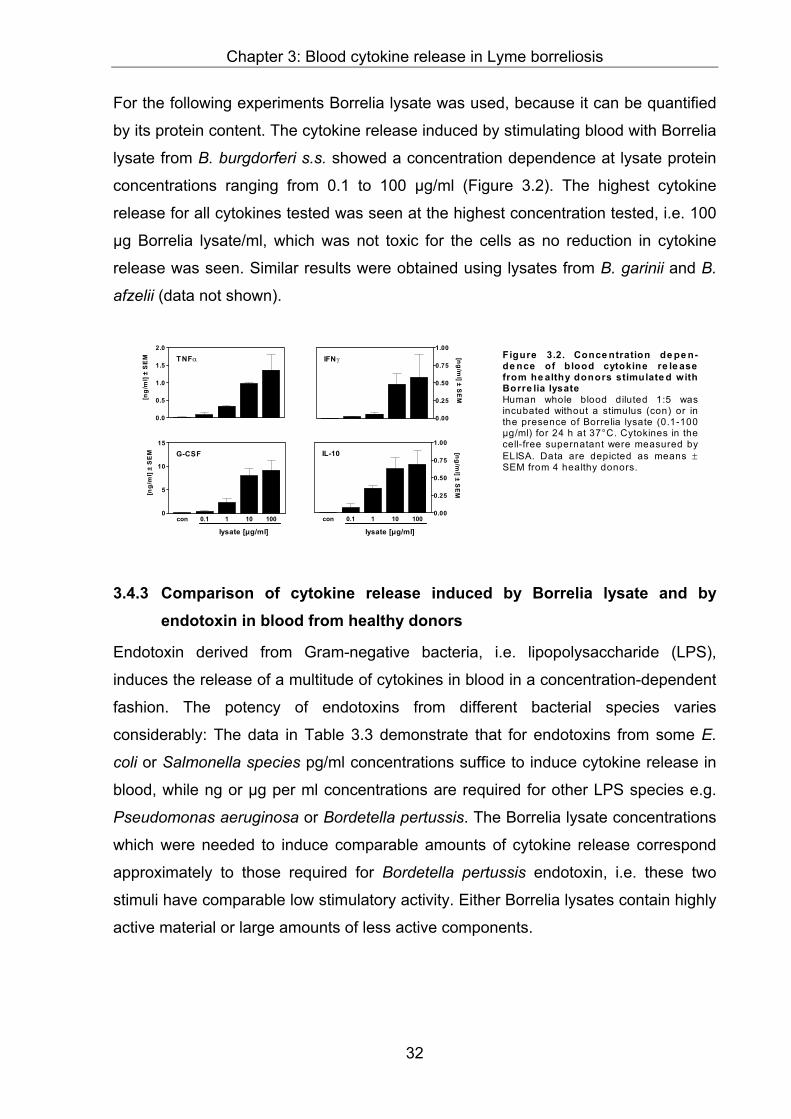
Chapter 3: Blood cytokine release in Lyme borreliosis
32
For the following experiments Borrelia lysate was used, because it can be quantified
by its protein content. The cytokine release induced by stimulating blood with Borrelia
lysate from B. burgdorferi s.s. showed a concentration dependence at lysate protein
concentrations ranging from 0.1 to 100 µg/ml (Figure 3.2). The highest cytokine
release for all cytokines tested was seen at the highest concentration tested, i.e. 100
µg Borrelia lysate/ml, which was not toxic for the cells as no reduction in cytokine
release was seen. Similar results were obtained using lysates from B. garinii and B.
afzelii (data not shown).
3.4.3 Comparison of cytokine release induced by Borrelia lysate and by endotoxin in blood from healthy donors
Endotoxin derived from Gram-negative bacteria, i.e. lipopolysaccharide (LPS),
induces the release of a multitude of cytokines in blood in a concentration-dependent
fashion. The potency of endotoxins from different bacterial species varies
considerably: The data in Table 3.3 demonstrate that for endotoxins from some E.
coli or Salmonella species pg/ml concentrations suffice to induce cytokine release in
blood, while ng or µg per ml concentrations are required for other LPS species e.g.
Pseudomonas aeruginosa or Bordetella pertussis. The Borrelia lysate concentrations
which were needed to induce comparable amounts of cytokine release correspond
approximately to those required for Bordetella pertussis endotoxin, i.e. these two
stimuli have comparable low stimulatory activity. Either Borrelia lysates contain highly
active material or large amounts of less active components.
0.0
0.5
1.0
1.5
2.0T NFα
[ng/
ml]
± S
EM IFNγ
0.00
0.25
0.50
0.75
1.00
[ng/ml] ± S
EM
con 0.1 1 10 1000
5
10
15
lysate [µg/ml]
G-CSF
[ng/
ml]
± S
EM
con 0.1 1 10 100
lysate [µg/ml]
IL-10
0.00
0.25
0.50
0.75
1.00
[ng/ml] ± S
EM
F igure 3.2. Conce ntration de pe n-de nce of blood cytokine re le asefrom he althy donors stimulate d withBorre lia lysateHuman whole blood diluted 1:5 wasincubated without a stimulus (con) or inthe presence of Borrelia lysate (0.1-100µg/ml) for 24 h at 37°C. Cytokines in thecell-free supernatant were measured byELISA. Data are depicted as means ±SEM from 4 healthy donors.

Chapter 3: Blood cytokine release in Lyme borreliosis
33
Table 3.3. Threshold of IL-1β-induction in human whole blooda
Borrelia lysate LPS
B. b
urgd
orfe
ri s.
s.
B. g
arin
ii
B. a
fzel
ii
S. e
nter
itidi
s
Kl
pneu
mon
iae
S. a
bortu
s.
equi
E. c
oli 0
26-B
6
B. p
ertu
ssis
P. a
erug
inos
a
Vibr
io
Cho
lera
e
IL-1β-treshold (pg/ml) 1000 1000 1000 10 10 10 1000 1000 10000 10000
aHuman whole blood diluted 1:5 in RPMI was incubated in the presence of Borrelia lysate (0.1 ng - 100 µg/ml) or various endotoxins (1 pg - 10 µg/ml) in 10fold serial dilutions for 24 h at 37°C. IL-1β in the cell-free supernatant was measured by ELISA. Data represent the lowest concentration of the given stimulus in pg/ml where blood from all 4 healthy donors released significant amounts of IL-1β.
To compare the pattern of cytokine release induced by LPS and Borrelia lysate the
concentrations of endotoxin from four different LPS preparations were adjusted to
induce the same levels of TNFα-release as seen with 10µg/ml Borrelia lysate. The
release of the cytokines TNFα, IFNγ, G-CSF and IL-10 induced by endotoxins from
Salmonella abortus equi (200 pg/ml), Escherichia coli (10 ng/ml), Klebsiella
pneumoniae (100 pg/ml) and Salmonella enteriditis (50 pg/ml) was uniform in blood
from healthy volunteers (Figure 3.3) suggesting that different endotoxins share a
leukocyte activation principle. However, a pronounced difference was seen between
the four LPS preparations and the Borrelia lysate: at concentrations which induced
the same TNFα-release as 10 µg/ml Borrelia lysate, endotoxins induced much more
IFNγ compared to Borrelia lysate. Instead, Borrelia lysate induced a 5 to 10 fold
higher release of the anti-inflammatory cytokines IL-10 and G-CSF than the LPS
preparations. The lysates from other Borrelia species, i.e. B. afzelii and B. garinii,
induced the same cytokine pattern as B. burgdorferi (data not shown). These findings
show that LPS predominantly induces the release of the pro-inflammatory cytokine
IFNγ, while Borrelia lysate is a stronger inducer of the anti-inflammatory cytokines IL-
10 and G-CSF. Such an inverse cytokine induction pattern demonstrates that the
immunostimulatory components of Borrelia burgdorferi differ from endotoxins.

Chapter 3: Blood cytokine release in Lyme borreliosis
34
3.4.4 Comparison of ex vivo cytokine release from borreliosis patients to healthy controls in response to Borrelia lysate
It was now of interest whether a similar immunomodulation by Borrelia might also be
detected in the patients’ blood. Therefore, the ex vivo cytokine response to Borrelia
lysate of a group of ten borreliosis patients was compared with that of 24 healthy
controls. In blood from patients with Lyme borreliosis, the cytokine release capacity
for TNFα (-61 ± 14.3%, p≤ 0.001), IFNγ (-92.0 ± 3.2%, p≤ 0.001) and G-CSF (-84 ±
7.0%, p≤ 0.001) in response to Borrelia lysate was significantly reduced compared to
healthy controls (Figure 3.4). However, again no difference between controls and
patients was seen with regard to the release of the anti-inflammatory cytokine IL-10
in stimulated blood. These data indicate that ex vivo stimulated blood from borreliosis
patients responds differently to LPS as well as to Borrelia lysate in comparison to
healthy volunteers.
0.0
0.2
0.4
0.6
0.8
1.0
1.2
***
control lysatehd lb hd lb
TNFα
[ng/
ml]
± S
EM
***0
1
2
3
hd lb hd lb
IFNγ
control lysate
[ng/ml]
± SEM
0
1.0
2.0
3.0
4.0
***
hd lb hd lb
G-CSF
control lysate
[ng/
ml]
± S
EM
0 .0
0.2
0.4
0.6
0.8
1.0
hd lb hd lb
IL-10
control lysate
[ng/ml]
± SEM
Figure 3.4. Ex v ivo cytokinere le ase capacity of blood fromhe althy donors or borreliosispatie nts stimulate d with Borre lialysateWhole blood, either from healthydonors (hd, n=24) or patients withLyme borreliosis (lb, n=10), di luted1:5 was incubated with 10 µgprotein/ml Borrelia lysate for 24 h at37°C. Cytokines in the cell-freesupernatant were measured byELISA. Data are shown asBox-and-Whiskers plots. P-values of* 0.001 were considered significantand are marked with ***.
0.0
0.2
0.4
0.6
0.8
TNFα
[ng/
ml]±
SEM
* **
0.0
0.2
0.4
0.6
0.8
1.0IFN γ [ng/m
l] ± SEM
0.0
2.5
5.0
7.5
10.0
*** *** *** ***A B C D
LPSlysate
G-CSF
con
[ng/
ml]±
SEM
** ** ** **0.0
0.2
0.4
0.6
0.8
1.0
A B C D
LPSlysate
IL-10
con
[ng/ml] ± S
EM
F igure 3.3. In v itro cytokine re le asecapacity of b lood from he althy donorsstimulate d with Borre lia lysate or LPSHuman whole blood diluted 1:5 was incubatedwithout a stimulus (con) or with either Borrelialysate (10 µg/ml), A; endotoxin from S. abortusequi (200 pg/ml), B; from E. coli (10 ng/ml), C;from Kl. pneumoniae (100 pg/ml) or D; from S.enteritid is (50 pg/ml) for 24 h at 37°C.Cytokines in the cell-free supernatant weremeasured by ELISA. Data are depicted asmeans * SEM from 4 healthy donors. P-valuesof 0.05, * 0.01 and * 0.001 versus lysate wereconsidered significant and are depicted as *, **and *** respectively.

Chapter 3: Blood cytokine release in Lyme borreliosis
35
3.5 Discussion
Our study suggests at persistent infection with Borrelia spirochetes is associated with
an attenuation of the release capacity of some cytokines, in particular the pro-
inflammatory cytokines TNFα and IFNγ, in blood. Such an attenuation was not only
seen with Borrelia specific antigen, but also after stimulation with the endotoxin of
other Gram-negative bacteria (LPS), thus indicating a more general underlying
mechanism.
The cytokine release capacity of patient blood and healthy controls can be used for
comparative studies and alterations have been detected in blood from patients with
multiple sclerosis (67), malignant melanoma (68), rheumatoid arthritis (69), multiple
myeloma (70), HIV (73) and children infected with enterohemorrhagic E. coli (74).
Especially because leukocyte counts are not affected in Borrelia infection (75, 76) it
seemed appropriate to take a similar approach to characterize possible changes of
the immune response in patients with persistent Borrelia infection.
There is no clear consensus on the accurate diagnosis of chronic infection and it is
not yet possible to separate the symptoms of the persistent infection from possible
sequalae of a successful eradication of the pathogen. Therefore we defined the
following inclusion criteria both for the patient group and for the control group: The
clinical diagnosis of persistent Lyme borreliosis set by an experienced practitioner in
an endemic area plus well established serology were used. Furthermore, patients
with further non-borrelia-associated diseases were excluded. The healthy control
group was tested seronegative for Borrelia antibodies and had no history of tick bites
or borreliosis.
Using these criteria, a significant attenuation of the pro-inflammatory cytokine
response of blood was found in the patient compared to the healthy control group.
Due to the exploratory character of the main study, a number of confounding factors
have to be considered: Obviously, the age of patient and control group differed and
cytokine release was not controlled for differences in leukocyte counts. Therefore, a
control study was performed using healthy patients of the same general practitioner
as parallel control group. This control experiment resulted in the same tendency
towards attenuated pro-inflammatory cytokine response, although the lower number
of blood donors did not allow to reach statistical significance. Thus, an influence of an
imbalance in patient and control group selection can not be finally excluded.

Chapter 3: Blood cytokine release in Lyme borreliosis
36
The immunostimulatory properties of Borrelia antigen have been investigated
extensively in the past, with live or heat-killed pathogen or purified antigen
preparations (77), (78). For our experimental approach, we needed a standardized
Borrelia stimulus. Heat-inactivated and sonified Borrelia induced similar cytokine
release patterns and displayed a similar concentration dependence. We also
compared the immunostimulatory properties of 3 different Borrelia strains (B.
burgdorferi s.s., B. garinii, B. afzelii), however, no significant differences were found
with regard to concentration dependence or pattern of cytokines induced. With a
relatively high concentration (at least 10 ng protein/ml) of the lysate, a measurable
cytokine release was induced that was quantitatively comparable to endotoxin from
Pseudomonas aeruginosa. Since e.g. in murine Borrelia infections, accumulations of
up to 105 spirochetes were found in different organs of infected mice (79) it is feasible
that high concentrations of antigens are present at the site of infection that induce
local cytokine formation.
Further, Borrelia lysate- and LPS-induced cytokine release is qualitatively different as
to the pattern of predominant cytokines released. Although Borrelia belong to the
group of Gram-negative bacteria (57), they lack the typical endotoxin LPS (80, 81).
Instead, Borrelia express lipoproteins, e.g. outer surface proteins (Osp), which have
the ability to induce the release of TNFα (42), IL-1β (43) and IL-6 (44) when
incubated with isolated leukocytes, which pointed towards a probably characteristic
pro-inflammatory nature of Borrelia antigen (82). However, when we compared
Borrelia lysate and LPS at a concentration which induced an equipotent TNFα-
release, we found that Borrelia lysate induced greater amounts of the anti-
inflammatory cytokines G-CSF and IL-10 than LPS. Our results which indicate that
Borrelia induce an anti-inflammatory response are corroborated by recent findings
showing IL-10 induction in monocytes by Borrelia antigen (46).
We are well aware of the fact that OspA and further Borrelia-derived components are
able to evoke a cytokine response from isolated peripheral blood monocytes (83),
(84). We repeated and confirmed these experiments also with our Borrelia lysate and
observed complete inhibition of TNFα-release after neutralization of CD14 in analogy
to previous work with other bacterial stimuli (85). However, under identical conditions,
the cytokine response of whole blood to Borrelia was not attenuated (data not
shown). This phenomenon is currently under further investigation. It implies that the

Chapter 3: Blood cytokine release in Lyme borreliosis
37
cytokine pattern released from whole blood induced by Borrelia lysate is unrelated to
these known CD14-mediated initiating mechanisms.
On the basis of published data and the study presented here, we propose to consider
the attenuated release capacity of white blood cells for pro-inflammatory cytokines
such as TNFα and IFNγ as a mechanism that weakens the immune response of
borreliosis patients towards circulating spirochetes. This might be due to a direct
recognition of Borrelia components by immunocompetent cells or represent a
consequence of an enhanced local production of the anti-inflammatory factor IL-10
as published by others (84). In any case, the reconstitution of the patients immune
competence represents an attractive target for supportive treatment to antibiosis in
chronic Lyme borreliosis.

Chapter 4: Model of Borrelia-induced immunomodulation
38
4 Borrelia burgdorferi induced immune anergy as a model
of persistence via immunosuppression
Isabel Diterich, Carolin Rauter, Carsten J. Kirschning# and Thomas Hartung
Biochemical Pharmacology, Faculty of Biology, University of Konstanz #Institute of Medical Microbiology, Immunology and Hygiene, TU Munich
submitted to Infection and Immunity
4.1 Abstract
If left untreated, infection with Borrelia burgdorferi s.l. may lead to chronic Lyme
borreliosis. It is still unknown how this pathogen manages to persist in the host in the
presence of competent immune cells. We recently reported that Borrelia suppress the
host’s immune response, so perhaps preventing their elimination (30). Here, we
further characterize Borrelia-induced immunomodulation in order to develop a model
of this anergy. We observed that different Borrelia preparations we tested, i.e. live,
heat-inactivated and sonicated Borrelia, could desensitize human blood monocytes,
as shown by attenuated cytokine release on re-stimulation with either stimulus. Next,
we investigated whether these Borrelia-specific stimuli render monocytes tolerant
towards another toll-like receptor (TLR)2-agonist such as lipoteichoic acid from
Gram-positive bacteria or towards the TLR4-agonist lipopolysaccharide. Cross–
tolerance was induced towards all tested stimuli. Furthermore, using primary bone
marrow cells from TLR2-deficient mice and from mice with a non-functional TLR4
(C3H/HeJ), we demonstrated that the TLR2 was required for tolerance induction by
Borrelia and identified IL-10 by neutralizing antibodies as the key mediator involved.
PBMC tolerized by Borrelia exhibited a reduced TLR2-mRNA level, which might
contribute to tolerance. In summary, we characterized tolerance induced by Borrelia
burgdorferi, i.e. a model of desensitization which might mirror the
Chapter 4: Model of Borrelia-induced immunomodulation
39
immunosuppression recently attributed to persistence of Borrelia in
immunocompetent hosts.
4.2 Introduction
Borrelia burgdorferi s.l. is the causative agent of Lyme borreliosis (LB), the most
common vector-borne disease in the USA (2) and in many European countries (1). If
infection with this pathogen is not treated adequately with antibiotics, it may lead to a
chronic multisystemic disorder which is difficult to cure. The mechanism how Borrelia
survive in their natural reservoir, and in human tissues remains unclear. Although
they are recognized by the host’s immune defense and occasionally induce strong
inflammatory reactions, Borrelia are often not eradicated. Thus, how Borrelia persist
in immunologically competent hosts is a key question in understanding the
pathogenesis of LB and remains an area of debate. It has been postulated that
Borrelia interact with the complement system, inactivating the complement regulatory
proteins FHL-1/reconectin and Factor H (86, 87). Others have proven by electron
microscopy, that Borrelia can hide themselves in immunopriviledged sites like the
collagen fibers of the connective tissue (88) or in human synovial cells (24). The
antigenic variation of the Borrelia burgdorferi outer membrane is also discussed as a
possible strategy to evade the immune response (26, 89, 90). Data from Gross et al.
suggest alternatively, that Borrelia induce an autoimmune process, as they could
identify a homology between the Borrelia outer surface protein A (OspA) and the
human lymphocyte-function-associated antigen-1 alpha (hLFA-1) and also found
cross reactive T-cells (29).
We investigated whether Borrelia modulate the host’s immune system in order to
persist. Blood cells from patients suffering from persistent Lyme borreliosis released
significantly lower levels of pro-inflammatory cytokines (i.e. TNFα and IFNγ) in
response to either a Borrelia-specific stimulus or to lipopolysaccharide (LPS) in
comparison to the cells from healthy volunteers (30). Based on their findings in
patient specimens, others have also described Borrelia as an immunomodulator,
supporting the hypothesis of Borrelia-induced immundysregulation (91), (92). In line,
Borrelia seem to influence the balance between the pro- and anti-inflammatory
immune response. We and others found that Borrelia not only induce pro-
inflammatory cytokines, but also lead to a strong formation of IL-10, which is an
important down-regulator of the pro-inflammatory immune response (30, 46, 84). In
Chapter 4: Model of Borrelia-induced immunomodulation
40
addition, the modulatory and regulatory capacity of IL-10 with regard to Borrelia-
induced cytokine release has been found in different in vitro models (93, 94). For
Borrelia lipoproteins the toll-like receptor 2 (TLR2) has been identified as the major
signal transducing receptor (95-99). However, recently the existence of TLR2-
independent, non-lipoprotein Borrelia components was postulated (100). In order to
address these conflicting results we tested the role of the TLR2 and TLR4 in the
recognition of different Borrelia-specific stimuli.
The phenomenon of endotoxin (LPS) tolerance has been investigated extensively in
vitro and in vivo (for review see (101)). It describes a status of macrophage
hyporesponsiveness after exposure to low LPS doses to a subsequent high or lethal
LPS dose (102, 103). Recently, similar desensitization experiments were reported
which demonstrated, that stimuli other than LPS, i.e. highly purified lipoteichoic acid
(LTA) (104) or macrophage-activating lipopeptide-2 (MALP-2) from mycoplasma
(105), can also render macrophages tolerant to subsequent re-stimulation.
Furthermore, it was shown in these same studies, that tolerance can also be induced
by two heterologous stimuli, independent of the receptor involved in their recognition
and signaling. In this case the appropriate term is cross-tolerance or hetero-
tolerance. To our knowledge it has not yet been investigated whether Borrelia
burgdorferi also has the capacity to desensitize macrophages. However, Borrelia-
induced hyporesponsiveness could represent a mechanism enabling the survival of
this pathogen in the host despite the presence of immune cells.
We tested this hypothesis in desensitization experiments with Borrelia-specific stimuli
and with other well characterized TLR2- and TLR4-agonists. Additionally, the
involvement of the TLR2 and the TLR4, as well as of endogenous IL-10 formation in
tolerance and cross-tolerance induction was addressed.
4.3 Material and Methods
4.3.1 Borrelia cultivation and preparation of Borrelia-specific stimuli
Borrelia burgdorferi sensu stricto (s.s.) (strain N40, kindly provided by T. Kamradt,
Berlin, Germany) was cultivated as described previously (30). Borrelia cultures
passaged fewer than 8 times after isolation from mice were grown to log-phase (≥
108/ml) and differentially prepared for stimulation experiments.

Chapter 4: Model of Borrelia-induced immunomodulation
41
For experiments with live Borrelia, B. burgdorferi numbers were determined by
microscopy using a Thoma-counting-chamber, with a modified depth of 0.02 mm.
The culture was adjusted to 5 x 10⁵ Borrelia/ml. Due to the low replication rate of
Borrelia and to their high requirements with regard to the culture conditions an
increase in bacterial numbers during the 24 h of subsequent incubation is
improbable.
Before heat-inactivation or sonication, Borrelia cultures were washed twice. Briefly,
cultures were centrifuged at 10000 g for 30 min, the supernatant was removed and
the pellet was resuspended with pyrogen-free saline solution. Subsequently, the
bacterial numbers were determined as described above. Finally, washed Borrelia
were either incubated for 30 min at 56°C for heat-inactivation, or they were sonicated
yielding Borrelia lysate as described elsewhere (30). Protein concentration of the
lysate was adjusted to a final concentration of 1 mg/ml. Remaining viable spirochetes
were excluded visually under the microscope after one week incubation under
standard cultivation conditions.
The amount of endotoxin units (EU) contained in 10 µg protein of sonicated Borrelia
and of 1 x 10⁷ heat-inactivated Borrelia was below the detection limit (0.1 EU and
0.05 EU respectively), assessed by Limulus amoebocyte lysate assay (LAL) (QCL-
1000, Charles River Endosafe, Charleston, SC, USA). The spike recovery (0.5 EU),
was 107% and 103%, respectively. Live Borrelia were only used for the first set of
experiments, because they represent a highly variable stimulus which is difficult to
standardize as they change their surface protein expression depending on cultivation
conditions (106). All the other experiments were carried out with the same batch of
heat-inactivated or sonicated Borrelia.
4.3.2 Isolation of human peripheral blood mononuclear cells
Peripheral blood mononuclear cells were isolated with cell preparation tubes
(Vacutainer CPT, sodium citrate, Becton Dickinson Biosciences, Heidelberg,
Germany) according to the manufacturer’s instructions. Briefly, blood from healthy
donors was centrifuged in the tubes for 20 minutes at 1650 g. PBMC, separated from
erythrocytes and neutrophils by the gel phase were transferred into 50 ml
polypropylene tubes (bio-one, Greiner, Frickenhausen, Germany) and washed twice
with RPMI 1640 (Bio Whittaker, Apen, Germany) supplemented with 2.5 IU/ml
heparin (Liquemin, Hoffmann LaRoche, Grenzach-Whylen, Germany). The overall

Chapter 4: Model of Borrelia-induced immunomodulation
42
cell count of PBMC (i.e. monocytes and lymphocytes) was determined with a Pentra60
(ABX Diagnostics, Montpellier, France). PBMC were adjusted to a final concentration
of 1 x 10⁷ PBMC/ml with RPMI 1640 supplemented with heparin. 100 µl of cells were
pipetted per well of a 96-well cell-culture plate (Greiner) and 10% autologous plasma
was added to each well.
4.3.3 Mice
Female C3H/HeJ-mice, characterized by a non functional TLR4 due to a natural point
mutation in the TLR4-gene (107) and the corresponding wildtype mouse strain
(C3H/HeN) were purchased from Charles Rivers Laboratories (Sulzfeld, Germany).
TLR2 deficient mice generated by homologous recombination were a generous gift
from Tularik, (South San Francisco, CA, USA) and the corresponding wildtype mice
(129Sv/B57BL/6) were bred in the animal facilities of the University of Konstanz at
24°C, 55% humidity, 12 h day-night rhythm on a diet of Altromin C 1310 (Altromin,
Lage, Germany). Mice were used for the experiments at 8 to 10 weeks of age.
4.3.4 Isolation of primary bone marrow cells from mice
Mice were killed by terminal pentobarbital anaesthesia (Narcoren, Merial,
Hallbergmoos, Germany). Femurs were lavaged with 10 ml ice-cold sterile PBS (Life
Technologies, Karlsruhe, Germany). The lavages were transferred to siliconized
glass tubes (Vacutainer, Bioscience, Heidelberg, Germany) for isolation of bone
marrow cells. Bone derived debris were removed by re-suspending the lavage and
transferring it into a new glass tube after 1 minute sedimentation. After centrifugation,
primary murine bone marrow cells were re-suspended in RPMI 1640 containing 10%
FCS (Biochrom, Berlin, Germany) and 100 IU/ml penicillin/streptomycin (APP-
laboratories, Linz, Austria). Cell counts were determined with a Pentra60 (see above)
and plated to 96-well culture plate (Greiner) at a density of 5 x 10⁵ cells per well.
4.3.5 In vitro desensitization and re-stimulation experiments
Immediately after isolation, cells (PBMC or primary murine bone marrow cells (BMC))
were desensitized with different concentrations of the following stimuli: live, heat-
inactivated or sonicated Borrelia (lysate), LTA from S. aureus (isolated and prepared
in house as described previously (108)), endotoxin (LPS from Salmonella abortus
equi) (Sigma-Aldrich, Seelze, Germany), or left untreated for the controls. The

Chapter 4: Model of Borrelia-induced immunomodulation
43
volume was adjusted to 220 µl with medium (RPMI supplemented with 100 IU/ml
penicillin/streptomycin, containing 10% FCS for the BMC and 2.5 IU/ml heparin for
the PBMC). After incubation for 24 h at 37°C in the presence of 5% CO2, the
supernatants were transferred into 96-well round-bottom plates (Greiner) and stored
at -70°C until cytokines were measured. The remaining adherent, tolerized,
monocytes in the plate were washed twice with RPMI 1640, and subsequently re-
stimulated with 1 ng/ml LPS, 10 µg/ml LTA, 10 µg/ml Borrelia lysate or 1 x 10⁶ heat-
inactivated Borrelia burgdorferi s.s. (Bb)/ml. 10% autologous plasma was added to
the PBMC. The final volume of the incubation was adjusted to 220 µl RPMI 1640
(supplemented as described above). After 24 h incubation at 37°C and 5% CO2 the
supernatants were stored at –70°C until cytokine measurement. To study the
involvement of mediators in tolerance induction, neutralizing antibodies (anti-IL-10
(Endogen, Eching, Germany), pan-specific anti-TGFβ (R&D Systems, Wiesbaden,
Germany), and polyclonal anti-mu-G-CSF-sheep IgG raised in our laboratory (109))
were added to the first 24 h incubation period.
4.3.6 MTT-assay
MTT (3-[4,5-dimethyl-2-thiazolyl]-2,5-diphenyl-2H-tetrazolium bromide, Sigma) stock
solution (5 mg/ml in PBS) was diluted 1:5 in RPMI 1640. 200 µl of this working
solution was added to the adherent cells in a 96-well culture-plate for 2 h at 37°C and
5% CO2. Cells with mitochondrial activity convert dissolved MTT to insoluble purple
formazan. After the incubation the supernatant was removed and the cells were lysed
for 10 minutes with 95% isopropanol, 5% formic acid. Absorbance of converted dye
was measured at 555 nm against 690 nm as reference wavelength.
4.3.7 Cytokine measurement in culture supernatant by ELISA
The concentrations of human and murine TNFα in the supernatants were measured
by in-house sandwich ELISA using commercially available antibody pairs and
recombinant standards. Monoclonal antibody pairs against human TNFα were
purchased from Endogen (Perbio Science, Bonn, Germany). Recombinant human
TNFα was a gift from Dr. S. Poole (National Institute for Biological Standards and
Controls, London, GB) and was used as standard. For the measurement of murine
TNFα, polyclonal antibodies from R&D Systems (Wiesbaden, Germany) and
standard from Pharmingen (BD Bioscience, Heidelberg, Germany) were used.

Chapter 4: Model of Borrelia-induced immunomodulation
44
Assays were carried out in flat-bottom, ultrasorbant 96-well plates (Nunc, Wiesbaden,
Germany). Binding of secondary biotinylated antibodies was detected with
horseradish-peroxidase-conjugated streptavidin (Biosource, Camarillo, CA, USA) and
3’,3’, 5’, 5’-tetramethylbenzidine solution (Sigma) used as substrate.
4.3.8 RNA-extraction and TLR2-mRNA-quantification
5 x 10⁶ PBMC/ml were pipetted into 24 well-culture plates in 1 ml medium. After 24 h
incubation at 37°C and 5% CO2 with 10 µg/ml lysate or without a stimulus the
supernatant was removed and cells were either lysed for RNA extraction or they were
washed and re-stimulated with lysate (10 µg/ml) or with LPS (1 ng/ml) for another 3
h. After removal of the supernatant, RNA was prepared from the adherent cells with
QIAmp RNA Blood Mini Kit (Qiagen, Hilden, Germany) according to the
manufacturer’s instructions, including DNA digestion with the RNase-free DNase-Set
(Qiagen).
6 µl RNA was reverse transcribed in a sample volume of 20 µl containing 2.5 µM
Oligo dT16 (Gibco BRL, custom primer), MgCl2 (5 mM), dNTP (1 mM each), RNase
inhibitor (1 U/µl), murine leukemia virus reverse transcriptase (2.5 U/µl) in PCR buffer
(all PE Applied Biosystems, Weiterstadt, Germany). Samples were incubated at 21°C
for 10 min, 42°C for 15 min, 94°C for 5 min and 5°C for 5 min in a GeneAmp PCR
System 2400 (PE Applied Biosystems).
For relative quantification real-time PCR was performed using a LightCycler rapid
thermal cycler system (Roche Diagnostics GmbH, Mannheim, Germany). The cDNA
for TLR2 and GAP-DH was amplified using LightCycler-FastStart DNA Master SYBR-
Green (Roche Diagnostics, Germany) according to the manufacturer’s protocol. The
sequence for the primers were ggc cag caa att acc tgt gtg (forward), agg cgg aca tcc
tga acc t (reverse) and gaa ggt gaa ggt cgg agt c (forward), gaa gat ggt gat ggg att tc
(reverse) for TLR2 and GAP-DH, respectively. The MgCl2 concentration was adjusted
to 2 mM for TLR2 and 4 mM for GAP-DH. The thermal cycling was performed
according to the manufacturer’s protocol (50 cycles) with an annealing temperature of
58°C and 65°C and an elongation time of 3 sec and 11 sec for TLR2 and GAP-DH,
respectively. The amplification was followed by a melting program which started at
65°C for 15 sec and then increased to 95°C at 0.1°C/sec. The specific melting
temperatures for TLR2 and GAP-DH products were 86°C and 86.5°C, respectively.
The TLR2 results were normalized with GAP-DH.

Chapter 4: Model of Borrelia-induced immunomodulation
45
4.3.9 Statistics
Data are shown either as mean ± SEM of 4 different blood donors or of primary cells
from 4-7 individual mice. To take into account the varying viability of the cells on day
2, TNFα-concentrations were divided by the mitochondrial activity assessed in the
MTT-assay. Repeated measure analysis of variance followed by Dunnett’s multiple
comparison test were performed using GraphPad Prism 3.00 (GraphPad Software,
San Diego, CA, USA). ≤ 0.05, 0.01 and 0.001 were considered significant and are
depicted as *, ** and ***, respectively.
4.4 Results
4.4.1 Borrelia-induced tolerance in human PBMC
We tested whether Borrelia are able to induce a state of hyporesponsiveness in
human monocytes in vitro. On the one hand, we used the same Borrelia
preparations, i.e. heat-inactivated, sonicated and live Borrelia, for pre-stimulation and
for re-stimulation. On the other hand, we tested all combinations of the different
Borrelia preparations in the two sequential incubation periods. When human PBMC
were pre-treated with either sonicated, heat-inactivated or live Borrelia for 24 h, they
showed a significantly reduced release of TNFα in response to a secondary
stimulation with the same or either of the other Borrelia preparations (Figure 4.1). In
contrast, cells which had not been stimulated during the first incubation period were
fully responsive to the second stimulus, as demonstrated by the non-pretreated
control cells. Thus, all Borrelia-specific stimuli tested could desensitize macrophages
to either the same or to another Borrelia-specific stimulus, which is termed Borrelia-
tolerance.

Chapter 4: Model of Borrelia-induced immunomodulation
46
4.4.2 Comparison of TNFα-inducing potency of different bacterial stimuli
We next tested TNFα-inducing capacity of different bacterial stimuli during 24 h
incubation in the in vitro model. Human PBMC treated with LPS, LTA or with two
Borrelia-specific stimuli (heat-inactivated Borrelia and Borrelia lysate) induced dose-
dependent TNFα-release as measured by ELISA. 1 ng/ml LPS, 10 µg/ml LTA, 1 x
106 heat-inactivated Bb/ml and 10 µg/ml Borrelia lysate induced comparable amounts
of TNFα in the range of 200-500 pg/ml. Therefore, these concentrations were used in
the subsequent re-stimulation experiments.
4.4.3 Borrelia-induced cross-tolerance to LTA and LPS
Next, we investigated whether Borrelia-specific stimuli - which according to literature
(95, 96, 98, 99) act via TLR2 - render macrophages tolerant towards another TLR2-
agonist such as lipoteichoic acid (LTA) from Gram-positive Staphylococcus aureus or
towards a TLR4-agonist, i.e. the lipopolysaccharide from Salmonella abortus equi.
PBMC were first treated overnight with either heat-inactivated Borrelia or Borrelia
lysate. Then, the cells were re-stimulated either with 10 µg/ml LTA or with 1 ng/ml
LPS. Figure 4.2a and 4.2b demonstrate, that pre-stimulation with Borrelia-specific
stimuli led to reduced responsiveness of the PBMC to re-stimulation with LTA as well
as with LPS. Tolerance induction was concentration dependent. The stronger the
0
1000
2000
3000
4000
5000
6000
7000
8000
***
TNFα
[pg/
ml]
± SE
M/O
D
***
***
a) 2nd stimulus: Borrelia lysate
0
1000
2000
3000
4000
5000
b) 2nd stimulus: heat-inact. Bb
* * *TNFα
[pg/
ml]
± SE
M/O
D
con 10 µg/ml 7 x 10⁵ 2 x 10⁵ 0
2500
5000
7500
10000
12500
Borrelialysate
heat-inact.Bb/ml
live Borrelia
c) 2nd stimulus: live Borrelia
TNFα
[pg/
ml]
± SE
M/O
D
1st stimulus:
*** *** ***
Figure 4.1. Borrelia-induced tolerance inhuman PBMC1 x 106 human PBMC from 4 healthy donors wereincubated with culture medium (con), 10 µg/mlBorrelia lysate, 7 x 10⁵ heat-inactivated or 2 x 10⁵
live Borrelia during 24 h, then washed andincubated for another 24 h in the presence of a) 10µg/ml Borrelia lysate, b) 7 x 10⁵ heat-inactivated orc) 2 x 10⁵ live Borrelia. The concentrations of TNFαwere measured by ELISA. Data are expressed asthe mean ± SEM divided by the OD assessed by theMTT-assay. The statistical significance wascompared with the non-tolerized (con) cells. *, p <0.05, ***, p < 0.001 based on ANOVA, followed byDunnett's multiple comparison test.

Chapter 4: Model of Borrelia-induced immunomodulation
47
stimulus during the pre-incubation period, the lower the TNFα-response to the
second stimulus. Thus, Borrelia induced cross–tolerance towards two different
heterologous stimuli, one also signaling via TLR2, the other TLR4-mediated.
4.4.4 LPS and LTA-induced cross-tolerance to Borrelia-specific stimuli
Stimulation of macrophages with LPS renders the cells tolerant to a subsequent LPS-
stimulation. This kind of tolerance has been known since the 1960-1970s (110-112).
Recently, we reported tolerance-induction by LTA, including cross-tolerance inducible
by LTA towards LPS and vice versa, indicating that cross-tolerance between TLR2
and TLR4-agonists is possible (104). Therefore, our results prompted us to test
whether cells rendered tolerant by LPS or LTA were also hyporesponsive to re-
stimulation with Borrelia lysate.
In the experiments shown in Figure 4.3, LPS or LTA were used for pre-stimulation.
For subsequent re-stimulation, Borrelia lysate was added to the cells. Similarly to the
previous experiments, cytokine release in response to the second stimulus was
decreased in a dose-dependent fashion depending on the concentration of the first
stimulus. The same results were obtained when heat-inactivated Borrelia were used
for re-stimulation instead of Borrelia lysate (data not shown).
0
500
1000
1500
2000
2500
3000
3500
a) 2nd stimulus: LTA
* * * *
TNF α
[pg/
ml]
± SE
M /
OD
con 0.1 1 10 1x10⁵ 1x1060
200
400
600
800
1000
1200
b) 2nd stimulus: LPS
1st stimulus: Borrelia lysate [µg/ml] heat-inact. Bb/ml
*** *** *** ***
TNFα
[pg/
ml]
± SE
M /
OD
Figure 4.2. Induction of Borreliacross-tolerance to LTA or LPS in humanPBMCHuman PBMC (1 x 10⁶/well) from 4 differentdonors were incubated with culture medium (con,black bars), 0.1, 1, 10 µg/ml Borrelia lysate (whitebars),1 x 105 and 1 x 10⁶ heat-inactivatedBorrelia (hatched bars ) during 24 h, then washedand incubated for another 24 h in the presence of10 µg/ml LTA (a) or 10 ng/ml LPS (b). Theconcentrations of TNFα were measured by ELISA.Data are expressed as the mean ± SEM dividedby the OD assessed by the MTT-test. Thestatistical significance was compared with thenon-tolerized (con) cells, *, p < 0.05, ***, p < 0.001based on ANOVA, followed by Dunnett's multiplecomparison test.
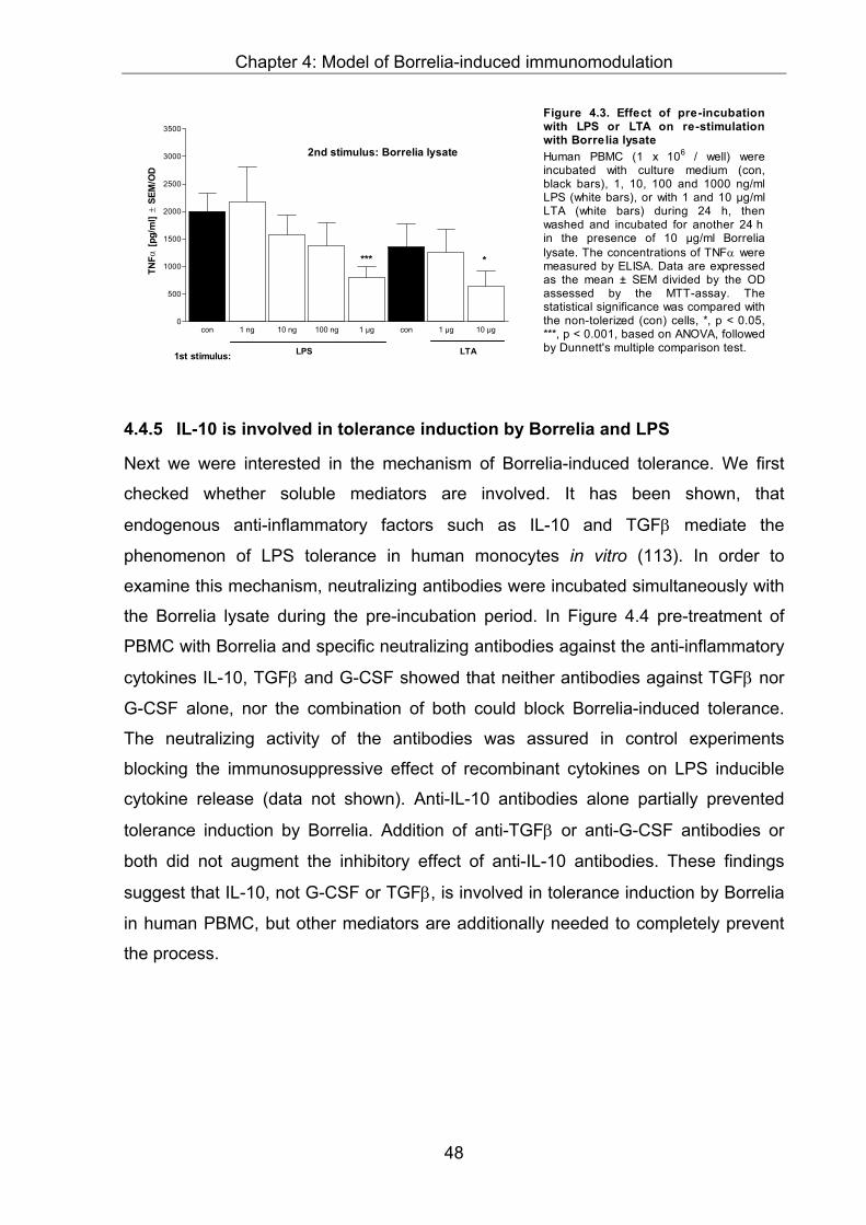
Chapter 4: Model of Borrelia-induced immunomodulation
48
4.4.5 IL-10 is involved in tolerance induction by Borrelia and LPS
Next we were interested in the mechanism of Borrelia-induced tolerance. We first
checked whether soluble mediators are involved. It has been shown, that
endogenous anti-inflammatory factors such as IL-10 and TGFβ mediate the
phenomenon of LPS tolerance in human monocytes in vitro (113). In order to
examine this mechanism, neutralizing antibodies were incubated simultaneously with
the Borrelia lysate during the pre-incubation period. In Figure 4.4 pre-treatment of
PBMC with Borrelia and specific neutralizing antibodies against the anti-inflammatory
cytokines IL-10, TGFβ and G-CSF showed that neither antibodies against TGFβ nor
G-CSF alone, nor the combination of both could block Borrelia-induced tolerance.
The neutralizing activity of the antibodies was assured in control experiments
blocking the immunosuppressive effect of recombinant cytokines on LPS inducible
cytokine release (data not shown). Anti-IL-10 antibodies alone partially prevented
tolerance induction by Borrelia. Addition of anti-TGFβ or anti-G-CSF antibodies or
both did not augment the inhibitory effect of anti-IL-10 antibodies. These findings
suggest that IL-10, not G-CSF or TGFβ, is involved in tolerance induction by Borrelia
in human PBMC, but other mediators are additionally needed to completely prevent
the process.
con 1 ng 10 ng 100 ng 1 µg con 1 µg 10 µg0
500
1000
1500
2000
2500
3000
3500
2nd stimulus: Borrelia lysate
1st stimulus: LPS
****
LTA
TNFα
[pg/
ml]
± SE
M/O
D
Figure 4.3. Effect of pre-incubationwith LPS or LTA on re-stimulationwith Borrelia lysateHuman PBMC (1 x 10⁶ / well) wereincubated with culture medium (con,black bars), 1, 10, 100 and 1000 ng/mlLPS (white bars), or with 1 and 10 µg/mlLTA (white bars) during 24 h, thenwashed and incubated for another 24 hin the presence of 10 µg/ml Borrelialysate. The concentrations of TNFα weremeasured by ELISA. Data are expressedas the mean ± SEM divided by the ODassessed by the MTT-assay. Thestatistical significance was compared withthe non-tolerized (con) cells, *, p < 0.05,***, p < 0.001, based on ANOVA, followedby Dunnett's multiple comparison test.

Chapter 4: Model of Borrelia-induced immunomodulation
49
4.4.6 TLR2-downregulation by Borrelia-induced tolerance
Recently published experiments from Wang et al. suggest that the TLR2 is
downregulated in synthetic bacterial lipopeptide-tolerance (114). We therefore
measured the TLR2-mRNA expression of human PBMC in Borrelia-induced
tolerance. 24 h stimulation of PBMC in the presence of Borrelia lysate (10 µg/ml) or
LPS (1 ng/ml) led to a significantly reduced TLR2-mRNA expression, which remained
attenuated after re-stimulating these tolerized cells (Figure 4.5). These data suggest
that downregulation of the TLR2 might contribute to suppressed TNFα-formation
upon re-stimulation in Borrelia-induced tolerance.
0
25
50
75
100
2nd stimulus: Borrelia lysate
* * * *
TNFα
[pg/
ml]/
OD
[% o
f NaC
l]
1st stimulus:Bb lysatea-IL-10a-TGFβ
a-G-CSF
----
+ + + + + + + ++-
- -- -
++-
++
+
+ +
++
++
- -- -
-
-
*** *** *** ***
Figure 4.4. Neutralization of IL-10, TGFβ andG-CSF during pre-incubation preventsestablishment of desensitization by BorrelialysatePBMC were cultured for 24 h in the presence of 10µg/ml Borrelia lysate and neutralizing anti-TGFβ (1µg/ml) and anti-IL-10 (10 µg/ml) monoclonalantibodies, 1% anti-mu-G-CSF-sheep IgG orcombinations of the neutralizing antibodies andserum. Controls were cultured without Borrelia lysate.After washing, cells were re-stimulated with 10 µg/mlBorrelia lysate for further 24 h at 37°C. Cytokines insupernatants were determined by ELISA. Thestatistical significance was compared with thenon-tolerized (con) cells, *, p < 0.05, ***, p < 0.001based on ANOVA, followed by Dunnett's multiplecomparison test.
0
100
200
300
400
500
600
700
800
900
1000
1st stimulus:
2nd stimulus:
medium
medium
medium
lysate
lysate
lysate
lysate
LPS
lysate LPS
*** ***
** **§§§
§§§
TLR2
mRN
A
Figure 4. 5. TLR2-mRNA expression inhuman PBMC5 x 106 PBMC from 4 different donors wereincubated with culture medium, 1ng/ml LPS or10 µg/ml Borrelia lysate during 24 h and eitherlysed or incubated for another 3 h in thepresence of medium, 10 µg/ml Borrelia lysateor 1 ng/ml LPS. The cDNA for TLR2 andGAP-DH was amplified and relativequantification was performed using aLightCycler. The TLR2 results were normalizedto GAP-DH. **, p< 0.01 and ***, p< 0.001 basedon ANOVA, followed by Bonferroni's multiplecomparison test. * vs. medium - medium, § vs.medium - lysate.

Chapter 4: Model of Borrelia-induced immunomodulation
50
4.4.7 TLR2 but not TLR4 is required for tolerance- and cross-tolerance-induction by Borrelia
To further investigate the role of TLR2 and TLR4 in Borrelia-induced tolerance, we
carried out desensitization experiments using primary murine bone marrow cells from
TLR2-knock-out mice and C3H/HeJ-mice (with a non-functional TLR4) and
corresponding wildtype cells. As expected there was no measurable TNFα- or IL-10-
release in TLR2-/- bone marrow macrophages after stimulation with either heat-
inactivated Borrelia or Borrelia lysate on the first day after 8 h or 24 h (data not
shown). Hereby, we confirmed data from others (95, 96, 98, 99), showing that
Borrelia-specific stimuli employ TLR2. However, cells from TLR2-/-mice responded to
LPS to the same extent as cells from wildtype mice, reflecting the normal responsivity
to a TLR4-agonist (data not shown). Cells from TLR2- plus TLR4-defective mice
obtained by crossing both strains, did neither release TNFα upon stimulation with
Borrelia, nor with LPS or LTA (data not shown). We controlled the responsiveness of
the cells to exclude that they were desensitized by activating them with other stimuli
such as CpG-oligonucleotides, a TLR9-mediated stimulus modeling bacterial DNA,
and by the phorbolesther PMA, a receptor-independent stimulus (data not shown).
Pre-incubation of cells from TLR2-/-mice with a Borrelia-specific stimulus had no
significant effect on their responsiveness to LPS (Figure 4.6b). They behaved similar
to saline-pretreated cells. This result indicates that the cells had not been
desensitized by heat-inactivated Borrelia and demonstrates that TLR2 is required for
Borrelia-induced tolerance. Furthermore, as expected, no TNFα-release could be
measured upon re-stimulation of the TLR2-deficient cells with either lysate (Figure
4.6d) or heat-inactivated Borrelia (data not shown) independent of the pre-incubation
stimulus. In contrast, re-stimulating wildtype and TLR2-/--cells after LPS pre-treatment
with LPS again, resulted as expected in strongly reduced TNFα-release, showing
that the cells had been rendered tolerant (Figure 4.6a). Thus, the TLR2 was not
required for LPS-induced tolerance. In line with our PBMC-results (Figure 4.1, Figure
4.3) this experiment shows that pre-stimulation of primary murine wildtype cells with
LPS or Borrelia lysate tolerized the cells to Borrelia lysate.

Chapter 4: Model of Borrelia-induced immunomodulation
51
0
2500
5000
7500
10000 2nd stimulus: LPSTLR2 +/+
*** ****** ******TN
Fα [p
g/m
l] ±
SEM
/OD
a
0
2500
5000
7500
10000
* *
b
TNFα
[pg/
ml]
± SE
M/O
D
2nd stimulus: LPSTLR2 -/-
con 1 10 1 3 100
10000
20000
30000
LPS [ng/ml] x 105 heat-inact. Bb/ml1st stimulus:
*** ***
*** ***
TNFα
[pg/
ml]
± SE
M/O
D
c 2nd stimulus: Borrelia lysateTLR2 +/+
con 1 10 1 3 100
2500
5000
7500
10000
LPS [ng/ml] x 105 heat-inact. Bb/ml1st stimulus:
d 2nd stimulus: Borrelia lysateTLR2 -/-
TNFα
[pg/
ml]
± SE
M/O
D
n.d. n.d. n.d. n.d. n.d. n.d.
Figure 4.6. Induction of tolerance andcross-tolerance in primary bone marrowcells from TLR2+/+ and TLR2-/- miceBone marrow cells (5 x 10⁵/well) from TLR2+/+
-mice (black bars, a, c) and TLR2-/--mice(hatched bars, b, d) were incubated withculture medium (con), 1 and 10 ng/ml LPS, orwith 1 x 10⁵, 3 x 10⁵, 1 x 10⁶ heat-inactivatedBorrelia during 24 h, then washed andincubated for another 24 h in the presence ofa) and b) 1 ng/ml LPS or c) and d) 10 µg/mlBorrelia lysate. The concentrations of TNFαwere measured by ELISA. Data are expressedas the mean ± SEM divided by the viabilityassessed by MTT-assay. The statisticalsignificance was compared with the nontolerized cells. *, p < 0.05, ***, p < 0.001. n.d.not detectable.
The same experiments were also conducted with primary murine bone marrow cells
from C3H/HeJ mice which lack a functional TLR4 and their corresponding wildtype
cells. Cells from both mouse strains responded similarly to Borrelia-specific stimuli
during the pre-incubation period (data not shown), confirming that TLR4 is not
required for signaling of the Borrelia-specific stimuli. Consequently, cells from
C3H/HeJ mice behaved like wildtype-cells, showing a reduced TNFα-release to re-
stimulation with heat-inactivated Borrelia after pre-treatment with the same Borrelia-
specific stimulus (Figure 4.7a). Furthermore, in line with our expectations, neither
tolerance to Borrelia (Figure 4.7b) nor to LPS (data not shown) could be induced by
LPS-pre-treatment in cells without a functional TLR4.
0
10000
20000
30000
40000
50000
60000
a) 2nd stimulus: Borrelia lysate
HeN
***
*** ***
******
***
TNFα
[pg/
ml]
± SE
M/O
D
con 1 10 0.1 1 10 1 3 10
10000
20000
30000
b) 2nd stimulus: Borrelia lysate
1st stimulus
***
HeJ
*** ******
***
***
LPS [ng/ml] lysate [µg/ml] x 105 heat-inact. Bb/ml
TNFα
[pg/
ml]
± SE
M/O
D
Figure 4.7. Induction of tolerance andcross-tolerance in primary bone marrowcells from C3H/HeN and C3H/HeJ miceBone marrow cells (5 x 10⁵/well) fromC3H/HeN-mice (black bars) and C3H/HeJ mice(hatched bars) were incubated with culturemedium (con), 1 and 10 ng/ml LPS, or with 1 x10⁵, 3 x 10⁵, 1 x 10⁶ heat-inactivated Borreliaduring 24 h, then washed and incubated foranother 24 h in the presence of 10 µg/ml Borrelialysate. The concentrations of TNFα weremeasured by ELISA. Data are expressed as themean ± SEM divided by the viability assessed byMTT-assay. The statistical significance wascompared with the non tolerized cells. ***, p <0.001 based on ANOVA, followed by Dunnett'smultiple comparison test.

Chapter 4: Model of Borrelia-induced immunomodulation
52
Hence, in the absence of TLR2, no cytokine release could be induced by Borrelia
and no state of hyporesponsiveness for re-stimulation with LPS was achieved. In line,
no desensitization to Borrelia stimuli could be induced by LPS in the absence of
functional TLR4.
4.5 Discussion
Understanding the immunopathology of LB is still a major challenge. Although
inducing strong immune activation, e.g. in phases of arthritis, the causative agent of
LB persists and leads to a chronic pathology in the immunocompetent host.
Noteworthy, the inflammatory episodes in LB are typically self-limiting and the site of
manifestation often changes e.g. between different joints. These phenomena suggest
counter-regulatory anti-inflammatory mechanisms and the long phases of latency
indicate phases of immune evasion.
We recently proposed a possible mechanism for survival of Borrelia in the
immunologically competent host: Since our ex vivo experiments with whole blood
from patients with LB revealed a significantly reduced capacity to release TNFα and
IFNγ in comparison to blood cells from healthy controls, we postulated that Borrelia
modulate the host’s immune system in order to evade immune clearance (30). There
are few reports about Borrelia acting as an immunomodulator (91, 92, 115). We
found further evidence for Borrelia-induced immunomodulation by demonstrating,
that Borrelia induce a stronger anti-inflammatory cytokine-response than endotoxins
from various other Gram-negative bacteria (30). These data are in line with findings
from other authors, indicating that Borrelia burgdorferi is a potent inducer of the anti-
inflammatory cytokine IL-10 (46, 84, 116). In addition, the modulatory and regulatory
capacity of IL-10 with regard to Borrelia-induced cytokine release has been found in
different in vitro models (93, 94).
In the present study, we examined whether tolerance could represent a possible
model for Borrelia-induced immunomodulation. First, we checked whether Borrelia
can render cells hyporesponsive and second we investigated some of the underlying
mechanisms involved in this immunomodulation. Borrelia could indeed render human
PBMC tolerant, i.e. unable to react to a second stimulation with Borrelia, as shown by
a reduced capacity of TNFα-release (Figure 4.1). All the tested settings led to a
significantly reduced TNFα-release as a result of pre-incubation with Borrelia,
compared to saline controls. As no major differences between live and killed Borrelia

Chapter 4: Model of Borrelia-induced immunomodulation
53
with respect to their ability to render the monocyte hyporesponsive could be
observed, we selected the more standardized stimuli (heat-inactivated Borrelia and
Borrelia lysate) for the subsequent experiments. Based on these experiments we
demonstrated, that Borrelia are able to modulate the monocytic immune response.
Next, assuming that Borrelia can induce a more general desensitization in the cell,
we investigated whether a monocyte rendered tolerant by Borrelia was also
hyporesponsive to a different stimulus. Differences in the potency of TNFα-induction
among the bacterial stimuli (Borrelia, LPS and LTA) were excluded by choosing
equipotent concentrations, i.e. concentrations which induced comparable amounts of
TNFα-release during 24 h of incubation.
According to literature, Borrelia signal via the TLR2 (95, 96, 98, 99). This could be
confirmed in our experiments with primary murine bone marrow cells from mice
lacking either a functional TLR2 or TLR4 or both. The findings in Figure 4.2 a, b
demonstrate that Borrelia desensitize monocytes in a more general manner, because
they also rendered PBMC hyporesponsive to subsequent stimulation with
heterologous stimuli such as the TLR2 agonist LTA or the TLR4 agonist LPS. The
extent of desensitization was concentration dependent. Using LPS or LTA to tolerize
the cells similarly led to hyporesponsiveness to Borrelia (Figure 4.3). We did not
observe any difference in cross-tolerance induction by the tested stimuli on the level
of TNFα-release. These findings confirm our data regarding cross-tolerance between
LTA and LPS, showing that the degree of hyporesponsiveness induced by the TLR2
or the TLR4 agonist, does not vary (104). Opposing results have been published by
others, which postulate that LPS pre-treatment is less effective in other cross-
tolerance models. Sato et al., for example describe poor tolerance induction by LPS
towards MALP-re-stimulation (105).
Tolerance experiments presented here suggest that the underlying mechanisms
which lead to Borrelia-tolerance seem to be very similar to those in tolerance induced
by cell wall components from other bacteria since no difference could be observed
between the tested combinations of tolerance and cross-tolerance. Furthermore,
cross-tolerance data indicate, that signal pathways shared by LPS and TLR2
agonists seem to be impaired. Similar heterologous tolerance phenomena have
recently also been described for macrophage-activating lipopeptides (MALP) from
mycoplasma (105), bacterial DNA (CpG) (117), Staphylococcus aureus (118), LTA

Chapter 4: Model of Borrelia-induced immunomodulation
54
(104), arabinose-capped lipoarabinomannan (119) and LPS. There is accumulating
evidence that tolerance induced by different TLR2- and TLR4-agonists shares
common intracellular signal transduction pathways (114), (105). However, recently
Jacinto et al. suggested unique TLR2 signaling components downstream of TLR2
and upstream of MyD88/IRAK in LTA tolerance by demonstrating that IRAK
expression and IRAK activity differs in LPS and LTA tolerance (120). Controversial
data are reported regarding the involvement of the TLR in LPS and non-LPS
tolerance and cross-tolerance. Our findings with primary murine bone marrow cells
show that in the absence of the Borrelia-recognition receptor, TLR2, no tolerance
could be induced (Figure 4.6b). Similarly the TLR4 was required for LPS-induced
tolerance (Figure 4.7b) indicating that these two receptors are essential in Borrelia-
and LPS-induced tolerance, respectively.
Differing results regarding the regulation of the TLR in tolerance have been published
(105, 119, 121). Wang et al. showed downregulation of TLR2 in tolerance induced by
synthetic bacterial lipopeptide (BLP), but not LPS (114). Our results regarding the
TLR2-mRNA are partially in line with these observations, since the TLR2 was
significantly downregulated in cells tolerized by Borrelia lysate upon re-stimulation
with the same stimulus. As a major difference to Wang et al., we also observed a
downregulation of the TLR2 by LPS. The opposing results could be ascribed to the
fact that cross-tolerance we observe is complete and likewise induced by the two
stimuli, unlike Wang et al. who report an incomplete LPS-induced cross-tolerance to
BLP. Our data suggest that downregulation of the TLR2 seems to occur in tolerance,
representing a probable explanation for the observed state of unresponsiveness to a
second stimulation.
We also addressed the involvement of soluble mediators. Data from Randow et al.
demonstrated that LPS-induced tolerance is mediated by endogenous cytokines
such as IL-10 and TGFβ (113). Our results with regard to tolerance induced by
Borrelia in the presence of IL-10-, TGFβ- and G-CSF-neutralizing antibodies (Figure
4.4) indicate that IL-10 is indeed involved in induction of Borrelia-tolerance. However,
tolerance induction could not be completely prevented by blocking the mediator,
suggesting that other mediators also play a role in this process. TGFβ, which also
contributed to LPS-tolerance in human PBMC (113), was apparently not required for
Borrelia-tolerance, nor was G-CSF. Further studies will be necessary to definitely

Chapter 4: Model of Borrelia-induced immunomodulation
55
settle this point, since our data oppose data from others, showing that peritoneal
macrophages from wildtype and IL-10-/- mice could be similarly tolerized by LPS and
MALP, suggesting that IL-10 was not involved (105). Own results from coculture
experiments with TLR2- and TLR4-deficient primary murine cells and the
corresponding wildtype cells stimulated with LTA and LPS also argue against soluble
factors responsible for suppression of TNFα upon secondary stimulation (104).
Our experiments comparing LPS and Borrelia-induced tolerance cannot provide final
evidence regarding differences and similarities of these two phenomena, since our
observations were restricted to TNFα-release, neglecting other mediators induced by
bacteria. So far, except for TLR-involvement there was little difference between both
phenomena. Taken together, in this study we characterized Borrelia-induced
desensitization in human monocytes which is cross-reactive to LPS and to LTA and
IL-10- and TLR2-dependent. Based on this ability of Borrelia to render cells
hyporesponsive we propose that this might mirror a mechanism how this human
pathogen avoids elimination and persists in the host leading to chronic disease.

Chapter 5: Filgrastim treatment in Lyme borreliosis
56
5 Experimental Therapy of Lyme borreliosis with
Granulocyte Colony-Stimulating Factor (Filgrastim)
Isabel Diterich, Carolin Rauter, Albrecht Wendel and Thomas Hartung
Biochemical Pharmacology, Faculty of Biology, University of Konstanz
submitted to Wien Klin Wochenschrift
5.1 Abstract
Therapy of late stage Lyme borreliosis (LB) is difficult due to the limited success of
antibiotic treatment in some cases. Here, we report an anecdotal observation of a
late stage LB patient, who was successfully treated by a combination therapy of the
cephalosporin antibiotic Ceftriaxone plus Filgrastim. As the patient became free of
any symptoms with this new regimen, we were interested to investigate the effects of
Filgrastim treatment of LB in more detail. Based on this case and on our recent
report, that Borrelia modulate the host’s immune response impairing their elimination
(30), we raised the hypothesis that Filgrastim might restore the pathogen-induced
dysfunction of immune competence. In order to study the therapeutic effects of
Filgrastim in Borrelia infection we carried out experiments in two different mouse
models. We first assessed the ankle swelling of Borrelia-infected immunocompetent
disease-susceptible C3H/HeN mice treated with Filgrastim compared to placebo-
treated controls and found that treatment had no beneficial effect on the pathology,
i.e. joint swelling. In the next experiment the bacterial load was determined by
quantitative real-time PCR. Less spirochetes were found in the bladder and in the
ankle of Filgrastim-treated C3H/HeN mice compared to the placebo-treated. In
severe combined immunodeficiency (SCID) mice infection again resulted in severe
arthritis in both groups, however, the numbers of Borrelia were less in all organs
tested of the Filgrastim-treated group. Taken together, the positive effects of
Filgrastim treatment in the patient case as well as in mice prompted us to propose a
clinical study on immunosupportive therapy of LB.

Chapter 5: Filgrastim treatment in Lyme borreliosis
57
5.2 Introduction
While antibiotic treatment of early stage LB has a high success rate (122), treatment
often fails in chronic stages (123-128), which are characterized by a broad variety of
symptoms that often only develop after many symptom-free years. When a patient
with symptoms and serology indicative of Lyme arthritis, who had failed in a first
antibiotic therapy with cephalosporin infusion (2 grams of Ceftriaxone daily for two
weeks) presented, we combined the repetition of this treatment with Filgrastim
application, i.e. with a recombinant hematopoietic growth factor in clinical use with
some prospects in strengthening host defense against infection. The therapeutic
benefit of this experimental approach prompted us to study the value of Filgrastim as
an adjuvant immunotherapy in preclinical settings. We reported recently that late
stage Lyme borreliosis patients were characterized by a reduced release of pro-
inflammatory cytokines in ex vivo stimulated whole blood in comparison to healthy
controls (30). This finding suggests that immunosuppression and persistence of
Borrelia might be causally linked. Since in particular Borrelia induced the
predominantly anti-inflammatory, immunosuppressive IL-10 in blood from healthy
donors (30), we raised the hypothesis that downregulation of immune response might
represent a virulence mechanism of Borrelia, lending support to explain the clinical
experience of reported antibiosis failure despite no documented resistance of
Borrelia. A typical scenario of transient amelioration of symptoms under antibiosis
might be explained by the fact that Borrelia numbers are reduced but not entirely
eradicated because the lack of endogenous immune defense allows persistence and
repopulation with recurrence of the symptoms.
Filgrastim (granulocyte colony-stimulating factor, G-CSF) is a recombinant,
hematopoetic growth factor that primarily recruits and primes neutrophilic
granulocytes, but also induces mono- and lymphocytosis in human volunteers (72). It
has immunomodulatory properties, acting both as a pro-inflammatory (129-131) and
as an anti-inflammatory cytokine (for review see (132, 133) (134)) possessing anti-
infectious properties (135, 136). In addition, this endogenous growth factor has been
shown to ameliorate the course of infection in more than 40 studies in non-
neutropenic animals (for review see (137, 138)).
There is accumulating evidence that neutrophilic granulocytes contribute to host
defense against Borrelia (39, 139-141). An obvious rationale in the case of LB might
either be attributed to an enhanced defense capacity of granulocytes by Filgrastim, or

Chapter 5: Filgrastim treatment in Lyme borreliosis
58
the recruitment of new, non-anergic leukocytes from the bone marrow. We therefore
investigated this issue in the mouse model of Lyme borreliosis (142) which
represents a model of the common form of LB in humans, and is accepted to study
the development and regulation of subacute LB. Joint swelling was monitored and
bacterial burden was quantified by real-time PCR during Filgrastim treatment of
Borrelia infection in two disease-susceptible mouse strains, one being
immunocompetent (C3H/HeN), the other one being genetically immunodeficient
(SCID).
5.3 Materials and Methods
5.3.1 Case report
A 51 year old patient with a history of frequent exposures to tick bites presented with
polyarthritis in the fingers and feet. Arthritic destruction of synovial clefts mainly in the
metacarpophalangial and in the proximal interphalangial joints of fingers and feet
could be demonstrated by X-ray. Low, but clearly positive, serum titers of Borrelia
IgG by ELISA and immunoblot (p100 +++) and a negative IgM-ELISA (both Max-
Pettenkofer-Institute, Munich, Germany) corroborated diagnosis of late stage Borrelia
infection. A standard two week i.v. treatment with 2 g/day Ceftriaxone (Rocephin,
Hoffmann LaRoche, Grenzach-Whylen, Germany) led to transient improvement of
symptoms, i.e. subjective decline of arthritis, that lasted for eight weeks. Then, the
inflammatory symptoms returned and became progressively worse, indicating that
the treatment had probably failed. We hypothesized that persistence of Borrelia might
be due to a disabled immunocompetence of the patient. Therefore, we tested
whether a complete eradication of the pathogen could be achieved by combining
immunosupportive treatment with antibiosis. The experimental treatment regimen,
applied with the informed consent of the patient, was as follows: First week 2 g
Ceftriaxone (Rocephin) i.v. daily, second week 480 µg s.c. Filgrastim (Neupogen,
Amgen, Thousand Oaks, USA) every second day, and third week 2 g Ceftriaxone
daily plus 300 µg Filgrastim every second day (Figure 5.1). Neutrophil counts were
determined by a Coulter STKS counter (Coulter, Krefeld, Germany).
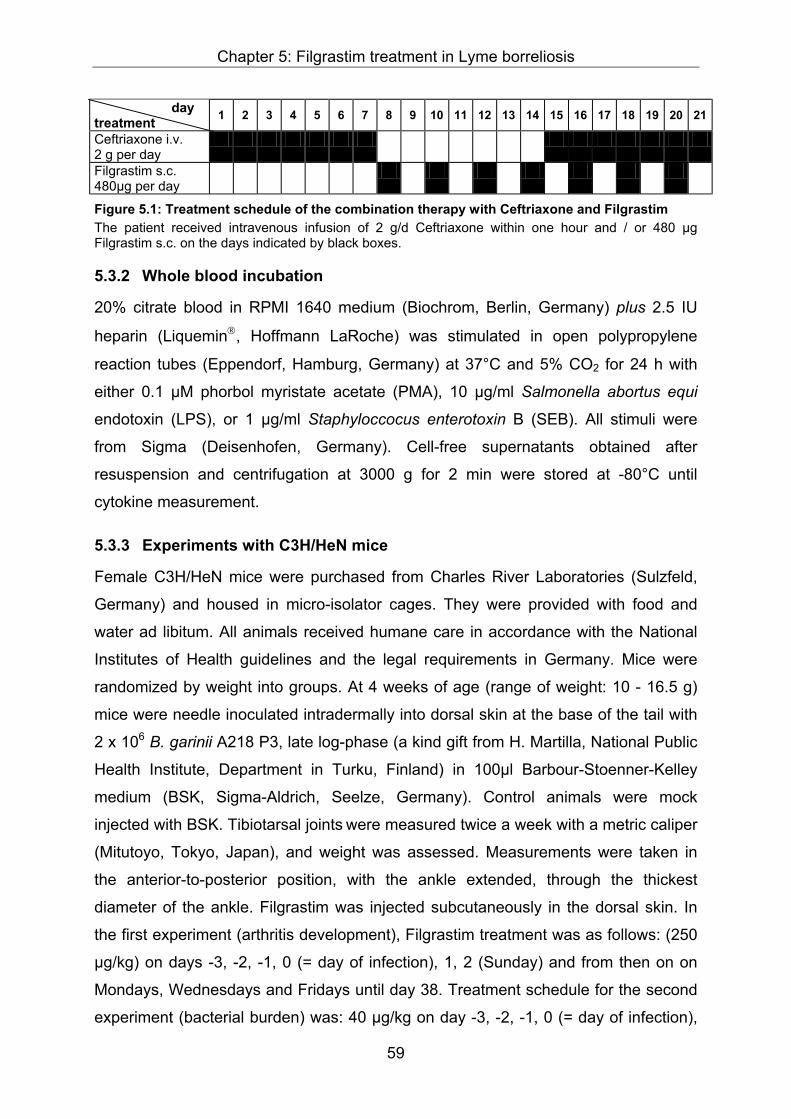
Chapter 5: Filgrastim treatment in Lyme borreliosis
59
day treatment 1 2 3 4 5 6 7 8 9 10 11 12 13 14 15 16 17 18 19 20 21
Ceftriaxone i.v. 2 g per day
Filgrastim s.c. 480µg per day
Figure 5.1: Treatment schedule of the combination therapy with Ceftriaxone and Filgrastim The patient received intravenous infusion of 2 g/d Ceftriaxone within one hour and / or 480 µg Filgrastim s.c. on the days indicated by black boxes.
5.3.2 Whole blood incubation
20% citrate blood in RPMI 1640 medium (Biochrom, Berlin, Germany) plus 2.5 IU
heparin (Liquemin, Hoffmann LaRoche) was stimulated in open polypropylene
reaction tubes (Eppendorf, Hamburg, Germany) at 37°C and 5% CO2 for 24 h with
either 0.1 µM phorbol myristate acetate (PMA), 10 µg/ml Salmonella abortus equi
endotoxin (LPS), or 1 µg/ml Staphyloccocus enterotoxin B (SEB). All stimuli were
from Sigma (Deisenhofen, Germany). Cell-free supernatants obtained after
resuspension and centrifugation at 3000 g for 2 min were stored at -80°C until
cytokine measurement.
5.3.3 Experiments with C3H/HeN mice
Female C3H/HeN mice were purchased from Charles River Laboratories (Sulzfeld,
Germany) and housed in micro-isolator cages. They were provided with food and
water ad libitum. All animals received humane care in accordance with the National
Institutes of Health guidelines and the legal requirements in Germany. Mice were
randomized by weight into groups. At 4 weeks of age (range of weight: 10 - 16.5 g)
mice were needle inoculated intradermally into dorsal skin at the base of the tail with
2 x 10⁶ B. garinii A218 P3, late log-phase (a kind gift from H. Martilla, National Public
Health Institute, Department in Turku, Finland) in 100µl Barbour-Stoenner-Kelley
medium (BSK, Sigma-Aldrich, Seelze, Germany). Control animals were mock
injected with BSK. Tibiotarsal joints were measured twice a week with a metric caliper
(Mitutoyo, Tokyo, Japan), and weight was assessed. Measurements were taken in
the anterior-to-posterior position, with the ankle extended, through the thickest
diameter of the ankle. Filgrastim was injected subcutaneously in the dorsal skin. In
the first experiment (arthritis development), Filgrastim treatment was as follows: (250
µg/kg) on days -3, -2, -1, 0 (= day of infection), 1, 2 (Sunday) and from then on on
Mondays, Wednesdays and Fridays until day 38. Treatment schedule for the second
experiment (bacterial burden) was: 40 µg/kg on day -3, -2, -1, 0 (= day of infection),

Chapter 5: Filgrastim treatment in Lyme borreliosis
60
1, 2 and, starting at day 3 with 250 µg/kg, every second day until day 12. Untreated
controls received saline-injections on the respective days. At the end of the
observation period, mice were killed by terminal pentobarbital anesthesia (Narcoren®,
Merial, Hallbergmoos, Germany).
5.3.4 Experiments with SCID mice
C.B-17.SCID mice were bred under specific pathogen-free conditions at the Max-
Planck-Institute for Immunobiology, Freiburg, Germany. Virulent, low-passage tick
isolate B. burgdorferi ZS7 was grown in BSK-medium and harvested as described
(143). 6 - 8 week old female mice were randomized by weight into 3 groups: group A:
uninfected control mice (n = 3), group B: infected mice (n = 7) and group C: infected
mice plus Filgrastim treatment (n = 7). Filgrastim (250 µg/kg) was injected
subcutaneously into the dorsal skin, on days -3, -2, -1, 0 (= day of infection), 1, 2
(Sunday) and from then on on Mondays, Wednesdays and Fridays until day 27.
Untreated controls received saline-injections on the respective days. Mice were
needle inoculated intradermally (s.c.) with 1 x 10⁵ B. burgdorferi ZS7. Control animals
were mock-injected with BSK. Mice were monitored for the development of clinical
arthritis under blinded conditions. The development of arthritis in the right and the left
tibiotarsal joints was scored as described (144) using swelling and reddening as
criteria: ++, severe; +, prominent; (+), moderate; +/-, mild; (+/-), mainly reddening
without significant swelling; and -, no clinical signs of arthritis. At the end of the
observation period (day 27 p.i.), mice were killed by terminal pentobarbital
anesthesia (Narcoren).
5.3.5 Murine leukocyte counts
Blood was obtained by cardiac puncture under terminal pentobarbital anesthesia.
White blood cell counts were determined microscopically in a Neubaur chamber after
erythrocyte lysis with Türk’s solution (Merck, Darmstadt, Germany). Leukocyte
differential counts of 100 cells each were done on May-Grünwald/Giemsa-stained
smears.
5.3.6 Determination of cytokine production by peritoneal lavage cells
Mice were killed by terminal pentobarbital anaesthesia. Peritoneal cavities were
lavaged with 10 ml ice-cold PBS (Life Technologies, Karlsruhe, Germany). The
lavage was transferred to siliconized glass tubes (Vacutainer, Becton Dickinson,

Chapter 5: Filgrastim treatment in Lyme borreliosis
61
Heidelberg, Germany) for isolation of peritoneal cells. After centrifugation, cells were
resuspended in medium (RPMI 1640, Bio Whittaker, Apen, Germany) containing
10% FCS (Biochrom) and 100 IU/ml penicillin/streptomycin (Biochrom) and plated to
96-well culture plate (bio-one, Greiner, Frickenhausen, Germany) at a density of 1 x
105 cells per well. Cells were stimulated immediately with pyrogen-free saline (Braun,
Melsungen, Germany), 0.01 µg/ml – 10 µg/ml LPS from Salmonella abortus equi (
Sigma) or 0.1 – 10 µg/ml Borrelia lysate (30). After incubation for 24 h at 37°C in the
presence of 5% CO2, the supernatants were transferred into 96-well round-bottom
plates (Greiner) and stored at -80°C until cytokines were measured.
5.3.7 Quantitative real-time PCR of Borrelia DNA in murine tissue
Ear tissues, contralateral rear ankle joints, lymph nodes, meninges, entire hearts,
brains and bladders were harvested from experimental mice at the end of the
observation period. Brain and heart samples were homogenized with a polytron
(PT1200; Kinematik, Lucerne, Switzerland) in order to test a representative aliquot
from these organs. DNA was prepared from the tissues with Qiagen DNeasyTM tissue
kit (Qiagen, Hilden, Germany) according to the manufacturer’s instructions. DNA
content was determined by measuring the absorbance at 260 nm against 280 nm
and adjusted to 10 µg DNA/ml. Quantification of Borrelia DNA was performed by real-
time PCR as described previously (145).
5.3.8 Cytokine ELISA
Monoclonal antibody pairs against human IFNγ were purchased from Endogen
(Biomar, USA / Endogen). Recombinant human IFNγ (a generous gift from Thomae
Biberach, Germany) was used as standard. For the measurement of murine (mu)
TNFα, polyclonal antibodies from R&D Systems (Wiesbaden, Germany) and
standard from Pharmingen (BD Bioscience, Heidelberg, Germany) were used. For
the detection of mu IL-10 the OptEia kit from PharMingen was used. Assays were
carried out in flat bottom, ultrasorbant 96-well plates (Nunc, Wiesbaden, Germany).
Binding of secondary biotinylated antibodies was detected with horseradish-
peroxidase-conjugated streptavidin (Biosource, Camarillo, CA, USA) and 3’,3’,5’,5’-
tetramethylbenzidine solution (Sigma) used as substrate.

Chapter 5: Filgrastim treatment in Lyme borreliosis
62
5.3.9 Statistics
Data in the figures and in the tables are given as means ± SEM. Analysis of bacterial
burden and leukocyte numbers were done with the unpaired t-test or the two-sided
Welch’s correction test for two groups. For experiments with more than two groups
one-way ANOVA was performed, followed by Bonferroni’s multiple comparison test
for selected groups or Dunnett’s test for comparison with the control group. All tests
were performed using GraphPad Prism 3.0 (GraphPad Software, San Diego, CA,
USA). P ≤ 0.05, p ≤ 0.01, and p ≤ 0.001 were considered significant and are depicted
as *, ** and *** respectively.
5.4 Results
5.4.1 Patient case report
The combination therapy of Ceftriaxone plus Filgrastim was well tolerated. Only after
the first injection of Filgrastim the patient reported acute but moderate pain in the
previously affected joints i.e. the shoulder, fingers and knees. Circulating neutrophil
counts increased from 1400 to 17000 cells/µl within 24 h after the first Filgrastim
injection. Monocyte numbers increased about two-fold, while there was little effect on
lymphocytes (Figure 5.2a). The plateau of neutrophil counts at about 17000 cells/µl
blood was maintained until one day after the end of treatment. The subjective
symptoms disappeared during the following six weeks after the treatment. The
patient reported that he was able to resume previously abandoned sporting activities
including mountain climbing and downhill skiing. Moreover, fine mechanical skills
needed for piano playing were restored. After three months, the Borrelia IgG titer was
negative. The intensity of the immunoblot at this time point was significantly reduced
(from +++ to +) and two years later it was negative. Eight years after treatment the
patient is still free of arthritic symptoms.

Chapter 5: Filgrastim treatment in Lyme borreliosis
63
0 2 4 6 8 100
1
2
3
4
22 240
5
10
15
20
25leukocytes
PMN
monocytes
lymphocytes
time after injection [h]
mon
ocyt
es [x
106 /m
l]ly
mph
ocyt
es [x
106 /m
l] leukocytes [x 106/m
l]PM
N [x 106/m
l]
Figure 5.2a. Leukocyte counts of aLyme borreliosis patient after firstinjection of 480 µg Filgrastim s.c.On day 8 of the therapy the patientwas injected with 480 µg Filgrastim s.c.at 8 a.m. (= time point 0). Blood waswithdrawn at the time points indicatedand the differential white blood cellcount was determined (PMN:polymorphonuclear neutrophil).
In order to monitor the immunological response of our patient, we followed the
attenuation of IFNγ-release capacity of whole blood taken at various time points after
Filgrastim injection, which is a well-known effect of this treatment (146). The release
of the lymphocyte-derived pro-inflammatory cytokine IFNγ was stimulated ex vivo by
different stimuli, i.e. PMA (0,1 µM), LPS (10 µg/ml) or SEB (1 µg/ml). As expected,
the inducible release of IFNγ in blood of the Filgrastim-treated patient was abrogated
from 2 h after Filgrastim injection onwards (Figure 5.2b), indicating that Filgrastim
treatment was effective.
5.4.2 Course of B. garinii-infection in C3H/HeN mice under Filgrastim treatment
This anecdotal therapeutic success of Filgrastim in the patient prompted us to carry
out further experimental studies. In order to address a possible effect of Filgrastim
more systematically, we carried out experimental treatments in different mouse
strains.
Arthritis-susceptible C3H/HeN mice were needle inoculated with Borrelia and arthritis
development was monitored by measuring joint swelling for 38 days. This is shown
0 2 4 6 8
0
25
50
75
100
LPSSEBPMA
22 24time after injection [h]
IFN
γ [n
g/m
l blo
od] Figure 5.2b. Ex vivo
IFNγ-release capacity of bloodfrom a Lyme borreliosis patientafter first injection of 480 µgFilgrastim s.c.The patient was injected with 480µg Filgrastim s.c. at 8 a.m., i.e. timepoint 0. Blood was withdrawn at thetime points indicated andstimulated with 10 µg/ml LPS, 1µg/ml SEB or 0.1 µM PMA for 24 hat 37°C. Cytokines in thesupernatant were determined byELISA.

Chapter 5: Filgrastim treatment in Lyme borreliosis
64
for infection with and without Filgrastim treatment in figure 5.3 in comparison to
uninfected control mice. There was no significant difference in ankle swelling
between the Filgrastim-treated and placebo-treated group. From day 21 p.i. ankle
swelling was similar in all infected mice. It peaked by day 28 and then resolved
continually. Thus, in spite of a slight delay in measurable arthritis manifestations,
Filgrastim treatment did not attenuate the extent of transient arthritis observed in
infected mice.
5.4.3 Determination of dissemination kinetics of B. garinii-infection in C3H/HeN mice
Kinetics of Borrelia dissemination into various tissues of inbred mouse strains have
been studied by several groups (50, 79, 147-149). It is well known that the course of
infection varies depending on the mouse strain and on the Borrelia strain used for
infection. We therefore characterized our model with regard to the spirochetal load
over the time, to determine when peak spirochete numbers are reached. C3H/HeN
mice were infected with B. garinii or mock-injected with BSK-medium. On day 1, 3,
22 and 43 post infection, bacterial load was assessed by quantitative real-time PCR
in four different organs from four infected mice and their respective controls. In order
to exclude the presence of inhibitors, which might interfere with the detection of
Borrelia DNA by PCR, spike experiments were carried out with all organs. No
interference could be seen, since spike recovery was around 100% for all the tissues.
The bacterial load of the tested organs differed strongly (Figure 5.4). Bladder and
ankle contained high numbers of spirochetes, whereas in the meninx and the lymph
node less spirochetes were detected. B. garinii spread quickly from the infection site
(base of the tail) to other tissues. The highest numbers of spirochetes were detected
at day 10 p.i. in all organs tested correlating with the evolving arthritis and declined
0 5 10 15 20 25 30 35 402.7
2.8
2.9
3.0
3.1
3.2
3.3
3.4
3.5
3.6
3.7
uninfectedB. garinii-infectedB. garinii- infected + Filgrastim
***
***
***
***
***
***
**
***
***
days after infection
ankl
e di
amet
er [m
m]
Figure 5.3. Effect of Filgrastimtreatment on ankle swellingfollowing infection of mice with B.gariniiC3H/HeN mice (n = 8) were inoculateds.c. in the base of the tail.Measurements were taken in theanterior-to-posterior position, with theankle extended, through the thickestdiameter of the ankle. Results aredepicted as mean diameter (mm) ±SEM, **, p ≤ 0.01 and ***. p ≤ 0.001vs. control.

Chapter 5: Filgrastim treatment in Lyme borreliosis
65
rapidly over time. In an additional long-term experiment, in spite of strong ankle
swelling around day 30 p.i., no Borrelia DNA at all could be detected by PCR on day
135 p.i. (data not shown), indicating that infection had completely been cleared by
the immune cells of the disease-susceptible inbred C3H/HeN mice.
In order to characterize the immune response of mice during the course of B. garinii-
infection, we determined leukocyte numbers and performed stimulation experiments
with isolated macrophages in vitro. Neither overall leukocyte numbers, nor monocyte,
lymphocyte, or neutrophil counts differed between infected and uninfected mice
during the observation period of 43 days (Figure 5.5). In addition, mice’s weights
were not influenced during the whole course of the experiment (data not shown).
These results confirm that Borrelia-infection in mice does not represent a severe
infection, which would interfere strongly with the host’s immune system. When
peritoneal lavage macrophages from infected or uninfected control mice were
stimulated with either LPS or Borrelia lysate for 24 h, there was no significant
difference in release of pro-inflammatory TNFα and anti-inflammatory IL-10 between
the two groups at any time point tested (i.e. day 1, 3, 10, 23, 43). The TNFα levels
induced by 10 µg/ml Borrelia lysate ranged from 100 to 200 pg/ml, and IL-10 levels
between 200 pg/ml and 300 pg/ml were measured. No measurable TNFα levels
could be detected in plasma of infected and uninfected mice.
1 3 10 22 430
1000
2000
3000
4000
5000
6000
bladderanklelymphnodemeninx
10000
20000
30000
40000
days after infection
Borr
elia
/ or
gan
Figure 5.4. Kinetics of spirochetedistribution in tissues from miceC3H/HeN mice were infected with 2 x 10⁶ B.garinii. At the time points indicated 4 mice pergroup were sacrificed and DNA was isolatedfrom selected tissues. The numbers ofspirochetes were determined by real-timePCR. The mean numbers of spirochetes ±SEM per organ are given.
0
1
2
3
4
1 3 10 22 43
uninfectedB. garinii -infected
days post infection
x 10
6 cel
ls/m
l
Figure 5.5. Leukocyte counts fromuninfected and B. garinii-infected C3H/HeN miceAt the indicated days post infection 4uninfected (dark symbols) and 4 infected(light symbols) mice were sacrificed andleukocyte counts in the blood weredetermined in a Neubaur chamber. The linecorresponds to the mean of the 4 values.

Chapter 5: Filgrastim treatment in Lyme borreliosis
66
5.4.4 Effect of Filgrastim treatment on B. garinii numbers in tissues of C3H/HeN mice
It has been shown, that symptomatic manifestations in both mice and humans are
strongly associated with the presence of bacteria, however, the levels of tissue
spirochetes and the severity of pathology do not correlate strictly with each other (50,
150). Therefore, we determined the effects of Filgrastim on spirochete burden in the
host’s tissues, instead of concentrating only on a possibly resolving arthritis.
Based on our results from the dissemination kinetics, day 12 p.i. was chosen to
terminate a further experiment, since by this time point joint inflammation had started
to evolve and spirochetes had reached maximum numbers in the organs. The
distribution of B. garinii in seven tested organs (ankle, bladder, lymph node, meninx,
ear, brain, heart) was not influenced uniformly by Filgrastim (Table 5.1). There was a
decrease in Borrelia numbers in the bladder and in the ankle of Filgrastim-treated
mice, however the reduction was not significant. The lymph node was similarly
infected with B. garinii in both groups, but it has to be taken into account that
spirochete levels were around the detection limit in this organ. Surprisingly, no
Borrelia at all could be detected in meninx, ear, brain, and heart.
Table 5.1. Borrelia numbers in selected tissues from C3H/HeN mice during Filgrastim treatment a
ankle bladder lymph node meninx ear brain heart C3H/HeN 5960 ± 1220 2280 ± 950 340 ± 180 n.d. n.d. n.d. n.d.
C3H/HeN + Filgrastim 3170 ± 1030* 560 ± 200* 460 ± 180* n.d. n.d. n.d. n.d. a Filgrastim-treated (treatment schedule see material and methods) and untreated mice were infected with 2 x 10⁶ B. garinii (A218) and sacrificed at 12 days p.i.. DNA was extracted from the tissues and amplified by real-time PCR as described in materials and methods. Borrelia numbers / organ were quantified with a standard curve. Means ± SEM from 8 mice are given, (* not significant vs. C3H/HeN, n.d. not detectable).
To control the effectiveness of Filgrastim treatment, leukocyte counts were
determined. Neutrophil numbers were increased in Filgrastim-treated compared to
untreated mice (3.5 x 10⁶ / ml ± 0.7 vs. 1.9 x 10⁶ / ml ± 0.3, p = 0.08). However, the
effect of recruiting neutrophils from the bone marrow was less pronounced than
expected (109).
5.4.5 Effect of Filgrastim treatment on B. burgdorferi infection in SCID mice
Murine Borrelia burgdorferi infection is often studied in severe combined
immunodeficient (SCID) mice which, despite lacking B- and T-cells, develop severe

Chapter 5: Filgrastim treatment in Lyme borreliosis
67
arthritis (148, 151). We were interested to investigate whether Filgrastim possesses
beneficial effects on arthritis development and spirochete clearance in these mice.
Therefore we repeated the experiments in C.B-17 SCID mice infected with B.
burgdorferi s.s. ZS7 at the Max-Planck-Institute in Freiburg. The same treatment
protocol as with immunocompetent mice was applied. Development of clinical
arthritis was again assessed by scoring the redness and degree of swelling of the
joint during the infection. Filgrastim-treated and untreated B. burgdorferi-infected
SCID mice developed similarly strong ankle swelling compared to the uninfected
control mice (Figure 5.6).
As in the experiment with C3H/HeN mice, Filgrastim treatment caused a slight delay
in the development of ankle swelling, however, from day 18 p.i. the extent of swelling
was the same in the two infected groups. Thus, Filgrastim treatment did not seem to
have an attenuating effect on Borrelia-induced ankle swelling in immunodeficient
SCID mice. By the end of the experiment (day 27 p.i.), spirochetal burden in 5
different organs was assessed by quantitative real-time PCR. Borrelia DNA could be
detected in all tested organs from both, Filgrastim treated and untreated B.
burgdorferi-infected mice. There was an 18-fold reduction in the numbers of Borrelia
in the pericardium and a 9-fold reduction in the ankles. Bladder, lymph node and
meninx from Filgrastim-treated mice contained 2-times less Borrelia than the
respective organs from untreated mice (Table 5.2). Thus, in general, Filgrastim-
treated SCID mice harbored fewer spirochetes in the investigated tissues than
untreated mice, indicating that Filgrastim treatment might improve elimination of
Borrelia from the tissues of immunodeficient mice.
0 5 10 15 20 25 30
uninfectedBb-infectedBb-infected + Filgrastim
-/-
± /-
+/-
± / ±
± / +
++ / ++
+ / +
days after infection
scor
es
Figure 5.6. Effect of Filgrastim treatment onankle swe lling of CB-17 SCID micefollowing infection with B. burgdorferi s.s.ZS7CB-17 SCID mice (n = 4 control group, n = 7,infected groups) were inoculated s.c. in thebase of the tail. The development of arthritis inthe right and the left tibiotarsal joints was scoredas described using swelling and reddening ascriteria: ++, severe; +, prominent; (+), moderate;+/-, mild; (+/-), mainly reddening withoutsignificant swelling, and -, no clinical signs ofarthritis. Results are depicted as the meandiameter (mm) ± SEM.

Chapter 5: Filgrastim treatment in Lyme borreliosis
68
Table 5.2. Borrelia numbers in tissues from SCID mice during Filgrastim treatment a ankle bladder lymph node meninx pericardium
SCID 7670 ± 4360 4640 ± 2040 2970 ± 1290 880 ± 260 3670 ± 3350 SCID +
Filgrastim 880 ± 170* 1950 ± 280* 1240 ± 510* 480 ± 230* 200 ± 80*
a Filgrastim-treated (treatment schedule described in material and methods) and untreated C.B.17-SCID mice were infected with 1 x 10⁵ B. burgdorferi s.s. (ZS7) and sacrificed at day 27 p.i.. Means ± SEM from 4 mice are given, * not significant vs. SCID (for further details see Table 5.1 legend).
5.5 Discussion
While early stages of LB respond to therapy with antibiotics, therapy of late stage LB
is still unsatisfactory. About 10% of all cases of late stage LB are therapy-resistant,
i.e. they cannot be cured with standard therapies (124, 152-154).
Immunosupportive therapy represents a novel promising approach in the treatment of
chronic LB. We chose Filgrastim for a combination therapy with antibiotics, due to its
unique anti-infectious and hematopoietic pharmacological properties, and the fact
that it is well tolerated on the other hand, as shown in many clinical trials (138). The
treatment regimen for the patient was set on the basis of previous experiences from
various studies treating healthy volunteers with Filgrastim (72, 146, 155, 156). The
major reservation was the intention to avoid a Herxheimer reaction, i.e. a stage of
hypertension, fever and rigors, subsequently followed by a stage of hypotension after
initiation of antibiosis in LB patients. Therefore, we started Filgrastim treatment after
one week of antibiotic therapy when the bacterial burden should have declined and a
Herxheimer reaction was less likely. During this week antibiotic treatment was
interrupted to avoid possible interferences. Finally, the treatment course was
completed with one week of combined therapy. The increasing experience of the
safety of Filgrastim treatment in infectious diseases and the experience from this first
case reduces these precautions retrospectively. Therefore, in future experimental
trials, interruption of antibiosis might not be necessary, though we still feel that
initiation of antibiosis should precede Filgrastim therapy. The injection of Filgrastim
every other day resulted from our experience with healthy volunteers, where both
neutrophilia and immunomodulation by Filgrastim lasted for 48 hours after single
injection of Filgrastim (146). Since the effects of Filgrastim on leukocytes other than
neutrophils were most prominent after 5 days of daily treatment, we would nowadays
consider this the most promising regimen to be combined with antibiosis (72).
Noteworthy a later study in healthy volunteers indicated that G-CSF treatment thrice
a week maintains its hematopoietic but not its anti-inflammatory effects (156). The

Chapter 5: Filgrastim treatment in Lyme borreliosis
69
case reported here represents a relatively mild case of chronic Lyme arthritis. It is not
clear whether positive effects of Filgrastim would also translate to more severe
cases, especially to patients with neuroborreliosis, where bacteria are difficult to
reach for leukocytes in the immunopriviledged central nervous system.
Our recent observation that blood from patients with chronic LB shows a suppressed
pro-inflammatory cytokine release when stimulated ex vivo (30) made it attractive to
speculate that the immunomodulatory effects of Filgrastim contributed the therapeutic
efficacy. Properties of Filgrastim which most likely enhanced the beneficial effects of
the treatment include amplified hematopoesis, which leads to an enforcement of the
innate immune system, allowing a greater number of non-anergic, naive activated
neutrophils to reach and kill bacteria even at remote sites of infection. The enhanced
anti-bacterial capacity might be especially efficient under the chosen conditions:
antibiotic pre-therapy would lead to lysis of bacteria thus releasing membrane
components that allow Filgrastim-activated neutrophils to be attracted by chemotactic
signals to eradicate the bacteria at poorly perfused, e.g. intraarticular locations.
Additional effects of Filgrastim on neutrophils which might also have contributed to
the therapeutic efficacy of the combination treatment include increased microbicidal
activity (157), enhanced oxidative burst (130, 158), and improved adherence and
phagocytic capacity (129, 159). It is well-known that repetitive treatment with
antibiotics increases the probability of Borrelia eradication and success of the second
course of treatment might be explained by this simple fact. However, it is very
intriguing that the patient reported warmth and pain for two days after the first two
injections of Filgrastim only in those joints which were affected by arthritic pain and
immobility over years before. This incident could be tentatively explained by a
successful granulocytic local attack on residual bacteria in the joints.
To investigate in more detail the effects of Filgrastim in Borrelia infection we
employed the mouse model of immunocompetent C3H/HeN and immunodeficient
SCID mice. Filgrastim could not attenuate edema formation, as monitored by ankle
swelling, neither in immunocompetent inbred mice, nor in SCID mice. However,
increasing evidence indicates that the degree of arthritis (assessed as ankle swelling)
does not necessarily correlate with histopathology. Anti-IL-12 treatment for example
did not affect the degree of ankle swelling, but arthritis documented by histopathology
was improved significantly (33). Wooten et al. found that ankle swelling provides only
information on the degree of edema within the ankle tissue, but neglects joint

Chapter 5: Filgrastim treatment in Lyme borreliosis
70
pathology (100). This notion is supported by recent findings indicating that ankle
swelling and arthritic lesion are two traits which are regulated independently by
genetic loci mapping to distinct locations (160). Histological analyses of the joints
could not be performed retrospectively. Thus we cannot rule out that, despite lacking
an effect on joint swelling, Filgrastim might have attenuated the inflammation in the
joint, i.e. sheath thickness and inflammatory cell infiltration.
Different authors observed that arthritis severity does not always correlate with
spirochete numbers in the organs from infected mice. A comparison between
different inbred mouse strains showed that, in spite of equal levels of spirochetes in
the ankles, the degree of arthritis pathology differed strongly (50) (161). Anti-IL-12
treatment reduced arthritis severity, but led to increased spirochetal load in the ear
(33) and IL-10-/- mice harbored less Borrelia in their ear, ankles and hearts, but
displayed a more severe arthritis compared to their wildtype littermates (53).
Consequently we next determined whether Filgrastim treatment altered the number
of spirochetes in infected mice. The bacterial burden was assessed by quantitative
real-time PCR (145).
Dissemination kinetics of Borrelia in mice have been studied extensively by many
different authors (50, 79, 147-149). However, it is well known, that on the one hand
Borrelia strains spread differently depending on the immunocompetence of the host,
and on the other hand mouse strains are differently susceptible depending on the
Borrelia strain used for infection (162-165). Our results on dissemination kinetics of
B. garinii in C3H/HeN mice are in line with data from others (50, 147-149), finding
peak spirochetal numbers by day 10 of infection.
The numbers of spirochetes in C3H/HeN mice were only moderately influenced by
immunosupportive Filgrastim treatment. Some organs of the Filgrastim-treated mice
appeared to contain less spirochetes at the time points tested (bladder, ankle), while
others (brain, ear, heart, and meninx) did not show any Borrelia. It is not clear
whether the chosen time point (day 12 p.i.) is representative for the whole course of
the infection and treatment. Noteworthy, the treatment dosage and administration
protocol was set following the case report. Possibly a lower dosage given daily would
have had stronger effects on arthritis development and / or bacterial load. The fact
that the effects of Filgrastim treatment on neutrophil numbers were less pronounced
than in a previous study, in which a corresponding dosis of murine G-CSF was given
on two consecutive days leading to a 5-fold increase indicates that the administration

Chapter 5: Filgrastim treatment in Lyme borreliosis
71
protocol was suboptimal. Importantly a high species cross-reactivity has been shown
by Moore et al. (166) making it unlikely that in our experiment the poor effectiveness
was due to the use of human Filgrastim in mice.
Additional experiments demonstrated that Borrelia-induced immune dysfunction and
longterm persistence cannot be investigated in immunocompetent mice: firstly,
Borrelia numbers declined over time and were no longer found at day 135, indicating
a long-lasting but, in contrast to humans, self-limiting infection; secondly, unlike
chronically infected patients, when testing the influence of Borrelia-infection on
macrophages of immunocompetent mice, no alteration of ex vivo inducible cytokine
production in infected mice was found, as was characteristic for LB patients (30),
suggesting that mice are not immunosuppressed by Borrelia. These results are in
line with data from Montgomery et al., who showed, that Borrelia neither induce a
global downregulation of phagocytic functions in mice (167) nor reduce inflammatory
activity in infected organs of these animals (168). It is attractive to speculate but
difficult to prove that the lack of immunomodulation is linked to the self-limiting
character of the infection in mice in contrast to humans.
Since the model of Borrelia infection in immunocompetent mice appeared to be
unsuitable to study persistence of Borrelia, we studied the effects of Filgrastim in
SCID mice, lacking B- and T-cells, which develop an array of symptoms such as
chronic progressive arthritis typical of LB in man (144) and which do not show
resolution of disease over time (66, 148). By treating SCID mice with Filgrastim a
reduction of spirochete levels in ankle, bladder, pericardium, lymphnode and meninx
was observed. Our observation that Filgrastim given without antibiotics to
immunodeficient mice, which do no possess an acquired immune system, resulted in
some reduction of bacterial burden, suggests that Filgrastim mainly acted on the
innate immune system, enhancing monocyte and neutrophil recruitment. This is of
special importance because neutrophils have been shown to play a critical role in
spirochetal clearance (140).
In conclusion, the concept of Borrelia-induced immune dysfunction as an explanation
of persistence and frequent antibiotic failure cannot be studied in the mouse model.
However, considering the unsatisfactory current antibiotic treatment, the positive
experience of an experimental therapy with Ceftriaxone plus Filgrastim in a LB
patient, and the tendency of enhanced elimination of Borrelia from mice with

Chapter 5: Filgrastim treatment in Lyme borreliosis
72
Filgrastim alone, are quite promising and encouraged an experimental clinical trial
which is currently underway.
Chapter 6: Discussion
73
6 Discussion Understanding the pathogenesis of a bacterial infection is of major importance when
ameliorating diagnostics and searching for preventive and therapeutic strategies.
This study provides insights into the pathogenesis of Lyme borreliosis, which could
help to clarify how the causative agent, B. burgdorferi, interacts with the host defense
in order to persist.
6.1 Borrelia-induced immunogenicity versus host predisposition
When investigating Borrelia-induced pathology it has to be taken into account that
the pathogen as well as the host contribute to the outcome of LB, thus when studying
host-pathogen interactions both aspects have to be considered.
6.1.1 Borrelia-induced immunogenicity
Several reports have been published demonstrating, that Borrelia-derived
components, such as the outer surface proteins (150, 169), the Erp-proteins (170),
and the variable major protein-like sequence expressed (VlsE) (171), contribute to
the immunogenicity of the pathogen and therefore affected LB development.
Experiments with low and high passaged Borrelia demonstrate, that with increasing
passage number outer surface protein expression varies and infectivity decreases
considerably (172-174). Further, complement regulator-acquiring surface protein
(CRASP) 1 and CRASP-2 Borrelia have been identified by Kraiczy et al.
demonstrating that these two outer suface proteins bind human complement
regulators and control complement activation on their surface, preventing the
formation of toxic activation products and conferring resistance to Borrelia (87).
Another property of Borrelia which contributes to the development of LB is its ability
to invade various host tissues. Several candidate bacterial ligands haven been
identified, including decorins, fibronectin, glycosaminolgycanes and β3-chain integrins
(for review see (175)), which might facilitate binding to the mammalian cells. Recent
studies underline the interplay between host and pathogen signals in both directions
showing that pro-inflammatory responses of the mammalian host trigger
recombination in the variable protein-like sequence expressed (vlsE) locus of the
spirochete resulting in the appearance of antigenically altered vlsE gene products
(176).

Chapter 6: Discussion
74
6.1.2 Host predisposition
More recently, there is accumulating evidence that host-derived factors play a critical
role in disease pathology (50). The control of Borrelia invasion in the human body is
characterized by the release of cytokines (78, 177, 178) and of oxygen radicals (38,
179) as well as the upregulation of adhesion molecules (180, 181). Depending on the
magnitude of the inflammatory response its result can either be the resolution of
infection or, if the immune response is excessive, damages such as arthritis. On the
other hand ineffective clearance of the spirochetes will occur if the response is too
weak. Thus, the strength of the immune response is decisive for the outcome of the
disease. Using genetically and immunologically manipulated mice the importance of
the host’s immune system has been investigated and led to the identification of
several host genes, controlling spirochete growth and influencing the severity of
arthritis, possibly by regulating the balance of pro- and anti-inflammatory responses
(reviewed in (182)). Steere et al. demonstrated that particular class II major
histocompatibility complex alleles represent important determinants for the host’s
immune response to B. burgdorferi and consequently for the pathology of LB.
According to their findings, the presence of HLA-DR4 and HLA-DR2 correlates with
the failure to respond to antibiotic therapy and consequently with chronic Lyme
arthritis (54). A genetic predisposition for chronic arthritis in humans has additionally
been associated with the appearance of Borrelia-specific T-cells that cross react with
self antigens (29). Recently the role of the TLR in Lyme borreliosis has been
addressed. Identifying Lyme disease vaccine low responders characterized by a
lower cell-surface expression of Toll-like receptor (TLR) 1, but normal expression of
TLR2, as compared to normal cells, Alexopoulou et al. demonstrated that defects in
the TLR1/2 signaling pathway may account for human hyporesponsiveness to OspA
vaccination (183).
In the present study, LB pathogenesis has been elucidated from two aspects: first,
the immune responsiveness of Borrelia-infected patients was characterized and
second, the effect of Borrelia on the immune response of healthy blood cells was
examined.
6.2 Hypotheses of Borrelia persistence
Although Borrelia-infection leads to activation of monocytes and granulocytes, and
further induces a prominent antibody and T helper (Th) cell cytokine response in

Chapter 6: Discussion
75
humans, no protective immunity is conferred, indicating that Borrelia-induced
activation of the immune system alone is not sufficient to eradicate the pathogen and
to protect against ongoing infection. Several hypotheses have been put forward to
explain the persistence of Borrelia in the human host:
It has been postulated that Borrelia interact with the complement system, inactivating
the complement regulatory proteins FHL-1/reconectin and Factor H (86, Kraiczy,
2002 #912). Others have proven that Borrelia can hide themselves in
immunoprivileged sites such as the collagen fibers of the connective tissue (88) and
in human synovial cells (24) as well as in the extracellular matrix (25). The antigenic
variation of the B. burgdorferi outer membrane is also discussed as a possible
strategy to evade the immune response (26, Zhang, 1997 #938, Wilske, 1996 #939).
Data from Gross et al. suggest alternatively, that Borrelia induce an auto-immune
process, as they could identify a homology between the Borrelia outer surface protein
A (OspA) and the human lymphocyte-function-associated antigen-1 alpha (hLFA-1)
and also found cross reactive T-cells (29).
6.2.1 Immunomodulation as a possible immune evasion strategy
In the present study a further hypothesis was raised postulating that the host’s
immune response is modulated by the pathogen such that the bacteria shift or
suppress it in a way that enables survival of the pathogen. Examples of this latter
type are known for viral (58), bacterial (59) and parasitic infections (60), and has
hence led to the concept of microbial cytokine-inducing or suppressing molecules
named modulins (61, 62). Our hypothesis of Borrelia-induced immunomodulation as
a possible immune evasion mechanism is based on the findings that ex vivo
stimulated whole blood from patients with chronic LB revealed a significantly reduced
capacity to release TNFα and IFNγ in comparison to blood cells from healthy controls
(see chapter 3). There are few other reports about Borrelia acting as an
immunomodulator: Mullegger et al. found a lack of IFNγ expression in the LB-specific
chronic skin lesions, i.e. acrodermatitis chronica atrophicans (ACA), and postulated
reduced effectiveness of spirochetal killing (91). Furthermore, the anti-inflammatory
influence of Borrelia is reflected in a decreased expression of MHC II markers on
Langerhans cells in skin of ACA patients (92). An unusual subset of T-cells,
characterized by secreting Th1 (IFNγ) and Th2 (IL-10) cytokines concomitantly, were
identified in the joints of LB patients in which Borrelia were present (115). Sigal et al.

Chapter 6: Discussion
76
designated Borrelia “a potent immunomodulator due to its observed potential for
immundysregulation” (82).
The idea of Borrelia-induced immunomodulation was extended by our findings, that
Borrelia are stronger inducers of an anti-inflammatory cytokine response than
endotoxins from various Gram-negative bacteria (see chapter 3), confering them the
capacity to influence the balance between the pro- and the anti-inflammatory immune
response. Our data are in line with findings from other authors, indicating that B.
burgdorferi is a potent inducer of the anti-inflammatory cytokine IL-10 (46, 84, 116).
In addition, the modulatory and regulatory capacity of IL-10 with regard to Borrelia-
induced cytokine release has been found in different in vitro models (93, Lisinski,
2002 #952). Evidence for the in vivo relevance of the modulatory capacity of IL-10
was provided by Brown et al. (53), showing that in the absence of IL-10 the
spirochetal burden in IL-10-/- mice was significantly lower than in wildtype mice. In
contrast, inflammation in the ankle was significantly higher. These results
demonstrate the key role of IL-10 in regulating Borrelia-infection in Lyme arthritis.
6.3 Borrelia lipoproteins and the Toll-like receptor 2
For Borrelia lipoproteins the Toll-like receptor 2 (TLR2) has been identified as the
major signal transducing receptor. Using TLR2-cotransfected mice macrophages
Brightbill et al. demonstrated, that Borrelia lipoprotein induced IL-12 production and
activation of iNOS is TLR2-mediated (98). At the same time Hirschfeld et al.
described that TLR2-transfection into a glioma cell-line confers responsiveness to
lipoproteins and sonicated Borrelia (95). Experiments transfecting CHO cells with
TLR2 renders them responsive to OspA, synthetical lipoproteins and live Borrelia as
assessed by induction of pro-inflammatory cytokines and NFκB-translocation (96).
Aliprantis et al. postulate that TLR2 is involved in Borrelia-induced cell-activation and
apoptosis (99). Further evidence for TLR2-involvement in recognition of Borrelia was
provided by Lorenz et al., who performed functional studies with 293T cells
transfected with the non-functional Arg753Gln polymorphic TLR2 gene in comparison
to the wildtype gene (97). The most convincing data that TLR2 is crucial for host
defense to Borrelia were obtained in TLR2-/- mice, which harbored up to 100-fold
more spirochetes in tissues than their wildtype littermates. However, since the
antibody response was not influenced by the absence of TLR2 it was postulated that
the acquired immune response to Borrelia does not require this receptor. The most

Chapter 6: Discussion
77
surprising result was that TLR2-/- mice developed greater ankle swelling than TLR2+/+
mice, suggesting TLR2-independent components to be involved in the inflammatory
response to Borrelia. Indeed, although being unresponsive to stimulation with
lipoproteins, when stimulating macrophages from TLR2-/- mice with high
concentrations (50 µg/ml) of sonicated Borrelia, they released TNFα and NO (100).
These findings are in line with our hypothesis that the TLR2 mediated response is
primarily of an anti-inflammatory nature.
In order to address these conflicting results, we tested the role of TLR2 and TLR4 in
the recognition of different Borrelia-specific stimuli. As demonstrated in stimulation-
experiments with bone marrow-derived macrophages from TLR2-/- mice, none of the
tested Borrelia-specific stimuli induced cytokine release in the absence of TLR2
(chapter 4). In contrast, despite a nonfunctional TLR4, stimulation of macrophages
from C3H/HeJ mice with the same Borrelia-specific stimuli resulted in TNFα-release
comparable to wildtype cells. Thus, our results substantiate the view, that TLR2 is the
major receptor responsible for Borrelia-induced cytokine production. It has to be
mentioned that we could confirm data from Wooten et al., inducing weak but clearly
measurable TNFα-release when stimulating macrophages from TLR2-deficient mice
with Borrelia lysate concentrations above 10 µg/ml. However, since these TLR2-
independent components, which have neither been isolated nor characterized to
date, appear to be immunogenic only in high concentrations their relevance still has
to be defined.
6.4 Borrelia-induced tolerance and cross-tolerance
The phenomenon of endotoxin (LPS) tolerance has been investigated extensively in
vitro and in vivo (for review see (101)). It describes a status of macrophage
hyporesponsiveness after exposure to low LPS doses to a subsequent high or lethal
LPS dose (102, 103). In this study we investigated whether B. burgdorferi also has
the capacity to desensitize macrophages, since Borrelia-induced
hyporesponsiveness could represent a mechanism enabling the survival of this
pathogen in the immunocompetent host (chapter 4). We tested this hypothesis in
desensitization experiments with Borrelia-specific stimuli. Indeed, similarly to the
effect of LPS pre-treatment in endotoxin tolerance, pre-treatment with Borrelia lysate
could render human PBMC tolerant, i.e. unable to react to a second stimulation with
Borrelia, as shown by a reduced capacity of TNFα-release. Thus, Borrelia are able to
Chapter 6: Discussion
78
modulate the monocytic immune response. They even induce a more general
desensitization, since they also rendered human PBMC hyporesponsive to
subsequent stimulation with heterologous stimuli such as the well characterized
TLR2 agonist LTA or the TLR4 agonist LPS. Recently, similar desensitization
experiments were reported which demonstrated, that stimuli other than LPS, i.e.
highly purified lipoteichoic acid (LTA) (104) or macrophage-activating lipopeptide-2
(MALP-2) from mycoplasma (105), can also render macrophages tolerant to
subsequent re-stimulation. Furthermore, it was shown in these same studies, that
tolerance can also be induced by two heterologous stimuli, independent of the
receptor involved in their recognition and signaling. In this case the appropriate term
is cross-tolerance or hetero-tolerance.
6.4.1 Toll-like receptors and signaltransduction pathways in Borrelia-induced tolerance
Several reports have been published, showing that tolerance induced by different
TLR2- and TLR4-agonists shares common intracellular signal transduction pathways:
Pre-treatment with synthetic bacterial lipoproteins (BLP) inhibits phosphorylation of
ERK, JNK and p38 Map-kinases and reduces NFκB-activation and NFκB-DNA
binding activity similarly to LPS pre-treatment (114). In MALP-2 induced cross-
tolerance to LPS NFκB-activation and c-Jun NH2 terminal kinase is suppressed
(105). Down regulation of IL-1 receptor associated kinases (IRAK) protein levels and
kinase activity correlates with development of LPS tolerance and with LPS induced
cross-tolerance to LTA, but not with LTA tolerance and LTA-induced cross-tolerance,
suggesting differences in tolerance mechanisms of these agonists. Unique TLR2
signaling components downstream of TLR2 and upstream of MyD88/IRAK are
postulated in LTA tolerance (120).
Our experiments with bone marrow lavage cells from TLR2-/- mice and from mice
lacking a functional TLR4 substantiate the view that common signaling molecules are
shared by TLR2 and TLR4. In contrast to LPS, pre-exposure to Borrelia induced
macrophage hyporesponsiveness in the absence of a functional TLR4. Instead, pre-
exposure to Borrelia lysate failed to confer refractoriness to TLR2-deficient cells,
whereas LPS-induced TNFα suppression was unaffected in these cells. Thus,
Borrelia-specific stimuli required the TLR2 to induce tolerance and respectively LPS
required the TLR4. Based on our findings that stimulation with Borrelia lysate, which

Chapter 6: Discussion
79
signals via TLR2, results in hyporesponsiveness to TLR4-mediated LPS signaling
and vice versa, we postulated that.
6.4.2 Regulation of the TLR in Borrelia-induced tolerance
Differing results regarding the regulation of the TLR in tolerance have been published
(105, 119, 121). Wang et al. showed downregulation of TLR2 in tolerance induced by
synthetic bacterial lipopeptide (BLP), but not LPS (114). Our results regarding the
TLR2-mRNA are partially in line with these observations, since the TLR2 was
significantly downregulated in cells tolerized by Borrelia lysate upon re-stimulation
with the same stimulus (chapter 4). As a major difference to Wang et al., we also
observed a downregulation of the TLR2 by LPS. The opposing results could be
ascribed to the fact that cross-tolerance we observe is complete and likewise induced
by the two stimuli, unlike Wang et al. who report an incomplete LPS-induced cross-
tolerance to BLP. Our data suggest that downregulation of the TLR2 seems to occur
in tolerance, representing a probable explanation for the observed state of
unresponsiveness to a second stimulation.
6.4.3 Role of soluble mediators in Borrelia-induced tolerance
Several reports have been published on the role of leukocyte-derived inflammatory
mediators in tolerance induction of macrophages. According to these publications
endogenous anti-inflammatory cytokines such as IL-10, TGFβ or IL-1 are considered
to be involved in LPS-induced tolerance by suppressing the formation of pro-
inflammatory cytokines factors (113, 184, 185)). Others postulate the existence of
soluble yet unidentified suppressor molecules of TNFα-expression after pre-exposure
of cells to endotoxin (186). Data from Randow et al. demonstrate that LPS-induced
tolerance is mediated by endogenous cytokines such as IL-10 and TGFβ (113). Our
results with regard to tolerance induced by Borrelia in the presence of IL-10-, TGFβ-
and G-CSF-neutralizing antibodies indicate that IL-10 is indeed involved in induction
of Borrelia-tolerance. However, tolerance induction could not be completely
prevented by blocking the mediator, suggesting that other mediators also play a role
in this process. TGFβ, which also contributed to LPS-tolerance in human PBMC
(113), was apparently not required for Borrelia-tolerance, nor was G-CSF. Further
studies will be necessary to definitely settle this point, since our data oppose data
from others, showing that peritoneal macrophages from wildtype and IL-10-/- mice
could be similarly tolerized by LPS and MALP, suggesting that IL-10 was not involved

Chapter 6: Discussion
80
(105). Own results from coculture experiments with TLR2- and TLR4-deficient
primary murine cells and the corresponding wildtype cells stimulated with LTA and
LPS also argue against soluble factors responsible for suppression of TNFα upon
secondary stimulation in this murine model (104).
6.5 Adjuvant immunotherapy in Lyme borreliosis
While LB at early stages can be managed with antibiotics, therapy of late stage LB
still is unsatisfactory. A small percentage of all late stage LB cases are therapy
resistant, i.e. they cannot be cured with standard therapies (124, 152-154). In this
work evidence has been provided that Borrelia modulates the human immune
response representing a possible mechanism to persist in the host. Based on this
finding that pathology of LB is associated with modulation of leukocyte functions,
adjuvant immunotherapy should be taken into account as an additional therapy to
vaccination and antibiosis. The reconstitution of the patients immune competence
with activating cytokines represents an attractive target for supportive treatment in
chronic LB. Possible candidates could be the endogenous products granulocyte
macrophage colony-stimulating factor (GM-CSF) and IFNγ, which possess strong
pro-inflammatory properties and have been shown to restore the systemic TNFα-
response to endotoxin in LPS-desensitized mice (187) or granulocyte colony-
stimulating factor (G-CSF, Filgrastim) which acts as well as a pro- as an anti-
inflammatory cytokine, recruits and primes neutrophils and induces mono- and
lymphocytosis in human volunteers (72). In the study presented here, G-CSF
(Filgrastim) was chosen for the combination therapy with antibiotics, due to its unique
anti-infectious and hematopoietic pharmacological properties on the one hand, and
the fact that it is well tolerated on the other hand, as shown in many clinical trials
(138).
6.5.1 Case report
We report the anecdotal observation of a late stage LB patient, who was successfully
treated with a combination therapy of a cephalosporin antibiotic plus Filgrastim
(chapter 5). Properties of Filgrastim which most likely contributed to the beneficial
effects of the treatment include amplified hematopoesis, which leads to an
enforcement of the innate immune system, allowing a greater number of activated
non-anergic, naive neutrophils to reach and kill bacteria even at remote sites of
infection. This is of special importance since it has been demonstrated that

Chapter 6: Discussion
81
neutrophilic granulocytes contribute substantially to host defense against Borrelia
(39, 139-141). The enhanced anti-bacterial capacity might be especially efficient
under the chosen conditions: antibiotic pre-therapy could lead to lysis of bacteria,
thus releasing membrane components that allow Filgrastim-activated neutrophils to
be attracted by chemotactic signals to eradicate the bacteria at poorly perfused, e.g.
intraarticular locations. Additional effects of Filgrastim on neutrophils which might
also have contributed to the therapeutic efficacy of the combination treatment include
increased microbicidal activity (157), enhanced oxidative burst (130, 158) and
improved adherence and phagocytic capacity (129, 159). Noteworthy, the patient
represented a relatively mild case of chronic Lyme arthritis. It is not clear whether
positive effects of Filgrastim would also translate to more severe cases, especially to
patients with neuroborreliosis, where bacteria are difficult to reach for leukocytes in
the immunopriviledged central nervous system. Another aspect which has to be
mentioned is that repetitive treatment with antibiotics increases per se the probability
of Borrelia eradication and success of the second course of treatment might be
explained by this simple fact. However, it is very intriguing that the patient reported
warmth and pain after the first two injections of Filgrastim for two days only in those
joints which were affected by arthritic pain and immobility over years before. This
incident could be tentatively explained by a successful granulocytic local attack on
residual bacteria in the joints.
6.5.2 Mouse model
The murine model of LB developed by Barthold and colleagues demonstrated that
certain strains of mice develop severe inflammatory arthritis 3 - 4 weeks after intra-
dermal injection of Borrelia with histopathological characteristics similar to the
subacute arthritis seen in humans (188). Therefore this mouse model is frequently
used to study pathology of Borrelia-infection and to find new treatment strategies in
LB. To investigate in more detail the effects of Filgrastim in Borrelia-infection we
carried out experiments with immunocompetent C3H/HeN and immunodeficient SCID
mice. Filgrastim could not attenuate edema formation, as monitored by ankle
swelling, neither in immunocompetent inbred mice, nor in SCID mice. However,
considering recent findings, it might have influenced lesion severity. Increasing
evidence indicates that the degree of arthritis (assessed as ankle swelling) does not
necessarily correlate with histopathology. Anti-IL-12 treatment for example did not
affect the degree of ankle swelling, but arthritis documented by histopathology was

Chapter 6: Discussion
82
improved significantly (33). Wooten et al. found that ankle swelling provides only
information on the degree of edema within the ankle tissue, but neglects joint
pathology (100). This notion is supported by recent findings indicating that ankle
swelling and arthritic lesion are two traits which are regulated independently by
genetic loci mapping to distinct locations (160). Histological analyses of the joints
could not be performed retrospectively. Thus we cannot rule out that, despite lacking
an effect on joint swelling, Filgrastim might have attenuated the inflammation in the
joint, i.e. sheath thickness and inflammatory cell infiltration.
Differing results have been published regarding the correlation between levels of
tissue spirochetes and severity of pathology. Some authors demonstrate that
development of arthritis in susceptible mouse strains correlates with higher numbers
of spirochetes in these hosts (147, 189). In contrast, comparison between different
inbred mouse strains showed that, in spite of equal levels of spirochetes in the
ankles, the degree of arthritis pathology differed strongly (50, 161). Anti-IL-12
treatment reduced arthritis severity, but led to increased spirochetal load in the ear
(33) and IL-10-/- mice harbored less Borrelia in their ear, ankles and hearts, but
displayed a more severe arthritis compared to their wildtype littermates (53). In our
experiments the numbers of spirochetes assessed by quantitative real-time PCR in
C3H/HeN mice were only moderately influenced by immunosupportive Filgrastim
treatment. Some organs of the Filgrastim-treated mice appeared to contain less
spirochetes at the time points tested (ankle, bladder, lymph node), while in others
(brain, ear, heart, and meninx) no Borrelia could be detected. The chosen time-point
might not be representative of the whole course of the infection. Furthermore
treatment dosage and administration protocol possibly account for the low effects on
arthritis development and bacterial load and could be varied in future studies.
Additional experiments demonstrated that Borrelia-induced immune dysfunction and
longterm persistence could not be investigated in immunocompetent mice: firstly,
Borrelia numbers declined over time and were no longer found at day 135, indicating
a long-lasting but, in contrast to humans, self-limiting infection; secondly, unlike
chronically infected patients, when testing the influence of Borrelia-infection on
macrophages of immunocompetent mice, no alteration of ex vivo inducible cytokine
production in infected mice was found, as was characteristic for LB patients (30),
suggesting that mice are not immunosuppressed by Borrelia. These results are in line
with data from Montgomery et al., who showed, that Borrelia neither induce a global

Chapter 6: Discussion
83
downregulation of phagocytic functions in mice (167) nor reduce inflammatory activity
in infected organs of these animals (168). It is attractive to speculate but difficult to
prove that the lack of immunomodulation is linked to the self-limiting character of the
infection in mice in contrast to humans.
Since the model of Borrelia-infection in immunocompetent mice appeared to be
unsuitable to study persistence of Borrelia, we studied the effects of Filgrastim in
SCID mice, lacking B- and T-cells, which develop an array of symptoms such as
chronic progressive arthritis typical of LB in man (144) and which do not show
resolution of disease over time (66, 148). By treating SCID mice with Filgrastim a
reduction of spirochete levels in the ankle, bladder, heart and meninx was observed.
Our observation that Filgrastim given without antibiotics to immunodeficient mice,
which do no possess an acquired immune system, resulted in some reduction of
bacterial burden, suggests that Filgrastim mainly acted on the innate immune system,
enhancing monocyte and neutrophil recruitment.
In conclusion, a novel approach in the treatment of chronic LB was tested based on
the concept that killing of Borrelia with antibiotics should be combined with
reactivation of immune cells which have been modulated by the presence of the
pathogen. Considering the unsatisfactory current antibiotic treatment, the positive
experience of an experimental therapy with Ceftriaxone plus Filgrastim in a LB
patient, and the enhanced elimination of Borrelia from SCID-mice, as well as the
tendency to beneficial effects in immunocompetent mice with Filgrastim alone are
quite promising and encouraged a clinical trial on the immunosupportive Filgrastim
therapy of LB which is currently underway.
Chapter 7: Summary
84
7 Summary If infection with Borrelia burgdorferi is not treated adequately with antibiotics in an
early stage, it may lead to Lyme borreliosis (LB), a chronic multisystemic disorder
which is difficult to cure. In some cases the pathogen survives in spite of antibiotic
treatments. It is challenging to understand why Borrelia are often not eradicated,
although being recognized by the host’s immune defense and occasionally inducing
a strong inflammatory reaction. Thus, it remains an area of debate how this pathogen
persists in human tissues. This question was addressed in the present thesis,
examining possible immune evasion mechanisms of Borrelia.
• Blood cells from patients suffering from persistent LB released significantly
lower levels of pro-inflammatory cytokines (TNFα and IFNγ) in response to
either Borrelia lysate or to lipopolysaccharide (LPS) in comparison to cells
from healthy volunteers.
• In blood from healthy volunteers Borrelia lysate led to strong production of
anti-inflammatory IL-10 and G-CSF, while inducing only low levels of pro-
inflammatory IFNγ, compared to LPS.
• Similar to endotoxin tolerance, different Borrelia preparations desensitized
human blood monocytes on re-stimulation with either stimulus.
• Borrelia-specific stimuli induced cross-tolerance towards heterelogous stimuli
such as lipopolysaccharid (LPS) and lipoteichoic acid (LTA) in human
monocytes.
• Toll-like receptor (TLR) 2 but not TLR4 was required for Borrelia-induced
tolerance and cross-tolerance, as shown in experiments with knock-out mice.
• PBMC tolerized by Borrelia lysate exhibited reduced TLR2-mRNA levels.
Further, IL-10 was identified as a key mediator involved in tolerance-induction
by Borrelia lysate.
• Combination of Filgrastim treatment with Ceftriaxone in a late stage LB-patient
who failed standard antibiotic therapy led to successful eradication of the
pathogen and complete regression of symptoms.
• The mouse model of Borrelia infection was set up and characterized in order
to study the therapeutic effects of Filgrastim in vivo.

Chapter 7: Summary
85
• Treating immunocompetent and immunodeficient SCID mice with Filgrastim,
as an immunosupportive therapy of LB did not attenuate the characteristic
ankle swelling induced by Borrelia infection.
• Regular application of Filgrastim led to an enhanced elimination of Borrelia
from various organs in SCID mice. In immunocompetent mice this effect was
less pronounced.
In summary, we propose that Borrelia modulate the host’s immune system in order to
evade clearance in the immunologically competent host. Tolerance could represent
the mechanism inhibiting host response thereby enabling survival and persistence of
the pathogen. Promising results were obtained testing a novel treatment strategy for
late stage LB, a combination of Filgrastim as an immunosupportive therapy with
antibiotics. The respective clinical trial based on these findings was recently started.

Chapter 8: Zusammenfassung
86
8 Zusammenfassung Die Infektion mit Borrelia burgdorferi, die nicht frühzeitig und angemessenen
behandelt wird, führt im Menschen zu einer chronischen Multisystem-Erkrankung, die
schwer zu therapieren ist, da Antibiotika teilweise versagen. Trotz intensiver
Forschung ist bisher noch nicht befriedigend geklärt, wie die Borrelien in einem
funktionierenden Immunsystem persistieren. In der vorliegenden Arbeit wurde dieser
Frage der Borrelienpersistenz nachgegangen.
• Im Blut chronisch erkrankter Borreliosepatienten wurde nach Stimulation mit
Borrelienlysat oder LPS im Vergleich zu einem gesunden Kontrollkollektiv eine
signifikant reduzierte TNFα- und IFNγ-Freisetzung nachgewiesen.
• Im Blut gesunder Spender stellten Borrelien im Vergleich zu LPS einen
starken Stimulus für die Freisetzung der anti-inflammatorischen Zytokine
G-CSF und IL-10 dar und nur einen schwachen für das pro-inflammatorische
Zytokin IFNγ.
• Die wiederholte Stimulation mit verschiedenen Borrelien-spezifischen Stimuli
induzierte eine Desensitivierung von humanen Blutmonozyten, vergleichbar
mit der „Endotoxin Toleranz“.
• Borrelien-spezifische Stimuli erzeugten mit LTA oder mit LPS eine
Kreuzdesensitivierung von humanen Blutmonozyten.
• Experimente mit Makrophagen von knockout-Mäusen ergaben, dass TLR2 -
nicht aber TLR4 - benötigt wird, um mit Borrelienlysat Desensitivierung und
Kreuzdesensitivierung zu induzieren.
• Bei der Borrelienlysat-induzierten Desensitivierung und Kreuzdesensitivierung
von humanen PBMC wurde die Toll-like receptor (TLR) 2-mRNA
herunterreguliert und es konnte die Beteiligung von IL-10 als Mediator
nachgewiesen werden, nicht aber von TGFβ oder G-CSF.
• Die Kombination des Immunaktivators Filgrastim mit Ceftriaxon bei einem
chronisch infizierten Borreliosepatienten und Therapieversager führte zu der
erfolgreichen Eradikation des Erregers und dem vollständigen Rückgang der
Krankheitssymptome.

Chapter 8: Zusammenfassung
87
• Das Mausmodell wurde zur in vivo Untersuchung der Borrelieninfektion
etabliert und charakterisiert, um anschließend Therapiestrategien testen zu
können.
• Der Einsatz von Filgrastim im Borreliose-Mausmodell als adjuvante
Immuntherapie hatte keinen Effekt auf die Entwicklung der Arthritis, weder in
der immundefizienten, noch in der immunkompetenten Maus.
• Die regelmäßige Gabe von Filgrastim vor und während der Borrelieninfektion
führte in der immundefizienten Maus zu einer deutlichen Reduktion der
Erregerlast in allen Organen, aber in nur einem Organ der immunkompetenten
Maus.
Aufgrund dieser Ergebnisse stellt die Modulation der Immunantwort eine mögliche
Erklärung für die Persistenz der Borrelien im Patienten dar, indem sie einerseits die
anti-inflammatorische Zytokinantwort der Immunzellen begünstigen und andererseits
einen Zustand der Desensitivierung im Makrophagen hervorrufen können. Aus dieser
Kenntnis der pathophysiologischen Defekte und des erfolgreichen Einzelbefundes,
zusammen mit den Daten aus der Maus, stellt der Einsatz von Immunaktivatoren
eine vielversprechende therapeutische Interventionen in der Borreliose dar. Die auf
diesen Befunden basierende experimentelle klinische Studie wurde kürzlich
gestartet.

Chapter 9: References
88
References
1. Strle, F., and M. Stantic-Pavlinic. 1996. Lyme disease in Europe. N Engl J Med 334:803. 2. Orloski, K. A., E. B. Hayes, G. L. Campbell, and D. T. Dennis. 2000. Surveillance for Lyme
disease-United States, 1992-1998. Mor Mortal Wkly Rep CDC Surveill Summ 49:1. 3. Maiwald, M., D. Hassler, and H. A. Zappe. 1996. Epidemiologie und Prophylaxe
zeckenübertragener Krankheiten. Allgemeinarzt 9:986. 4. Hassler, D., L. Zöller, M. Haude, D. Hufnagel, and H. G. Sonntag. 1992. Lyme-Borreliose in
einem europäischen Endemiegebiet. Dtsch med Wschr 117:767. 5. Oehme, R., K. Hartelt, H. Backe, S. Brockmann, and P. Kimmig. 2002. Foci of tick-borne
diseases in southwest Germany. Int J Med Microbiol 291 Suppl 33:22. 6. Afzelius, A. 1921. Erythema chronicum migrans. Acta Derm Venereo (Stockholm) 2:120. 7. Bannwarth, A. 1941. Chronische lymphocytäre Meningitis, entzündliche Polyneuritis und
"Rheumatismus". Ein Beitrag zum Problem "Allergie und Nervensystem". Arch Psychiatr Nervenkr 113:284.
8. Hellerstrom, S. 1950. Erythema chronicum migrans Afzelius with meningitis. Southern Med J 43:330.
9. Steere, A. C., S. E. Malawista, D. R. Snydman, R. E. Shope, W. A. Andiman, M. R. Ross, and F. M. Steele. 1977. Lyme arthritis: an epidemic of oligoarticular arthritis in children and adults in three connecticut communities. Arthritis Rheum 20(1):7.
10. Burgdorfer, W., A. G. Barbour, S. F. Hayes, J. L. Benach, E. Grunwaldt, and J. P. Davis. 1982. Lyme disease-a tick-borne spirochetosis? Science 216(4552):1317.
11. Fraser, C. M., S. Casjens, W. M. Huang, G. G. Sutton, R. Clayton, R. Lathigra, O. White, K. A. Ketchum, R. Dodson, E. K. Hickey, M. Gwinn, B. Dougherty, J. F. Tomb, R. D. Fleischmann, D. Richardson, J. Peterson, A. R. Kerlavage, J. Quackenbush, S. Salzberg, M. Hanson, R. vanVugt, N. Palmer, M. D. Adams, J. Gocayne, J. C. Venter, and e. al. 1997. Genomic sequence of a Lyme disease spirochaete, Borrelia burgdorferi. Nature 390(6660):580.
12. Strle, F. 1999. Lyme borreliosis in Slovenia. Zentralbl Bakteriol 289:643. 13. Gray, J. S., O. Kahl, J. N. Robertson, M. Daniel, A. Estrada-Pena, G. Gettinby, T. G.
Jaenson, P. Jensen, F. Jongejan, E. Korenberg, K. Kurtenbach, and P. Zeman. 1998. Lyme borreliosis habitat assessment. Zentralbl Bakteriol 287:211.
14. Ai, C. X., R. J. Hu, K. E. Hyland, Y. X. Wen, Y. G. Zhang, Q. C. Qiu, D. Y. Li, X. D. Liu, Z. X. Shi, J. H. Zhao, and et al. 1990. Epidemiological and aetiological evidence for transmission of Lyme disease by adult Ixodes persulcatus in an endemic area in China. Int J Epidemiol 19:1061.
15. Dennis, D. T. 1998. Epidemiology, ecology, and prevention of Lyme disease. American College of Physicians, Philadelphia, PA.
16. O'Connell, S., M. Granstrom, J. S. Gray, and G. Stanek. 1998. Epidemiology of European Lyme borreliosis. Zentralbl Bakteriol 287:229.
17. Hassler, D. 1997. Langzeitbeobachtung zum Krankheitsbild der Lyme-Borreliose in einem Endemiegebiet. Daten zur Vektorökologie, Epidemiologie, Serologie und Klinik, Therapie und Therapiekontrolle, Habilitationsschrift.
18. Piesman, J., T. N. Mather, R. J. Sinsky, and A. Spielman. 1987. Duration of tick attachment and Borrelia burgdorferi transmission. J Clin Microbiol 25(3):557.
19. Kahl, O., C. Janetzki-Mittmann, J. S. Gray, R. Jonas, J. Stein, and R. de Boer. 1998. Risk of infection with Borrelia burgdorferi sensu lato for a host in relation to the duration of nymphal Ixodes ricinus feeding and the method of tick removal. Zentralbl Bakteriol 287:41.
20. Franz, J. K., S. Priem, M. G. Rittig, G. R. Burmester, and A. Krause. 1999. Studies on the pathogenesis and treatment of Lyme arthritis. Wien Klin Wochenschr 111:981.
21. Busch, U., C. Hizo-Teufel, R. Bohmer, V. Fingerle, D. Rossler, B. Wilske, and V. Preac-Mursic. 1996. Borrelia burgdorferi sensu lato strains isolated from cutaneous Lyme borreliosis biopsies differentiated by pulsed-field gel electrophoresis. Scand J Infect Dis 28:583.
22. Rijpkema, S., D. Golubic, M. Molkenboer, N. Verbeek-De Kruif, and J. Schellekens. 1996. Identification of four genomic groups of Borrelia burgdorferi sensu lato in Ixodes ricinus ticks collected in a Lyme borreliosis endemic region of northern Croatia. Exp Appl Acarol 20:23.
23. van der Heijden, I. M., B. Wilbrink, S. G. Rijpkema, L. M. Schouls, P. H. Heymans, J. D. van Embden, F. C. Breedveld, and P. P. Tak. 1999. Detection of Borrelia burgdorferi sensu stricto by reverse line blot in the joints of Dutch patients with Lyme arthritis. Arthritis Rheum 42:1473.

Chapter 9: References
89
24. Girschick, H. J., H. I. Huppertz, H. Russmann, V. Krenn, and H. Karch. 1996. Intracellular persistence of Borrelia burgdorferi in human synovial cells. Rheumatol Int 16:125.
25. Haupl, T., G. Hahn, M. Rittig, A. Krause, C. Schoerner, U. Schonherr, J. R. Kalden, and G. R. Burmester. 1993. Persistence of Borrelia burgdorferi in ligamentous tissue from a patient with chronic Lyme borreliosis. Arthritis Rheum 36(11):1621.
26. Seiler, K. P., and J. J. Weis. 1996. Immunity to Lyme disease: protection, pathology and persistence. Curr Opin Immunol 8(4):503.
27. Restrepo, B. I., and A. G. Barbour. 1994. Antigen diversity in the bacterium B. hermsii through "somatic" mutations in rearranged vmp genes. Cell 78:867.
28. Kamradt, T., A. Krause, and G. R. Burmester. 1995. A role for T cells in the pathogenesis of treatment-resistant Lyme arthritis. Mol Med 1(5):486.
29. Gross, D. M., T. Forsthuber, M. Tary-Lehmann, C. Etling, K. Ito, Z. A. Nagy, J. A. Field, A. C. Steere, and B. T. Huber. 1998. Identification of LFA-1 as a candidate autoantigen in treatment-resistant Lyme arthritis. Science 281(5377):703.
30. Diterich, I., L. Härter, D. Hassler, A. Wendel, and T. Hartung. 2001. Modulation of cytokine release in ex vivo stimulated blood from borreliosis patients. Infect Immun 69:687.
31. Kang, I., S. W. Barthold, D. H. Persing, and L. K. Bockenstedt. 1997. T-helper-cell cytokines in the early evolution of murine Lyme arthritis. Infect Immun 65(8):3107.
32. Gross, D. M., A. C. Steere, and B. T. Huber. 1998. T helper 1 response is dominant and localized to the synovial fluid in patients with Lyme arthritis. J Immunol 160:1022.
33. Anguita, J., D. H. Pering, M. Rincón, S. W. Barthold, and E. Fikrig. 1996. Effect of anti-interleukin 12 treatment on murine Lyme borreliosis. J Clin Invest 97(4):1028.
34. Montgomery, R. R., and S. E. Malawista. 1996. Entry of Borrelia burgdorferi into macrophages is end-on and leads to degradation in lysosomes. Infect Immun 64:2867.
35. Rittig, M. G., A. Krause, T. Haupl, U. E. Schaible, M. Modolell, M. D. Kramer, E. Lutjen-Drecoll, M. M. Simon, and G. R. Burmester. 1992. Coiling phagocytosis is the preferential phagocytic mechanism for Borrelia burgdorferi. Infect Immun 60(10):4205.
36. Suhonen, J., K. Hartiala, and M. K. Viljanen. 1998. Tube phagocytosis, a novel way for neutrophils to phagocytize Borrelia burgdorferi. Infect Immun 66(7):3433.
37. Rittig, M. G., T. Haupl, A. Krause, M. Kressel, P. Groscurth, and G. R. Burmester. 1994. Borrelia burgdorferi-induced ultrastructural alterations in human phagocytes: a clue to pathogenicity? J Pathol 173:269.
38. Modolell, M., U. E. Schaible, M. Rittig, and M. M. Simon. 1994. Killing of Borrelia burgdorferi by macrophages is dependent on oxygen radicals and nitric oxide and can be enhanced by antibodies to outer surface proteins of the spirochete. Immunol Lett 40:139.
39. Suhonen, J., K. Hartiala, H. Tuominen-Gustafsson, and M. K. Viljanen. 2000. Borrelia burgdorferi-Induced Oxidative Burst, Calcium Mobilization, and Phagocytosis of Human Neutrophils Are Complement Dependent. J Infect Dis 181:195.
40. Benach, J. L., H. B. Fleit, G. S. Habicht, J. L. Coleman, E. M. Bosler, and B. P. Lane. 1984. Interactions of phagocytes with the Lyme disease spirochete: role of the Fc receptor. J Infect Dis 150:497.
41. Montgomery, R. R., M. H. Nathanson, and S. E. Malawista. 1994. Fc- and non-Fc-mediated phagocytosis of Borrelia burgdorferi by macrophages. J Infect Dis 170:890.
42. Defosse, D. L., and R. C. Johnson. 1992. In vitro and in vivo induction of tumor necrosis factor alpha by Borrelia burgdorferi. Infect Immun 60(3):1109.
43. Habicht, G. S., G. Beck, J. L. Benach, J. L. Coleman, and K. D. Leichtling. 1985. Lyme disease spirochetes induce human and murine interleukin 1 production. J Immunol 134(5):3147.
44. Tai, K. F., Y. Ma, and J. J. Weis. 1994. Normal human B lymphocytes and mononuclear cells respond to the mitogenic and cytokine-stimulatory activities of Borrelia burgdorferi and its lipoprotein OspA. Infect Immun 62(2):520.
45. Anguita, J., S. W. Barthold, S. Samanta, J. Ryan, and E. Fikrig. 1999. Selective anti-inflammatory action of interleukin-11 in murine lyme disease: Arthritis decreases while carditis persists. J Infect Dis 179(3):734.
46. Giambartolomei, G. H., V. A. Dennis, and M. T. Philipp. 1998. Borrelia burgdorferi stimulates the production of interleukin-10 in peripheral blood mononuclear cells from uninfected humans and rhesus monkeys. Infect Immun 66(6):2691.
47. Onrust, S. V., and K. L. Goa. 2000. Adjuvanted Lyme disease vaccine: a review of its use in the management of Lyme disease. Drugs 59:281.
48. Katavolos, P., P. M. Armstrong, J. E. Dawson, and S. R. Telford, 3rd. 1998. Duration of tick attachment required for transmission of granulocytic ehrlichiosis. J Infect Dis 177:1422.

Chapter 9: References
90
49. Krause, P. J., A. Spielman, S. R. Telford, 3rd, V. K. Sikand, K. McKay, D. Christianson, R. J. Pollack, P. Brassard, J. Magera, R. Ryan, and D. H. Persing. 1998. Persistent parasitemia after acute babesiosis. N Engl J Med 339:160.
50. Brown, C. R., and S. L. Reiner. 1998. Clearance of Borrelia burgdorferi may not be required for resistance to experimental Lyme arthritis. Infect Immun 66:2065.
51. Kurtenbach, K., H. S. Sewell, N. H. Ogden, S. E. Randolph, and P. A. Nuttall. 1998. Serum complement sensitivity as a key factor in Lyme disease ecology. Infect Immun 66:1248.
52. Keane-Myers, A., and S. P. Nickell. 1995. Role of IL-4 and IFN-gamma in modulation of immunity to Borrelia burgdorferi in mice. J immunol 155 (4):2020.
53. Brown, J. P., J. F. Zachary, C. Teuscher, J. J. Weis, and R. M. Wooten. 1999. Dual Role of Interleukin-10 in Murine Lyme Disease: Regulation of Arthritis Severity and Host Defense. Infect Immun 67:5142.
54. Steere, A. C., E. Dwyer, and R. Winchester. 1990. Association of chronic Lyme arthritis with HLA-DR4 and HLA-DR2 alleles [published erratum appears in N Engl J Med 1991 Jan 10;324(2):129]. N Engl J Med 323:219.
55. Steere, A. C., R. E. Levin, P. J. Molloy, R. A. Kalish, J. H. Abraham, 3rd, N. Y. Liu, and C. H. Schmid. 1994. Treatment of Lyme arthritis. Arthritis Rheum 37:878.
56. Simon, M. M., Y. Bauer, W. Zhong, H. Hofmann, and R. Wallich. 1999. Lyme disease: pathogenesis and vaccine development. Zentralbl Bakteriol 289:690.
57. Steere, A. C. 1989. Lyme disease. N Engl J Med 321(9):586. 58. Hsu, D. H., R. de Waal Malefyt, D. F. Fiorentino, M. N. Dang, P. Vieira, J. de Vries, H.
Spits, T. R. Mosmann, and K. W. Moore. 1990. Expression of interleukin-10 activity by Epstein-Barr virus protein BCRF1. Science 250(4982):830.
59. Beuscher, H. U., F. Rodel, A. Forsberg, and M. Rollinghoff. 1995. Bacterial evasion of host immune defense: Yersinia enterocolitica encodes a suppressor for tumor necrosis factor alpha expression. Infect Immun 63:1270.
60. Osborne, J., and E. Devaney. 1999. Interleukin-10 and antigen-presenting cells actively suppress Th1 cells in BALB/c mice infected with the filarial parasite Brugia pahangi. Infect Immun 67:1599.
61. Henderson, B., S. Poole, and M. Wilson. 1996. Microbial/host interactions in health and disease: who controls the cytokine network? Immunopharmacology 35(1):1.
62. Henderson, B., and M. Wilson. 1995. Modulins: a new class of cytokine-inducing, pro-inflammatory bacterial virulence factor. Inflamm Res 44:187.
63. Wilske, B., V. Preac-Mursic, U. B. Gobel, B. Graf, S. Jauris, E. Soutschek, E. Schwab, and G. Zumstein. 1993. An OspA serotyping system for Borrelia burgdorferi based on reactivity with monoclonal antibodies and OspA sequence analysis. J Clin Microbiol 31(2):340.
64. Kamradt, T., B. Lengl-Janssen, A. F. Strauss, G. Bansal, and A. C. Steere. 1996. Dominant recognition of a Borrelia burgdorferi outer surface protein A peptide by T helper cells in patients with treatment-resistant Lyme arthritis. Infect Immun 64:1284.
65. Yin, Z., J. Braun, L. Neure, P. Wu, U. Eggens, A. Krause, T. Kamradt, and J. Sieper. 1997. T cell cytokine pattern in the joints of patients with Lyme arthritis and its regulation by cytokines and anticytokines. Arthritis Rheum 40 (1):69.
66. Brown, C. R., and S. L. Reiner. 1999. Genetic control of experimental Lyme arthritis in the absence of specific immunity. Infect Immun 67:1967.
67. Dettke, M., P. Scheidt, H. Prange, and H. Kirchner. 1997. Correlation between interferon production and clinical disease activity in patients with multiple sclerosis. J Clin Immunol 17:293.
68. Elsasser-Beile, U., S. von Kleist, W. Stahle, C. Schurhammer-Fuhrmann, J. S. Monting, and H. Gallati. 1993. Cytokine levels in whole blood cell cultures as parameters of the cellular immunologic activity in patients with malignant melanoma and basal cell carcinoma. Cancer 71:231.
69. Haddad, A., J. Bienvenu, and P. Miossec. 1998. Increased production of a Th2 cytokine profile by activated whole blood cells from rheumatoid arthritis patients. J Clin Immunol 18:399.
70. Muller, K., E. B. Herner, A. Stagg, K. Bendtzen, and P. Woo. 1998. Inflammatory cytokines and cytokine antagonists in whole blood cultures of patients with systemic juvenile chronic arthritis. Br J Rheumatol 37:562.
71. Chernoff, A. E., E. V. Granowitz, L. Shapiro, E. Vannier, G. Lonnemann, J. B. Angel, J. S. Kennedy, A. R. Rabson, S. M. Wolff, and C. A. Dinarello. 1995. A randomized, controlled trial of IL-10 in humans. Inhibition of inflammatory cytokine production and immune responses. J Immunol 154:5492.

Chapter 9: References
91
72. Hartung, T., W. D. Doecke, D. Bundschuh, M. A. Foote, F. Gantner, C. Hermann, A. Lenz, S. Milwee, B. Rich, B. Simon, H. D. Volk, S. von Aulock, and A. Wendel. 1999. Effect of filgrastim treatment on inflammatory cytokines and lymphocyte functions. Clin Pharmacol Ther 66:415.
73. Hartung, T., D. L. Pitrak, M. Foote, E. M. Shatzen, S. C. Verral, and A. Wendel. 1998. Filgrastim restores interleukin-2 production in blood from patients with advanced human immunodeficiency virus infection. J Infect Dis 178(3):686.
74. Westerholt, S., T. Hartung, M. Tollens, A. Gustrau, M. Oberhoffer, H. Karch, B. Klare, K. Pfeffer, P. Emmrich, and R. Oberhoffer. 2000. Inflammatory and immunological parameters in children with haemolytic uremic syndrome (hus) and gastroenteritis-pathophysiological and diagnostic clues [In Process Citation]. Cytokine 12:822.
75. Stiernstedt, G., G. Eriksson, W. Enfors, H. Jorbeck, B. Svenungsson, B. Skoldenberg, and M. Granstrom. 1986. Erythema chronicum migrans in Sweden: clinical manifestations and antibodies to Ixodes ricinus spirochete measured by indirect immunofluorescence and enzyme-linked immunosorbent assay. Scand J Infect Dis 18:217.
76. Weber, K., and O. Braun-Falco. 1974. [Blood picture changes in erythema chronicum migrans] (in German). Hautarzt 25:611.
77. Krause, A., G. R. Burmester, A. Rensing, C. Schoerner, U. E. Schaible, M. M. Simon, P. Herzer, M. D. Kramer, and R. Wallich. 1992. Cellular immune reactivity to recombinant OspA and flagellin from Borrelia burgdorferi in patients with Lyme borreliosis. Complexity of humoral and cellular immune responses. J Clin Invest 90(3):1077.
78. Ma, Y., and J. J. Weis. 1993. Borrelia burgdorferi outer surface lipoproteins OspA and OspB possess B-cell mitogenic and cytokine-stimulatory properties. Infect Immun 61(9):3843.
79. Pahl, A., U. Kuhlbrandt, K. Brune, M. Rollinghoff, and A. Gessner. 1999. Quantitative detection of Borrelia burgdorferi by real-time PCR. J Clin Microbiol 37:1958.
80. Hardy, P. H. J., and J. Levin. 1983. Lack of endotoxin in Borrelia hispanica and Treponema pallidum. Proc Soc Exp Biol Med 174(1):47.
81. Takayama, K., R. J. Rothenberg, and A. G. Barbour. 1987. Absence of lipopolysaccharide in the Lyme disease spirochete, Borrelia burgdorferi. Infect Immun 55(9):2311.
82. Sigal, L. H. 1997. Lyme disease: a review of aspects of its immunology and immunopathogenesis. Annu Rev Immunol 15:63.
83. Radolf, J. D., L. L. Arndt, D. R. Akins, L. L. Curetty, M. E. Levi, Y. Shen, L. S. Davis, and M. V. Norgard. 1995. Treponema pallidum and Borrelia burgdorferi lipoproteins and synthetic lipopeptides activate monocytes/macrophages. J Immunol 154:2866.
84. Giambartolomei, G. H., V. A. Dennis, B. L. Lasater, and M. T. Philipp. 1999. Induction of pro- and anti-inflammatory cytokines by Borrelia burgdorferi lipoproteins in monocytes is mediated by CD14. Infect Immun 67(1):140.
85. Fan, X., F. Stelter, R. Menzel, R. Jack, I. Spreitzer, T. Hartung, and C. Schutt. 1999. Structures in Bacillus subtilis are recognized by CD14 in a lipopolysaccharide binding protein-dependent reaction. Infect Immun 67:2964.
86. Kraiczy, P., C. Skerka, M. Kirschfink, P. F. Zipfel, and V. Brade. 2001. Mechanism of complement resistance of pathogenic Borrelia burgdorferi isolates. Int Immunopharmacol 1:393.
87. Kraiczy, P., C. Skerka, M. Kirschfink, P. F. Zipfel, and V. Brade. 2002. Immune evasion of Borrelia burgdorferi: insufficient killing of the pathogens by complement and antibody. Int J Med Microbiol 291 Suppl 33:141.
88. de Koning, J., D. J. Tazelaar, J. A. Hoogkamp-Korstanje, and J. D. Elema. 1995. Acrodermatitis chronica atrophicans: a light and electron microscopic study. J Cutan Pathol 22:23.
89. Zhang, J. R., J. M. Hardham, A. G. Barbour, and S. J. Norris. 1997. Antigenic variation in Lyme disease borreliae by promiscuous recombination of VMP-like sequence cassettes. Cell 89:275.
90. Wilske, B., U. Busch, V. Fingerle, S. Jauris-Heipke, V. Preac Mursic, D. Rossler, and G. Will. 1996. Immunological and molecular variability of OspA and OspC. Implications for Borrelia vaccine development. Infection 24:208.
91. Mullegger, R. R., G. McHugh, R. Ruthazer, B. Binder, H. Kerl, and A. C. Steere. 2000. Differential expression of cytokine mRNA in skin specimens from patients with erythema migrans or acrodermatitis chronica atrophicans. J Invest Dermatol 115:1115.
92. Silberer, M., F. Koszik, G. Stingl, and E. Aberer. 2000. Downregulation of class II molecules on epidermal langerhans cells in lyme borreliosis. Br J Dermatol 143:786.

Chapter 9: References
92
93. Murthy, P. K., V. A. Dennis, B. L. Lasater, and M. T. Philipp. 2000. Interleukin-10 modulates proinflammatory cytokines in the human monocytic cell line THP-1 stimulated with Borrelia burgdorferi lipoproteins. Infect Immun 68:6663.
94. Lisinski, T. J., and M. B. Furie. 2002. Interleukin-10 inhibits proinflammatory activation of endothelium in response to Borrelia burgdorferi or lipopolysaccharide but not interleukin-1beta or tumor necrosis factor alpha. J Leukoc Biol 72:503.
95. Hirschfeld, M., C. J. Kirschning, R. Schwandner, H. Wesche, J. H. Weis, R. M. Wooten, and J. J. Weis. 1999. Cutting edge: inflammatory signaling by Borrelia burgdorferi lipoproteins is mediated by toll-like receptor 2. J Immunol 163:2382.
96. Lien, E., T. J. Sellati, A. Yoshimura, T. H. Flo, G. Rawadi, R. W. Finberg, J. D. Carroll, T. Espevik, R. R. Ingalls, J. D. Radolf, and D. T. Golenbock. 1999. Toll-like receptor 2 functions as a pattern recognition receptor for diverse bacterial products. J Biol Chem 274:33419.
97. Lorenz, E., J. P. Mira, K. L. Cornish, N. C. Arbour, and D. A. Schwartz. 2000. A novel polymorphism in the toll-like receptor 2 gene and its potential association with staphylococcal infection. Infect Immun 68:6398.
98. Brightbill, H. D., D. H. Libraty, S. R. Krutzik, R. B. Yang, J. T. Belisle, J. R. Bleharski, M. Maitland, M. V. Norgard, S. E. Plevy, S. T. Smale, P. J. Brennan, B. R. Bloom, P. J. Godowski, and R. L. Modlin. 1999. Host defense mechanisms triggered by microbial lipoproteins through toll-like receptors. Science 285:732.
99. Aliprantis, A. O., R. B. Yang, M. R. Mark, S. Suggett, B. Devaux, J. D. Radolf, G. R. Klimpel, P. Godowski, and A. Zychlinsky. 1999. Cell activation and apoptosis by bacterial lipoproteins through toll-like receptor-2. Science 285:736.
100. Wooten, R. M., Y. Ma, R. A. Yoder, J. P. Brown, J. H. Weis, J. F. Zachary, C. J. Kirschning, and J. J. Weis. 2002. Toll-like receptor 2 is required for innate, but not acquired, host defense to Borrelia burgdorferi. J Immunol 168:348.
101. Lehner, M. D., and T. Hartung. 2002. Endotoxin tolerance - mechanisms and beneficial effects in bacterial infection. Reviews of Physiology, Biochemistry, and Pharmacology 144:96.
102. Granowitz, E. V., R. Porat, J. W. Mier, S. F. Orencole, G. Kaplanski, E. A. Lynch, K. Ye, E. Vannier, S. M. Wolff, and C. A. Dinarello. 1993. Intravenous endotoxin suppresses the cytokine response of peripheral blood mononuclear cells of healthy humans. J Immunol 151:1637.
103. Takasuka, N., T. Tokunaga, and K. S. Akagawa. 1991. Preexposure of macrophages to low doses of lipopolysaccharide inhibits the expression of tumor necrosis factor-alpha mRNA but not of IL-1 beta mRNA. J Immunol 146:3824.
104. Lehner, M. D., S. Morath, K. S. Michelsen, R. R. Schumann, and T. Hartung. 2001. Induction of cross-tolerance by lipopolysaccharide and highly purified lipoteichoic acid via different toll-like receptors independent of paracrine mediators. J Immunol 166:5161.
105. Sato, S., F. Nomura, T. Kawai, O. Takeuchi, P. F. Muhlradt, K. Takeda, and S. Akira. 2000. Synergy and cross-tolerance between toll-like receptor (TLR) 2- and TLR4-mediated signaling pathways. J Immunol 165:7096.
106. Carroll, J. A., C. F. Garon, and T. G. Schwan. 1999. Effects of environmental pH on membrane proteins in Borrelia burgdorferi. Infect Immun 67:3181.
107. Poltorak, A., X. He, I. Smirnova, M. Y. Liu, C. V. Huffel, X. Du, D. Birdwell, E. Alejos, M. Silva, C. Galanos, M. Freudenberg, P. Ricciardi-Castagnoli, B. Layton, and B. Beutler. 1998. Defective LPS signaling in C3H/HeJ and C57BL/10ScCr mice: mutations in Tlr4 gene. Science 282:2085.
108. Morath, S., A. Geyer, and T. Hartung. 2001. Structure-function relationship of cytokine induction by lipoteichoic acid from Staphylococcus aureus. J Exp Med 193:393.
109. Barsig, J., D. S. Bundschuh, T. Hartung, A. Bauhofer, A. Sauer, and A. Wendel. 1996. Control of fecal peritoneal infection in mice by colony-stimulating factors. J Infect Dis 174(4):790.
110. Neter, E. 1969. Endotoxins and the immune response. Curr Top Microbiol Immunol 47:82. 111. Greisman, S. E., E. J. Young, and F. A. Carozza, Jr. 1969. Mechanisms of endotoxin
tolerance. V. Specificity of the early and late phases of pyrogenic tolerance. J Immunol 103:1223.
112. Brooke, M. S. 1965. Conversion of immunological paralysis to immunity by endotoxin. Nature 206:635.
113. Randow, F., U. Syrbe, C. Meisel, D. Krausch, H. Zuckermann, C. Platzer, and H. D. Volk. 1995. Mechanism of endotoxin desensitization: involvement of interleukin 10 and transforming growth factor beta. J Exp Med 181:1887.
Chapter 9: References
93
114. Wang, J. H., M. Doyle, B. J. Manning, Q. D. Wu, S. Blankson, and H. P. Redmond. 2002. Induction of bacterial lipoprotein tolerance is associated with suppression of toll-like receptor 2 expression. J Biol Chem 19:19.
115. Pohl-Koppe, A., K. E. Balashov, A. C. Steere, E. L. Logigian, and D. A. Hafler. 1998. Identification of a T cell subset capable of both IFN-gamma and IL-10 secretion in patients with chronic Borrelia burgdorferi infection. J Immunol 160:1804.
116. Giambartolomei, G. H., V. A. Dennis, B. L. Lasater, P. K. Murthy, and M. T. Philipp. 2002. Autocrine and exocrine regulation of interleukin-10 production in THP-1 cells stimulated with Borrelia burgdorferi lipoproteins. Infect Immun 70:1881.
117. Schwartz, D. A., C. L. Wohlford-Lenane, T. J. Quinn, and A. M. Krieg. 1999. Bacterial DNA or oligonucleotides containing unmethylated CpG motifs can minimize lipopolysaccharide-induced inflammation in the lower respiratory tract through an IL-12-dependent pathway. J Immunol 163:224.
118. Kreutz, M., U. Ackermann, S. Hauschildt, S. W. Krause, D. Riedel, W. Bessler, and R. Andreesen. 1997. A comparative analysis of cytokine production and tolerance induction by bacterial lipopeptides, lipopolysaccharides and Staphyloccous aureus in human monocytes. Immunology 92:396.
119. Medvedev, A. E., P. Henneke, A. Schromm, E. Lien, R. Ingalls, M. J. Fenton, D. T. Golenbock, and S. N. Vogel. 2001. Induction of tolerance to lipopolysaccharide and mycobacterial components in Chinese hamster ovary/CD14 cells is not affected by overexpression of Toll-like receptors 2 or 4. J Immunol 167:2257.
120. Jacinto, R., T. Hartung, C. McCall, and L. Li. 2002. Lipopolysaccharide- and lipoteichoic acid-induced tolerance and cross-tolerance: distinct alterations in IL-1 receptor-associated kinase. J Immunol 168:6136.
121. Nomura, F., S. Akashi, Y. Sakao, S. Sato, T. Kawai, M. Matsumoto, K. Nakanishi, M. Kimoto, K. Miyake, K. Takeda, and S. Akira. 2000. Cutting edge: endotoxin tolerance in mouse peritoneal macrophages correlates with down-regulation of surface toll-like receptor 4 expression. J Immunol 164:3476.
122. Wormser, G. P., R. B. Nadelman, R. J. Dattwyler, D. T. Dennis, E. D. Shapiro, A. C. Steere, T. J. Rush, D. W. Rahn, P. K. Coyle, D. H. Persing, D. Fish, and B. J. Luft. 2000. Practice guidelines for the treatment of Lyme disease. The Infectious Diseases Society of America. Clin Infect Dis 31 Suppl 1:1.
123. Steere, A. C., R. T. Schoen, and E. Taylor. 1987. The clinical evolution of Lyme arthritis. Ann Intern Med 107(5):725.
124. Preac-Mursic, V., K. Weber, H. W. Pfister, B. Wilske, B. Gross, A. Baumann, and J. Prokop. 1989. Survival of Borrelia burgdorferi in antibiotically treated patients with Lyme borreliosis. Infection 17:355.
125. Limbach, F. X., B. Jaulhac, X. Puechal, H. Monteil, J. L. Kuntz, Y. Piemont, and J. Sibilia. 2001. Treatment resistant Lyme arthritis caused by Borrelia garinii. Ann Rheum Dis 60:284.
126. Stricker, R. B., J. Burrascano, and E. Winger. 2002. Longterm decrease in the CD57 lymphocyte subset in a patient with chronic Lyme Disease. Ann Agric Environ Med 9:111.
127. Stricker, R. B., and E. E. Winger. 2001. Decreased CD57 lymphocyte subset in patients with chronic Lyme disease. Immunol Lett 76:43.
128. Oksi, J., M. Marjamaki, J. Nikoskelainen, and M. K. Viljanen. 1999. Borrelia burgdorferi detected by culture and PCR in clinical relapse of disseminated Lyme borreliosis. Ann Med 31:225.
129. Liles, W. C., J. E. Huang, J. A. van Burik, R. A. Bowden, and D. C. Dale. 1997. Granulocyte colony-stimulating factor administered in vivo augments neutrophil-mediated activity against opportunistic fungal pathogens. J Infect Dis 175:1012.
130. Allen, R. C., P. R. Stevens, T. H. Price, G. S. Chatta, and D. C. Dale. 1997. In vivo effects of recombinant human granulocyte colony-stimulating factor on neutrophil oxidative functions in normal human volunteers. J Infect Dis 175:1184.
131. Bober, L. A., M. J. Grace, C. Pugliese-Sivo, A. Rojas-Triana, T. Waters, L. M. Sullivan, and S. K. Narula. 1995. The effect of GM-CSF and G-CSF on human neutrophil function. Immunopharmacology 29:111.
132. Hartung, T. 1998. Anti-inflammatory effects of granulocyte colony-stimulating factor. Curr Opin Hematol 5:221.
133. Hartung, T. 1999. Immunomodulation by colony-stimulating factors. In: Blaustein MP, Greger R, Grunicke H, Jahn R, Lederer WJ, Mendell LM, Miyajima A, Pette D, Schultz G, Schweiger M.
134. Boneberg, E. M., and T. Hartung. 2002. Molecular aspects of anti-inflammatory action of G-CSF. Inflamm Res 51:119.
Chapter 9: References
94
135. Nelson, S. 1994. Role of granulocyte colony-stimulating factor in the immune response to acute bacterial infection in the nonneutropenic host: an overview. Clin Infect Dis 18 Suppl 2:S197.
136. Dale, D. C., W. C. Liles, W. R. Summer, and S. Nelson. 1995. Review: granulocyte colony-stimulating factor--role and relationships in infectious diseases. J Infect Dis 172:1061.
137. Hartung, T., S. von Aulock, and A. Wendel. 1998. Role of granulocyte colony-stimulating factor in infection and inflammation. Med Microbiol Immunol (Berl) 187(2):61.
138. Hartung, T., J. Gaviria Milton, S. M. Garrido, and R. K. Root. 2001. G-CSF and GM-CSF. Lippincott Wiiliams & Wilkins, Philadelphia.
139. Lusitani, D., S. E. Malawista, and R. R. Montgomery. 2002. Borrelia burgdorferi are susceptible to killing by a variety of human polymorphonuclear leukocyte components. J Infect Dis 185:797.
140. Barthold, S. W., and M. de Souza. 1995. Exacerbation of Lyme arthritis in beige mice. J Infect Dis 172:778.
141. Morrison, T. B., J. H. Weis, and J. J. Weis. 1997. Borrelia burgdorferi outer surface protein A (OspA) activates and primes human neutrophils. J Immunol 158:4838.
142. Barthold, S. W., M. S. deSouza, J. L. Janotka, A. L. Smith, and D. H. Persing. 1993. Chronic Lyme borreliosis in the laboratory mouse. Am J Pathol 143 (3):959.
143. Schaible, U. E., M. D. Kramer, K. Eichmann, M. Modolell, C. Museteanu, and M. M. Simon. 1990. Monoclonal antibodies specific for the outer surface protein A (OspA) of Borrelia burgdorferi prevent Lyme borreliosis in severe combined immunodeficiency (scid) mice. Proc Natl Acad Sci U S A 87:3768.
144. Schaible, U. E., S. Gay, C. Museteanu, M. D. Kramer, G. Zimmer, K. Eichmann, U. Museteanu, and M. M. Simon. 1990. Lyme borreliosis in the severe combined immunodeficiency (scid) mouse manifests predominantly in the joints, heart, and liver. Am J Pathol 137:811.
145. Rauter, C., R. Oehme, I. Diterich, M. Engele, and T. Hartung. 2002. Distribution of clinically relevant borrelia genospecies in ticks assessed by a novel, single-run, real-time PCR. J Clin Microbiol 40:36.
146. Hartung, T., W. D. Docke, F. Gantner, G. Krieger, A. Sauer, P. Stevens, H. D. Volk, and A. Wendel. 1995. Effect of granulocyte colony-stimulating factor treatment on ex vivo blood cytokine response in human volunteers. Blood 85(9):2482.
147. Barthold, S. W., D. H. Persing, A. L. Armstrong, and R. A. Peeples. 1991. Kinetics of Borrelia burgdorferi dissemination and evolution of disease after intradermal inoculation of mice. Am J Pathol 139(2):263.
148. Barthold, S. W., C. L. Sidman, and A. L. Smith. 1992. Lyme borreliosis in genetically resistant and susceptible mice with severe combined immunodeficiency. Am J Trop Med Hyg 47 (5):605.
149. Wang, G., C. Ojaimi, R. Iyer, V. Saksenberg, S. A. McClain, G. P. Wormser, and I. Schwartz. 2001. Impact of genotypic variation of Borrelia burgdorferi sensu stricto on kinetics of dissemination and severity of disease in C3H/HeJ mice. Infect Immun 69:4303.
150. Wooten, R. M., and J. J. Weis. 2001. Host-pathogen interactions promoting inflammatory Lyme arthritis: use of mouse models for dissection of disease processes. Curr Opin Microbiol 4:274.
151. Schaible, U. E., M. D. Kramer, C. Museteanu, G. Zimmer, H. Mossmann, and M. M. Simon. 1989. The severe combined immunodeficiency (scid) mouse. A laboratory model for the analysis of Lyme arthritis and carditis. J Exp Med 170:1427.
152. Pfister, H. W., V. Preac-Mursic, B. Wilske, E. Schielke, F. Sorgel, and K. M. Einhaupl. 1991. Randomized comparison of ceftriaxone and cefotaxime in Lyme neuroborreliosis. J Infect Dis 163(2):311.
153. Stanek, G., J. Klein, R. Bittner, and D. Glogar. 1990. Isolation of Borrelia burgdorferi from the myocardium of a patient with longstanding cardiomyopathy. N Engl J Med 322(4):249.
154. Strle, F., Y. Cheng, J. Cimperman, V. Maraspin, S. Lotric-Furlan, J. A. Nelson, M. M. Picken, E. Ruzic-Sabljic, and R. N. Picken. 1995. Persistence of Borrelia burgdorferi sensu lato in resolved erythema migrans lesions. Clin Infect Dis 21:380.
155. Boneberg, E. M., L. Hareng, F. Gantner, A. Wendel, and T. Hartung. 2000. Human monocytes express functional receptors for granulocyte colony- stimulating factor that mediate suppression of monokines and interferon- gamma. Blood 95:270.
156. von Aulock, S., E. M. Boneberg, and T. Hartung. 2000. Intermittent G-CSF (filgrastim) treatment cannot induce lymphocytosis in volunteers. Clin Pharmacol Ther 68:104.
157. Vecchiarelli, A., C. Monari, F. Baldelli, D. Pietrella, C. Retini, C. Tascini, D. Francisci, and F. Bistoni. 1995. Beneficial effect of recombinant human granulocyte colony-stimulating factor

Chapter 9: References
95
on fungicidal activity of polymorphonuclear leukocytes from patients with AIDS. J Infect Dis 171:1448.
158. Mur, E., A. Zabernigg, W. Hilbe, W. Eisterer, W. Halder, and J. Thaler. 1997. Oxidative burst of neutrophils in patients with rheumatoid arthritis: influence of various cytokines and medication. Clin Exp Rheumatol 15:233.
159. Hoglund, M., L. Hakansson, and P. Venge. 1997. Effects of in vivo administration of G-CSF on neutrophil functions in healthy volunteers. Eur J Haematol 58:195.
160. Weis, J. J., B. A. McCracken, Y. Ma, D. Fairbairn, R. J. Roper, T. B. Morrison, J. H. Weis, J. F. Zachary, R. W. Doerge, and C. Teuscher. 1999. Identification of quantitative trait loci governing arthritis severity and humoral responses in the murine model of Lyme disease. J Immunol 162:948.
161. Ma, Y., SeilerKP, EichwaldEJ, WeisJH, TeuscherC, and WeisJJ. 1998. Distinct characteristics of resistance to Borrelia burgdorferi-induced arthritis in C57BL/6N mice. Infect Immun 66 (1):161.
162. Zeidner, N. S., B. S. Schneider, M. D. Dolan, and J. Piesman. 2001. An analysis of spirochete load, strain, and pathology in a model of tick-transmitted Lyme borreliosis. Vector borne Zoonotic Dis 1:35.
163. Purser, J. E., and S. J. Norris. 2000. Correlation between plasmid content and infectivity in borrelia burgdorferi. Proc Natl Acad Sci U S A 97:13865.
164. Barthold, S. W. 1991. Infectivity of Borrelia burgdorferi relative to route of inoculation and genotype in laboratory mice. J Infect Dis 163:419.
165. Isogai, E., H. Isogai, K. Kimura, S. Hayashi, T. Kubota, T. Nishikawa, A. Nakane, and N. Fujii. 1996. Cytokines in the serum and brain in mice infected with distinct species of Lyme disease Borrelia. Microb Pathog 21:413.
166. Moore, M. A. 1991. The clinical use of colony stimulating factors. Annu Rev Immunol 9:159. 167. Montgomery, R. R., R. E. Palmarozza, D. S. Beck, E. Ngo, K. A. Joiner, and M. S.E. 2000.
Functional competence of peritoneal macrophages in murine lyme borreliosis. Inflammation 24:277.
168. Montgomery, R. R., X. M. Wang, and S. E. Malawista. 2001. Murine Lyme disease: no evidence for active immune down-regulation in resolving or subclinical infection. J Infect Dis 183:1631.
169. Brandt, M. E., B. S. Riley, J. D. Radolf, and M. V. Norgard. 1990. Immunogenic integral membrane proteins of Borrelia burgdorferi are lipoproteins. Infect Immun 58:983.
170. Stevenson, B., J. L. Bono, T. G. Schwan, and P. Rosa. 1998. Borrelia burgdorferi erp proteins are immunogenic in mammals infected by tick bite, and their synthesis is inducible in cultured bacteria. Infect Immun 66:2648.
171. McDowell, J. V., S. Y. Sung, L. T. Hu, and R. T. Marconi. 2002. Evidence That the Variable Regions of the Central Domain of VlsE Are Antigenic during Infection with Lyme Disease Spirochetes. Infect Immun 70:4196.
172. Schwan, T. G., W. Burgdorfer, and C. F. Garon. 1988. Changes in infectivity and plasmid profile of the Lyme disease spirochete, Borrelia burgdorferi, as a result of in vitro cultivation. Infect Immun 56(8):1831.
173. Norris, S. J., C. J. Carter, J. K. Howell, and A. G. Barbour. 1992. Low-passage-associated proteins of Borrelia burgdorferi B31: characterization and molecular cloning of OspD, a surface-exposed, plasmid-encoded lipoprotein. Infect Immun 60:4662.
174. Schwan, T. G., and J. Piesman. 2000. Temporal Changes in Outer Surface Proteins A and C of the Lyme Disease- Associated Spirochete, Borrelia burgdorferi, during the Chain of Infection in Ticks and Mice. J Clin Microbiol 38:382.
175. Coburn, J. 2001. Adhesion mechanisms of the Lyme disease spirochete, Borrelia burgdorferi. Curr Drug Targets Infect Disord 1:171.
176. Anguita, J., V. Thomas, S. Samanta, R. Persinski, C. Hernanz, S. W. Barthold, and E. Fikrig. 2001. Borrelia burgdorferi-induced inflammation facilitates spirochete adaptation and variable major protein-like sequence locus recombination. J Immunol 167:3383.
177. Schulze, J., S. Breitner-Ruddock, H. vonBriesen, and V. Brade. 1996. High- and low-level cytokine induction in human peripheral blood mononuclear cells by different Borrelia burgdorferi strains. Med Microbiol Immunol 185(1):31.
178. Talkington, J., and S. P. Nickell. 1999. Borrelia burgdorferi spirochetes induce mast cell activation and cytokine release. Infect Immun 67:1107.
179. Ma, Y., K. P. Seiler, K. F. Tai, L. Yang, M. Woods, and J. J. Weis. 1994. Outer surface lipoproteins of Borrelia burgdorferi stimulate nitric oxide production by the cytokine-inducible pathway. Infect Immun 62(9):3663.

Chapter 9: References
96
180. Kerr, J. R. 1999. Cell adhesion molecules in the pathogenesis of and host defence against microbial infection. Mol Pathol 52:220.
181. Coburn, J., M. Medrano, and C. Cugini. 2002. Borrelia burgdorferi and its tropisms for adhesion molecules in the joint. Curr Opin Rheumatol 14:394.
182. Weis, J. J. 2002. Host-pathogen interactions and the pathogenesis of murine Lyme disease. Curr Opin Rheumatol 14:399.
183. Alexopoulou, L., V. Thomas, M. Schnare, Y. Lobet, J. Anguita, R. T. Schoen, R. Medzhitov, E. Fikrig, and R. A. Flavell. 2002. Hyporesponsiveness to vaccination with Borrelia burgdorferi OspA in humans and in TLR1- and TLR2-deficient mice. Nat Med 1:1.
184. Cavaillon, J. M., C. Pitton, and C. Fitting. 1994. Endotoxin tolerance is not a LPS-specific phenomenon: partial mimicry with IL-1, IL10, and TGFb. J Endotox Res 1:21.
185. Karp, C. L., M. Wysocka, X. Ma, M. Marovich, R. E. Factor, T. Nutman, M. Armant, L. Wahl, P. Cuomo, and G. Trinchieri. 1998. Potent suppression of IL-12 production from monocytes and dendritic cells during endotoxin tolerance. Eur J Immunol 28:3128.
186. Flach, R., and F. U. Schade. 1997. Peritoneal macrophages from endotoxin-tolerant mice produce an inhibitor of tumor necrosis factor alpha synthesis and protect against endotoxin shock. J Endotoxin res 4:241.
187. Bundschuh, D. S., J. Barsig, T. Hartung, F. Randow, W. D. Docke, H. D. Volk, and A. Wendel. 1997. Granulocyte-macrophage colony-stimulating factor and IFN-gamma restore the systemic TNF-alpha response to endotoxin in lipopolysaccharide-desensitized mice. J Immunol 158(6):2862.
188. Barthold, S. W., D. S. Beck, G. M. Hansen, G. A. Terwilliger, and K. D. Moody. 1990. Lyme borreliosis in selected strains and ages of laboratory mice. J Infect Dis 162 (1):133.
189. Yang, L., J. H. Weis, E. Eichwald, C. P. Kolbert, D. H. Persing, and J. J. Weis. 1994. Heritable susceptibility to severe Borrelia burgdorferi-induced arthritis is dominant and is associated with persistence of large numbers of spirochetes in tissues. Infect Immun 62:492.