immunologic methods module one precipitation and agglutination methods cls 404 immunology
Post on 19-Dec-2015
225 views
TRANSCRIPT
Immunologic Methods
Module One
Precipitation and Agglutination Methods
CLS 404 Immunology
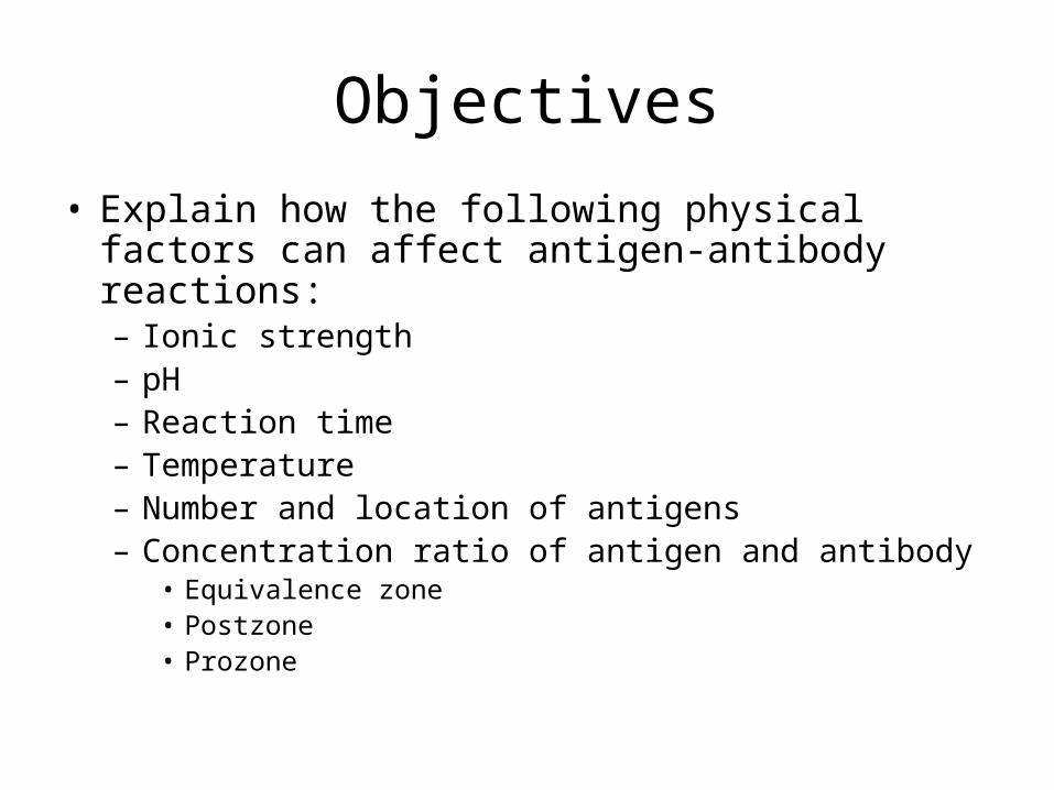
Objectives
• Explain how the following physical factors can affect antigen-antibody reactions:– Ionic strength– pH– Reaction time– Temperature– Number and location of antigens– Concentration ratio of antigen and antibody
• Equivalence zone• Postzone• Prozone

Objectives
• Explain the principle of each test method presented, and give a clinical use for each.
• Differentiate between precipitation and agglutination reactions.
• Interpret the results of Ouchterlony double diffusion.

Antigen/Antibody Reactions
Review – How do antigens and antibodies interact?

Antigen / Antibody Reactions
• Antigen and antibody bind with “Lock and Key” fit.• Affinity – the total attractive force that draws antibody to
antigen.• Avidity – how “tightly” antigen and antibody bind.
AG AB

Antigen / Antibody Reactions
• Antigen / antibody reactions are readily reversible.
• Free Ag + Free Ab Ag-Ab complex

Antigen / Antibody Reactions
• May be visualized when lattice structures form.– Sensitization: antibody attaches to antigen– Agglutination or precipitation: antibody attaches to
antigens on different cells, resulting in cross-linking between cellsY
Y Y
Y
Y
Y
Antigen/antibody reactions are influenced by:

Ionic Strength
• Shielding – charges that surround the Fab portion of an antibody, blocking antigen/antibody binding
• Zeta potential – the difference in electrical charge between the surface of a cell and the outer layer of the ionic cloud that surrounds the cell in an electrolyte solution.
– Keeps cells too far apart to allow lattice formation

- - - - - - - - - - - - -
Y
YYYYYJ
Zeta Potential

Ag/Ab binding is influenced by:
• pH- optimal 6.5 to 7.5• Reaction time- depends on the immunoglobulin
class and test medium– If the incubation time is too short, there will not be time for Ag/Ab
interaction and lattice formation (false negative).– Prolonged incubation favors the free antigen and antibody state,
also resulting in false negative test results.
• Temperature- depends on the immunoglobulin class – IgM reacts best at room temperature or colder. – IgG reacts best at 36-38oC (very narrow temperature range).
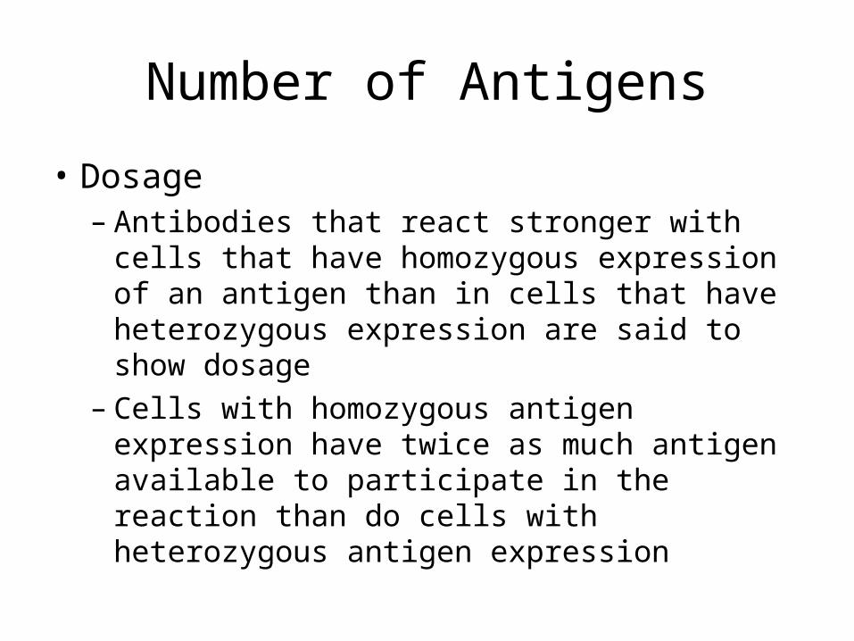
Number of Antigens
• Dosage– Antibodies that react stronger with cells that
have homozygous expression of an antigen than in cells that have heterozygous expression are said to show dosage
– Cells with homozygous antigen expression have twice as much antigen available to participate in the reaction than do cells with heterozygous antigen expression

If there is an anti- antibody, which of these cells will yield the
stronger reaction with it?
Homozygous Heterozygous
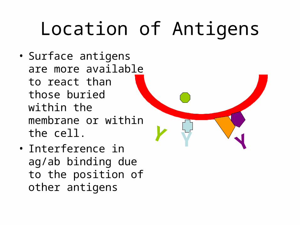
Location of Antigens• Surface antigens are
more available to react than those buried within the membrane or within the cell.
• Interference in ag/ab binding due to the position of other antigens
YYY
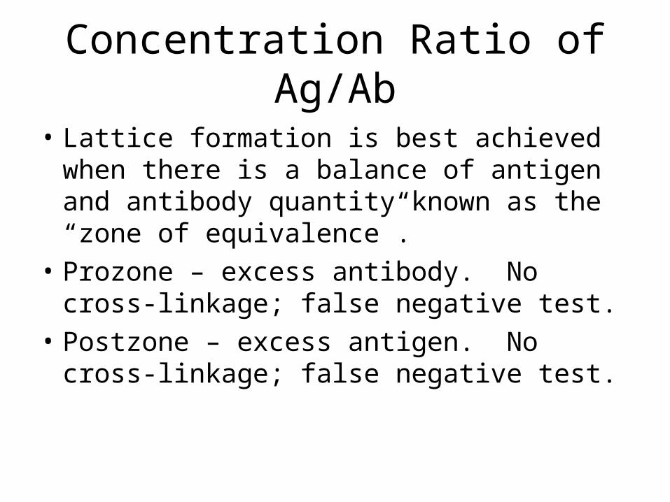
Concentration Ratio of Ag/Ab
• Lattice formation is best achieved when there is a balance of antigen and antibody quantity known as the “zone of equivalence”.
• Prozone – excess antibody. No cross-linkage; false negative test.
• Postzone – excess antigen. No cross-linkage; false negative test.

Zone of Equivalence
Y
Y
Y
YY
Y Y
Y
Y
Y
Y
Y
Y
YY
YY
YY
Y
Y
Y
Y
YY
Y
Y
Prozone – antibody excess
Postzone-antigen excess

Precipitation Based Methods
Soluble antigen combines with antibody to form aggregates which
precipitate out of solution.

Nephelometry
• Antibody reagent is combined with patient sample.
• If antigen is present in the patient’s sample, Ag/Ab complexes will form and precipitate out of solution.
Y
Y Y

Nephelometry
• When light is passed through the solution, the precipitates cause the light to refract at various angles.
• The light that is scattered at a particular angle is measured. This corresponds to the level of antigen in the sample.
• Nephelometry can be used to detect serum protein levels.
Light source
Detector

Double ImmunodiffusionOuchterlony Method
• Testing performed in agar gel.
• Antigen is placed in one well.
• Antibody is place in the other well.
• Each diffuses through gel for 12 -48 hours.
• If antibody is specific to that antigen, a precipitin line will form where the two meet.
AG
AB

Double ImmunodiffusionOuchterlony Method
• The density of the precipitin line correlates with the amount of Ag/Ab complexes formed.
• The pattern of precipitin lines can be interpreted.– Identity– Partial Identity– Non-identity
• This test may be used to identify:– fungal antigens– antibodies to nuclear antigens

Double ImmunodiffusionIdentity
• Known antibody with multiple specificities is placed in center well. • Known antigen is placed in an outside well. • Patient specimen containing antigen is placed in other outside well. •The curved line indicates the unknown patient antigen is the same as the known antigen.

Double ImmunodiffusionNonidentity
•Here, the precipitin lines intersect.
•The patient’s antigen is not the same as the known antigen.

Double ImmunodiffusionPartial Identity
• The antigens share one epitope that reacts with the antibody
• One of the antigens is more complex, having a second unique epitope that also reacts with the antibody.
• The precipitin lines merge, with a spur created that points to the simpler antigen.

Double ImmunodiffusionPartial Identity
D AG
Anti-D
Da AG
Da Db
DdDc
D antigen
“Shared”, simpler antigen
Complex antigen

Immunoelectrophoresis
• Electrophoresis is used to separate serum proteins on a gel according to differences in electrical charge.
• Following electrophoresis, known antibody is applied to the gel. – Antibody diffuses through the gel.– Precipitates form where Ag/Ab complexes have been
trapped in the gel.
• The gel is washed to remove any unprecipitated proteins, and then stained to aid in visualization of the precipitin bands.

Immunoelectrophoresis
Figure 6-7Kuby Immunology , 6th ed©2007, WH Freeman & Co.Used with permission
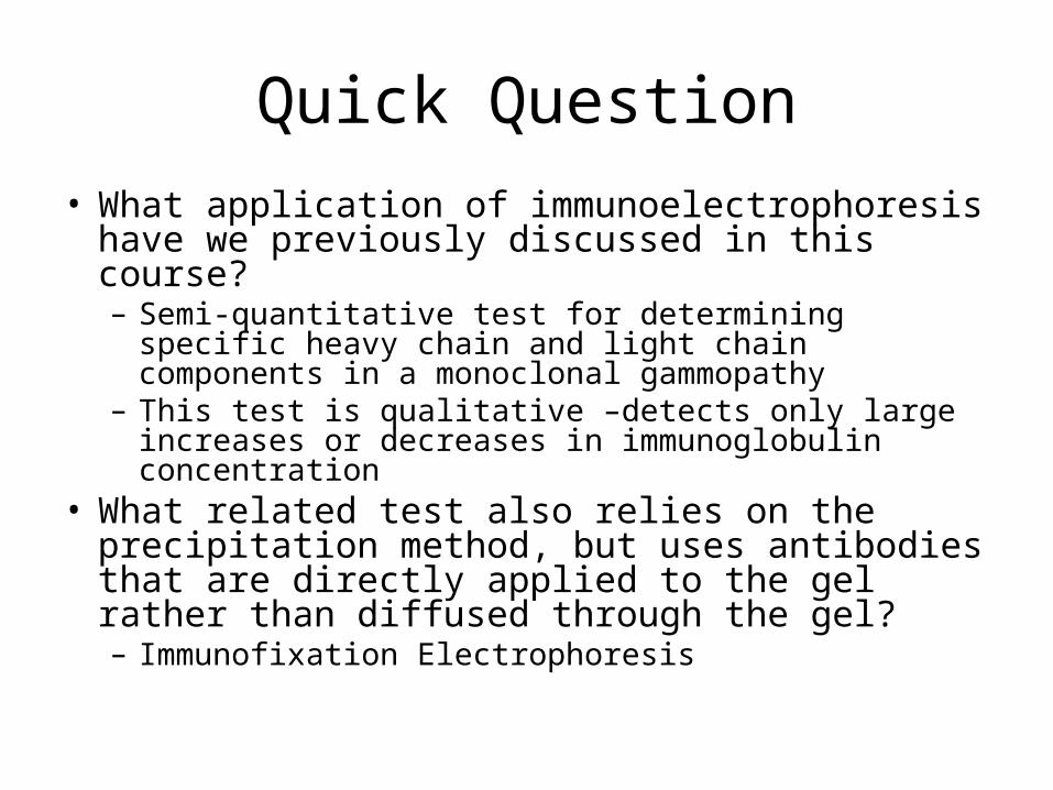
Quick Question
• What application of immunoelectrophoresis have we previously discussed in this course?– Semi-quantitative test for determining specific heavy
chain and light chain components in a monoclonal gammopathy
– This test is qualitative –detects only large increases or decreases in immunoglobulin concentration
• What related test also relies on the precipitation method, but uses antibodies that are directly applied to the gel rather than diffused through the gel?– Immunofixation Electrophoresis

Western Blot
• Known antigens are electrophoresed to separate them.
• The separated components are transferred to nitrocellulose paper by blotting the gel.
• The patient’s serum is applied to the paper.• If the patient has antibodies to any of the
antigens on the paper, it will form a precipitate.• Paper is washed and stained.• The patient’s antibody pattern is compared to a
known antibody pattern.
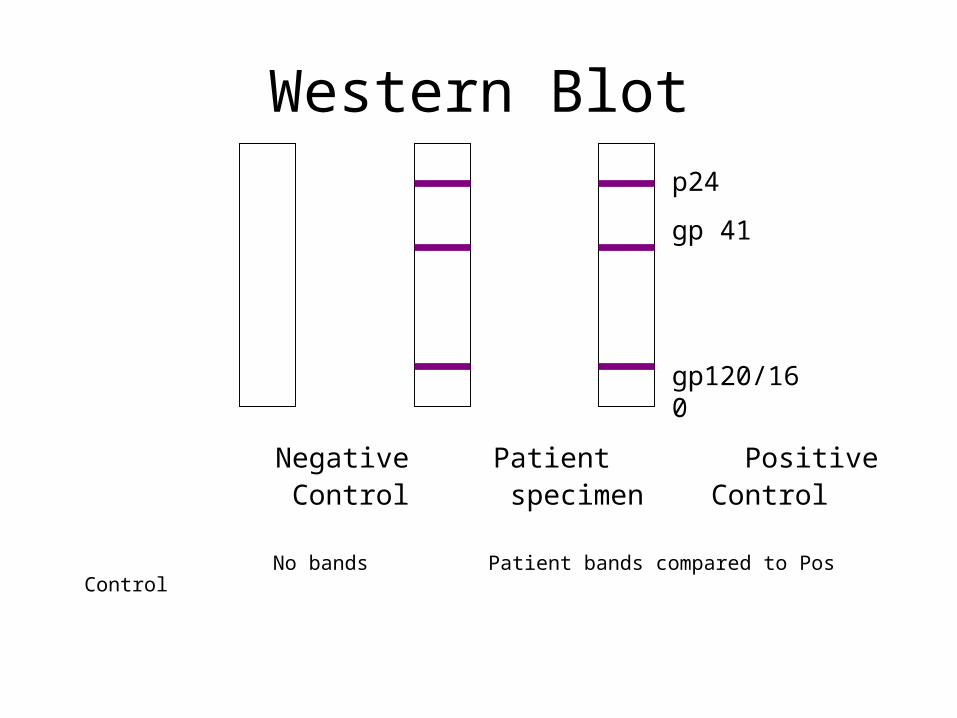
Western Blot
Negative Patient Positive Control specimen Control
No bands Patient bands compared to Pos Control
p24
gp 41
gp120/160
Western Blot
• If a patient has antibodies to more than one antigen that an organism possesses, it is highly likely that patient has been infected by the organism.
• Western Blot is used as a confirmatory test for HIV infection.

Agglutination Based Methods
Antibodies cause the cross-linking of particulate antigen, usually
found on a cell.

Direct Agglutination
• The antigen is a natural part of the solid’s surface.
• Often performed at room temperature.
• May use centrifugation to bring antigen and antibody into closer proximity.

Direct Agglutination
• Can be used to detect antigen (using known antibody as a reagent) or antibody (using known antigen as the reagent) – Bacterial antigens/antibodies– RBC antigens/antibodies
• Known as hemagglutination

Quick Question
• Why is IgM the most effective agglutinin?– Because of its
structure (pentamer) and larger size, it can easily bridge between cells
YYYYYJ

Passive AgglutinationAntigens on a carrier molecule, such as latex, combine with patient’s sample for antibody detection.

Reverse Passive Agglutination
•Antibody is bound to the carrier molecule, which is then mixed with patient’s sample to detect antigen.
•Uses for passive agglutination include ID of bacteria, measuring hormone and drug levels, and measuring levels of some proteins.

Inhibition of Agglutination
• Antibody reagent is combined with patient’s specimen.
• If patient’s specimen contains that antigen, the antigens will react with the antibodies.
• Reagent antigen is added.• A positive reaction will show
no agglutination, because the antibodies were bound to the patient antigen before the reagent antigen was added.
• A negative reaction shows agglutination between reagent antibodies and antigen.
YYY
YY
Y
•Uses include detecting viral antibodies and detecting hormones.
The End
After completing the assignments for this module, please continue to
Module 2, “Labeled Methods”.