immunologic evaluation of drug allergy · in general, it is accepted feia sensitivity for bpo is...
TRANSCRIPT

251© Copyright The Korean Academy of Asthma, Allergy and Clinical Immunology • The Korean Academy of Pediatric Allergy and Respiratory Disease http://e-aair.org
INTRODUCTION
Immunological reactions to drugs, also known as hypersensi-tivity drug reactions (HDR) are considered within the category B of adverse drug reactions where the mechanism is related with the subject abnormal response to the drug.1 This is in con-trast to type A reactions that occur in normal individuals and are usually dose-related. Within the category B, HDR are those mediated by immunological mechanisms and may contribute up to one third of all reactions.2-4 In the last years there has been a growing interest in this area of knowledge with an increase in the scientific production and worldwide activities dedicated to it.5 This manuscript will focus on the immunological evaluation of HDR. For this purpose it is needed to analyse in certain detail what are the mechanisms involved. In addition drugs reactions to biological agents will be also included, given the increasing importance of these in the elicitation of reactions.6
CLASSIFICATION OF ALLERGIC DRUG REACTIONS
Allergic reactions can be produced by any of the four immu-nologic mechanisms proposed by Gell and Coombs.7 Type I re-actions, also called immediate-type reactions, occur usually within less than one hour after drug administration and are mediated by drug-specific IgE antibodies. Classical examples
Immunologic Evaluation of Drug AllergyEnrique Gómez,1 Maria Jose Torres,2 Cristobalina Mayorga,1 Miguel Blanca2*
1Research Laboratory, 2Allergy Service, Carlos Haya Hospital, Málaga, Spain
are anaphylaxis and urticaria induced by betalactams antibiot-ics, otherwise the most frequent drugs involved in immediate hypersensitivity reactions.8,9
Type II (cytotoxic reactions) and Type III (immune complex reactions), are usually mediated by IgG or IgM specific antibod-ies.7 Although with the classical drugs (organic compounds) it has been difficult to prove their involvement in Type III reac-tions, this mechanism is gaining interest in the last years with the use of biological agents, with some evidence pointing out that this can be the case.6 Type IV reactions, mediated by effec-tor drug-specific-T cells, are also known as delayed hypersensi-tivity reactions (DHR) since they appear from hours to days af-ter the drug intake.10 We will refer to this group in this review as non-immediate allergic reactions. In the immunological evalu-ation we also include a group that although inflammatory me-diators are released, same as those involved in IgE-mediated reactions, non-specific immunological mechanisms take part, as occurs in reactions induced by NSAIDs.11 These are in fact
ReviewAllergy Asthma Immunol Res. 2012 September;4(5):251-263.http://dx.doi.org/10.4168/aair.2012.4.5.251pISSN 2092-7355 • eISSN 2092-7363
Hypersensitivity drug reactions (HDR) consist of an individual abnormal response with the involvement of the immunological system. In addition to specific immunological mechanisms where specific antibodies or sensitised T cells participate, release of inflammatory mediators by non-specific immunological recognition may also occur. Within this category are one of the most common groups of drugs, the non-steroidal anti-inflammatory drugs. In addition to chemical drugs new emerging ones with an increasing protagonism are biological agents like humanised antibodies and others. For IgE dependent reactions both in vivo and in vitro tests can be used for the immunological evaluation. Sensitivity of these is not optimal and very often a drug provocation test must be considered for knowing the mechanism involved and/or establishing the diagnosis. For non-immediate reac-tions also both in vivo and in vitro tests can be used. Sensitivity for in vivo tests is generally low and in vitro tests may be needed for the immunolog-ical evaluation. Immunohistochemical studies of the affected tissue enable a more precise classification of non-immediate reactions. The monitor-ization of the acute response of the reactions has given clues for understanding these reactions and has promising results for the future of the im-munological evaluation of HDR.
Key Words: Drug; immunology; inflammatory cell; diagnosis
This is an Open Access article distributed under the terms of the Creative Commons Attribution Non-Commercial License (http://creativecommons.org/licenses/by-nc/3.0/) which permits unrestricted non-commercial use, distribution, and reproduction in any medium, provided the original work is properly cited.
Correspondence to: Miguel Blanca, MD, PhD, Allergy Service, 1 Floor Pavillon 6, Carlos Haya Hospital (Pabellon C), Plaza del Hospital Civil s/n, 29009 Málaga, Spain.Tel: +34-951-290190; Fax: +34-951-290200; E-mail: [email protected]: January 10, 2012; Accepted: February 1, 2012•There are no financial or other issues that might lead to conflict of interest.

Gómez et al.
Allergy Asthma Immunol Res. 2012 September;4(5):251-263. http://dx.doi.org/10.4168/aair.2012.4.5.251
Volume 4, Number 5, September 2012
252 http://e-aair.org
the most common group of drugs involved in HDR.12 In gener-al, all these reactions are included within hypersensitivity reac-tions, being the term allergic reactions reserved for those where an immunological mechanism takes part.13
For those where specific immunological mechanisms are in-volved, a working classification has been adapted from the for-mer proposed by Levine14 considering the immediate reactions as those occurring within one hour after the drug intake and non-immediate reactions as those who occur later than one hour after the drug administration including both accelerated and delayed reactions.15 In general, these reactions can occur from hours to days or even weeks after drug intake and are char-acterized by a wider range of clinical manifestations than im-mediate reactions.10 In Fig. 1, it is shown a general pictogram of both immediate and non-immediate reactions with the cell in-volved. Detailed analysis of these will be given through the text.
IMMEDIATE TYPE IGE MEDIATED REACTIONS
General considerationsTypical reactions included within this group are anaphylaxis
and urticaria. Betalactams antibiotics continue to be the most frequent drugs that induce these reactions.16,17 The facility for binding spontaneously to endogenous proteins renders them to some extent immunogenic and able to induce IgE antibod-ies.9,16 In addition, immediate reactions have been reported by many drugs with an increasing list although at a much lower proportion than betalactams. Relevant emerging drugs are NSAIDs and quinolones.18,19
In the case of betalactams several determinants generated from benzyl penicillin have been proposed (BPO and MDM).8,14 Moreover, other betalactams provide determinants that must be considered in the immunological evaluation.16,20-25 The more relevant for the diagnosis of immediate reactions is amoxicillin and recently clavulanic acid as well as cephalosporins.20,22,25 Sec-ond relevant drugs are NSAIDs that although in most instances induce non immunological mediated reactions, up to 30% of subjects can develop urticaria/angioedema or anaphylaxis with a selective response mediated by an IgE mechanism.18 Pyrazo-lones are the most relevant drugs although propionic acid de-rivatives, particularly ibuprofen, followed by the aryl acetic acid derivative diclofenac are gaining in importance in this type of reactions.12,18,26 Weather ibuprofen itself or some of its metabo-lites are the responsible for IgE mediated reactions is not known at present.
In vivo diagnosisSkin testing is the most sensitive tests for the diagnosis of im-
mediate reactions to betalactams.27 General principles for skin testing with these and with the rest of the drugs are provided by the ENDA group.20 Maximal recommended concentrations are for BPO 5×10⁻⁵ M for MDM 2×10⁻² M and for amoxicillin and clavulanic acid 20 mg/mL. With soluble cephalosporins con-centrations of 20 mg/mL can produce false positive results in some of them due to an irritant effect and 2 mg/mL is recom-mended.20 Working concentrations for the rest of drugs involved in immediate reactions are also provided.28
Sensitivity for skin testing with betalactams and with the rest
Fig. 1. Different clinical manifestation and the immunological mechanism involved in immediate and non-immediate reactions to drugs.
Immature
Drug (hapten)Inflammation mediators Keratinocytes
MHC-1
Immature dendritic cells (Langerhans)
Eosinophil Mast cells
MHC-IINK
MØ
Lymph nodeMature dendritic
ll IgE
E-selectinChemokines IP10, CCL17, ITAC, MCP1, Mig
MØ
cells
Naïve T lymphocyteCytotoxic lymphocytes
IL-4
Plasmatic cells
IL-12Fas/FasL
Perforin
Granzyme B
B lymphocytesT lymphocytes (CLA+, CCR/CXCR+)
y
Memory T lymphocytes
Memory B lymphocytes

Immunologic Evaluation of Drug Allergy
Allergy Asthma Immunol Res. 2012 September;4(5):251-263. http://dx.doi.org/10.4168/aair.2012.4.5.251
AAIR
253http://e-aair.org
of the drugs is time dependent after allergic episode occurrence with subjects converting from positive to negative as time elaps-es.29-31 In vivo sensitivity is not optimal what implies that in the event of a clear positive history, if skin test negative, subject can still be allergic.16,17 Only in a minor proportion of cases in vitro tests can be positive with skin test negative but up to 20%-30% of patients may need a control administration of the drug for confirming the diagnosis.32,33
In vitro diagnosisMost assays developed for drug specific IgE detection includ-
ing the commercial ones consisted on the quantitation of IgE using radiolabelled anti IgE antibodies (RAST) or more recently enzyme (ELISA) or fluoroenzyme (FEIA) assays. The principle consists in a solid phase to which the hapten conjugated to a carrier protein is bound covalently. The carrier proteins can be HSA or other molecules like polylisines or aliphatic spacers.34-36 Although HSA has been used for many years37 this has not shown to be the most suitable carrier being preferable in many in-stances others with a high capacity for hapten fixation and ex-position to IgE antibodies.34-36,38,39 Since last years the radiola-belled method has been substituted for the ELISA and later with the FEIA although no sufficient comparatives studies has been made so far. In general, it is accepted FEIA sensitivity for BPO is reasonable compared to skin testing,40 however, for oth-er betalactams, including amoxicillin, important differences in sensitivity exist.39 This is particularly relevant in the cases of cephalosporins where sensitivity non higher than 20% has been reported.41 This can be due to the not inclusion of the culprit cephalosporin. For many years only cefaclor has been available for in vitro testing and it is well known that for this betalactam the side chain at R1 position is very important in the specific IgE recognition.25,42-44 Similar assays have been developed for other drugs, being in most cases experimental prototypes that need further validations in a sufficient number of positives con-trols.45-52
An alternative solid phase used for many drugs has been ep-oxy activated sepharose to which drugs bind covalently. Classi-cal drugs used have been cephalosporins,28,29 quinolones,30,31 and muscle relaxants32 with different sensitivity and specificity results.
It is relevant to note that drug allergy IgE mediated can be an occupational disease affecting workers involved in health care, pharmacy and industries producing or manufacturing pencil-lins, cephallosporins53-55 as well as other antibiotics.56 In this sense the group of Park has published several reports using the methodology outlined above for diagnosing these cases.53-56
BASOPHYL ACTIVATION TEST (BAT)
The flow cytometry technology facilities exploit the capacity of basophyls to be activated after the interaction of the hapten
with specific IgE antibodies on their surface.57 The principle is based on the basophyl property for expressing in their cell mem-brane di novo or increased upon activation different markers, being the most widely used CD63 and CD203c.57,58 Although this methodology is actually mostly reserved to specialised cen-tres involved in drug allergy, its advantage is that different drugs can be used that are not available for skin testing, have anti-in-flammatory properties and/or lack the ability to be conjugated to a solid phase support in an efficient way as occurs with corti-coids,59 quinolones,60 contrast media,61 dipyrone,62 anaesthet-ics,63,64 omeprazol,65 cyclosporine66 as well as many other drugs.67 Most studies have been carried out with betalactams antibiot-ics.68,69 With this technique sensitivity approaching 60% have been obtained with cases detectable only by the BAT assay be-ing negative to both intradermal testing and the in vitro immu-noassays.68 The potential use of these techniques deserves fur-ther studies with the possibility of including more drugs for proving the existence of specific IgE antibodies.67
CELLULAR ANTIGEN STIMULATION TEST (CAST)
Alternative to the BAT, the CAST is based on the quantitation of sulfidoleukotrienes released in the supernatant after baso-phyl stimulation.70 In general this technique has been used in the last years but did not show clear advantages compared to basophyl activation.57 Important to note is that the flow cytom-eter is not required for their performance but only a system for quantifying the histamine released.57,70 However, no agreement has been made about when and how this technique must sub-stitute the others available or under which circumstances this is indicated.70
NON-IMMEDIATE TYPE T CELLS DEPENDENT REACTIONS
General considerationsThese reactions are produced of sensitised T cells that recog-
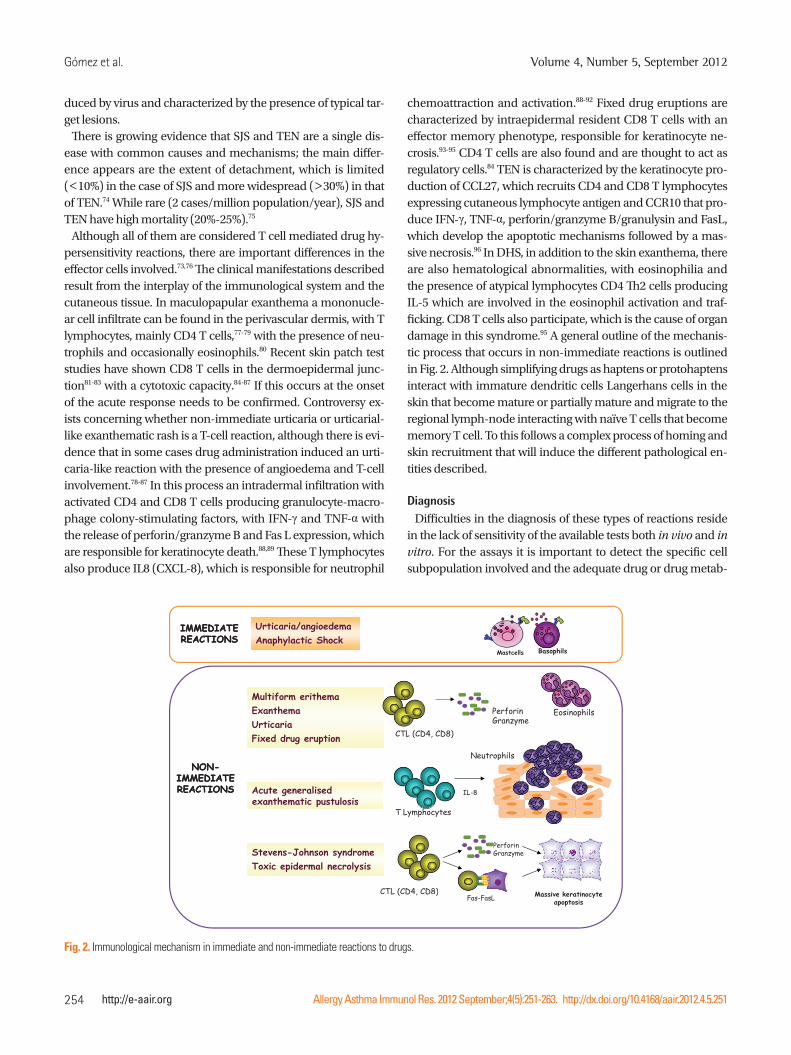
nise drugs as xenobiotics and induce an immunological effec-tor response.7 Although T cells reactions can occur virtually with any drug in any organ,71 the skin is the most frequent target in-volved.10,72,73 Clinical entities are shown in Fig. 2. They ranks from maculopapular exanthema to non-immediate urticaria and other less common but more severe entities such as AGEP, drug rash with eosinophilia and systemic symptoms, drug hy-persensitivity syndrome (DHS), Stevens-Johnson syndrome (SJS), and Toxic epidermal necrolysis (TEN), fixed drug erup-tion and contact dermatitis as well as organ specific reactions.72,73 Although maculopapular exanthema is the most common re-ported reaction, it can sometimes be intense, accompanied with subcutaneous angioedema, and persists for several weeks despite discontinuation of treatment.73 DHS and bullous reac-tions with mucosal involvement are considered severe diseas-es.10,72 Erythema multiform, which is less severe, is usually in-
Gómez et al.
Allergy Asthma Immunol Res. 2012 September;4(5):251-263. http://dx.doi.org/10.4168/aair.2012.4.5.251
Volume 4, Number 5, September 2012
254 http://e-aair.org
duced by virus and characterized by the presence of typical tar-get lesions.
There is growing evidence that SJS and TEN are a single dis-ease with common causes and mechanisms; the main differ-ence appears are the extent of detachment, which is limited (<10%) in the case of SJS and more widespread (>30%) in that of TEN.74 While rare (2 cases/million population/year), SJS and TEN have high mortality (20%-25%).75
Although all of them are considered T cell mediated drug hy-persensitivity reactions, there are important differences in the effector cells involved.73,76 The clinical manifestations described result from the interplay of the immunological system and the cutaneous tissue. In maculopapular exanthema a mononucle-ar cell infiltrate can be found in the perivascular dermis, with T lymphocytes, mainly CD4 T cells,77-79 with the presence of neu-trophils and occasionally eosinophils.80 Recent skin patch test studies have shown CD8 T cells in the dermoepidermal junc-tion81-83 with a cytotoxic capacity.84-87 If this occurs at the onset of the acute response needs to be confirmed. Controversy ex-ists concerning whether non-immediate urticaria or urticarial-like exanthematic rash is a T-cell reaction, although there is evi-dence that in some cases drug administration induced an urti-caria-like reaction with the presence of angioedema and T-cell involvement.78-87 In this process an intradermal infiltration with activated CD4 and CD8 T cells producing granulocyte-macro-phage colony-stimulating factors, with IFN-γ and TNF-α with the release of perforin/granzyme B and Fas L expression, which are responsible for keratinocyte death.88,89 These T lymphocytes also produce IL8 (CXCL-8), which is responsible for neutrophil
chemoattraction and activation.88-92 Fixed drug eruptions are characterized by intraepidermal resident CD8 T cells with an effector memory phenotype, responsible for keratinocyte ne-crosis.93-95 CD4 T cells are also found and are thought to act as regulatory cells.84 TEN is characterized by the keratinocyte pro-duction of CCL27, which recruits CD4 and CD8 T lymphocytes expressing cutaneous lymphocyte antigen and CCR10 that pro-duce IFN-γ, TNF-α, perforin/granzyme B/granulysin and FasL, which develop the apoptotic mechanisms followed by a mas-sive necrosis.96 In DHS, in addition to the skin exanthema, there are also hematological abnormalities, with eosinophilia and the presence of atypical lymphocytes CD4 Th2 cells producing IL-5 which are involved in the eosinophil activation and traf-ficking. CD8 T cells also participate, which is the cause of organ damage in this syndrome.95 A general outline of the mechanis-tic process that occurs in non-immediate reactions is outlined in Fig. 2. Although simplifying drugs as haptens or protohaptens interact with immature dendritic cells Langerhans cells in the skin that become mature or partially mature and migrate to the regional lymph-node interacting with naïve T cells that become memory T cell. To this follows a complex process of homing and skin recruitment that will induce the different pathological en-tities described.
DiagnosisDifficulties in the diagnosis of these types of reactions reside
in the lack of sensitivity of the available tests both in vivo and in vitro. For the assays it is important to detect the specific cell subpopulation involved and the adequate drug or drug metab-
IMMEDIATE IMMEDIATE Urticaria/angioedemaIMMEDIATE IMMEDIATE REACTIONSREACTIONS
Urticaria/angioedemaAnaphylactic Shock
Mastcells Basophils
Multiform erithema Exanthema Perforin EosinophilsUrticariaFixed drug eruption
Neutrophils
CTL (CD4, CD8)Granzyme
Eosinophils
NONNON--IMMEDIATE IMMEDIATE REACTIONSREACTIONS Acute generalised
exanthematic pustulosis
p
IL-8
St J h d
exanthematic pustulosisT Lymphocytes
Perforin Stevens-Johnson syndromeToxic epidermal necrolysis
CTL (CD4, CD8)
Granzyme
Massive keratinocyte L ( D , D )Fas-FasL Massive keratinocyte
apoptosis
Fig. 2. Immunological mechanism in immediate and non-immediate reactions to drugs.

Immunologic Evaluation of Drug Allergy
Allergy Asthma Immunol Res. 2012 September;4(5):251-263. http://dx.doi.org/10.4168/aair.2012.4.5.251
AAIR
255http://e-aair.org
olites eliciting the immunological response.15,76 Other difficul-ties are the lack of knowledge of the cofactors that may have been present at the time of the reaction, and the possibility that symptoms attributed to the drug were not immunologically mediated.73
Skin testsReliable skin test procedures for the diagnosis of non-imme-
diate reactions are generally lacking and test concentrations are often unknown or poorly validated for many drugs.28 Delayed-reading intradermal and/or patch tests 24-48 hours after the drug application have been used for many years.97,98 The main advantage of drug patch tests is that they can be performed with any commercially available drug, while intradermal tests are more sensitive, but they need to be performed with a solu-ble injectable or a pure form, sterile preparation of the drug.97 Both intradermal and patch tests have been widely used in the diagnosis of non-immediate reactions to betalactams, with re-ported sensitivity ranging from 2.6% to 37.8%.99 Further evi-dence seems to indicate that skin test sensitivity is lower than previously believed.100-102
Lymphocyte transformation testIn the last years a considerable effort has been made in the re-
introduction of lymphocyte transformation test assays in the evaluation of DHR.103,104 This is based on the principle that T cells can proliferate in the presence of a specific antigen. The usefulness of this test in the diagnosis of non-immediate reac-tions has been debated pointing out that it depends on the drug involved in the reaction.105 Reports published to date have been characterized by small series, a wide range of drugs and differ-ent clinical entities and show and overall sensitivity rating 60% to 70% with a rather low specificity (85%).103,104,106,107 However, this test is not available everywhere and is still considered a re-search tool. In a study by our group, 57% of patients with a non-immediate reaction to betalactams had a positive lymphocyte transformation test to at least one of the penicillins tested.104 A recent study analysing the role of dendritic cells in the enhance-ment of amoxicillin-specific lymphocyte proliferation in pa-tients with non-immediate reactions to amoxicillin showed that compared to traditional antigen-presenting cells such as B cells or monocytes, dendritic cells improved lymphocyte trans-formation test sensitivity.108 Similar results have been obtained for non-immediate reactions with other drugs such as heparins and contrast media.109,110
Immunopathological studiesBecause in many instances DHR affect the skin, samples can
be taken from the lesions at both the acute reaction as well as after positive drug provocation or a delayed skin test. While the immunohistochemistry findings help in the investigation of underlying immunologic mechanism, they do not provide in-
formation about the drug involved or even discriminate be-tween different types of reactions. The most common finding is a mononuclear cell infiltrate composed mainly of activated T cells expressing activation markers such as CD69, IL-2R (CD25) and HLA-DR, and the skin-homing receptor cutaneous lym-phocyte antigen in both CD4 and CD8 T cells with a predomi-nance of one of them depending on the clinical manifestation.15 In maculopapular exanthema, for example, a predominant perivascular mononuclear cell infiltrate with increased num-bers of eosinphils in the papillary dermis has been found, with up to 20% of the cells expressing perforin and granzyme B and enhanced IL12 expression.76 SJS/TEN, in contrast, is character-ized by the presence of many dead keratinocytes with a mini-mum T-cell infiltration, probably due to the loss of superficial skin layers.15,73 In AGEP, keratinocytes express high levels of IL-8, a chemokine that recruits neutrophils to the epidermis.80,88,89 Fig. 3 represents examples of the haematoxylin eosin staining and immunohistochemistry for cell marker of lymphocytes subpopulations as well as effector molecules involved in the lessional skin process.
Drug provocation studiesBecause intradermal or patch testing have non optimal sensi-
tivity in patients with non-immediate reactions, a large propor-tion of patients need to be given the drug to establish a diagno-sis or, perhaps more often, to confirm tolerance.111 Drug provo-cation testing is the best tool by which a causal relationship be-tween drug administration and a non-immediate reaction is established.15 It involves the careful administration of a suspect agent in a specialized centre and close monitoring for symp-toms, in particular skin manifestations. Drug provocation test-ing, however, is not generally recommended and is contraindi-cated in some cases such as generalized bullous fixed drug eruptions, AGEP, SJS, TEN, DHS/DIHS, systemic vasculitis, spe-cific organ manifestations (blood-cytopenia, hepatitis, nephri-tis, pneumonitis) and drug-induced autoimmune diseases.75,111
Of all the drugs suspected to cause non-immediate reactions, betalactams have been the most extensively studied.100,112,113 Most patients who develop an exanthematic reaction after be-talactam administration and have negative skin tests can toler-ate drug in a drug provocation test.87,102 Nonetheless, some pa-tients with clear non-immediate reactions are diagnosed by a positive drug provocation test. This indicates that drug provo-cation testing is the most important diagnostic tool in the par-ticular case of exanthematic reactions to betalactams.113
NON-ALLERGIC HYPERSENSITIVITY REACTIONS: NSAIDS
General considerationsNon-allergic hypersensitivity reactions refer to adverse drug
reactions that are not mediated by specific immunological mechanisms, this is IgE or T cell dependent responses. This

Gómez et al.
Allergy Asthma Immunol Res. 2012 September;4(5):251-263. http://dx.doi.org/10.4168/aair.2012.4.5.251
Volume 4, Number 5, September 2012
256 http://e-aair.org
group of reactions is commonly described as intolerant, pseu-doallergic, or idiosyncratic reactions although the term non-al-lergic hypersensitivity is actually used for integrating all these terms.13 The most representative group of drugs is made by NSAIDs.12,114 These are medicaments of variable chemical com-position that antagonize inflammation by interfering with the function of cyclooxygenases. Cyclooxygenases are enzymes that participate in the conversion of arachidonic acid into pros-taglandins and thromboxanes, which generate strong media-tors of the inflammatory process. This inhibition results in a shunting of arachidonic acid metabolism toward the 5-lipoxy-genase pathway, resulting in the increased release of cysteinyl leukotrienes and a decreased production of prostaglandin E2 mediators that could be involved in the pathogenesis of these reactions.11,115,116
NSAIDs are responsible for 21%-25% of reported adverse drug events which include immunologic and non-immunologic hy-persensitivity reactions.11 A recent study indicates that these fig-ures are even higher.117 Depending on the timing, symptom-atology and putative mechanism of the reactions there are sev-eral subtypes of hypersensitivity to NSAIDs.115,118,119 The follow-ing categories are actually recognised:
1. NSAID-exacerbated respiratory disease presently designat-ed as aspirin-exacerbate respiratory disease.
2. NSAID-exacerbated cutaneous disease, in particular, urti-caria and angioedema in patients with chronic idiopathic urticaria. In analogy to aspirin-exacerbate respiratory dis-ease, it could be called NSAID or aspirin-exacerbated cuta-neous disease.
3. Multiple NSAID-triggered urticaria, angioedema, and ana-phylaxis in patients without other underlying disease.
4. Urticaria, angioedema, and anaphylaxis induced by a single NSAID. In turn these can be divided into IgE or T cell de-pendent reactions.120
DiagnosisBecause it is estimated that in the 70% of cases with NSAID
hypersensitivity non-specific immunological mechanism are involved,12 only in 1/3 of the cases will be theoretically possible to apply in vivo or in vitro specific immunological tests based on the capacity of IgE or T cells. For this, the in vitro test has fo-cused on the mediator release determination.121-123 The capaci-ty for histamine release or other mediators such as leukotrienes or ECP from eosinphils has been used for the development of
Contact Eccema
H-EClinic CD3 CD4 CD8 CD45RO CLA PerforinGranzyme - B
Skin test Exanthema
Exanthema
Steven-Johnson
FDE
Fig. 3. Haematoxylin-eosin and for immunohistochemical stains of lymphocyte subpopulations (CD3, CD4, CD8, CD45RO), skin homing receptor (CLA), and cytotoxic markers (Granzyme B and Perforin) in skin biopsies from different delayed reactions, taken during the acute phase.

Immunologic Evaluation of Drug Allergy
Allergy Asthma Immunol Res. 2012 September;4(5):251-263. http://dx.doi.org/10.4168/aair.2012.4.5.251
AAIR
257http://e-aair.org
in vitro assays. Although extensive research has been made these tests cannot be recommended for routine diagnosis.124 These are outlined below.
Sulfidoleukotrienes release assayBecause aspirin can induce LTC4 release from peripheral
blood leukocytes of sensitive patients, measurement of sulfido-leukotriene release has been tested for the diagnosis of aspirin-exacerbate respiratory disease but the results are inconsis-tent.116,121,125-127
Basophyl activation test (BAT)As basophyls can be activated by both specific and non-spe-
cific mechanisms and since their involvement has been dem-onstrated in this type of reactions,128 the measurement of cell surface molecule CD63 upon in vitro drug challenge has been proposed for in vitro diagnosis of aspirin-exacerbate respirato-ry disease.123 However low sensitivity and specificity were vari-able with no firm conclusion on the use of this test for the diag-nosis has been provided.70
Challenge testsIn patients with a history of reaction to a single NSAID and no
additional exposure to a second NSAID, skin testing is possible and may reveal a selective sensitization, although until now it has been shown in a very low number of cases and only with some drugs like pyrazolones.12,18 IgE tests are not commercially available.11,18,129 It may be convenient to confirm the diagnosis by oral challenge, although this should be done cautiously be-cause low concentrations of the drug may already cause symp-toms. If the results are positive, another NSAID of a different chemical group should be tested to demonstrate cross-re-sponse.130 A history of systemic anaphylaxis would be a contra-indication to perform the provocation tests with the incriminat-ed drug.12,131
The oral provocation test is the “gold standard” for the diagno-sis, although it should not be performed during an urticaria or airways exacerbation. According to the EAACI/GA2LEN guide-line,132 subjects should be challenged under single-blind, place-bo-controlled conditions, after at least 1-2 weeks without any skin eruptions. Acetyl-salicylic acid challenges are recommend-ed to be done during two consecutive days, administering on the first day 4 capsules of placebo and on the second day expo-nentially increasing doses of acetyl-salicylic acid (71, 117, 312, and 500 mg) at 1.5-2 hour intervals, up to a cumulative dose of 1,000 mg of acetyl-salicylic acid. The challenge procedure is in-terrupted, if cutaneous reactions appear or when other symp-toms of NSAID hypersensitivity develop. Challenge protocols for NSAIDs other than acetyl-salicylic acid are available in the literature.132 Most patients react to doses of acetyl-salicylic acid between 325 and 650 mg, and the time interval between acetyl-salicylic acid intake and onset of hives is generally no longer
than one hour. The sensitivity of challenge is not 100%; in fact negative results of challenges with the suspected NSAIDs have been reported, even in cross-reactors.133
HYPERSENSITIVITY REACTIONS TO BIOLOGICAL AGENTS
General considerationsBiological agents are new medicaments with increasing appli-
cations that are progressively implicated in hypersensitivity re-actions.6 They are not synthetic organic chemicals (xenobiotics) being structurally similar to autologous proteins. Different mech-anisms have been proposed to be involved in the reactions in-duced by these agents that have been reviewed in detail else-where.134 Because many of them affect inflammatory processes, the immune effect can be induced by their activities; however, they can produce true allergic reactions mediated by specific immunological mechanisms.6,134 Amongst the biological agents considered within this group are cytokines, antibodies against cytokines, receptors, cell surface markers and fusion proteins as cytokine receptors or cellular ligands.6
Reactions induced by these agents belong to all the different immunological mechanisms described in the classic reactions by Gell and Coombs.7 Anaphylactic reactions,135-138 cytotoxic re-actions like immune haemolytic anaemia and thrombocytope-nia,88,89 immune complex like diseases139-143 and T cell respons-es144-146 have been reported. We will refer in more detail to Type I and Type IV reactions.
The ability of these reagents for inducing true allergic reactions is related with their ability of being recognised as different by the immunological system. Thus chimeric antibodies (ended by -ximab, are more immunogenic than those partially (-zum-ab) and whole humanises (-mumab).84 The best studied model is cetuximab, an antibody used for treating different neopla-sias.147 It has been shown that many of these patients are pri-marily sensitised to a carbohydrate, α-Gal present in different natural sources as meat or induced after tick bites.148,149 In the case of non-immediate reactions from mild to severe responses to these agents have been reported indicating that these have been recognised by T cells which are involved in the response.146
Common reactions induced by these drugs are fever, chills, nausea, vomiting, headaches and diarrhea that sometimes may mimic an allergy reaction.149 But these usually may appear after the first administration and are often controlled by symptomat-ic medications. In the case of anaphylactic reactions these usu-ally appear after several therapeutic courses and can be often severe and potentially fatal.
DiagnosisBecause these medicaments are proteins it is not difficult to
work with them both in vivo and in vitro for the diagnostic eval-uation. Different studies have shown positive skin tests136,147-149 and in vitro specific IgE antibodies for immediate reactions136

Gómez et al.
Allergy Asthma Immunol Res. 2012 September;4(5):251-263. http://dx.doi.org/10.4168/aair.2012.4.5.251
Volume 4, Number 5, September 2012
258 http://e-aair.org
and also T cell responses.144 The general principles outlined in this section are valid for approaching to these patients if specif-ic immunological mechanisms are the responsible of the reac-tions.6,7
MONITORIZATION OF THE ACUTE RESPONSE
The progress in molecular biology and genetics has enable to monitor the acute response and define to what extent Type I or Type IV reactions occur in more detail.76 This approach is not new but has been recently refined.76 Although the boosted IgE response after an anaphylactic reaction can be monitored149 the most useful is the quantitation of classical mediators for imme-diate reactions like histamine/histamine metabolites and trypt-ase.150 The first can be determined in peripheral blood or as N-methyl histamine metabolite in urine or other body fluids like nasal lavage and the second in peripheral blood or affected or-gan because as a protein it is not excreted in urine.150-152 For their evaluation it is very important to take the sample at the optimal time of release. Histamine peaks only for few minutes in peripheral blood150,151 but a clearance in urine show a peak at 3-5 hours after the episode that normalised in the subsequent 24 hours. On the contrary tryptase after being released in the affected organ pass to peripheral circulation with an optimal peak during the 2-4 hours following the acute episode that fur-ther returns to normal levels in a maximum of 24 hours.151 Re-lease of these mediators is compatible with an acute immediate reaction but it is not exclusively for an IgE mechanism since these can be also released in NSAIDs cross intolerance as well as other responses.11
Very often in allergic reactions it is not clearly known which is the specific immunological mechanism involved. This is partic-ularly relevant for T cell mediated reactions.76 Therefore the monitorization of T cell dependent reactions has shown an in-crease in IFN-γ and the transcription factors T-bet with a down regulation of IL-4 and GATA 3, which is indicative of aTh1 re-sponse.153 On the contrary in IgE mediated reactions the oppo-site was observed with a polarised pattern of response toward a Th2 with up-regulation of IL-4 and GATA-3 and a down regula-tion of IFN-γ and T-bet that normalised as reaction subsides.153,154
The monitorization can be carried out not only in peripheral blood but also in skin, the organ mainly involved in non-imme-diate drug reactions, showing a parallelism between the results found in the two compartments with the participation of differ-ent T cell subsets depending on the clinical entity.154 Studies in more detail have shown that skin homing receptors78,155 were increased in both peripheral blood and skin whereas the corre-sponding chemokines ligands were expressed only in skin. In this work it was observed an increase in CCR6 and CCR10 with their corresponding ligands CCL20 and CCL27 responsible for skin homing and CXCR3 with its corresponding ligands CXCL9 and CXCR10 related with a Th1 response in cases with maculo-
papular exanthema.155 This integrated approach can be more deeply studied with a complete view of the genetic expression by DNA microarrays.156 The determination of the different markers involved in drug allergic reactions can be crucial for discriminating from virus diseases with similar clinical mani-festations although different immunological mechanism are underlying.157
CONCLUDING REMARKS
In spite of the difficulties for the immunological evaluation of HDR, increasing interest and progress has been made in the last years that has enabled a better understanding and manage-ment of HDR. In the cases of IgE mediated reactions, betalac-tams continue to be the drugs most frequent involved, being side chain penicillin determinants like amoxicillin needed both in vivo and in vitro in the immunological evaluation. Other drugs can also induce IgE mediated reactions, being fluorqui-nolones a group of antibiotics with increasing relevance. Al-though experimental prototypes have been developed for the determination of specific IgE antibodies to many drugs, the BAT is an assay quite useful for identify the culprit drug. In the case of non-immediate reactions, the number of entities is greater than in immediate reactions and the way how T cells and cells of the innate immune system interact followed by a specific recruitment of effector T cells to the affected organ is better known actually. The progress made in molecular biology and genetics is contributing to these advances. Monitorization of the acute response in non-immediate reactions offers great promises.
Concerning NSAIDs increasing evidence is showing that these are the most relevant group of medicaments involved in adverse drug reactions, with a non-immunological mechanism involved in the higher proportion of patients. These are desig-nated as hypersensitivity reactions with absence en allergic mechanism and patients are cross-intolerant, reacting to differ-ent NSAIDs non-chemically related. Because there are no in vivo or in vitro tests for the diagnosis, the immunological evalu-ation in these cases required a drug provocation test to estab-lish the diagnosis of cross-intolerance, selective responder or tolerant cases.
ACKNOWLEDGMENTS
The study was funded by FIS-Thematic Networks and Co-op-erative Research Centres (RIRAAF/RD07/0064), Junta de Anda-luc´a (CTS 06603, PI-0545-2010) and FIS (09/01768, PS09/00966).
REFERENCES
1. Rawlins MD, Thompson JW. Mechanisms of adverse drug reactions.

Immunologic Evaluation of Drug Allergy
Allergy Asthma Immunol Res. 2012 September;4(5):251-263. http://dx.doi.org/10.4168/aair.2012.4.5.251
AAIR
259http://e-aair.org
In: Davies DM, editor. Textbook of adverse drug reactions. Oxford: Oxford University Press; 1977.
2. Demoly P, Hillaire-Buys D. Classification and epidemiology of hy-persensitivity drug reactions. Immunol Allergy Clin North Am 2004;24:345-56, v.
3. Gomes ER, Demoly P. Epidemiology of hypersensitivity drug reac-tions. Curr Opin Allergy Clin Immunol 2005;5:309-16.
4. Thong BY, Tan TC. Epidemiology and risk factors for drug allergy. Br J Clin Pharmacol 2011;71:684-700.
5. Romano A, Pichler WJ, Blanca M. Highlights of the 4th Drug Hy-persensitivity Meeting--Rome, April 22-25, 2010. Preface. J Allergy Clin Immunol 2011;127:S59.
6. Pichler WJ. Adverse side-effects to biological agents. Allergy 2006; 61:912-20.
7. Gell PGH, Coombs RRA. Clinical aspects of immunology. 2nd ed. Oxford: Blackwell Scientific Publications; 1968.
8. Weiss ME, Adkinson NF. Immediate hypersensitivity reactions to penicillin and related antibiotics. Clin Allergy 1988;18:515-40.
9. Dewdney JM. Immunolgy of antibiotics. In: Sela M, editor. The an-tigens. New York: Academic Press; 1977.
10. Pichler WJ. Delayed drug hypersensitivity reactions. Ann Intern Med 2003;139:683-93.
11. Kowalski ML, Makowska JS, Blanca M, Bavbek S, Bochenek G, Bousquet J, Bousquet P, Celik G, Demoly P, Gomes ER, Niz. ankows-ka-Mogilnicka E, Romano A, Sanchez-Borges M, Sanz M, Torres MJ, De Weck A, Szczeklik A, Brockow K. Hypersensitivity to non-steroidal anti-inflammatory drugs (NSAIDs) - classification, diag-nosis and management: review of the EAACI/ENDA(#) and GA-2LEN/HANNA*. Allergy 2011;66:818-29.
12. Doña I, Blanca-López N, Cornejo-García JA, Torres MJ, Laguna JJ, Fernández J, Rosado A, Rondón C, Campo P, Agúndez JA, Blanca M, Canto G. Characteristics of subjects experiencing hypersensi-tivity to non-steroidal anti-inflammatory drugs: patterns of re-sponse. Clin Exp Allergy 2011;41:86-95.
13. Johansson SG, Bieber T, Dahl R, Friedmann PS, Lanier BQ, Lockey RF, Motala C, Ortega Martell JA, Platts-Mills TA, Ring J, Thien F, Van Cauwenberge P, Williams HC. Revised nomenclature for aller-gy for global use: Report of the Nomenclature Review Committee of the World Allergy Organization, October 2003. J Allergy Clin Im-munol 2004;113:832-6.
14. Levine BB. Immunochemical mechanisms of drug allergy. Annu Rev Med 1966;17:23-38.
15. Torres MJ, Mayorga C, Blanca M. Nonimmediate allergic reactions induced by drugs: pathogenesis and diagnostic tests. J Investig Al-lergol Clin Immunol 2009;19:80-90.
16. Blanca M, Vega JM, Garcia J, Miranda A, Carmona MJ, Juarez C, Terrados S, Fernandez J. New aspects of allergic reactions to beta-lactams: crossreactions and unique specificities. Clin Exp Allergy 1994;24:407-15.
17. Blanca M. Allergic reactions to penicillins. A changing world? Al-lergy 1995;50:777-82.
18. Canto MG, Andreu I, Fernandez J, Blanca M. Selective immediate hypersensitivity reactions to NSAIDs. Curr Opin Allergy Clin Im-munol 2009;9:293-7.
19. Blanca-López N, Andreu I, Torres Jaén MJ. Hypersensitivity reac-tions to quinolones. Curr Opin Allergy Clin Immunol 2011;11:285-91.
20. Torres MJ, Blanca M, Fernandez J, Romano A, Weck A, Aberer W, Brockow K, Pichler WJ, Demoly P. Diagnosis of immediate allergic
reactions to beta-lactam antibiotics. Allergy 2003;58:961-72.21. Blanca M, Romano A, Torres MJ, Demoly P, DeWeck A. Continued
need of appropriate betalactam-derived skin test reagents for the management of allergy to betalactams. Clin Exp Allergy 2007;37: 166-73.
22. Torres MJ, Ariza A, Mayorga C, Doña I, Blanca-Lopez N, Rondon C, Blanca M. Clavulanic acid can be the component in amoxicillin-clavulanic acid responsible for immediate hypersensitivity reac-tions. J Allergy Clin Immunol 2010;125:502-5.e2.
23. Torres MJ, Ariza A, Fernández J, Moreno E, Laguna JJ, Montañez MI, Ruiz-Sanchez AJ, Blanca M. Role of minor determinants of amoxicillin in the diagnosis of immediate allergic reactions to amoxicillin. Allergy 2010;65:590-6.
24. Sánchez-Morillas L, Pérez-Ezquerra PR, Reaño-Martos M, Lagu-na-Martínez JJ, Sanz ML, Martínez LM. Selective allergic reactions to clavulanic acid: a report of 9 cases. J Allergy Clin Immunol 2010; 126:177-9.
25. Antunez C, Blanca-Lopez N, Torres MJ, Mayorga C, Perez-Inestro-sa E, Montañez MI, Fernandez T, Blanca M. Immediate allergic re-actions to cephalosporins: evaluation of cross-reactivity with a panel of penicillins and cephalosporins. J Allergy Clin Immunol 2006;117:404-10.
26. Chaudhry T, Hissaria P, Wiese M, Heddle R, Kette F, Smith W. Oral drug challenges in NSAID-induced urticaria, angioedema and anaphylaxis. Intern Med J. Forthcoming 2011.
27. Blanca M, Mayorga C, Torres MJ, Warrington R, Romano A, De-moly P, Silviu-Dan F, Moya M, Fernandez J, Juárez C. Side-chain-specific reactions to betalactams: 14 years later. Clin Exp Allergy 2002;32:192-7.
28. Brockow K, Romano A, Blanca M, Ring J, Pichler W, Demoly P. General considerations for skin test procedures in the diagnosis of drug hypersensitivity. Allergy 2002;57:45-51.
29. Blanca M, Torres MJ, García JJ, Romano A, Mayorga C, de Ramon E, Vega JM, Miranda A, Juarez C. Natural evolution of skin test sensi-tivity in patients allergic to beta-lactam antibiotics. J Allergy Clin Immunol 1999;103:918-24.
30. Fernandez T, Torres MJ, R RP, Fuentes MS, Robles S, Mayorga C, Blanca M. Decrease of selective immunoglobulin E response to amoxicillin despite repeated administration of benzylpenicillin and penicillin V. Clin Exp Allergy 2005;35:1645-50.
31. Fernández TD, Torres MJ, Blanca-López N, Rodríguez-Bada JL, Gomez E, Canto G, Mayorga C, Blanca M. Negativization rates of IgE radioimmunoassay and basophil activation test in immediate reactions to penicillins. Allergy 2009;64:242-8.
32. Torres MJ, Mayorga C, Cornejo-Garcia JA, Romano A, Blanca M. IgE antibodies to penicillin in skin test negative patients. Allergy 2002;57:965.
33. Torres MJ, Mayorga C, Leyva L, Guzman AE, Cornejo-García JA, Juarez C, Blanca M. Controlled administration of penicillin to pa-tients with a positive history but negative skin and specific serum IgE tests. Clin Exp Allergy 2002;32:270-6.
34. Blanca M, Mayorga C, Perez E, Suau R, Juarez C, Vega JM, Carmo-na MJ, Perez-Estrada M, Garcia J. Determination of IgE antibodies to the benzyl penicilloyl determinant. A comparison between poly-L-lysine and human serum albumin as carriers. J Immunol Methods 1992;153:99-105.
35. Garcia JJ, Blanca M, Moreno F, Vega JM, Mayorga C, Fernandez J, Juarez C, Romano A, de Ramon E. Determination of IgE antibod-ies to the benzylpenicilloyl determinant: a comparison of the sen-

Gómez et al.
Allergy Asthma Immunol Res. 2012 September;4(5):251-263. http://dx.doi.org/10.4168/aair.2012.4.5.251
Volume 4, Number 5, September 2012
260 http://e-aair.org
sitivity and specificity of three radio allergo sorbent test methods. J Clin Lab Anal 1997;11:251-7.
36. Moreno F, Blanca M, Mayorga C, Terrados S, Moya M, Pérez E, Suau R, Vega JM, García J, Miranda A, Carmona MJ. Studies of the specificities of IgE antibodies found in sera from subjects with al-lergic reactions to penicillins. Int Arch Allergy Immunol 1995;108: 74-81.
37. Ceska M, Eriksson R, Varga JM. Radioimmunosorbent assay of al-lergens. J Allergy Clin Immunol 1972;49:1-9.
38. Edwards RG, Spackman DA, Dewdney JM. Development and use of three new radioallergosorbent tests in the diagnosis of penicillin allergy. Int Arch Allergy Appl Immunol 1982;68:352-7.
39. Blanca M, Mayorga C, Sanchez F, Vega JM, Fernandez J, Juarez C, Suau R, Perez E. Differences in serum IgE antibody activity to ben-zylpenicillin and amoxicillin measured by RAST in a group of pen-icillin allergic patients. Allergy 1991;46:632-8.
40. Blanca M, Mayorga C, Torres MJ, Reche M, Moya MC, Rodriguez JL, Romano A, Juarez C. Clinical evaluation of Pharmacia CAP Sys-tem RAST FEIA amoxicilloyl and benzylpenicilloyl in patients with penicillin allergy. Allergy 2001;56:862-70.
41. Fontaine C, Mayorga C, Bousquet PJ, Arnoux B, Torres MJ, Blanca M, Demoly P. Relevance of the determination of serum-specific IgE antibodies in the diagnosis of immediate beta-lactam allergy. Allergy 2007;62:47-52.
42. Kim SH, Choi JH, Park HS. Heterogeneity of the IgE response to al-lergenic determinants of cefaclor in serum samples from patients with cefaclor-induced anaphylaxis. Ann Allergy Asthma Immunol 2005;94:700-4.
43. Sánchez-Sancho F, Perez-Inestrosa E, Suau R, Montañez MI, May-orga C, Torres MJ, Romano A, Blanca M. Synthesis, characteriza-tion and immunochemical evaluation of cephalosporin antigenic determinants. J Mol Recognit 2003;16:148-56.
44. Montannez MI, Mayorga C, Torres MJ, Ariza A, Blanca M, Perez-Inestrosa E. Synthetic approach to gain insight into antigenic de-terminants of cephalosporins: in vitro studies of chemical struc-ture-IgE molecular recognition relationships. Chem Res Toxicol 2011;24:706-17.
45. Laroche D, Chollet-Martin S, Léturgie P, Malzac L, Vergnaud MC, Neukirch C, Venemalm L, Guéant JL, Roland PN. Evaluation of a new routine diagnostic test for immunoglobulin e sensitization to neuromuscular blocking agents. Anesthesiology 2011;114:91-7.
46. Kautz O, Schumann H, Degerbeck F, Venemalm L, Jakob T. Severe anaphylaxis to the antiseptic polyhexanide. Allergy 2010;65:1068-70.
47. Venemalm L, Degerbeck F, Smith W. IgE-mediated reaction to mepivacaine. J Allergy Clin Immunol 2008;121:1058-9.
48. Ebo DG, Venemalm L, Bridts CH, Degerbeck F, Hagberg H, De Clerck LS, Stevens WJ. Immunoglobulin E antibodies to rocuroni-um: a new diagnostic tool. Anesthesiology 2007;107:253-9.
49. Garvey LH, Krøigaard M, Poulsen LK, Skov PS, Mosbech H, Vene-malm L, Degerbeck F, Husum B. IgE-mediated allergy to chlorhex-idine. J Allergy Clin Immunol 2007;120:409-15.
50. Florvaag E, Johansson SG, Oman H, Venemalm L, Degerbeck F, Dybendal T, Lundberg M. Prevalence of IgE antibodies to mor-phine. Relation to the high and low incidences of NMBA anaphy-laxis in Norway and Sweden, respectively. Acta Anaesthesiol Scand 2005;49:437-44.
51. Burgdorff T, Venemalm L, Vogt T, Landthaler M, Stolz W. IgE-me-diated anaphylactic reaction induced by succinate ester of methyl-
prednisolone. Ann Allergy Asthma Immunol 2002;89:425-8.52. Magnan A, Venemalm L, Porri F, Vervloet D. Anaphylactic reaction
to rifamycin SV: presence of specific IgE antibodies. J Allergy Clin Immunol 1999;103:954-6.
53. Kim JE, Kim SH, Choi GS, Ye YM, Park HS. Detection of specific IgE antibodies to cefotiam-HSA conjugate by ELISA in a nurse with occupational anaphylaxis. Allergy 2010;65:791-2.
54. Suh YJ, Lee YM, Choi JH, Suh CH, Nahm DH, Park HS. Heteroge-neity of IgE response to cefteram pivoxil was noted in 2 patients with cefteram-induced occupational asthma. J Allergy Clin Immu-nol 2003;112:209-10.
55. Park HS, Kim KU, Lee YM, Choi JH, Lee JH, Park SW, Jang AS, Park CS. Occupational asthma and IgE sensitization to 7-aminocepha-losporanic acid. J Allergy Clin Immunol 2004;113:785-7.
56. Choi GS, Sung JM, Lee JW, Ye YM, Park HS. A case of occupational asthma caused by inhalation of vancomycin powder. Allergy 2009; 64:1391-2.
57. de Weck AL, Sanz ML, Gamboa PM, Aberer W, Bienvenu J, Blanca M, Demoly P, Ebo DG, Mayorga L, Monneret G, Sainte-Laudy J. Di-agnostic tests based on human basophils: more potentials and per-spectives than pitfalls. Int Arch Allergy Immunol 2008;146:177-89.
58. Abuaf N, Rostane H, Rajoely B, Gaouar H, Autegarden JE, Leyna-dier F, Girot R. Comparison of two basophil activation markers CD63 and CD203c in the diagnosis of amoxicillin allergy. Clin Exp Allergy 2008;38:921-8.
59. Aranda A, Mayorga C, Ariza A, Doña I, Blanca-Lopez N, Canto G, Blanca M, Torres MJ. IgE-mediated hypersensitivity reactions to methylprednisolone. Allergy 2010;65:1376-80.
60. Aranda A, Mayorga C, Ariza A, Doña I, Rosado A, Blanca-Lopez N, Andreu I, Torres MJ. In vitro evaluation of IgE-mediated hypersen-sitivity reactions to quinolones. Allergy 2011;66:247-54.
61. Gamboa PM, Sanz ML, Caballero MR, Antépara I, Urrutia I, Jáu-regui I, González G, Diéguez I, De Weck AL. Use of CD63 expres-sion as a marker of in vitro basophil activation and leukotriene de-termination in metamizol allergic patients. Allergy 2003;58:312-7.
62. Gómez E, Blanca-Lopez N, Torres MJ, Requena G, Rondon C, Can-to G, Blanca M, Mayorga C. Immunoglobulin E-mediated imme-diate allergic reactions to dipyrone: value of basophil activation test in the identification of patients. Clin Exp Allergy 2009;39:1217-24.
63. Ebo DG, Bridts CH, Hagendorens MM, Mertens CH, De Clerck LS, Stevens WJ. Flow-assisted diagnostic management of anaphylaxis from rocuronium bromide. Allergy 2006;61:935-9.
64. Mertes PM, Aimone-Gastin I, Guéant-Rodriguez RM, Mouton-Faivre C, Audibert G, O’Brien J, Frendt D, Brezeanu M, Bouaziz H, Guéant JL. Hypersensitivity reactions to neuromuscular blocking agents. Curr Pharm Des 2008;14:2809-25.
65. Gamboa PM, Sanz ML, Urrutia I, Jáuregui I, Antépara I, Diéguez I, De Weck AL. CD63 expression by flow cytometry in the in vitro di-agnosis of allergy to omeprazole. Allergy 2003;58:538-9.
66. Ebo DG, Bridts CH, Stevens WJ. IgE-mediated anaphylaxis from chlorhexidine: diagnostic possibilities. Contact Dermatitis 2006; 55:301-2.
67. Sanz ML, Gamboa PM, Antépara I, Uasuf C, Vila L, Garcia-Avilés C, Chazot M, De Weck AL. Flow cytometric basophil activation test by detection of CD63 expression in patients with immediate-type reactions to betalactam antibiotics. Clin Exp Allergy 2002;32: 277-86.
68. Torres MJ, Padial A, Mayorga C, Fernández T, Sanchez-Sabate E, Cornejo-García JA, Antúnez C, Blanca M. The diagnostic interpre-

Immunologic Evaluation of Drug Allergy
Allergy Asthma Immunol Res. 2012 September;4(5):251-263. http://dx.doi.org/10.4168/aair.2012.4.5.251
AAIR
261http://e-aair.org
tation of basophil activation test in immediate allergic reactions to betalactams. Clin Exp Allergy 2004;34:1768-75.
69. De Week AL, Sanz ML, Gamboa PM, Aberer W, Sturm G, Bilo MB, Montroni M, Blanca M, Torres MJ, Mayorga L, Campi P, Manfredi M, Drouet M, Sainte-Laudy J, Romano A, Merk H, Weber JM, Jer-mann TM; ENDA (European Network for Drug Allergy). Diagnosis of immediate-type beta-lactam allergy in vitro by flow-cytometric basophil activation test and sulfidoleukotriene production: a mul-ticenter study. J Investig Allergol Clin Immunol 2009;19:91-109.
70. Sanz ML, Gamboa PM, Mayorga C. Basophil activation tests in the evaluation of immediate drug hypersensitivity. Curr Opin Allergy Clin Immunol 2009;9:298-304.
71. Bobadilla-González P, Pérez-Rangel I, García-Menaya JM, Sán-chez-Vega S, Cordobés-Durán C, Zambonino-Carreiras MA. Type IV reaction due to phenylephrine administered nasally with cross-reactivity with ethylephrine. J Investig Allergol Clin Immunol 2011; 21:69-72.
72. Roujeau JC. Clinical heterogeneity of drug hypersensitivity. Toxi-cology 2005;209:123-9.
73. Mayorga C, Torres MJ, Fernandez J, Canto G, Blanca M. Cutaneous symptoms in drug allergy: what have we learnt? Curr Opin Allergy Clin Immunol 2009;9:431-6.
74. Auquier-Dunant A, Mockenhaupt M, Naldi L, Correia O, Schröder W, Roujeau JC; SCAR Study Group. Correlations between clinical patterns and causes of erythema multiforme majus, Stevens-John-son syndrome, and toxic epidermal necrolysis: results of an inter-national prospective study. Arch Dermatol 2002;138:1019-24.
75. Rzany B, Mockenhaupt M, Baur S, Schröder W, Stocker U, Mueller J, Holländer N, Bruppacher R, Schöpf E. Epidemiology of erythe-ma exsudativum multiforme majus, Stevens-Johnson syndrome, and toxic epidermal necrolysis in Germany (1990-1992): structure and results of a population-based registry. J Clin Epidemiol 1996; 49:769-73.
76. Mayorga C, Pena RR, Blanca-López N, López S, Martin E, Torres MJ. Monitoring the acute phase response in non-immediate aller-gic drug reactions. Curr Opin Allergy Clin Immunol 2006;6:249-57.
77. Tsuge I, Okumura A, Kondo Y, Itomi S, Kakami M, Kawamura M, Nakajima Y, Komatsubara R, Urisu A. Allergen-specific T-cell re-sponse in patients with phenytoin hypersensitivity; simultaneous analysis of proliferation and cytokine production by carboxyfluo-rescein succinimidyl ester (CFSE) dilution assay. Allergol Int 2007; 56:149-55.
78. Torres MJ, Mayorga C, Fernández TD, Cornejo-García JA, Antúnez C, Valenzuela M, Del Prado MF, Rodriguez-Pena R, Blanca M. T cell assessment in allergic drug reactions during the acute phase according to the time of occurrence. Int J Immunopathol Pharma-col 2006;19:119-30.
79. Torres MJ, Mayorga C, Cornejo-Garcia JA, Lopez S, Chaves P, Ron-don C, Fernandez T, Blanca M. Monitoring non-immediate aller-gic reactions to iodine contrast media. Clin Exp Immunol 2008; 152:233-8.
80. Pichler WJ, Yawalkar N, Britschgi M, Depta J, Strasser I, Schmid S, Kuechler P, Naisbitt D. Cellular and molecular pathophysiology of cutaneous drug reactions. Am J Clin Dermatol 2002;3:229-38.
81. Barbaud AM, Béné MC, Reichert-Penetrat S, Jacquin-Petit MA, Schmutz JL, Faure GC. Immunocompetent cells and adhesion molecules in 14 cases of cutaneous drug reactions induced with the use of antibiotics. Arch Dermatol 1998;134:1040-1.
82. Yawalkar N, Shrikhande M, Hari Y, Nievergelt H, Braathen LR, Pi-
chler WJ. Evidence for a role for IL-5 and eotaxin in activating and recruiting eosinophils in drug-induced cutaneous eruptions. J Al-lergy Clin Immunol 2000;106:1171-6.
83. Hertl M, Bohlen H, Jugert F, Boecker C, Knaup R, Merk HF. Pre-dominance of epidermal CD8+ T lymphocytes in bullous cutane-ous reactions caused by beta-lactam antibiotics. J Invest Dermatol 1993;101:794-9.
84. Rozieres A, Vocanson M, Saïd BB, Nosbaum A, Nicolas JF. Role of T cells in nonimmediate allergic drug reactions. Curr Opin Allergy Clin Immunol 2009;9:305-10.
85. Rozieres A, Hennino A, Rodet K, Gutowski MC, Gunera-Saad N, Berard F, Cozon G, Bienvenu J, Nicolas JF. Detection and quantifi-cation of drug-specific T cells in penicillin allergy. Allergy 2009;64: 534-42.
86. Hertl M, Jugert F, Merk HF. CD8+ dermal T cells from a sulpha-methoxazole-induced bullous exanthem proliferate in response to drug-modified liver microsomes. Br J Dermatol 1995;132:215-20.
87. Padial A, Antunez C, Blanca-Lopez N, Fernandez TD, Cornejo-Garcia JA, Mayorga C, Torres MJ, Blanca M. Non-immediate reac-tions to beta-lactams: diagnostic value of skin testing and drug provocation test. Clin Exp Allergy 2008;38:822-8.
88. Schaerli P, Britschgi M, Keller M, Steiner UC, Steinmann LS, Moser B, Pichler WJ. Characterization of human T cells that regulate neu-trophilic skin inflammation. J Immunol 2004;173:2151-8.
89. Sidoroff A, Halevy S, Bavinck JN, Vaillant L, Roujeau JC. Acute gen-eralized exanthematous pustulosis (AGEP)--a clinical reaction pattern. J Cutan Pathol 2001;28:113-9.
90. Britschgi M, Pichler WJ. Acute generalized exanthematous pustu-losis, a clue to neutrophil-mediated inflammatory processes or-chestrated by T cells. Curr Opin Allergy Clin Immunol 2002;2:325-31.
91. Britschgi M, Steiner UC, Schmid S, Depta JP, Senti G, Bircher A, Burkhart C, Yawalkar N, Pichler WJ. T-cell involvement in drug-in-duced acute generalized exanthematous pustulosis. J Clin Invest 2001;107:1433-41.
92. Padial MA, Alvarez-Ferreira J, Tapia B, Blanco R, Mañas C, Blanca M, Bellón T. Acute generalized exanthematous pustulosis associ-ated with pseudoephedrine. Br J Dermatol 2004;150:139-42.
93. Shiohara T, Mizukawa Y. Fixed drug eruption: a disease mediated by self-inflicted responses of intraepidermal T cells. Eur J Derma-tol 2007;17:201-8.
94. Mizukawa Y, Yamazaki Y, Shiohara T. In vivo dynamics of intraepi-dermal CD8+ T cells and CD4+ T cells during the evolution of fixed drug eruption. Br J Dermatol 2008;158:1230-8.
95. Wu Y, Farrell J, Pirmohamed M, Park BK, Naisbitt DJ. Generation and characterization of antigen-specific CD4+, CD8+, and CD4+ CD8+ T-cell clones from patients with carbamazepine hypersensi-tivity. J Allergy Clin Immunol 2007;119:973-81.
96. Chung WH, Hung SI, Yang JY, Su SC, Huang SP, Wei CY, Chin SW, Chiou CC, Chu SC, Ho HC, Yang CH, Lu CF, Wu JY, Liao YD, Chen YT. Granulysin is a key mediator for disseminated keratinocyte death in Stevens-Johnson syndrome and toxic epidermal necroly-sis. Nat Med 2008;14:1343-50.
97. Barbaud A. Drug patch testing in systemic cutaneous drug allergy. Toxicology 2005;209:209-16.
98. Barbaud A. Skin testing in delayed reactions to drugs. Immunol Allergy Clin North Am 2009;29:517-35.
99. Lammintausta K, Kortekangas-Savolainen O. The usefulness of skin tests to prove drug hypersensitivity. Br J Dermatol 2005;152:

Gómez et al.
Allergy Asthma Immunol Res. 2012 September;4(5):251-263. http://dx.doi.org/10.4168/aair.2012.4.5.251
Volume 4, Number 5, September 2012
262 http://e-aair.org
968-74.100. Romano A, Di Fonso M, Papa G, Pietrantonio F, Federico F, Fabrizi
G, Venuti A. Evaluation of adverse cutaneous reactions to amino-penicillins with emphasis on those manifested by maculopapular rashes. Allergy 1995;50:113-8.
101. Blanca-López N, Zapatero L, Alonso E, Torres MJ, Fuentes V, Mar-tínez-Molero MI, Blanca M. Skin testing and drug provocation in the diagnosis of nonimmediate reactions to aminopenicillins in children. Allergy 2009;64:229-33.
102. Padial A, Posadas S, Alvarez J, Torres MJ, Alvarez JA, Mayorga C, Blanca M. Nonimmediate reactions to systemic corticosteroids suggest an immunological mechanism. Allergy 2005;60:665-70.
103. Nyfeler B, Pichler WJ. The lymphocyte transformation test for the diagnosis of drug allergy: sensitivity and specificity. Clin Exp Aller-gy 1997;27:175-81.
104. Luque I, Leyva L, José Torres M, Rosal M, Mayorga C, Segura JM, Blanca M, Juárez C. In vitro T-cell responses to beta-lactam drugs in immediate and nonimmediate allergic reactions. Allergy 2001; 56:611-8.
105. Jurado-Palomo J, Cabañas R, Prior N, Bobolea ID, Fiandor-Román AM, López-Serrano MC, Quirce S, Bellón T. Use of the lymphocyte transformation test in the diagnosis of DRESS syndrome induced by ceftriaxone and piperacillin-tazobactam: two case reports. J In-vestig Allergol Clin Immunol 2010;20:433-6.
106. Trcka J, Seitz CS, Bröcker EB, Gross GE, Trautmann A. Aminopeni-cillin-induced exanthema allows treatment with certain cephalo-sporins or phenoxymethyl penicillin. J Antimicrob Chemother 2007;60:107-11.
107. Schnyder B, Pichler WJ. Skin and laboratory tests in amoxicillin- and penicillin-induced morbilliform skin eruption. Clin Exp Aller-gy 2000;30:590-5.
108. Rodriguez-Pena R, Lopez S, Mayorga C, Antunez C, Fernandez TD, Torres MJ, Blanca M. Potential involvement of dendritic cells in delayed-type hypersensitivity reactions to beta-lactams. J Aller-gy Clin Immunol 2006;118:949-56.
109. Lopez S, Torres MJ, Rodríguez-Pena R, Blanca-Lopez N, Fernan-dez TD, Antunez C, Canto G, de Luque V, Mayorga C. Lymphocyte proliferation response in patients with delayed hypersensitivity re-actions to heparins. Br J Dermatol 2009;160:259-65.
110. Antunez C, Barbaud A, Gomez E, Audonnet S, Lopez S, Guéant-Rodriguez RM, Aimone-Gastin I, Gomez F, Blanca M, Guéant JL. Recognition of iodixanol by dendritic cells increases the cellular response in delayed allergic reactions to contrast media. Clin Exp Allergy 2011;41:657-64.
111. Aberer W, Bircher A, Romano A, Blanca M, Campi P, Fernandez J, Brockow K, Pichler WJ, Demoly P; European Network for Drug Al-lergy (ENDA); EAACI interest group on drug hypersensitivity. Drug provocation testing in the diagnosis of drug hypersensitivity reactions: general considerations. Allergy 2003;58:854-63.
112. Terrados S, Blanca M, Garcia J, Vega J, Torres MJ, Carmona MJ, Mi-randa A, Moya M, Juarez C, Fernandez J. Nonimmediate reactions to betalactams: prevalence and role of the different penicillins. Al-lergy 1995;50:563-7.
113. Romano A, Blanca M, Torres MJ, Bircher A, Aberer W, Brockow K, Pichler WJ, Demoly P; ENDA; EAACI. Diagnosis of nonimmediate reactions to beta-lactam antibiotics. Allergy 2004;59:1153-60.
114. Gamboa PM. The epidemiology of drug allergy-related consulta-tions in Spanish Allergology services: Alergologica-2005. J Investig Allergol Clin Immunol 2009;19 Suppl 2:45-50.
115. Szczeklik A, Nizankowska-Mogilnicka E, Sanak M. Hypersensitivi-ty to aspirin and nonsteroidal anti-inflammatory drugs. In: Adkin-son NF Jr, Bochner BS, Busse WW, Holgate ST, Lemanske RF Jr, Si-mons FER, editors. Middleton’s allergy: principles & practice. 7th ed. New York: Mosby Elsevier; 2009. 1227-43.
116. Gray PA, Warner TD, Vojnovic I, Del Soldato P, Parikh A, Scadding GK, Mitchell JA. Effects of non-steroidal anti-inflammatory drugs on cyclo-oxygenase and lipoxygenase activity in whole blood from aspirin-sensitive asthmatics vs healthy donors. Br J Pharmacol 2002;137:1031-8.
117. Doña I, Torres MJ, García-Nuñez I, Gómez F, Salas M, Rondón C, Canto MG, Blanca M. Hypersensitivity reactions to drugs: patterns of responses, drug involved and temporal variation in a large serie of patients evaluated. J Investig Allergol Clin Immunol. Forthcom-ing 2012.
118. Stevenson DD, Sanchez-Borges M, Szczeklik A. Classification of allergic and pseudoallergic reactions to drugs that inhibit cycloox-ygenase enzymes. Ann Allergy Asthma Immunol 2001;87:177-80.
119. Makowska JS, Grzegorczyk J, Bienkiewicz B, Wozniak M, Kowalski ML. Systemic responses after bronchial aspirin challenge in sensi-tive patients with asthma. J Allergy Clin Immunol 2008;121:348-54.
120. Pérez-Calderón R, Gonzalo-Garijo MA, Pérez-Rangel I, Sánchez-Vega S, Zambonino MA. Fixed drug eruption due to nabumetone in a patient with previous fixed drug eruptions due to naproxen. J Investig Allergol Clin Immunol 2011;21:153-4.
121. Celik G, Bavbek S, Misirligil Z, Melli M. Release of cysteinyl leukot-rienes with aspirin stimulation and the effect of prostaglandin E(2) on this release from peripheral blood leucocytes in aspirin-in-duced asthmatic patients. Clin Exp Allergy 2001;31:1615-22.
122. Sanz ML, Gamboa P, de Weck AL. A new combined test with flow-cytometric basophil activation and determination of sulfidoleu-kotrienes is useful for in vitro diagnosis of hypersensitivity to aspi-rin and other nonsteroidal anti-inflammatory drugs. Int Arch Al-lergy Immunol 2005;136:58-72.
123. Gamboa P, Sanz ML, Caballero MR, Urrutia I, Antépara I, Esparza R, de Weck AL. The flow-cytometric determination of basophil ac-tivation induced by aspirin and other non-steroidal anti-inflam-matory drugs (NSAIDs) is useful for in vitro diagnosis of the NSAID hypersensitivity syndrome. Clin Exp Allergy 2004;34:1448-57.
124. Kowalski ML. Diagnosis of aspirin sensitivity in aspirin exacerbat-ed respiratory disease. In: Pawankar R, Holgate ST, Rosenwasser LJ, editors. Allergy frontiers: diagnosis and health economics. To-kyo, New York: Springer; 2009. 349-72.
125. Mewes T, Riechelmann H, Klimek L. Increased in vitro cysteinyl leukotriene release from blood leukocytes in patients with asthma, nasal polyps, and aspirin intolerance. Allergy 1996;51:506-10.
126. May A, Weber A, Gall H, Kaufmann R, Zollner TM. Means of in-creasing sensitivity of an in vitro diagnostic test for aspirin intoler-ance. Clin Exp Allergy 1999;29:1402-11.
127. Pierzchalska M, Mastalerz L, Sanak M, Zazula M, Szczeklik A. A moderate and unspecific release of cysteinyl leukotrienes by aspi-rin from peripheral blood leucocytes precludes its value for aspi-rin sensitivity testing in asthma. Clin Exp Allergy 2000;30:1785-91.
128. Szczeklik A, Stevenson DD. Aspirin-induced asthma: advances in pathogenesis, diagnosis, and management. J Allergy Clin Immu-nol 2003;111:913-21; quiz 22.
129. Liccardi G, Cazzola M, De Giglio C, Manfredi D, Piscitelli E, D’Amato M, D’Amato G. Safety of celecoxib in patients with adverse skin re-actions to acetaminophen (paracetamol) and other non-steroidal

Immunologic Evaluation of Drug Allergy
Allergy Asthma Immunol Res. 2012 September;4(5):251-263. http://dx.doi.org/10.4168/aair.2012.4.5.251
AAIR
263http://e-aair.org
anti-inflammatory drugs. J Investig Allergol Clin Immunol 2005; 15:249-53.
130. Quiralte J, Blanco C, Delgado J, Ortega N, Alcntára M, Castillo R, Anguita JL, Sáenz de San Pedro B, Carrillo T. Challenge-based clin-ical patterns of 223 Spanish patients with nonsteroidal anti-inflam-matory-drug-induced-reactions. J Investig Allergol Clin Immunol 2007;17:182-8.
131. Gonzalo-Garijo MA, Cordobés-Duran C, Lamilla-Yerga AM, More-no-Gastón I. Severe immediate reaction to nabumetone. J Investig Allergol Clin Immunol 2007;17:274-6.
132. Nizankowska-Mogilnicka E, Bochenek G, Mastalerz L, Swierczyn@-ska M, Picado C, Scadding G, Kowalski ML, Setkowicz M, Ring J, Brockow K, Bachert C, Wöhrl S, Dahlén B, Szczeklik A. EAACI/GA-2LEN guideline: aspirin provocation tests for diagnosis of aspirin hypersensitivity. Allergy 2007;62:1111-8.
133. Schubert B, Grosse Perdekamp MT, Pfeuffer P, Raith P, Bröcker EB, Trautmann A. Nonsteroidal anti-inflammatory drug hypersensi-tivity: fable or reality? Eur J Dermatol 2005;15:164-7.
134. Hausmann OV, Seitz M, Villiger PM, Pichler WJ. The complex clini-cal picture of side effects to biologicals. Med Clin North Am 2010; 94:791-804, xi-ii.
135. Baudouin V, Crusiaux A, Haddad E, Schandene L, Goldman M, Loirat C, Abramowicz D. Anaphylactic shock caused by immuno-globulin E sensitization after retreatment with the chimeric anti-interleukin-2 receptor monoclonal antibody basiliximab. Trans-plantation 2003;76:459-63.
136. Vultaggio A, Matucci A, Nencini F, Pratesi S, Parronchi P, Rossi O, Romagnani S, Maggi E. Anti-infliximab IgE and non-IgE antibod-ies and induction of infusion-related severe anaphylactic reac-tions. Allergy 2010;65:657-61.
137. Chung CH, Mirakhur B, Chan E, Le QT, Berlin J, Morse M, Murphy BA, Satinover SM, Hosen J, Mauro D, Slebos RJ, Zhou Q, Gold D, Hatley T, Hicklin DJ, Platts-Mills TA. Cetuximab-induced anaphy-laxis and IgE specific for galactose-alpha-1,3-galactose. N Engl J Med 2008;358:1109-17.
138. Kwan JM, Reese AM, Trafeli JP. Delayed autoimmune hemolytic anemia in efalizumab-treated psoriasis. J Am Acad Dermatol 2008; 58:1053-5.
139. Bishara AI, Hagmeyer KO. Acute profound thrombocytopenia fol-lowing abciximab therapy. Ann Pharmacother 2000;34:924-30.
140. Curtis BR, Swyers J, Divgi A, McFarland JG, Aster RH. Thrombocy-topenia after second exposure to abciximab is caused by antibod-ies that recognize abciximab-coated platelets. Blood 2002;99:2054-9.
141. Ashraf-Benson S, Wall GC, Veach LA. Serum sickness-like reaction associated with efalizumab. Ann Pharmacother 2009;43:383-6.
142. Gamarra RM, McGraw SD, Drelichman VS, Maas LC. Serum sick-ness-like reactions in patients receiving intravenous infliximab. J Emerg Med 2006;30:41-4.
143. Pilette C, Coppens N, Houssiau FA, Rodenstein DO. Severe serum sickness-like syndrome after omalizumab therapy for asthma. J Al-lergy Clin Immunol 2007;120:972-3.
144. Torres MJ, Chaves P, Doña I, Blanca-López N, Canto G, Mayorga C, Blanca M. T-cell involvement in delayed-type hypersensitivity re-
actions to infliximab. J Allergy Clin Immunol 2011;128:1365-7.e1.145. Salama M, Lawrance IC. Stevens-Johnson syndrome complicating
adalimumab therapy in Crohn’s disease. World J Gastroenterol 2009;15:4449-52.
146. Moneret-Vautrin DA, Morisset M, Vignaud JM, Kanny G. T cell mediated allergy to abciximab. Allergy 2002;57:269-70.
147. Jacquenet S, Moneret-Vautrin DA, Bihain BE. Mammalian meat-induced anaphylaxis: clinical relevance of anti-galactose-alpha-1,3-galactose IgE confirmed by means of skin tests to cetuximab. J Allergy Clin Immunol 2009;124:603-5.
148. Commins SP, James HR, Kelly LA, Pochan SL, Workman LJ, Perza-nowski MS, Kocan KM, Fahy JV, Nganga LW, Ronmark E, Cooper PJ, Platts-Mills TA. The relevance of tick bites to the production of IgE antibodies to the mammalian oligosaccharide galactose-al-pha-1,3-galactose. J Allergy Clin Immunol 2011;127:1286-93.e6.
149. Commins SP, Satinover SM, Hosen J, Mozena J, Borish L, Lewis BD, Woodfolk JA, Platts-Mills TA. Delayed anaphylaxis, angioede-ma, or urticaria after consumption of red meat in patients with IgE antibodies specific for galactose-alpha-1,3-galactose. J Allergy Clin Immunol 2009;123:426-33.
150. Moreno F, Blanca M, Fernandez J, Ferrer A, Mayorga C, del Caño A, Aguilar F, Juarez C, Garcia J. Determination of inflammatory mark-ers in allergic reactions to drugs. Allergy Proc 1995;16:119-22.
151. Fernandez J, Blanca M, Moreno F, Garcia J, Segurado E, del Cano A, Aguilar F. Role of tryptase, eosinophil cationic protein and hista-mine in immediate allergic reactions to drugs. Int Arch Allergy Im-munol 1995;107:160-2.
152. Blanca M, Campo P, Rondon C, Cornejo J, Doña I, Haggeman L, Torres M, Melendez L, Rodriguez-Bada J, Canto G. Response to nasal challenge with lysine-aspirin in subjects with hypersensitivi-ty reactions to NSAIDs with respiratory versus cutaneous involve-ment [abstract]. J Allergy Clin immunol 2011;127 Suppl:AB192. Abstract no. 737.
153. Posadas SJ, Leyva L, Torres MJ, Rodriguez JL, Bravo I, Rosal M, Fer-nandez J, Juarez C, Blanca M. Subjects with allergic reactions to drugs show in vivo polarized patterns of cytokine expression de-pending on the chronology of the clinical reaction. J Allergy Clin Immunol 2000;106:769-76.
154. Cornejo-Garcia JA, Fernandez TD, Torres MJ, Carballo M, Hernan I, Antunez C, Blanca M, Mayorga C. Differential cytokine and tran-scription factor expression in patients with allergic reactions to drugs. Allergy 2007;62:1429-38.
155. Fernandez TD, Mayorga C, Torres MJ, Cornejo-Garcia JA, López S, Chaves P, Rondon C, Blanca M. Cytokine and chemokine expres-sion in the skin from patients with maculopapular exanthema to drugs. Allergy 2008;63:712-9.
156. Bellón T, Alvarez L, Mayorga C, Morel E, Torres MJ, Martín-Díaz MA, Díaz R, Radial A, Carballo M, Blanca M. Differential gene ex-pression in drug hypersensitivity reactions: induction of alarmins in severe bullous diseases. Br J Dermatol 2010;162:1014-22.
157. Torres MJ, Corzo JL, Leyva L, Mayorga C, Garcia-Martin FJ, An-tunez C, Posadas S, Jurado A, Blanca M. Differences in the immu-nological responses in drug- and virus-induced cutaneous reac-tions in children. Blood Cells Mol Dis 2003;30:124-31.