immunohistochemistry stain offerings - arup … · trusted pathologists. invaluable answers.™...
TRANSCRIPT

TRUSTED PATHOLOGISTS.INVALUABLE ANSWERS.™
immunohistochemistry stain offerings
Information in this brochure is current as of March 2017. All content is subject to change. Please contact ARUP Client Services at (800) 522-2787 with any questions or concerns.
MARCH 2017www.aruplab.com/ap-ihc
TRUSTED PATHOLOGISTS.INVALUABLE ANSWERS.™
immunohistochemistry stain offerings
Information in this brochure is current as of February 2018. All content is subject to change. Please contact ARUP Client Services at (800) 522-2787 with any questions or concerns.
FEBRUARY 2018www.aruplab.com/ap-ihc

patients. answers. results.®A laboratory test is more than a number; it is a person, an answer, a diagnosis.®
ARUP LABORATORIESAs a nonprofit, academic institution of the University of Utah and its Department of Pathology, ARUP believes in collaborating, sharing knowledge, and contributing to laboratory science in ways that benefit our clients and their patients.
Our test menu is one of the broadest in the industry, encompassing more than 3,000 tests, including highly specialized and esoteric assays. We offer comprehensive testing in the areas of genetics, molecular oncology, pediatrics, and pain management, among others.
ARUP’s clients include many of the nation’s university teaching hospitals and children’s hospitals, as well as multihospital groups, major commercial laboratories, and group purchasing organizations. We do not compete with our clients for physician office business, choosing instead to support clients’ existing test menus by offering highly complex assays and accompanying consultative support so clients can provide exceptional patient care in their local communities.
Offering analytics, consulting, and decision support services, ARUP provides clients with the utilization management tools necessary to prosper in this time of value-based care. Our UM+ program helps clients control utilization, reduce costs, and improve patient care. In addition, ARUP is a worldwide leader in innovative laboratory research and development, led by the efforts of the ARUP Institute for Clinical and Experimental Pathology®.
ARUP’s reputation for quality is supported by our ability to meet or exceed the requirements of multiple regulatory and accrediting agencies and organizations. ARUP participates in the CAP laboratory accreditation program and has CLIA certification through the Centers of Medicare and Medicaid Services. In December 2016, ARUP earned accreditation to the ISO 15189:2012 standard under CAP.
. . . . . . . . . . . . . . . . . . . . . . . . . . . . . . . . . . . . . . . . . . .
We believe in collaborating, sharing knowledge, and contributing to laboratory science in ways that provide the best value for the patient. Together, ARUP and its clients will improve patient care today and in the future.
www.aruplab.com/ap-ihc 1
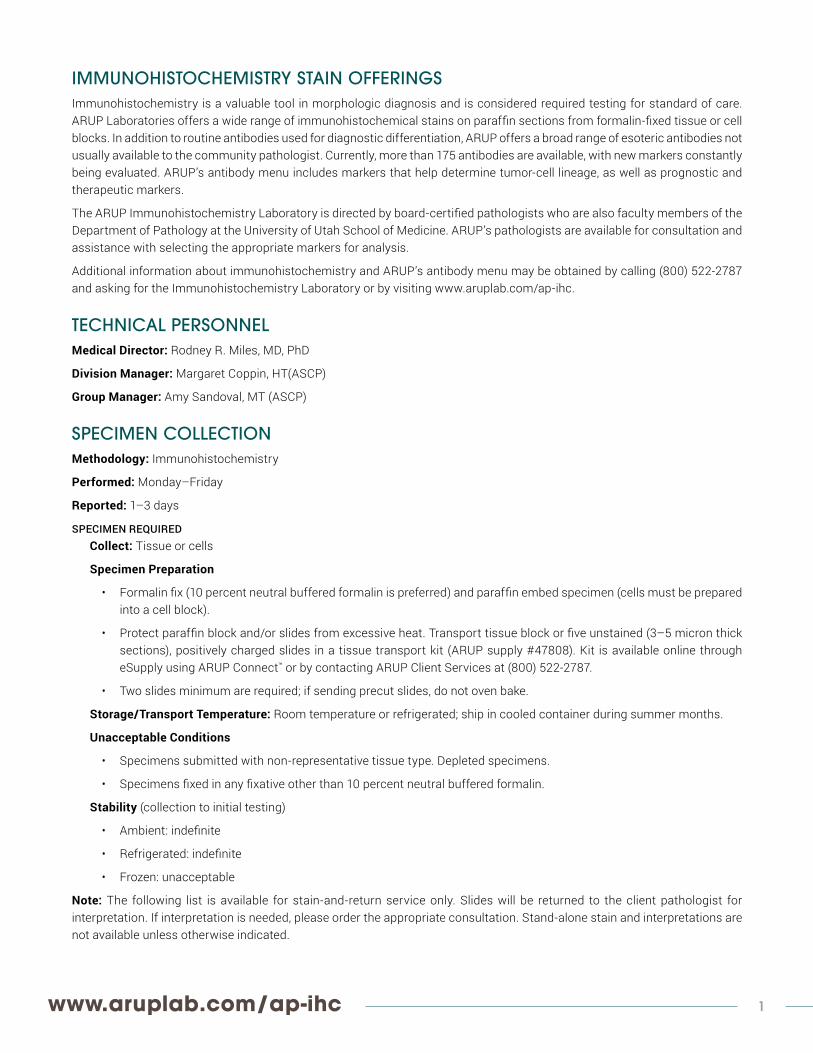
IMMUNOHISTOCHEMISTRY STAIN OFFERINGSImmunohistochemistry is a valuable tool in morphologic diagnosis and is considered required testing for standard of care. ARUP Laboratories offers a wide range of immunohistochemical stains on paraffin sections from formalin-fixed tissue or cell blocks. In addition to routine antibodies used for diagnostic differentiation, ARUP offers a broad range of esoteric antibodies not usually available to the community pathologist. Currently, more than 175 antibodies are available, with new markers constantly being evaluated. ARUP’s antibody menu includes markers that help determine tumor-cell lineage, as well as prognostic and therapeutic markers.
The ARUP Immunohistochemistry Laboratory is directed by board-certified pathologists who are also faculty members of the Department of Pathology at the University of Utah School of Medicine. ARUP’s pathologists are available for consultation and assistance with selecting the appropriate markers for analysis.
Additional information about immunohistochemistry and ARUP’s antibody menu may be obtained by calling (800) 522-2787 and asking for the Immunohistochemistry Laboratory or by visiting www.aruplab.com/ap-ihc.
TECHNICAL PERSONNELMedical Director: Rodney R. Miles, MD, PhD
Division Manager: Margaret Coppin, HT(ASCP)
Group Manager: Amy Sandoval, MT (ASCP)
SPECIMEN COLLECTIONMethodology: Immunohistochemistry
Performed: Monday–Friday
Reported: 1–3 days
SPECIMEN REQUIREDCollect: Tissue or cells
Specimen Preparation
• Formalin fix (10 percent neutral buffered formalin is preferred) and paraffin embed specimen (cells must be prepared into a cell block).
• Protect paraffin block and/or slides from excessive heat. Transport tissue block or five unstained (3–5 micron thick sections), positively charged slides in a tissue transport kit (ARUP supply #47808). Kit is available online through eSupply using ARUP Connect™ or by contacting ARUP Client Services at (800) 522-2787.
• Two slides minimum are required; if sending precut slides, do not oven bake.
Storage/Transport Temperature: Room temperature or refrigerated; ship in cooled container during summer months.
Unacceptable Conditions
• Specimens submitted with non-representative tissue type. Depleted specimens.
• Specimens fixed in any fixative other than 10 percent neutral buffered formalin.
Stability (collection to initial testing)
• Ambient: indefinite
• Refrigerated: indefinite
• Frozen: unacceptable
Note: The following list is available for stain-and-return service only. Slides will be returned to the client pathologist for interpretation. If interpretation is needed, please order the appropriate consultation. Stand-alone stain and interpretations are not available unless otherwise indicated.

2 For the most up-to-date information on Immunohistochemistry Stain Offerings, please visit:
QUICK REFERENCE FOR STAIN AND RETURN ONLY Breast/Endometrium/Ovary/Testes• Estrogen• Inhibin• PAX8• Progesterone
Differentiation Markers• B72.3 (breast)• Breast 2 (GCDFP-15)• DOG1• Estrogen (breast, ovary, and
endometrium)• HSA (liver)• Inhibin• MDM2 • P40• P504S (prostate)• P63• PAP (prostate)• PAX2• PAX8• PD-L1• PIN4 (prostate)• PSA (prostate)• RCC• TFE3
Epithelial Markers• AE1/AE3• Ber-EP4• Beta-Catenin• Calretinin• CAM 5.2 LMW• CDX2• CEA (monoclonal)• CEA (polyclonal)• CK 5/6• CK 7• CK 19• CK 20• E-cadherin• EMA• ERA (MOC-31)• Keratin 903 (HMW)• PIN4 (prostate)• TTF-1• WT-1 (N-terminus)
Germ-Cell Tumors/Placenta Markers• Human chorionic gonadotropin
(hCG)• Human placental lactogen (HPL)• Placental alkaline phosphatase
(PLAP)• Oct-3/4• SALL4
Hematopoietic Markers• ALK-1• BCL-2• BCL-6• Beta F1• BOB-1• CD1a• CD2 (AB75)• CD3• CD4 (1F6)• CD5• CD7• CD8• CD10 (calla)• CD14• CD15 (Leu M1)• CD19• CD20 (L26, Leu16)• CD21• CD23• CD25• CD30 (Ki-1)• CD31• CD33• CD34 (QBEND10)• CD35• CD42b• CD43 (L60, Leu 22)• CD44• CD45 (LCA)• CD52 (CAMPATH-1)• CD56 (NCAM)• CD57 (Leu 7)• CD61 (GPIIIa)• CD68 (KP1)• CD79a• CD117 (c-kit)• CD123• CD138 (plasma)• CD163• Cyclin D1 (SP4) • DBA.44• Factor XIIIa (factor XIII)• Glycophorin A• Granzyme B• IgA• IgD• IgG• IgG4• IgM• IRF4/MUM1• Kappa• Ki-67 (MIB-1)• Lambda• Mast-cell tryptase• Myeloperoxidase (MPO)• Oct-2
• p21• PAX-5• PD1• TdT• TIA-1• TRAP
Histiocytic• CD1a• CD68 (KP1)• Lysozyme (muramidase)
Liver• Alpha-1-antichymotrypsin (a-1-
ACT)• Alpha-1-antitrypsin (a-1-AT)• Alpha fetoprotein (AFP)• Arginase 1• Glypican 3• HSA (liver)
Melanocytic Markers• HMB-45• Melan A (MART1)• MITF• S-100
Mesenchymal Markers• Vimentin
Microbial Markers• Helicobacter pylori• Pneumocystis jiroveci
(Pneumocystis carinii)• Toxoplasmosis
Mucinous Markers• Muc-1 glycoprotein• Muc-4 glycoprotein• Muc-5AC glycoprotein
Muscle Marker• Caldesmon (h-CD)• Desmin• Muscle specific actin (MSA)• Myoglobin• Myosin• MYF-4• Smooth muscle actin (SMA)
Nervous System Markers• ATRX• CD56 (NCAM)• GFAP• IDH1• NeuN• Neurofilament (68kD)• PGP 9.5• PHF-Tau• S-100• aSynuclein• Ubiquitin
Neuroendocrine Markers• CD56 (NCAM)• CD57 (Leu 7)• Chromogranin A• NSE• PGP 9.5• Synaptophysin
Oncogene/Tumor Suppressor Markers• p16• p53
Pancreas• Gastrin
Peripheral Neuroectodermal Markers• CD99 (013) (Ewing sarcoma)
Pituitary Markers• ACTH• Human growth hormone (HGH)• Prolactin
Prognostic Markers• BAf47/Ini-1• ERBB2 (HercepTest)• HercepTest (Refer to ERBB2) • Ki-67 (MIB-1)• Mismatch repair by IHC (HNPCC)
(includes MLH1, MSH2, MSH6, and PMS2)—not available as stain and return
• p16• p53• WT-1
Thyroid/Parathyroid Markers• Calcitonin• Parathyroid hormone (PTH)• Thyroglobulin• TTF-1
Vascular Markers• C4d• Calponin• CD31• CD34 (QBEND10)• Collagen IV• D2-40• Glut-1• Procollagen I
Viral Markers• Adenovirus• CMV• HBsAg• HHV8• HSV I/HSV II• SV40 (BK virus)

www.aruplab.com/ap-ihc 3
THE FOLLOWING STAINS ARE AVAILABLE WITH INTERPRETATIONstain test # description
ALK (D5F3) 2007324
• The D5F3 monoclonal ALK antibody provides increased sensitivity, which can more accurately identify ALK-rearranged lung adenocarcinoma with high reproducibility, sensitivity, and specificity.
• Facilitates the routine identification of ALK-rearranged lung adenocarcinomas in clinical practice and detects lung cancers that may be responsive to ALK inhibitors
ALK (D5F3) with reflex to FISH if equivocal or positive 2011431 See ALK (D5F3)
ER/PR panel 0049210• Prognostic for breast cancer• Predictive for response of breast cancers and endometrial cancers to hormonal therapy• Differentiates endocervical from endometrial adenocarcinomas
ERBB2 (HercepTest) 0049174 • Aids in identifying breast cancer patients eligible for Herceptin therapy
ERBB2 (HercepTest) with reflex to FISH if 2+ 0049178 See ERBB2.
HNPCC 0049302 See Mismatch Repair (MSI).
IDH1 (R132H) point mutation 2007357
• Distinguishes primary from secondary glioblastoma multiform (GBM)• IDH1 mutations occur in approximately 70 percent of astrocytomas and oligodendroglial tumors• Allows the highly sensitive and specific discrimination of various tumors, such as astrocytoma from
primary glioblastomas or diffuse astrocytoma grade II from pilocytic astrocytoma or ependymoma
Ki-67 (MIB-1) 2007182
• Proliferation index indicator• Determines growth fraction• Aids in differentiating melanoma from nevus cells for sentinel node biopsy• Distinguishes benign and malignant adrenocortical tumors
Lynch syndrome 0049302 See Mismatch Repair (MSI).
Mismatch repair (Lynch syndrome; HNPCC) 0049302
• Microsatellite Instability (MSI)• MLH1, MSH2, MSH6, and PMS2• Mismatch repair proteins (MMR)• Used in the work up of Lynch syndrome (hereditary non-polyposis colorectal cancer or HNPCC)
Mismatch repair with reflex to BRAF Codon 600 mutation 2002327 • Distinguishes sporadic from Lynch (HNPCC)-associated colorectal cancers with abnormal MLH1
immunostaining
Mismatch repair with reflex to MLH1 promoter methylation 2005270 • Distinguishes sporadic from Lynch (HNPCC)-associated non-colorectal cancers with abnormal MLH1
immunostaining
p53 0049250 • Tumor suppressor protein; prognostic indicator
PD-L1 22C3 2013284 • FDA-approved test which aids in prediction of response to KEYTRUDA (pembrolizumab) for patients with non-small cell lung cancer (NSCLC)
PD-L1 28-8 2013684 • FDA-approved test which aids in the prediction of response to nivolumab (OPDIVO) for patients with non-squamous non-small cell lung cancer (NSCLC) or melanoma.
ROS1 with reflex to FISH if equivocal or positive 2008414
• Detects ROS1 fusion proteins by immunohistochemistry (IHC) using ROS1 clone D4D6 on FFPE tumor tissue
• Reflexes to FISH for confirmation if IHC result is equivocal
SDHB 2006948• SDHB by immunohistochemistry is used as a screening tool in directing testing algorithms for SDH
mutation. A negative result is highly suggestive of an SDH complex mutation but should be confirmed by molecular analysis.

4 For the most up-to-date information on Immunohistochemistry Stain Offerings, please visit:
antibody test # description
ACTH 2003427 • Adrenocorticotropic hormone; subclassifies pituitary adenomas
Adenovirus 2003430 • Specific to all subtypes of adenovirus
AE1/AE3 2003433 • Cytokeratin antibody cocktail for acidic and basic cytokeratins
ALK-1 2003439
• Anaplastic lymphoma kinase 1• Reacts with the NPM-ALK fusion protein expressed by t(2;5) positive anaplastic large-cell lymphomas as well as variant
ALK translocations• Not for lung cancers; refer to ALK (D5F3) in the stains with interpretations section.
Alpha synuclien 2003419 • aSynuclien (SNCA); demonstrates Lewy bodies in brain cells associated with Parkinson and Alzheimer disease
Alpha-1-antichymotrypsin 2003418 • Alpha 1 ACT; aids in identifying hepatomas and some germ-cell neoplasms
• Histiocyte marker for normal/neoplastic tissue
Alpha-1-antitrypsin 2003424 • Alpha 1 AT; expressed by cells of histolytic origin
• Aids in identifying germ-cell and histolytic neoplasms, as well as embryonal and some lung carcinomas
Alpha fetoprotein 2003436• Expressed by neoplastic liver and gonad tissue• Aids in identifying bladder carcinomas, yolk-sac tumors, some germ-cell tumors, and a high proportion of hepatocellular
carcinoma
Arginase-1 2011890 • Aides in the distinction of HCC from other hepatocellular and non-hepatocellular mass lesions, as well as in cases of metastatic carcinoma and other benign and malignant nonhepatocellular mimics
ATRX 2014499
• Expression of ATRX is implicated in cancer pathogenesis and is useful in the diagnosis of astrocytic gliomas. Its specificity and prevalence in lower-grade gliomas with an IDH-mutation argue for thorough characterization of associated signaling networks to facilitate therapeutic development.
• Mutation or loss of alpha-thalassemia/mental retardation syndrome X-linked (ATRX) expression has been described in anaplastic gliomas. ATRX loss is a hallmark of astrocytic tumors and defines a subgroup of astrocytic tumors with a favorable prognosis.
B72.3 2003445 • Tumor-associated glycoprotein (TAG.72); recognizes tumor-associated oncofetal antigen• Aids in identifying adenocarcinomas and breast carcinomas
BAF47/INI1 2003448 • Indicative of a tumor-suppressor role• Heterozygous tumors in the soft tissues of the head and neck
BCL-2 2004513
• B-cell lymphoma-2• Proto-oncogene• Overexpression increases life span in B cells• Aids in identifying colorectal adenomas and carcinomas• Distinguishes follicular lymphoma from reactive follicles
BCL-6 2003457• Transcription factor important in germinal center formation• Expressed in germinal center origin lymphomas, including some large-cell lymphomas, Burkitt lymphoma, and Hodgkin
lymphoma (nodular, lymphocyte predominant)
Ber-EP4 2003463
• Epithelial cell-membrane glycoprotein• Differentiates mesothelial from epithelial cells• Aids in identifying mammary Paget disease, lung adenocarcinomas, trichoepitheliomas, dermatofibromas, basal-cell
carcinomas, and other carcinomas
Beta-Catenin 2003454
• Binds to cytoplasmic region of e-cadherin molecule• Plays a role in cell adhesion, signal transmission, and actin cytoskeleton anchoring• Aids in identifying skin, liver, ovary, brain, prostate, and some breast cancers, as well as endometrial, ovarian, and colon
carcinomas
Beta F-1 2003466• Beta framework 1; BF-1; recognizes T-cell receptor (TCR) beta subunit• Aids in characterizing alpha-beta T-cell receptors from T-cell clones or polyclonal populations of T cells• Aids in diagnosing T-cell lineage neoplasms
BK Virus 2004137 See SV-40.
BOB-1 2003442 • B-cell oct-binding protein 1; OBF-1; expressed in spleen and peripheral blood leukocytes, B cells, and germinal centers• Aids in differentiating Hodgkin lymphomas (typically weak to negative) and B-cell lymphomas
Breast 2 (GCDFP-15) 2003472
• Gross cystic disease fluid protein-15; produced by cells with apocrine function• Differentiation marker for mammary carcinomas• Extramammary Paget disease
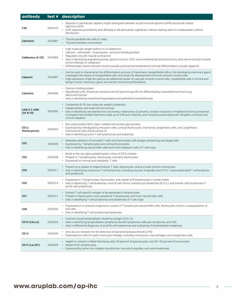
THE FOLLOWING STAINS ARE AVAILABLE AS STAIN AND RETURN ONLY (NO INTERPRETATION)
www.aruplab.com/ap-ihc 5
antibody test # description
C4D 2003475
• Deposits in peritubular capillary might distinguish between acute humoral rejection (AHR) and acute cellular rejection (ACR)
• AHR: deposits prominently and diffusely in the peritubular capillaries; intense staining seen in a widespread, uniform distribution
Calcitonin 2003481 • Thyroid parafollicular cells (C cells)• Thyroid medullary carcinomas
Caldesmon (h-CD) 2003484
• High molecular weight isoform is H-caldesmon• Calcium-, calmodulin-, tropomyosin-, and actin-binding protein• Regulates smooth muscle contraction• Aids in identifying angioleiomyomas, glomus tumors, GIST, some endometrial stromal tumors, and uterine smooth muscle
tumors (benign or malignant)• Differentiates uterine smooth muscle (usually positive) and endometrial stromal differentiation (usually negative)
Calponin 2003487
• Can be used to characterize the differentiation process of mammary myoepithelial cells in the developing mammary gland, investigate the nature of myoepithelial cells, and study the development of human smooth muscle cells
• High expression might be used as an additional marker of vascular smooth muscle cells, myopithelial cells in normal and benign human mammary gland, and certain stromal myofibroblasts
Calretinin 2003490
• Calcium-binding protein• Mesothelial cells: 89 percent sensitive and 90 percent specific for differentiating mesotheliomas from lung
adenocarcinomas• Aids in identifying mesothelial hyperplasia and epithelioid mesotheliomas
CAM 5.2 LMW (CK 8/18) 2003493
• Cytokeratin 8/18, low molecular weight cytokeratin• Hepatocellular and renal-cell carcinomas• Aids in identifying neuroendocrine carcinoma, melanomas (3 percent), nuclear inclusions of rhabdoid tumors (composed
of tangled intermediate filaments made up of CK8 and vimentin), and mutations associated with idiopathic cirrhosis and chronic hepatitis
CD1a (histiocytosis) 2003502
• Non-polymorphic MHC class I-related cell-surface glycoprotein• Expressed by interdigitating reticulum cells, cortical thymocytes, thymomas, langerhans cells, and Langerhans
histiocytosis cells (histiocytosis X)• Aids in identifying some T-cell lymphomas and leukemias
CD2 2003505• Mediates adhesion of activated T cells and thymocytes with antigen-presenting and target cells• Expressed by T lymphocytes and cortical thymocytes• Aids in identifying natural killer cells and most malignant cells of T-cell origin
CD3 2003508• Binds to the non-glycosylated epsilon chain of CD3 complex• Present in T-lymphocytes, thymocytes, and early thymocytes• Expressed on normal and neoplastic T cells
CD4 2003511• Present on a subset of helper/inducer T cells, thymocytes, and at a lower level on monocytes• Aids in identifying cutaneous T-cell lymphoma, including mycosis fungoides and HTLV-1-associated adult T-cell leukemia
and lymphoma
CD5 2003514• Expressed in T lymphocytes, thymocytes, and subset of B lymphocytes in lymph nodes• Aids in identifying T-cell leukemias, most B-cell chronic lymphocytic leukemias (B-CLL), and mantle-cell lymphomas/T-
and B-cell lymphomas
CD7 2003517• Earliest T-cell-specific antigen to be expressed in lymphocytes• Present in thymocytes, most peripheral T lymphocyte, and most natural killer cells• Aids in identifying T-cell lymphomas and leukemias of T-cell origin
CD8 2003520• Expressed on a cytotoxic/suppressor subset of T-lymphocyte natural killer cells, thymocytes, and on a subpopulation of
null cells• Aids in identifying T-cell lymphomas/leukemias
CD10 (CALLA) 2003523• Common acute lymphoblastic leukemia antigen (CALLA)• Aids in identifying lymphoblastic lymphoma, Burkitt lymphoma, follicular lymphoma, and CML• Aids in differential diagnosis of small B-cell lymphomas and subtyping of lymphoblastic leukemias
CD14 2003526 • Acts as a co-receptor for the detection of bacterial lipolysaccharide (LPS)• Expressed on cells of myelo-monocytic llineage, including monocytes, macrophages, and Langerhans cells
CD15 (Leu M1) 2003529• Hapten X; present in Reed-Sternberg cells, 90 percent of granulocytes, and 30–60 percent of monocytes• Absent from lymphocytes• Expressed by some non-Hodgkin lymphomas, mycosis fungoides, and some leukemias
6 For the most up-to-date information on Immunohistochemistry Stain Offerings, please visit:
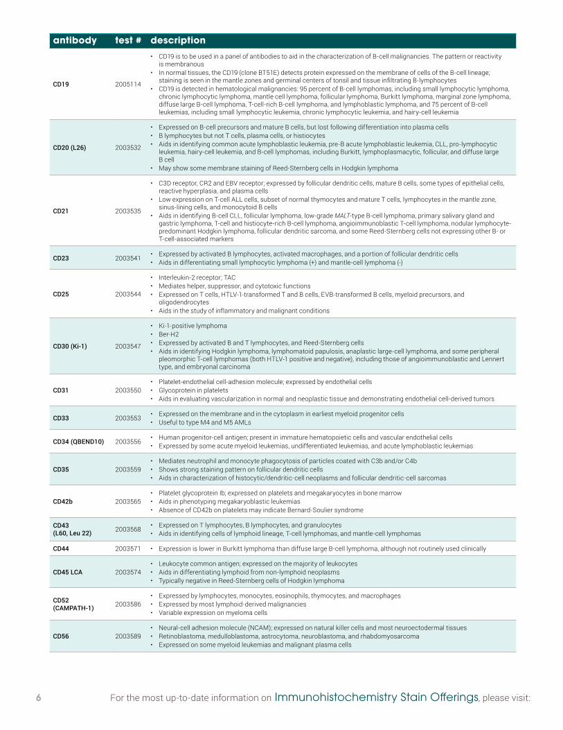
antibody test # description
CD19 2005114
• CD19 is to be used in a panel of antibodies to aid in the characterization of B-cell malignancies. The pattern or reactivity is membranous
• In normal tissues, the CD19 (clone BT51E) detects protein expressed on the membrane of cells of the B-cell lineage; staining is seen in the mantle zones and germinal centers of tonsil and tissue infiltrating B-lymphocytes
• CD19 is detected in hematological malignancies: 95 percent of B-cell lymphomas, including small lymphocytic lymphoma, chronic lymphocytic lymphoma, mantle cell lymphoma, follicular lymphoma, Burkitt lymphoma, marginal zone lymphoma, diffuse large B-cell lymphoma, T-cell-rich B-cell lymphoma, and lymphoblastic lymphoma, and 75 percent of B-cell leukemias, including small lymphocytic leukemia, chronic lymphocytic leukemia, and hairy-cell leukemia
CD20 (L26) 2003532
• Expressed on B-cell precursors and mature B cells, but lost following differentiation into plasma cells• B lymphocytes but not T cells, plasma cells, or histiocytes• Aids in identifying common acute lymphoblastic leukemia, pre-B acute lymphoblastic leukemia, CLL, pro-lymphocytic
leukemia, hairy-cell leukemia, and B-cell lymphomas, including Burkitt, lymphoplasmacytic, follicular, and diffuse large B cell
• May show some membrane staining of Reed-Sternberg cells in Hodgkin lymphoma
CD21 2003535
• C3D receptor, CR2 and EBV receptor; expressed by follicular dendritic cells, mature B cells, some types of epithelial cells, reactive hyperplasia, and plasma cells
• Low expression on T-cell ALL cells, subset of normal thymocytes and mature T cells, lymphocytes in the mantle zone, sinus-lining cells, and monocytoid B cells
• Aids in identifying B-cell CLL, follicular lymphoma, low-grade MALT-type B-cell lymphoma, primary salivary gland and gastric lymphoma, T-cell and histiocyte-rich B-cell lymphoma, angioimmunoblastic T-cell lymphoma, nodular lymphocyte-predominant Hodgkin lymphoma, follicular dendritic sarcoma, and some Reed-Sternberg cells not expressing other B- or T-cell-associated markers
CD23 2003541 • Expressed by activated B lymphocytes, activated macrophages, and a portion of follicular dendritic cells• Aids in differentiating small lymphocytic lymphoma (+) and mantle-cell lymphoma (-)
CD25 2003544
• Interleukin-2 receptor; TAC• Mediates helper, suppressor, and cytotoxic functions• Expressed on T cells, HTLV-1-transformed T and B cells, EVB-transformed B cells, myeloid precursors, and
oligodendrocytes• Aids in the study of inflammatory and malignant conditions
CD30 (Ki-1) 2003547
• Ki-1-positive lymphoma• Ber-H2• Expressed by activated B and T lymphocytes, and Reed-Sternberg cells• Aids in identifying Hodgkin lymphoma, lymphomatoid papulosis, anaplastic large-cell lymphoma, and some peripheral
pleomorphic T-cell lymphomas (both HTLV-1 positive and negative), including those of angioimmunoblastic and Lennert type, and embryonal carcinoma
CD31 2003550• Platelet-endothelial cell-adhesion molecule; expressed by endothelial cells• Glycoprotein in platelets• Aids in evaluating vascularization in normal and neoplastic tissue and demonstrating endothelial cell-derived tumors
CD33 2003553 • Expressed on the membrane and in the cytoplasm in earliest myeloid progenitor cells• Useful to type M4 and M5 AMLs
CD34 (QBEND10) 2003556 • Human progenitor-cell antigen; present in immature hematopoietic cells and vascular endothelial cells• Expressed by some acute myeloid leukemias, undifferentiated leukemias, and acute lymphoblastic leukemias
CD35 2003559• Mediates neutrophil and monocyte phagocytosis of particles coated with C3b and/or C4b• Shows strong staining pattern on follicular dendritic cells• Aids in characterization of histocytic/dendritic-cell neoplasms and follicular dendritic-cell sarcomas
CD42b 2003565• Platelet glycoprotein Ib; expressed on platelets and megakaryocytes in bone marrow• Aids in phenotyping megakaryoblastic leukemias• Absence of CD42b on platelets may indicate Bernard-Soulier syndrome
CD43 (L60, Leu 22) 2003568 • Expressed on T lymphocytes, B lymphocytes, and granulocytes
• Aids in identifying cells of lymphoid lineage, T-cell lymphomas, and mantle-cell lymphomas
CD44 2003571 • Expression is lower in Burkitt lymphoma than diffuse large B-cell lymphoma, although not routinely used clinically
CD45 LCA 2003574• Leukocyte common antigen; expressed on the majority of leukocytes• Aids in differentiating lymphoid from non-lymphoid neoplasms• Typically negative in Reed-Sternberg cells of Hodgkin lymphoma
CD52 (CAMPATH-1) 2003586
• Expressed by lymphocytes, monocytes, eosinophils, thymocytes, and macrophages• Expressed by most lymphoid-derived malignancies• Variable expression on myeloma cells
CD56 2003589• Neural-cell adhesion molecule (NCAM); expressed on natural killer cells and most neuroectodermal tissues• Retinoblastoma, medulloblastoma, astrocytoma, neuroblastoma, and rhabdomyosarcoma• Expressed on some myeloid leukemias and malignant plasma cells
www.aruplab.com/ap-ihc 7
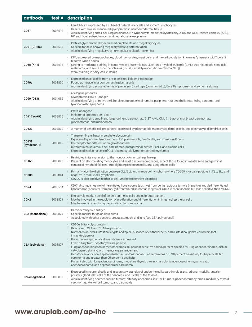
antibody test # description
CD57 2003592
• Leu-7, HNK1; expressed by a subset of natural killer cells and some T lymphocytes• Reacts with myelin-associated glycoprotein in neuroectodermal tissue• Aids in identifying small-cell lung carcinoma, NK lymphocyte-mediated cytotoxicity, AIDS and AIDS-related complex (ARC),
NK and T-cell subset tumors, and neural-tissue neoplasms
CD61 (GPIIIa) 2003595• Platelet glycoprotein IIIa; expressed on platelets and megakaryocytes• Specific for cells showing megakaryoblastic differentiation• Aids in identifying megakaryocytic/megakaryoblastic leukemias
CD68 (KP1) 2003598
• KP1; expressed by macrophages, blood monocytes, mast cells, and the cell population known as “plasmacytoid T cells” in reactive lymph nodes
• Strong to moderate staining in acute myeloid leukemia (AML), chronic myeloid leukemia (CML), true histiocytic neoplasia, melanoma, and some B-cell neoplasms (usually small lymphocytic lymphoma [SLL])
• Weak staining in hairy-cell leukemia
CD79a 2003800• Expressed on all B cells from pre-B cells until plasma-cell stage• Found as intracellular component in plasma cells• Aids in identifying acute leukemia of precursor B-cell type (common ALL), B-cell lymphomas, and some myelomas
CD99 (O13) 2004055
• MIC2 gene products• Glycoprotein HBA 71 antigen• Aids in identifying primitive peripheral neuroectodermal tumors, peripheral neuroepitheliomas, Ewing sarcoma, and
lymphoblastic lymphoma
CD117 (c-kit) 2003806
• Proto-oncogene• Inhibitor of apoptotic cell death• Aids in identifying small- and large-cell lung carcinomas, GIST, AML, CML (in blast crisis), breast carcinomas,
glioblastomas, and melanomas
CD123 2003809 • A marker of dendric cell precursors: expressed by plasmactoid monocytes, dendric cells, and plasmacytoid dendritic cells
CD138 (syndecan-1) 2003812
• Transmembrane heparin sulphate glycoprotein• Expressed by normal lymphoid cells, IgG plasma cells, pre-B cells, and immature B cells• Co-receptor for differentiation growth factors• Differentiates squamous cell carcinomas, postgerminal-center B cells, and plasma cells• Expressed in plasma cells of CLL, plasmacytoid lymphomas, and myelomas
CD163 2003815• Restricted in its expression to the monocytic/macrophage lineage • Present on all circulating monocytes and most tissue macrophages, except those found in mantle zone and germinal
centers of lymphoid follicles, interdigitating reticulum cells, and Langerhans cells
CD200 2012844• Primarily aids the distinction between CLL/SLL and mantle cell lymphoma where CD200 is usually positive in CLL/SLL and
negative in mantle cell lymphoma• CD200 is also positive in other B-cell lymphoproliferative disorders
CDK4 2005534 • CDK4 distinguishes well-differentiated liposarcoma (positive) from benign adipose tumors (negative) and dedifferentiated liposarcoma (positive) from poorly differentiated sarcomas (negative); CDK4 is more specific but less sensitive than MDM2
CDX2 2003821• Exclusively marks nuclei of colonic epithelial cells and colorectal cancers• May be involved in the regulation of proliferation and differentiation in intestinal epithelial cells• May be used in identifying metastatic colon carcinoma
CEA (monoclonal) 2003824• Carcinoembryonic antigen• Specific marker for colon carcinoma• Associated with other cancers: breast, stomach, and lung (see CEA polycolonal)
CEA (polyclonal) 2003827
• CD56e; biliary glycoprotein 1• Reacts with CEA and CEA-like proteins• Normal colon: small intestinal crypts and apical surfaces of epithelial cells, small intestinal goblet-cell mucin (not
intracytoplasmic)• Breast: some epithelial cell membranes expressed• Liver: biliary tract; hepatocytes are positive• Lung adenocarcinomas or mesotheliomas: 85 percent sensitive and 96 percent specific for lung adenocarcinoma, diffuse
cytoplasmic staining with membrane enhancement• Hepatocellular or non-hepatocellular carcinomas: canalicular pattern has 50–90 percent sensitivity for hepatocellular
carcinoma and greater than 95 percent specificity• Present also with lung adenocarcinoma, medullary thyroid carcinoma, colonic adenocarcinoma, pancreatic
adenocarcinoma, and hepatocellular carcinoma
Chromogranin A 2003830
• Expressed in neuronal cells and in secretory granules of endocrine cells: parathyroid gland, adrenal medulla, anterior pituitary gland, islet cells of the pancreas, and C cells of the thyroid
• Aids in identifying neuroendocrine tumors: pituitary adenomas, islet-cell tumors, phaeochromocytomas, medullary thyroid carcinomas, Merkel-cell tumors, and carcinoids

8 For the most up-to-date information on Immunohistochemistry Stain Offerings, please visit:
antibody test # description
CK 5/6 2003851
• Cytokeratins 5 and 6; stratified squamous epithelial cytokeratin• Aids in diagnosing low-differentiated pavement epithelium carcinoma, adenocarcinoma, and mesothelioma• Differentiates epithelial mesotheliomas (positive-cytoplasmic staining with perinuclear enhancement) from lung
adenocarcinoma (89 percent sensitive and 95 percent specific)
CK 7 2003854
• Cytokeratin 7; reacts with most glandular and transitional epithelia: breast, lung, bladder, female genital tract (endometrium and fallopian tube), gastrointestinal tract (gallbladder, hepatic ducts, and pancreatic ducts), urinary tract, and bile duct
• Present with subtypes of ovarian, pulmonary, and breast adenocarcinomas, transitional-cell carcinomas, tumors of female genital tract (endometrium and fallopian tube), urothelial carcinomas, breast carcinomas, and lung carcinomas
CK 19 2003845 • Reacts with a large number of epithelial cell types, including many ductal and glandular epithelia• Aids in identification of many benign and malignant epithelial lesions
CK 20 2003848
• Cytokeratin 20; expressed in intestinal epithelium, gastric foveolar epithelium, some endocrine cells of the upper portions of the pyloric glands, urethelium, and Merkel cells in epidermis
• Aids in identifying colorectal carcinoma, adenomas of the gallbladder and bile ducts, ductal cell adenocarcinomas of the pancreas, mucinous ovarian tumors, transitional-cell carcinomas, and Merkel-cell carcinomas of the skin
• Gastrointestinal adenocarcinomas express CK20 to a lesser degree
c-MET 2008652
• Tumors derived from c-Met expressing epithelia are usually positive; these include colorectal carcinomas, gastric adenocarcinomas, and non-small cell lung carcinomas
• In gastric cancer and non-small cell lung carcinoma, it has been determined that c-MET drives the cancer • It has also been found that c-MET is a resistance pathway in lung cancer for EGFR inhibitors -This antibody may be used
to aid in the identification of normal and neoplastic c-MET expressing cells. The pattern of reactivity is cytoplasmic/membranous
CMV 2003833 • Cytomegalovirus; reacts with the delayed and early DNA-binding protein p52• Does not crossreact with other herpesviruses or adenoviruses
c-MYC 2008317• c-MYC expression has been described in a variety of cancers including breast cancer, prostate cancer, lymphoma, lung,
and colon cancers. • The c-MYC antibody may be used to characterize lymphomas. The pattern of reactivity is nuclear.
Collagen IV 2003839 • Reacts with basement membranes in kidney, skin, striated and smooth muscle, spleen, lymph node, lung, placenta, and tendon
CXCL13 2008622
• B-lymphocyte chemoattractant / B-Cell attracting chemokine-1 (BLC/BCA-1)• CXC chemokine family controlling the organization of B cells within follicles of lymphoid tissues such as spleen, lymph
nodes, and Peyer’s patches.• In T-lymphocytes, CXCL13 expression is thought to reflect a germinal center origin of the Tcell.• Useful marker in the diagnosis of angioimmunoblastic T-cell lymphoma; when used in a panel it can differentiate it from
other proliferative T-cell lymphoma.
Cyclin D1 (SP4) 2003842• B-cell lymphoma-1• Mantle-cell lymphoma, various carcinomas (strong staining in carcinomas), multiple myelomas, some parathyroid
adenomas, and parathyroid carcinomas
D2-40 2003857 • High sensitivity and specificity for lymphatic endothelium• Can be used as a reliable lymphatic endothelial-cell marker in the evaluation of lymphatic involvement in tumors
DBA.44 (Hairy Cell Leukemia) 2003860
• Developed against the B-cell antigen• Aids in identifying hairy-cell leukemia (particularly hairy cytoplasmic processes), some follicular center-cell lymphomas,
high-grade B-cell lymphomas, and splenic lymphomas with villous lymphocytes
Desmin 2003863
• Intermediate filament present in smooth and striated muscle• Expressed in reactive mesothelial cells, myoblasts, myofibroblasts (variable), endometrial stroma, and smooth muscle
cells• Aids in identifying smooth muscle tumors (leiomyosarcomas), myogenic sarcomas, striated muscle tumors
(rhabdomyosarcoma), PNET, neuroblastomas, and intra-abdominal desmoplastic small round-cell tumors
DOG1 2010168• Shown to be highly specific and sensitive in the diagnosis of GIST• Approximately 4–15% of GIST will stain weakly or be negative for CD117 by IHC; in the vast majority of these cases, DOG1 is
expressed by IHC.
E-cadherin 2003869
• Cellular adhesion molecule; loss associated with invasive carcinoma• Differentiates LCIS from DCIS in indeterminate breast carcinoma• Reduced expression in invasive bladder cancer and ductal carcinoma• No expression in lobular carcinoma and LCIS
EMA 2003872
• Epithelial membrane antigen; prognostic• Expressed by almost all glandular and ductal epithelial cells, including breast and pancreas, activated T cells, monocytes,
some B cells, follicular dendritic cells, and perineurial cells• Aids in identifying most adenocarcinomas, anaplastic large-cell lymphomas, epithelioid sarcomas, meningiomas, some
mesotheliomas, myelomas, Paget disease, plasmacytomas, squamous-cell tumors, and metastatic carcinomas• Associated with invasion in pancreatic tumors

www.aruplab.com/ap-ihc 9
antibody test # description
ERA (MOC-31) 2003875 • Epithelial-related antigen (MOC-31); aids in identifying adenocarcinomas, squamous-cell carcinomas, adenomas, small-cell lung cancers, carcinoids, adenocystic carcinomas, and carcinosarcomas
ERBB2 (HercepTest) 2007332
• This test code is for stain-and-return service only; see above in available stains with interpretation section for alternate test code.
• Aids in identifying breast cancer patients eligible for Herceptin therapy
ERG 2012555 • Prostate marker• May be used to aid in the identification of prostate adenocarcinomas through the detection of truncated ERG
Estrogen 2004516• Estrogen receptor-alpha; prognostic for breast cancer• Predictive for response of breast cancers to hormonal therapy• Differentiates endocervical from endometrial adenocarcinomas
Factor XIIIa 2003878
• Blood pro-enzyme identified in platelets, megakaryocytes, and fibroblast-like mesenchymal or histiocytic cells present in the placenta, uterus, and prostate
• Present in monocytes, macrophages, and dermal dendritic cells• Aids in differentiating dermatofibromas, dermatosarcoma protuberans, and desmoplastic malignant melanomas• Positive in capillary hemangioblastomas, hemangioendotheliomas, hepatocellular carcinomas, hemangiopericytomas,
xanthogranulomas, glomus tumors, and meningiomas
Fli-1 2003887
• Friend leukemia integration-1; Friend leukemia insertion site 1• Anti-apoptotic activity• Expressed in heart, lung, spleen, and thymus• Aids in identifying erythroleukemias, lymphoblastic lymphomas, and Ewing sarcomas
Gastrin 2003896 • Expressed in G cells of the pyloric antrum• Aids in identifying G-cell hyperplasia and gastrin-secreting tumors
GATA3 2012558• Breast marker• Can be used in a panel of antibodies for diagnosis of unknown primary carcinoma when carcinomas of the breast or
bladder are a possibility. The pattern of reactivity should be nuclear.
GFAP 2003899• Glial fibrillary acidic protein; expressed in astrocytes and some CNS ependymal cells• Identifies astrocytomas and ependymomas• Many neural tumors, such as neuroblastomas, schwannomas, and extra-CNS tumors, do not stain
GLUT-1 (Glucose Transporter-1) 2003905 • Involved in glucose transport across epithelial and endothelial barrier tissues
• Stains the membrane of normal erythrocytes in various normal and neoplastic tissues
Glycophorin A 2003908 • Expressed in erythroid cells• Identifies M6 subtype of acute myeloblastic leukemia, erythroleukemia, and erythroblasts
Glypican 3 2011925 • Useful tumor marker for the diagnosis of hepatocellular carcinoma (HCC), hepatoblastoma, melanoma, testicular germ cell tumors, and Wilms’ tumor
Granzyme B 2007173• Granzyme B has been found to be expressed in the neoplastic counterparts of cytolytic CTL and NK-cells; therefore,
granzyme B may be a valuable tool in the diagnosis of T-cell/NK-cell lymphomas with cytotoxic phenotypes. High percentages of cytotoxic T-cells have been shown to be an unfavorable prognostic indicator in Hodgkin disease.
HBME-1 2003914
• Anti-mesothelial cell; has been demonstrated to immunostain the membrane and cytoplasm of normal plural and peritoneal mesothelial cells and of neoplastic epithelial mesothelioma cells
• Although both the membrane and cytoplasm of epithelial mesothelioma cells stain positive, the thick membrane staining pattern is found to be a more diagnostically useful marker of malignant mesothelioma.
Helicobacter pylori 2003941 • Campylobacter pylori; reacts with antigens of the H. pylori organism
HercepTest See ERBB2 (HercepTest).
HHV8 2003932 • Human herpes virus type 8 (latent nuclearantigen); aids in identifying multicentric Castleman disease, angioimmunoblastic lymphadenopathies, and Kaposi sarcoma
HMB45 (Melanoma Antibody)
2003935• Melanoma-specific antigen; expressed in junctional cells, blue-nevus cells, and fetal and neonatal melanocytes• Reacts with the majority of melanomas and other tumors with melanoma/melanocytic differentiation, including melanotic
schwannoma clear-cell sarcoma
HSA 2003923• Hepatocyte specific antigen: Hep Par-1; expressed in hepatocytes• Differentiates hepatocellular carcinomas and metastatic carcinomas• Differential diagnosis of hepatocellular carcinomas, cholangiocarcinomas, and hepatoblastomas
HSV I/HSV II 3000101 • Reacts with antigens common to HSV types 1 and 2; reacts with all the major glycoproteins present in the viral envelope.• HSV I/II by IHC will aid in identifying tissue infected with the herpes simplex virus
Human chorionic gonadotropin 2003920 • Beta-hCG; expressed on placental trophoblasts
• Aids in identifying trophoblastic germ-cell tumor
10 For the most up-to-date information on Immunohistochemistry Stain Offerings, please visit:
antibody test # description
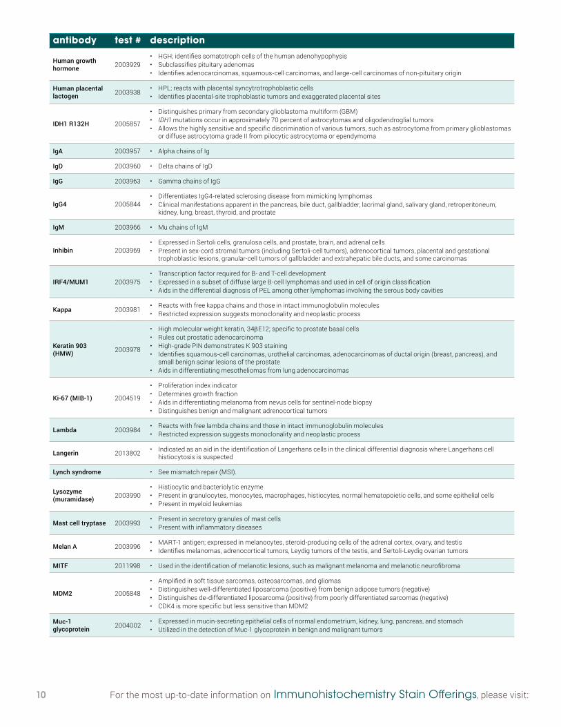
Human growth hormone 2003929
• HGH; identifies somatotroph cells of the human adenohypophysis• Subclassifies pituitary adenomas• Identifies adenocarcinomas, squamous-cell carcinomas, and large-cell carcinomas of non-pituitary origin
Human placental lactogen 2003938 • HPL; reacts with placental syncytrotrophoblastic cells
• Identifies placental-site trophoblastic tumors and exaggerated placental sites
IDH1 R132H 2005857
• Distinguishes primary from secondary glioblastoma multiform (GBM)• IDH1 mutations occur in approximately 70 percent of astrocytomas and oligodendroglial tumors• Allows the highly sensitive and specific discrimination of various tumors, such as astrocytoma from primary glioblastomas
or diffuse astrocytoma grade II from pilocytic astrocytoma or ependymoma
IgA 2003957 • Alpha chains of Ig
IgD 2003960 • Delta chains of IgD
IgG 2003963 • Gamma chains of IgG
IgG4 2005844• Differentiates IgG4-related sclerosing disease from mimicking lymphomas • Clinical manifestations apparent in the pancreas, bile duct, gallbladder, lacrimal gland, salivary gland, retroperitoneum,
kidney, lung, breast, thyroid, and prostate
IgM 2003966 • Mu chains of IgM
Inhibin 2003969• Expressed in Sertoli cells, granulosa cells, and prostate, brain, and adrenal cells• Present in sex-cord stromal tumors (including Sertoli-cell tumors), adrenocortical tumors, placental and gestational
trophoblastic lesions, granular-cell tumors of gallbladder and extrahepatic bile ducts, and some carcinomas
IRF4/MUM1 2003975• Transcription factor required for B- and T-cell development• Expressed in a subset of diffuse large B-cell lymphomas and used in cell of origin classification• Aids in the differential diagnosis of PEL among other lymphomas involving the serous body cavities
Kappa 2003981 • Reacts with free kappa chains and those in intact immunoglobulin molecules• Restricted expression suggests monoclonality and neoplastic process
Keratin 903 (HMW) 2003978
• High molecular weight keratin, 34βE12; specific to prostate basal cells• Rules out prostatic adenocarcinoma• High-grade PIN demonstrates K 903 staining• Identifies squamous-cell carcinomas, urothelial carcinomas, adenocarcinomas of ductal origin (breast, pancreas), and
small benign acinar lesions of the prostate• Aids in differentiating mesotheliomas from lung adenocarcinomas
Ki-67 (MIB-1) 2004519
• Proliferation index indicator• Determines growth fraction• Aids in differentiating melanoma from nevus cells for sentinel-node biopsy• Distinguishes benign and malignant adrenocortical tumors
Lambda 2003984 • Reacts with free lambda chains and those in intact immunoglobulin molecules• Restricted expression suggests monoclonality and neoplastic process
Langerin 2013802 • Indicated as an aid in the identification of Langerhans cells in the clinical differential diagnosis where Langerhans cell histiocytosis is suspected
Lynch syndrome • See mismatch repair (MSI).
Lysozyme (muramidase) 2003990
• Histiocytic and bacteriolytic enzyme• Present in granulocytes, monocytes, macrophages, histiocytes, normal hematopoietic cells, and some epithelial cells• Present in myeloid leukemias
Mast cell tryptase 2003993 • Present in secretory granules of mast cells• Present with inflammatory diseases
Melan A 2003996 • MART-1 antigen; expressed in melanocytes, steroid-producing cells of the adrenal cortex, ovary, and testis• Identifies melanomas, adrenocortical tumors, Leydig tumors of the testis, and Sertoli-Leydig ovarian tumors
MITF 2011998 • Used in the identification of melanotic lesions, such as malignant melanoma and melanotic neurofibroma
MDM2 2005848
• Amplified in soft tissue sarcomas, osteosarcomas, and gliomas• Distinguishes well-differentiated liposarcoma (positive) from benign adipose tumors (negative)• Distinguishes de-differentiated liposarcoma (positive) from poorly differentiated sarcomas (negative)• CDK4 is more specific but less sensitive than MDM2
Muc-1 glycoprotein 2004002 • Expressed in mucin-secreting epithelial cells of normal endometrium, kidney, lung, pancreas, and stomach
• Utilized in the detection of Muc-1 glycoprotein in benign and malignant tumors

www.aruplab.com/ap-ihc 11
antibody test # description
Muc-4 glycoprotein 2004008
• Stains stomach, colon, and the endothelial cells of small blood vessels and capillaries• Strong positive staining in colon polyps, colon carcinoma, and gastric adenocarcinoma• Positive staining also demonstrated in lung adenocarcinoma and ovarian mucinous adenocarcinoma
Muscle specific actin 2004011
• Present in skeletal, cardiac, smooth muscle, and myoepithelial cells• Identifies soft tissue tumors with muscle differentiation (leiomyomas, leiomyosarcomas, and rhabdomyosarcomas),
some pleomorphic liposarcomas, the majority of glomus tumors, occasional desmoid tumors, and myofibroblasts in some lesions
Myeloperoxidase 2004014• MPO; reacts with myeloperoxidase from granulocytes• Aids in differentiating lymphoid leukemias from myeloid leukemias• Identifies granulocytic sarcomas
Myf-4 2004017• Myogenin; expressed early in skeletal muscle differentiation• Is a sensitive and specific marker for rhabdomyosarcoma• Is more specific than desmin and muscle-specific actin and more sensitive than myoglobin
Myoglobin 2004031• Oxygen-binding protein• Expressed by striated muscle (cardiac and skeletal)• Present in rhabdomyosarcoma and other tumors with skeletal-muscle differentiation
Myosin 2004034 • Contractile protein; expressed in smooth muscle (non-sarcomeric) and skeletal muscle (sarcomeric) forms• Aids in muscle differentiation
Napsin A 2008716• Napsin A is highly specific in adenocarcinomas of lung and is useful in distinguishing primary lung adenocarcinomas from
adenocarcinomas of other organs.• The pattern of reactivity is cytoplasmic.
NeuN 2004046• Aids in the definitive identification of neuronal elements in ganglion-cell tumors or hamartomas, in which a distinction
between atypical glial cells and neurons may be difficult• May be used, similarly, for the study of neuronal loss in epilepsy, neurodegenerative diseases, or other conditions
Neurofilament (68kD) 2004049
• Cytoskeletal element in nerve axons/dendrites• Reacts with neurons, neuronal processes, peripheral nerves, sympathetic ganglion cells, and adrenal medulla• Identifies neuroblastoma and gangliomas
NSE 2004052
• Neuron-specific enolase; expressed by neuronal or neuroendocrine cells and their tumors: neuroblastomas and retinoblastomas
• May label non-neuronal tumors: meningiomas, medulloblastomas, astrocytomas, glioblastomas, oligoastrocytomas, oligodendrogliomas, pituitary adenomas, schwannomas, ependymomas, meningosarcomas, gliosarcomas, small-cell lung cancer, melanomas, and germ-cell tumors
Oct-2 2004061 • Octamer-binding transcription factor 2; aids in differentiating Hodgkin lymphomas (typically weak to negative) and B-cell lymphomas
Oct-3/4 2004058
• Octomer transcription factors 3 and 4; expressed by embryonic stem cells and germ cells• Has been reported to be expressed in germ-cell tumors and their metastases, which exhibit features of pluripotentiality,
including seminoma/dysgerminoma/germinoma and embryonial carcinoma• Has been proposed as a useful marker for germ-cell tumors and to assist in establishing a germ-cell origin for some
metastatic tumors of uncertain primary origin
p16 2004064 • F-12; negative regulator of the cell cycle• Prognostic significance (breast, colon, stomach, lung, and pituitary)
p21 2004067• WAF1-CIP1; inhibits and blocks cell-cycle progression• Present in melanomas, pancreatic carcinomas, cervical carcinomas, thymomas, thyroid carcinomas, breast carcinomas,
head and neck carcinomas, colon carcinomas, and Hodgkin lymphoma
p40 2010142 • Recognizes an epitope unique to the p40 protein and may have applications in cases where p63 has traditionally been used• Frequently used for lung squamous cell carcinoma, bladder, breast, prostate, and head and neck cancers
p53 2004522 • Tumor-suppressor protein; prognostic indicator
p57 2005542 • Used as an aid in identification of complete hydatidiform mole (CHM) (no nuclear labeling of cytotrophoblasts) from partial hydatidiform mole (PHM) and hydropic abortion
p63 2004073 • Differentiates prostatic adenocarcinoma and benign prostatic tissue• Also distinguishes poorly differentiated squamous-cell carcinoma from small-cell carcinoma or adenocarcinoma
P504S (AMACR) 2004076
• Prostate• α-Methylacyl-CoA racemase (AMCAR)• Specific for prostate adenocarcinomas• Detected in two premalignant lesions: high-grade prostatic intraepithelial neoplasia (PIN) and atypical adenomatous
hyperplasia

12 For the most up-to-date information on Immunohistochemistry Stain Offerings, please visit:
antibody test # description
PAP 2004079• Prostate acid phosphatase; reacts with prostatic epithelial cells and hyperplastic prostate• Present in carcinomas of the prostate and metastatic cells of prostate carcinoma, bladder carcinomas, and carcinoid
tumors
Parathyroid Hormone 2004118 • PTH; reacts with parathyroid epithelial cells
• Present in adenomas and primary and secondary hyperplasias
PAX5 2004082
• Member of the paired box family• B-cell-specific activator protein (BSAP)• Expressed in pro-, pre-, and mature B cells, but not in plasma cells• Present in pre B-cell acute lymphoblastic leukemias and classical Hodgkin lymphomas (typically weak)• Aids in differential diagnosis of lymphoplasmacytic lymphomas or plasmacytomas
PAX8 2010787
• Expressed in a high percentage of ovarian serous, endometroid, and clear cell carcinomas, but only rarely in primary ovarian mucinous adenocarcinomas
• Important marker of ovarian cancer and a useful marker for the differential diagnosis in lung and neck tumors, or tumors at distant sites where primary lung carcinoma, breast carcinoma, or thyroid carcinoma are possibilities.
PD1 2004085 • Angioimmunoblastic T-cell lymphomas are the only hematopoietic tumors that are positive for PD1 protein.• In tonsil and lymph tissues, the protein is expressed on T cells and some B cells of the light zone of germinal centers.
PD-L1 2011158• Clone E1L3N• Expressed in several tumor types, including melanoma, ovary, colon, lung, breast, and renal cell carcinoma• Additional research links PD-L1 expression to cancers associated with viral infections
PHF-Tau 2004094 • Paired helical filament-tau; tau abnormally phosphorlated in Alzheimer disease• Main component in paired helical filaments (PHFs) and neurofibrillary tangles
PIN4 2010045
• Prostate multiplex stain containing basal cell cocktail (34βE12/p63) and AMACR (P504s)• Has been reported to provide advantages in sensitivity over the use of p63 or anti-keratin (34βE12) alone in the detection
of prostatic basal cells• The two components of this cocktail not only augment but also complement each other in basal cell detection.
PGP 9.5 2004091 • Protein gene product 9.5; expressed in neurons, neuroendocrine cells, and melanocytes• Present in neuronal neoplasias (carcinoid tumors)
Placental alkaline phosphatase 2004097 • PLAP; expressed by placenta
• Present in most germ-cell tumors, and breast, lung, stomach, pancreas, and ovarian carcinomas
Pneumocystis javii (Pneumocystis carinii)
2004103 • Detects presence of Pneumocystis javii in infected tissue and free trophozoites
Procollagen I 2004106• Secreted by fibroblasts into the extracellular matrix, where it is cleaved to form collagen• Expression and secretion of procollagen are important features of the wound-healing and tissue-repair processes to which
the desmoplastic stroma of malignancy have sometimes been compared
Progesterone 2004525• Identifies A and B forms of progesterone• Predictive of response to hormone therapy for breast carcinoma and endometrial cancer• Aids in differentiating endocervical from endometrial adenocarcinomas
Prolactin 2004109 • Produced in the anterior pituitary gland• Subclassifies pituitary adenomas
Prostate Triple Stain 2010045
• Prostate multiplex stain containing basal cell cocktail (34βE12/p63) and AMACR (P504s)• Has been reported to provide advantages in sensitivity over the use of p63 or anti-keratin (34βE12) alone in the detection
of prostatic basal cells• The two components of this cocktail not only augment but also complement each other in basal cell detection.
PSA 2004112• Prostate-specific antigen; expressed by prostatic glandular epithelial cells and periurethral and perianal glands• Present in prostatic carcinomas, tumors of the colon, liver, lung, parotid, adrenal, and ovary, and, rarely, in metaplasias of
the bladder walls
RCC 2004124
• Renal-cell carcinoma; localized along the brush border of the pars-convolute and pars-recta segments of the proximal tubule and focally along the luminal surface of Bowman capsule
• In normal tissues, localized along the luminal surface of breast lobules and ducts, the luminal surface of the epididymal tubular epithelium, within the cytoplasm of the parathyroid parenchymal cells, and focally within the colloid of thyroid follicles
S-100 2004127
• Brain protein composed of S-100a and S-100b; expressed in neural crest (Schwann cells, melanocytes, and glial cells), chondrocytes, adipocytes, myoepithelial cells, macrophages, Langerhans cells, and dendritic cells
• Present in 95 percent of melanomas (including desmoplastic and spindle-cell tumors), 50 percent of malignant peripheral nerve-sheath tumors, clear-cell sarcomas, and occasional breast and undifferentiated carcinomas

www.aruplab.com/ap-ihc 13
antibody test # description
SALL4 2005432
• Sal-like 4; highly sensitive marker for gonadal seminoma/dysgerminoma, embryonal carcinomas, and yolk sac tumor as well as their metastatic form, including those metastasizing to the CNS
• An immunohistochemical panel, including SALL4, OCT4, and CD30, helps solve this diagnostic difficulty; germinoma will be positive for both SALL4 and OCT4 but negative for CD30, whereas embryonal carcinoma will show SALL4+/OCT4+/CD30+ profile, and yolk sac tumor will show SALL4+/OCT4−/CD30− profile.
STAT6 2013251 • Aids in diagnosis of solitary fibrous tumor
Smad4 2006403
• May be useful in the diagnosis of pancreatic cancer, juvenile polyposis syndrome, and hereditary hemorrhagic telangiectasia syndrome.
• The pattern of reactivity is mostly cytoplasmic but sometimes nuclear expression is seen in many cell types with highest expression levels in placenta and gastrointestinal tract.
Smooth muscle actin 2004130
• SMA; reacts with the alpha-smooth muscle isoform• Present in smooth muscle cells of vessels, parenchymes, myoepithelial cells, pericytes, and some stromal cells in the
intestine, testis, and ovary• Aids in differentiating leiomyosarcoma from rhabdomyosarcoma
SOX11 2012561• Lymphoma/hematopoietic marker• Will stain those cases of Mantle-cell lymphoma that are negative for the cyclin D1 stain, thereby aiding in a more timely
diagnosis of MCL.
SV-40 2004137 • Simian virus 40; closely related to BK virus and JC virus• Used to identify all polyomavirus infections due to cross-reactivity between SV-40 and BK or JC virus
Synaptophysin 2004139 • Labels neuroendocrine cells and neurons in the brain, spinal cord, and retina• Present in neuroendocrine tumors and neuroendocrine tumors of epithelial type
TdT 2004142
• Used in subtyping of blastic leukemias• Positive in all acute lymphoblastic leukemia (ALL) except Burkitt and B-cell FAB L-3• Positive in lymphoblastic crisis of chromic myelogenous leukemia (CML-BC-ALL) and lymphoblastic lymphoma• Some non-lymphocytic leukemias express positivity, but there is less intensity and greater variability
TFE3 2010688
• Indicated in the clinical diagnosis of malignancy as an aid in the recognition of Xp11 translocation in renal cell carcinoma and alveolar soft-part sarcoma.
• Also reported in transitional renal cell carcinoma, lung adenocarcinoma, papillary thyroid carcinoma, melanoma, and mesothelioma.
Thyroglobulin 2004145 • Protein synthesized by the follicular epithelial cells of the thyroid; aids in the localization of thyroglobulin in hyperplastic and neoplastic thyroid and in monitoring of patients after treatment for follicular carcinomas
TIA-1 2004148• T-cell intracytoplasmic antigen; reacts with 50–60 percent of CD8 lymphocytes, 10 percent of CD4 lymphocytes,
monocytes, granulocytes, activated CD4 T cells, activated NK cells, and con A-activated thymocytes• Aids in differentiating T-cell leukemias and lymphomas from B-cell leukemias and lymphomas
Toxoplasma gondii 2004157 • Detects the presence of Toxoplasma gondii in infected tissues
TRAP 2004160 • Tartrate-resistant acid phosphatase; found in hairy cells, osteoclasts, activated macrophages, and giant cells• Useful as a marker for hairy-cell leukemia in bone marrow
TTF-1 2004166
• Thyroid transcription factor-1; expressed in lung and thyroid epithelial cells• Present in pulmonary small-cell carcinomas, some pulmonary non-small-cell carcinomas, papillary carcinomas, follicular
carcinomas and goiter, thyroid medullary carcinomas, and thyroid papillary carcinomas• Aids in differentiating pulmonary adenocarcinomas from breast carcinomas
Ubiquitin 2004169 • Detects intracellular ubiquinated filamentous inclusions in the periphery of senile plaques, neuro-fibrillary tangles in Alzheimer disease, and Lewy bodies in Parkinson disease
Vimentin 2004181 • Aids in identifying melanomas and schwannomas
WT-1 (N-terminus) 2004184 • Aids in identifying Wilms’ tumor and mesotheliomas

www.aruplab.com | www.arupconsult.com
ARUP LABORATORIES500 Chipeta WaySalt Lake City, UT 84108-1221Phone: (800) 522-2787Fax: (801) 583-2712www.aruplab.com
© 2018 ARUP LaboratoriesBD-TS-004, Rev 12, February 2018
ARUP is a nonprofit enterprise of the University of Utah and its Department of Pathology.