immunohistochemical analysis of glutamate, cholecystokinin...
TRANSCRIPT

J. Biosci., Vol. 18, Number 2, June 1993, pp 229-238. © Printed in India.
Immunohistochemical analysis of glutamate, cholecystokinin andvasoactive intestinal polypeptide in the lateral geniculate complex ofalbino rat: A developmental study
SHASHI WADHWA and GEETA JOTWANI Department of Anatomy, All India Institute of Medical Sciences, Ansari Nagar, New Delhi 110 029, India MS received 29 August 1992; revised 1 February 1993 Abstract. The lateral geniculate nuclear complex of albino rats was investigated withrespect to the development of neurotransmitters/neuromodulators such as glutamate,cholecystokinin and vasoactive intestinal polypeptide at gestational day 18, variouspostnatal age periods and in the adult using immunohistochemical methods. The studyshows the unequivocal presence of and the sequential changes in the profile of glutamatewhile cholecystokinin and vasoactive intestinal polypeptide are not demonstrable at anyof the age periods. Glutamate is seen both in the cells and fibres from 40 postnatal dayonwards and immunoreactivity is more intense in the adult. The findings are discussedwith relevance to the role of neurotransmitters in development. Keywords. Neurotransmitter/neuromodulator; development; lateral geniculate nuclearcomplex; albino rat; immunohistochemistry.
1. Introduction
The lateral geniculate nuclear (LGN) complex in the rat receives major afferentinputs from the retina and cerebral cortex (Montero and Guillery 1968). Additonalafferents from other regions are also present (Hoover and Jacobwitz 1979; Kromerand Moore 1980; Ohara et al 1980; Pasquier and Villar 1982; Reese 1984). Somedetails of the neurochemical circuitry in this nucleus have been analysed in themammals. Excitatory amino acids (Canzek et al 1981; Anderson et al 1987) andtaurine (Pasantes Morales et al 1975) are said to be involved in the transmission ofretinal inputs to the LGN complex. Radioimmunoassay (Duner et al 1954) andimmunohistochemical (Brecha et al 1987) studies have indicated the presence ofsubstance Ρ (SP) immunoreactivity in the ganglion cell population in the retina aswell as in the terminals in LGN and superior colliculus. Three distinct transmitterspecific brain stem afferents — serotonergic from raphe nuclei (Pasquier and Villar1982), cholinergic from pontomesencephalic tegmental field (Mesulam et al 1983)and noradrenergic from locus coeruleus (De Lima and Singer 1987) are known toproject to the mammalian LGN. In the adult rat, however, immunocytochemicalstudy has revealed the presence of only one neuropeptide-enkephalin (ENK) in thedorsal division (dLGN), which is concerned with visual processing while aminergicand peptidergic neurotransmitters/neuromodulators such as serotonin (SER),neuropeptide Υ (NPY), vasoactive intestinal polypeptide (VIP) including ENK arepresent in the ventral division (vLGN) and the intergeniculate leaflet (IGL), which Abbreviations used: LGN, Lateral geniculate nuclear; SP, substance Ρ; ENK, enkephalin; dLGN, dorsalLGN;SER, serotonin; VIP, vasoactive intestinal polypeptide; vLGN, ventral LGN; IGL, intergeniculateleaflet; CCK,cholecystokinin; DPN, days postnatal.
229

230 Shashi Wadhwa and Geeta Jotwani probably regulate the endocrine and behavioural events in response to light (Mantyh and Kemp 1983).
In the developing albino rat LGN complex, however, we have earlier studied SP, Leu-ENK and SER profiles and observed SP immunoreactivity to be present in the dLGN and found it to increase from 1 DPN to 20 DPN but decrease thereafter. SER and Leu-ENK fibres and terminals, on the other hand, seen occasionally at 1, 5 and 10 DPN were better visualised from 20 DPN and gradually increased at laterage periods (Wadhwa et al 1990).
In the present study, we have undertaken to analyse the developmental profile of glutamte, cholecystokinin (CCK) and VIP in the albino rat LGN complex in anattempt to see if there are any changes in the neurotransmitter/neuropeptide patterns with age. No studies on developmental changes of these neurotransmitter/ neuropeptide profiles in the geniculate complex are available in primates either. 2. Materials and methods Two to three albino rats (Wistar strain) of either sex, mostly male, from each age period of 18 day gestation, 1, 5, 10, 20 and 40 days postnatal (DPN) and 1 or 2 adults were used for each neurotransmitter/neuropeptide. The body and brain weights of the animals used in the study are given in table 1. The animals were bred and raised in appropriate environmental surroundings with ad libitum diet and water intake and a 12 h day and night cycle.
Table 1. Body and brain weight parameters of rats.
2.1 Fixation The 18 day gestation (El8) and 1 DPN animals were decapitated after a very brief ether anaesthesia and immersion fixed. The postnatal and adult animals wereanaesthetised by ether and the brain perfused through the vascular route for 30- 45 min and postfixed in the same fixative overnight. For studying glutamateperfusion was done with 5% carbodiimide followed by 5% glutaraldehyde in 0·1 M PBS at pH 7·2. For localising CCK and VIP, 4% paraformaldehyde and 1% glutaraldehyde in 0·1 Μ PBS at pH 7·4 was followed by borate buffer at pH 11·0. 2.2 Immunohistochemical procedure Following fixation the tissues were equilibrated with 30% sucrose in 0·1 M

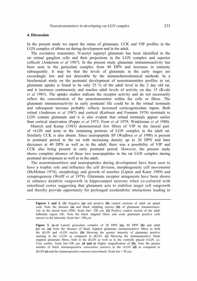
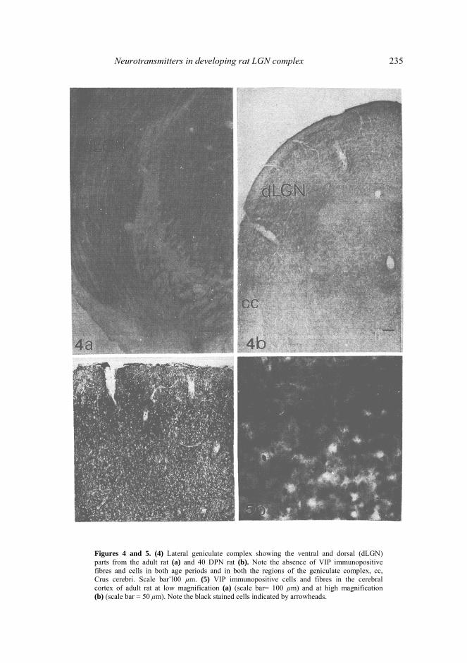
Neurotransmitters in developing rat LGN complex 231 phosphate buffer. Cryostat sections (30–35 μm) of diencephalon with lateral geniculate nuclear complex were cut coronally. The sections were incubated for immunohistochemical staining with polyclonal antibodies against CCK, VIP and monoclonal antibodies against glutamate (Incstar USA) at dilutions of 1:20, 1:20 and 1:100 respectively for a period of 48 h using avidin biotin technique and 3, 3'- diaminobenzidine (DAB) as the chromogen. In the control sections incubation with primary antibody was omitted; cervical spinal cord and habenula known to have glutamate as well as cerebral cortex known to contain VIP and CCK were taken as positive controls from the same and adult animals. Some sections were also treated with nickel II sulphate in 0·1 Μ imidiazole and 0·2 Μ acetate buffer alongwith DAB for intensification of staining. 3. Results 3.1 Glutamate immunoreactivity 3.1a E18, 1, 5, 10 and 20 days: No glutamate immunoreactivity was demonstrable at these age periods (figure 3a). However, the positive control sections of adultspinal cord and habenula processed simultaneously showed immunoreactivity (figures lb, 2). The negative control sections of spinal cord showed no immunoreactivity in the dorsal horn (figure la). 3.1b Forty days and adult: At both these age periods the immunopositive glutamate fibres were seen to be present in the vLGN and dLGN parts of geniculate nucleus as well as in the IGL. The immunopositive cell bodies were also observed. The cytoplasm of these neurons showed the brown precipitate of immunoreaction while the nuclear space was unstained. The immunoreactivity was more intense in the adult as compared to 40-day rats (figure 3b, c) and in the vLGN as compared to dLGN (figure 3d, e). 3.2 VIP No immunoreactivity was seen in the LGN complex at any of the age periods studied (figure 4) although the positive controls from adult cerebral cortex showed VIP immunopositive neurons and their processes in layers 2 and 3 predominantly (figure 5). 3.3 CCK immunoreactivity No CCK immunoreactivity was observed in the LGN complex at any of the ageperiods studied (figure 6a). The positive controls of adult cerebral cortex, however,showed CCK immunopositive neurons and their processes in layers 2 and 3predominantly (figure 7a). The negative control section of LGN and cortex showedno immunoreactivity at all (figures 6b, 7b).

232 Shashi Wadhwa and Geeta Jotwani
Figure 1 and 2.
Neurotransmitters in developing rat LGN complex 233 4. Discussion In the present study we report the status of glutamate, CCK and VIP profiles in the LGN complex of albino rat during development and in the adult.
The excitatory transmitter, N-acetyl aspartyl glutamate has been identified in the rat retinal ganglion cells and their projections in the LGN complex and superior colliculi (Anderson et al 1987). In the present study glutamate immunoreactivity has been seen in the geniculate complex from 40 DPN and increases in intensitysubsequently. It may be that the levels of glutamate in the early stages areexceedingly low and not detectable by the immunohistochemical methods. In abiochemical study on the postnatal development of neurotransmitter profiles in rat, glutamate uptake is found to be only 25 % of the adult level in the 2 day old rat, and it increases continuously and reaches adult levels of activity on day 15 (Kvale et al 1983). The uptake studies indicate the receptor activity and do not necessarilyreflect the concentration of the neurotransmitter within the cells or fibres. Theglutamate immunoreactivity in early postnatal life could be in the retinal terminals and subsequent increase probably reflects increased corticogeniculate inputs. Both retinal (Anderson et al 1987) and cortical (Karlssen and Fonnum 1978) terminals to LGN contain glutamate and it is also evident that retinal terminals appear earlier than cortical innervation (Poppe et al 1973; Frost et al 1979; Winkleman et al 1980).
Mantyh and Kemp (1983) demonstrated few fibres of VIP in the lateral partof vLGN and none in the remaining portions of LGN complex in the adult rat. Similarly CCK is also absent. Since neuropeptide SP (Wadhwa et al 1990) is present in postnatal period in the rat with increasing density up to 20 DPN and later decreases at 40 DPN as well as in the adult, there was a possibility of VIP and CCK also being present in early postnatal period. However, the present study shows complete absence of these two neuropeptides in the rat LGN complex during postnatal development as well as in the adult.
The neurotransmitters and neuropeptides during development have been seen to have a trophic role and influence the cell division, morphogenetic cell movements (McMohan 1974), morphology and growth of neurites (Lipton and Kater 1989) and synaptogenesis (Wolff et al 1979). Glutamate receptor antagonists have been shown to enhance dendrite outgrowth in hippocampal neurons when co-cultured with entorhinal cortex suggesting that glutamate acts to stabilize target cell outgrowth and thereby provide opportunity for prolonged axodendritic interactions leading to
Figures 1 and 2. (1) Negative (a) and positive (b) control sections of adult rat spinal cord. Note the absence (a) and black stippling (arrow) (b) of glutamate immunoreacti- vity in the dorsal horn (DH). Scale bar= 100 μm. (2) Positive control section of the adult habenula region (H). Note the black stippled fibres and some glutamate positive cells (arrow) in the habenula. Scale bar= 100 μm. Figure 3. (a–c) Lateral geniculate complex of 20 DPN (a), 40 DPN (b) and adult (c) rat. (a) Note the absence of black stippled glutamate immunoreactive fibres in both the dLGN and vLGN nuclei, (b) Showing the greater intensity of glutamate positivestaining in the vLGN as compared to dLGN. (c) Showing the immunopositive blackstippled glutamate fibres, both in the dLGN as well as in the ventrally placed vLGN. cc, Crus cerebri. Scale bar=100 μm. (d and e) Higher magnification of (b). Note the greater number of black immunopositive varicosities (arrows) in the vLGN (d) as compared to dLGN (e) and the immunopositive neurons (arrowhead). Scale bar = 50 μm.

234 Shashi Wadhwa and Geeta Jotwani
Figure 3. For caption, see page no. 233.
Neurotransmitters in developing rat LGN complex 235
Figures 4 and 5. (4) Lateral geniculate complex showing the ventral and dorsal (dLGN)parts from the adult rat (a) and 40 DPN rat (b). Note the absence of VIP immunopositive fibres and cells in both age periods and in both the regions of the geniculate complex, cc, Crus cerebri. Scale bar=l00 µm. (5) VIP immunopositive cells and fibres in the cerebral cortex of adult rat at low magnification (a) (scale bar= 100 µm) and at high magnification (b) (scale bar = 50 μm). Note the black stained cells indicated by arrowheads.

236 Shashi Wadhwa and Geeta Jotwani
Figures 6 and 7. (6) Adult rat LGN complex showing the absence of staining for CCK in the stained (a) and negative control section (b). Scale bar=100 µm. (7) Adult rat cerebral cortex showing the presence of CCK immunopositive neuron (black) and fibres in the stained section (a) and absence of staining in the negative control section of the cortex (b). Scale bar = 50 µm.
synaptic differentiation (Mattson et al 1988). It is also known that exposure of developing tadpole to NMDA receptor antagonists prevents normal segregation of retinotectal synapses (Cline and Constantine-Paton 1989) and addition of

Neurotransmitters in developing rat LGN complex 237 exogenous NMDA to the tadpoles sharpens the retinotectal map (Cline and Constantine-Paton 1990). In the rat LGN, the level of glutamate before eye opening (14 DPN), when segregation of retinal inputs occurs (Land and Lund 1979; Jeffery 1984), is probably inactivated or balanced by GABA. GABA is shown to increase continuously from birth to 24 DPN and decrease thereafter to adult levels in the rat LGN (Kvale et al 1983). This seems to be corroborated by the reduction seen in dendritic outgrowth inhibition and neurotoxic actions of glutamate when GABA receptors are simultaneously activated in the hippocampal-entorhinal co-cultures (Mattson and Kater 1989).
It is evident from the present study and our earlier work (Wadhwa et al 1990) that SP, SER, Leu-ENK and glutamate appear sequentially in the rat LGN. For the neurotransmitters to play roles in neural development, it is essential that both neurotransmitters and their receptors are properly paired in time and space. Whiledata pertaining to the neurotransmitter development in rat LGN is substantiallyincreasing, our knowledge regarding receptor expression is still quite limited. Increasing evidence is being provided for spatial and temporal integration of neurotransmitter signals in the development of neural circuitry. However, there is also strong evidence that different receptors and transmitters are not expressed simultaneously and may be expressed transiently and their unique presence very likely plays a crucial role in functional neuronal circuits (Mattson and Hauser 1991). Acknowledgement This work was financially supported by the Indian Council of Medical Research, New Delhi. References
Anderson K, Borja Μ A, Cotman C W, Moffett J R, Namboodiri Μ A A and Neale J Η 1987 N-Acetyl
aspartyl glutamate identified in the rat ganglion cells and their projections in the brain; Brain Res. 411 172–177
Brecha N, Johnson D, Bolz J, Sharma S and Parnavelas J G 1987 Substance P-immunoreactive retinal ganglion cells and their central axon terminals in the rabbit; Nature (London) 327 155–158
Canzek V, Wolfensberger M, Amstar V and Cuenod Μ 1981 In vivo release of glutamate and aspartate following optic nerve stimulation; Nature (London) 293 572–574
Cline Η Τ and Constantine-Paton Μ 1989 NMDA receptor antagonists disrupt the retinotectal topographic map; Neuron 3 413–426
Cline Η Τ and Constantine-Paton Μ 1990 NMDA receptor agonists and antagonists alter retinal ganglion cell arbor structure in the developing frog retinotectal projection; J. Neurosci. 10 1197–1216
De Lima A D and Singer W Τ I 1987 The serotonergic fibres in the dorsal lateral geniculate nucleus of the cat: Distribution and synaptic connections demonstrated with immunocytochemistry; J. Comp. Neurol. 258 339–358
Duner Η, Von Euler V S and Pernow Β 1954 Catecholamines and substance Ρ in the mammalian eye; Acta Physiol. Scand. 31 113–118
Frost D O, So K-F and Schneider G Ε 1979 Postnatal development of retinal projections in Syrian hamsters: a study using autoradiographic and anterograde degeneration techniques; Neuroscience 4 1649–1677
Hoover D Β and Jacobwitz D Μ 1979 Neurochemical and histochemical studies of the effect of a lesion in the nucleus cuneiformis on the cholinergic innervation of discrete areas of the rat brain; Brain Res.170 113–122

238 Shashi Wadhwa and Geeta Jotwani Jeffery G 1984 Retinal ganglion cell death and terminal field retraction in the developing rodent visual
system; Dev. Brain Res. 13 81–96 Karlssen L R and Fonnum F 1978 Evidence for glutamate as a neurotransmitter in the corticofugal
fibres to the dorsal lateral geniculate body and the superior colliculus in rats; Brain Res. 151 457–467 Kromer L F and Moore R Υ 1980 A study of the organisation of the locus coeruleus projections to the
lateral geniculate nuclei in the albino rat; Neuroscience 5 255–271 Kvale I, Fosse V Μ and Fonnum F 1983 Development of neurotransmitter parameters in lateral
geniculate body, superior colliculus and visual cortex of the albino rat; Dev. Brain Res. 7 137–145 Land Ρ W and Lund R D 1979 Development of rat's uncrossed retinotectal pathway and it's relation to
plasticity studies; Science 205 698–700 Lipton S A and Kater S Β 1989 Neurotransmitter regulation of neuronal outgrowth, plasticity and
survival; Trends. Neurosci. 12 265–270 Mantyh Ρ W and Kemp J A 1983 The distribution of putative neurotransmitters in the lateral geniculate
nucleus of the rat; Brain Res. 288 344–348 Mattson Μ Ρ and Hauser Κ F 1991 Spatial and temporal integration of neurotransmitter signals in the
development of neural circuitry: a critique; Neurochem. Int. 19 17–24 Mattson Μ Ρ, Lee R Ε, Adams Μ Ε, Guthrie Ρ Β and Kater S Ρ 1988 interactions between entorhinal
axons and target hippocampal neurons: a role for glutamate in the development of hippocampal circuitry; Neuron 1 865–867
Mattson Μ Ρ and Kater S Β 1989 Excitatory and inhibitory neurotransmitters in the generation and degeneration of hippocampal neuroarchiterture; Brain Res. 478 337–348
McMohan A 1974 Chemical messengers in development; Science 185 1012–1021 Mesulam Μ Μ, Mufson Ε J, Wainer Β Η and Levey A I 1983 Central cholinergic pathways in the rat:
An overview based on an alternative nomenclature (Ch I-Ch 6); Neuroscience 10 1185–1201 Montero V Μ and Guillery R W 1968 Degeneration in the dorsal lateral geniculate nucleus of the rat
following interruption of the retinal or cortical connections; J. Comp. Neurol. 134 211–242 Ohara Ρ Τ, Sefton A J and Leiberman A R 1980 Mode of termination of afferents from the thalamic
reticular nucleus in the dorsal lateral geniculate nucleus of the rat; Brain Res. 197 503–506 Pasantes-Morales H, Salceda R and Lopez-Colomba Α Μ 1975 The role of taurine in retina : factors
affecting its release; in Taurine (eds) R Huxtable and A Barbeau (New York: Raven Press) pp 191–200 Pasquier D A and Villar Μ J 1982 Specific serotonergic projections to the lateral geniculate body from
the lateral cell groups of the dorsal raphe nucleus; Brain Res. 249 142–146 Poppe Η, Bruckner G and Beisold D 1973 Postnatale entwicklung der synaptischen Kontaktzonen in
dorsalen Corpus geniculatum laterale der Ratte; Ζ. Microsk. Anat. Forsch. 87 457–464 Reese B E 1984 The projection from the superior colliculus to the dorsal lateral geniculate nucleus in the
rat; Brain Res. 305 162–168 Wadhwa S, Rath S, Jotwani G and Bijlani V 1990 Development of substance P, Leu-enkephalin and
serotonin profiles in the lateral geniculate nuclear complex of albino rat; Neurosci. Lett. 120 146–150 Winkleman E, Brauer Κ and Werner L 1980 Postnatal development of afferents in the rat LGB; in
Ontogenesis of the brain (eds) S Trigan and F Stastny (Praha CSSR: Universita Kariova) Vol. 3, pp 83–89
Wolff J R, Joo F, Dames W and Feher Ο 1979 Induction and maintenance of free postsynaptic membrane thickenings in adult superior cervical ganglion; J. Neurocytol 8 549–563