immunofluorescence guide · 2019-11-27 · 2 introduction 2 immunofluorescence workflow 4...
TRANSCRIPT
2 Introduction 2 Immunofluorescence Workflow 4 Immunofluorescence Selection Guide 6 Pioneering in IHC/IF Technology
8 Choosing a Detection System 10 Considerations for IF Detection
12 Fluorophore-Conjugated Secondary Antibodies
14 VectaFluor™ Ready-To-Use Antibody Reagents –VectaFluor™R.T.U.AntibodyKits –VectaFluor™DuetImmunofluorescenceDoubleLabelingKits –VectaFluor™ExcelAmplifiedStainingSystem
18 Fluorophore-Conjugated Streptavidin/Avidin Reagents 19 Anti-Streptavidin and Anti-Avidin Antibody Reagents
20 Secondary and Teritiary Detection Reagents1
–BiotinylatedandUnconjugatedSecondaryAntibodies –Enzyme-ConjugatedSecondaryAntibodies –AvidinandStreptavidinEnzymeConjugates
22 Species on Species Detection (Mouse) 23 Mouse on Mouse (M.O.M.®) Immunodetection Kits
24 Mounting Media 25 VECTASHIELD® Antifade Mounting Media –VECTASHIELD®AntifadeMountingMedium –VECTASHIELD®HardSet™AntifadeMountingMedium –VECTASHIELD®MountingMediaandFluorophoreCompatibility –VECTASHIELD®MountingMediaFormatsandApplications
28 VectaCell™ Products for Live Cell Imaging –VectaCell™TroloxAntifadeReagent –LiveCellImagingofOrganelles
29 Accessory Reagents –VECTABOND®ReagentTissueSectionAdhesive –ImmEdge™HydrophobicBarrierPen –ImmPrint™HistologyPen –ControlAntibodies –AntigenUnmaskingSolutions
30 Blocking Background Signal –TrueVIEW™AutofluorescenceQuenchingKit –BLOXALL®EndogenousHRP/APBlockingSolution –Avidin/BiotinandStreptavidin/BiotinBlockingKits –NormalSera –Animal-FreeBlockingSolutions –GeneralProteinBlockingReagents
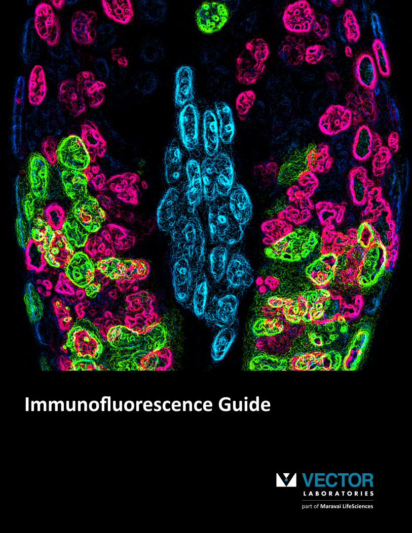
Front cover: Fluorescent images with neon effect showing successive proliferation within the bulb of a hair follicle. Proliferating cells labeled for CldU (red), IdU (green) with cells dividing twice taking up both labels (yellow). Epidermal nuclei (blue) and dermal papilla nuclei (cyan) labeled with DAPI. Image provided by Nigel Hammond (Dixon Lab). Faculty of Biology, Medicine and Health, University of Manchester, Manchester, UK.
VectorLaboratorieswasfoundedonagrowingportfolioofpurifiedlectinsandlectinconjugatesthathelpedtopioneerlectinhistochemistry.Theseproductsremainakeycomponentofourbusinesstoday.Intheearly1980s,weleveragedourexpertiseinhistochemistrytorevolutionizethefieldofIHCwiththecommercializationofantibody-basedavidin-biotinreagentsandtheintroductionoftheVECTASTAIN®ABCsystem.ThissystemenabledroutinelaboratoryuseofIHCwithanystandardbrightfieldmicroscope.FollowingthesuccessoftheABCkits,VectorLaboratorieshascontinuedtointroducemanynovelandinnovativeproductstosupportresearchendeavorsforcellandtissueantigenvisualization.TheseincludetheImmPRESS™micropolymerreagents,MouseonMouse(M.O.M.®)detectionsystems,uniqueImmPACT™enzymesubstrates,andVECTASHIELD®AntifadeMountingMediaforfluorescenceapplications.
Table of Contents
Helpingyoureachnewvisualizationfrontiersinyourresearch: thisisourmission.Sinceourfoundingin1976,aprimarydrivingprinciplehasbeentodevelopandmanufacturelabelinganddetectiontechnologiesthatmakeIFas easy as ABC.
Reliableandreproduciblereagentsthatinstilltrust andconfidence.
Simpleandrobustproductdesignsthatstreamlineworkflowsandallowelucidationofcomplexbiologicalsystems.
Aknowledgebaseofover100yearsofcombinedIFexperiencetohelpyouacceleratethepaceofdiscovery.
It’s as simple as that.
Making IF as easy as ABC
A.B.C.
Dorsal root ganglia cells (neurons and satelite glia): Beta III tubulin (ms), DyLight® 488 Anti-Mouse IgG, • S100 (rb), DyLight® 594 Anti-Rabbit IgG. Mounted in VECTASHIELD® HardSet™ Antifade Mounting Medium with DAPI. Image courtesy of Dr. Emma East, Department of Life Sciences, The Open University, Milton Keynes, UK.
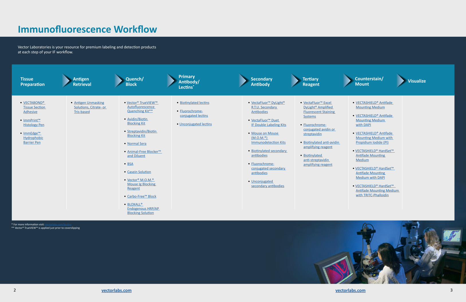
Immunofluorescence WorkflowVectorLaboratoriesisyourresourceforpremiumlabelinganddetectionproducts ateachstepofyourIFworkflow.
2 3vectorlabs.comvectorlabs.com
AntigenRetrieval
Tertiary Reagent
Secondary Antibody
Primary Antibody/Lectins*
Quench/Block
Counterstain/Mount
• AntigenUnmasking Solutions,Citrate-orTris-based
• VectaFluor™ Excel DyLight®Amplified FluorescentStaining Systems
• Fluorochrome- conjugatedavidinorstreptavidin
• Biotinylatedanti-avidinamplifyingreagent
• Biotinylated anti-streptavidin amplifyingreagent
• VectaFluor™DyLight® R.T.U.Secondary Antibodies
• VectaFluor™ Duet IFDoubleLabelingKits
• MouseonMouse(M.O.M.®) ImmunodetectionKits
• Biotinylatedsecondaryantibodies
• Fluorochrome- conjugatedsecondaryantibodies
• Unconjugated secondaryantibodies
• Biotinylatedlectins
• Fluorochrome- conjugatedlectins
• Unconjugatedlectins
• Vector®TrueVIEW™AutofluorescenceQuenchingKit**
• Avidin/Biotin BlockingKit
• Streptavidin/Biotin BlockingKit
• NormalSera
• Animal-FreeBlocker™andDiluent
• BSA
• CaseinSolution
• Vector®M.O.M.® MouseIgBlocking Reagent
• Carbo-Free™Block
• BLOXALL® EndogenousHRP/APBlockingSolution
• VECTASHIELD®AntifadeMountingMedium
• VECTASHIELD®AntifadeMountingMedium withDAPI
• VECTASHIELD®AntifadeMountingMediumwithPropidiumIodide(PI)
• VECTASHIELD®HardSet™AntifadeMounting Medium
• VECTASHIELD®HardSet™AntifadeMounting MediumwithDAPI
• VECTASHIELD®HardSet™AntifadeMountingMediumwithTRITC-Phalloidin
• VECTABOND® TissueSection Adhesive
• ImmPrint™ HistologyPen
• ImmEdge™ Hydrophobic BarrierPen
*Formoreinformationvisit:vectorlabs.com/lectins**Vector®TrueVIEW™isappliedjustpriortocoverslipping
VisualizeTissue Preparation

Followthesimplestepsbelowtochoosethemostappropriatelabeling anddetectionsolutionforyourexperiment.
Choose Signal Amplification with Biotin-based Systems (Step 2, Option C) •Multipleroundsofamplificationpossible (withbiotin-basedsystems)
Choose Mounting Media with or without a Counterstain •VECTASHIELD®AntifadeMountingMedia, withorwithoutcounterstain
Visualize •Fluorescencemicroscope•Viewusingappropriate excitation/emissionfilters
Primaryantibody
Fluorophore- conjugated secondaryantibody
Amplifierantibody
Biotinylatedanti-avidin/streptavidin
Fluorophore
Biotin
Legend
Fluorophore-conjugated avidin/ streptavidin
Choose Primary Antibody•Specificforantigenofinterest•Considertissuespeciesand preparation(fixation)
•Considerantigenretrieval requirements
Blocking Reagents•Choicesdeterminedbytheoptions selectedinSteps1-2
•Streptavidin/BiotinBlockingKit (ifusingbiotin/streptavidinsystem)
•Avidin/BiotinBlockingKit•NormalSera(fromthespeciesof secondaryantibody)
•M.O.M.®MouseIgBlockingReagent•R.T.U.Animal-FreeBlocker™andDiluent•BSA•CaseinSolution
Choose Secondary Antibody and Tertiary Detection System• Choosefluorophorebasedon wavelengthsavailableinmicroscope
• Fluorophore-conjugated secondaryantibodyor biotinylatedsecondaryantibody
• Considersensitivityrequirements• Considerspeciesofprimaryantibody• Considertissuespecies
Two Step Highestsensitivity.Non-biotinbased.•VectaFluor™ExcelAmplifiedFluorescent StainingSystem(AmplifierAntibody +fluorescenttertiaryantibody)
Two Step Biotin-based.• Biotinylatedsecondaryantibody andfluorophoreconjugatedavidin orstreptavidin.
•SeeStep3foradditionalamplification
One Step Single-label. Fast.Convenient.•Fluorophore-conjugatedsecondary antibodies
•VectaFluor™R.T.U.DyLight®Labeled SecondaryAntibodies
VECTABOND® Reagent (TissueSectionAdhesive)
SECONDARYDETECTIONSYSTEMS
1
2
3
5
6
or
or
Immunofluorescence Selection Guide
Option A
Option C
Option D
or
vectorlabs.comvectorlabs.com
Option B
Prostate: Anti-PSA (goat), VectaFluor™ DyLight® 488 Anti-Goat IgG. Mounted in VECTASHIELD® HardSet™ Mounting Medium.
4 5
One Step Dual-labeltwo-antigendetection. Fast.Convenient.•VectaFluor™DuetIFDoubleLabelingKits
Reduce Autofluorescence from Aldehyde Fixation •Vector®TrueVIEW™Autofluorescence QuenchingKit
4
Vector®TrueVIEW™ QuenchingAction
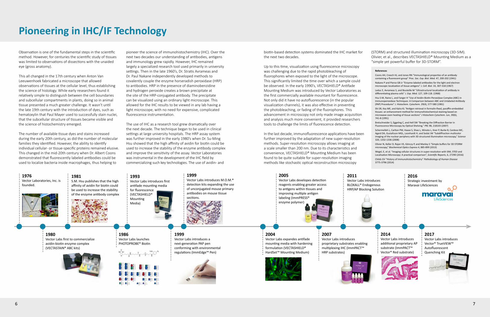
Pioneering in IHC/IF Technology
Observation is one of the fundamental steps in the scientific method. However, for centuries the scientific study of tissues was limited to observations of dissections with the unaided eye (gross anatomy).
This all changed in the 17th century when Anton Van Leeuwenhoek fabricated a microscope that allowed observations of tissues at the cellular level, thus establishing the science of histology. While early researchers found it relatively simple to distinguish between the cell boundaries and subcellular compartments in plants, doing so in animal tissue presented a much greater challenge. It wasn’t until the late 19th century with the introduction of dyes, such as hematoxylin that Paul Mayer used to successfully stain nuclei, that the subcellular structure of tissues became visible and the science of histochemistry emerged.
The number of available tissue dyes and stains increased during the early 20th century, as did the number of molecular families they identified. However, the ability to identify individual cellular- or tissue-specific proteins remained elusive. This changed in the mid-20th century when Dr. Albert Coons demonstrated that fluorescently labeled antibodies could be used to localize bacteria inside macrophages, thus helping to
1980VectorLabsfirsttocommercializeavidin-biotinenzymecomplex(VECTASTAIN®ABCkits)
1986VectorLabslaunchesPHOTOPROBE®Biotin
1993VectorLabsintroducesfirstantifademountingmedia forfluorescence (VECTASHIELD® Mounting Media)
1999VectorLabsintroducesanext-generationPAPpenconformingwithenvironmentalregulations(ImmEdge™Pen)
2007VectorLabsintroducesproprietarysubstratesenablingmultiplexingIHC(ImmPACT™HRPsubstrates)
2011VectorLabsintroducesBLOXALL®Endogenous HRP/APBlockingSolution
2014VectorLabsintroduces additionalproprietaryAP substrate(ImmPACT™Vector®Redsubstrate)
1981S.M.Hsupublishesthatthehighaffinityofavidinforbiotincouldbeusedtoincreasethestability oftheenzymeantibodycomplex
pioneer the science of immunohistochemistry (IHC). Over the next two decades our understanding of antibodies, antigens and immunology grew rapidly. However, IHC remained largely a specialized research tool used primarily in university settings. Then in the late 1960’s, Dr. Stratis Avrameas and Dr. Paul Nakane independently developed methods to covalently couple the enzyme horseradish peroxidase (HRP) to antibodies. HRP in the presence of diaminobenzidine and hydrogen peroxide creates a brown precipitate at the site of the HRP-conjugated antibody. The precipitate can be visualized using an ordinary light microscope. This allowed for the IHC results to be viewed in any lab having a light microscope, with no need for expensive, complicated fluorescence instrumentation.
The use of IHC as a research tool grew dramatically over the next decade. The technique began to be used in clinical settings at large university hospitals. The HRP assay system was further improved in the early 1980’s when Dr. Su-Ming Hsu showed that the high affinity of avidin for biotin could be used to increase the stability of the enzyme antibody complex and improve the sensitivity of the assay. Vector Laboratories was instrumental in the development of the IHC field by commercializing such key technologies. The use of avidin- and
biotin-based detection systems dominated the IHC market for the next two decades.
Up to this time, visualization using fluorescence microscopy was challenging due to the rapid photobleaching of fluorophores when exposed to the light of the microscope. This significantly limited the time over which a sample could be observed. In the early 1990’s, VECTASHIELD® Antifade Mounting Medium was introduced by Vector Laboratories as the first commercially available mountant for fluorescence. Not only did it have no autofluorescence (in the popular visualization channels), it was also effective in preventing the photobleaching, or fading of the fluorophores. This advancement in microscopy not only made image acquisition and analysis much more convenient, it provided researchers tools to challenge the limits of fluorescence detection.
In the last decade, immunofluorescence applications have been further improved by the adaptation of new super-resolution methods. Super-resolution microscopy allows imaging at a scale smaller than 200 nm. Due to its characteristics and convenience, VECTASHIELD® Mounting Medium has been found to be quite suitable for super-resolution imaging methods like stochastic optical reconstruction microscopy
References
CoonsAH,CreechHJ,andJonesRN“Immunologicalpropertiesofanantibodycontainingafluorescentgroup”Proc. Soc. Exp. Biol. Med.47,200-202(1941)
NakanePandPierceGBJr“Enzyme-labeledantibodiesforthelightandelectronmicroscopiclocalizationoftissueantigens” J. Cell. Biol.33,307-318(1967)
LeducE,AvrameasS,andBouteilleM“Ultrastructurallocalizationofantibodyindifferentiatingplasmacells”J. Exp. Med.127,109-118.(1968)
HsuS-M,RaineL,andFangerH“UseofAvidin-Biotin-PeroxidaseComplex(ABC)inImmunoperoxidaseTechniques:AComparisonbetweenABCandUnlabeledAntibody(PAP)Procedures”J. Histochem. Cytochem. 29(4),577-580(1981)
ShiSR,KeyME,andKalraKL“Antigenretrievalinformalin-fixed,paraffin-embeddedtissues:anenhancementmethodforimmunohistochemicalstainingbasedonmicrowaveovenheatingoftissuesections”J Histochem Cytochem.Jun,39(6), 741-8(1991)
BretschneiderS,EggelingC,andHellSW“BreakingtheDiffractionBarrierinFluorescenceMicroscopybyOpticalShelving,”PRL98,218103(2007)
SchermellehL,CarltonPM,HaaseS,ShaoL,WinotoL,KnerP,BurkeB,CardosoMC,AgardDA,GustafssonMGL,LeonhardtH,andSedatJW“Subdiffractionmulticolorimagingofthenuclearperipherywith3Dstructuredilluminationmicroscopy,”Science 320,1332-1336(2008)
OlivierN,KellerD,RajanVS,GönczyP,andManleyS“Simplebuffersfor3DSTORMmicroscopy,”Biochemical Optics Express4,885-899(2013)
Wegel,E,etal.“Imagingcellularstructuresinsuper-resolutionwithSIM,STEDandLocalisationMicroscopy:Apracticalcomparison”,Scientific Reports,6,27290(2016)
ChildsGV“HistoryofImmunohistochemistry”Pathobiology of Human Disease 3775-3796(2014)
(STORM) and structured illumination microscopy (3D-SIM). Olivier, et al., describes VECTASHIELD® Mounting Medium as a “simple yet powerful buffer for 3D-STORM”.
6 7
2004VectorLabsexpandesantifademountingmediawithhardeningformulation(VECTASHIELD® HardSet™MountingMedium)
2005VectorLabsdevelopesdetectionreagentsenablinggreateraccesstoantigenswithintissuesand improvingmultipleantigenlabeling(ImmPRESS® enzymepolymer)
2016StrategicinvestmentbyMaravaiLifeSciences
1999 VectorLabsintroducesM.O.M.®detectionkitsexpandingtheuseofunconjugatedmouseprimaryantibodiesonmousetissuesections.
1976VectorLaboratories,Inc.isfounded.
2017VectorLabsintroduces Vector®TrueVIEW™AutofluorescentQuenchingKit
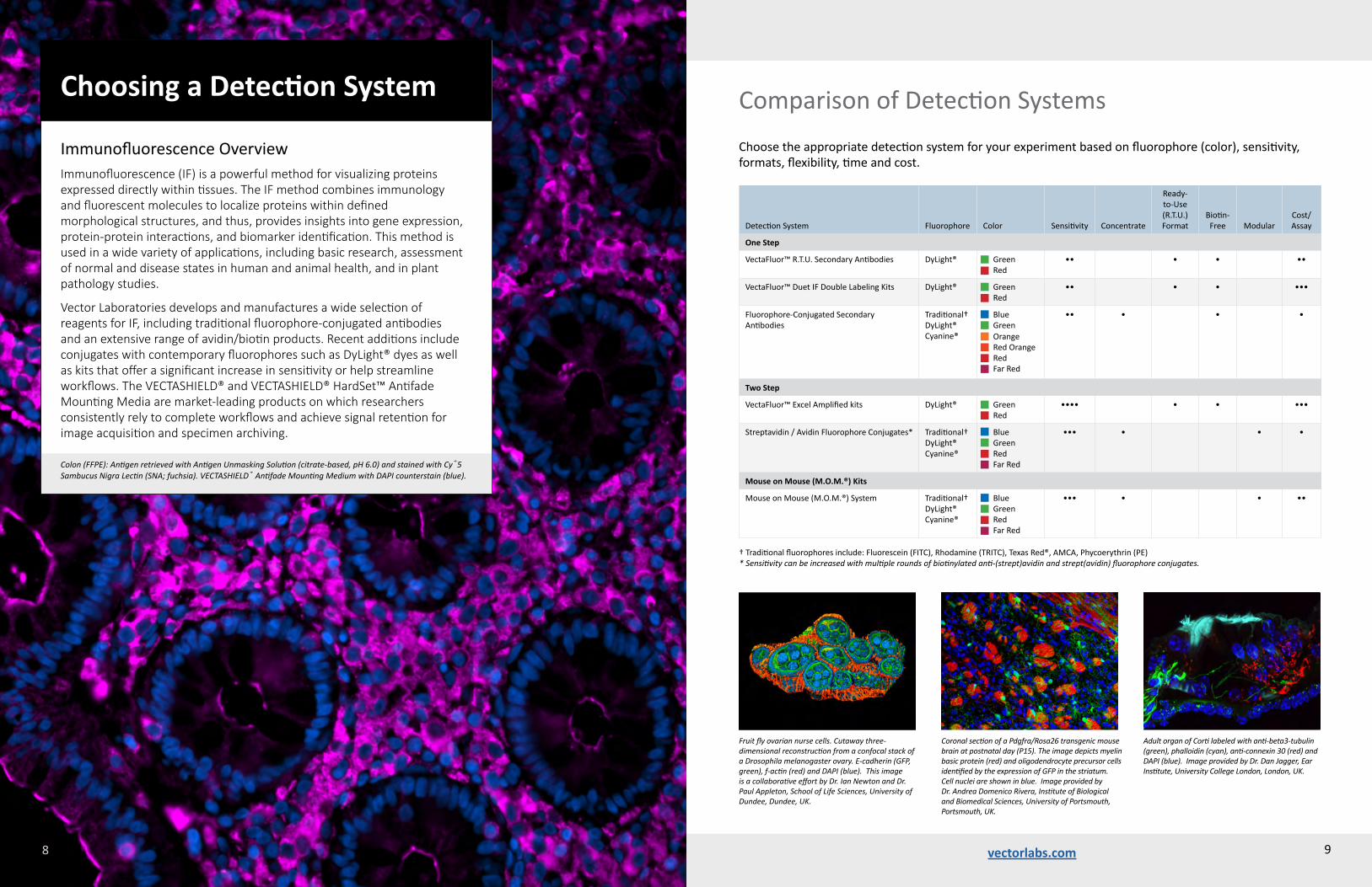
Colon (FFPE): Antigen retrieved with Antigen Unmasking Solution (citrate-based, pH 6.0) and stained with Cy®5 Sambucus Nigra Lectin (SNA; fuchsia). VECTASHIELD® Antifade Mounting Medium with DAPI counterstain (blue).
ImmunofluorescenceOverviewImmunofluorescence (IF) is a powerful method for visualizing proteins expressed directly within tissues. The IF method combines immunology and fluorescent molecules to localize proteins within defined morphological structures, and thus, provides insights into gene expression, protein-protein interactions, and biomarker identification. This method is used in a wide variety of applications, including basic research, assessment of normal and disease states in human and animal health, and in plant pathology studies.
Vector Laboratories develops and manufactures a wide selection of reagents for IF, including traditional fluorophore-conjugated antibodies and an extensive range of avidin/biotin products. Recent additions include conjugates with contemporary fluorophores such as DyLight® dyes as well as kits that offer a significant increase in sensitivity or help streamline workflows. The VECTASHIELD® and VECTASHIELD® HardSet™ Antifade Mounting Media are market-leading products on which researchers consistently rely to complete workflows and achieve signal retention for image acquisition and specimen archiving.
Choosing a Detection System
DetectionSystem Fluorophore Color Sensitivity Concentrate
Ready-to-Use(R.T.U.) Format
Biotin-Free Modular
Cost/Assay
One Step
VectaFluor™R.T.U.SecondaryAntibodies DyLight® GreenRed
•• • • ••
VectaFluor™DuetIFDoubleLabelingKits DyLight® GreenRed
•• • • •••
Fluorophore-ConjugatedSecondaryAntibodies
Traditional†DyLight®Cyanine®
BlueGreenOrange RedOrangeRedFarRed
•• • • •
Two Step
VectaFluor™ExcelAmplifiedkits DyLight® GreenRed
•••• • • •••
Streptavidin/AvidinFluorophoreConjugates* Traditional†DyLight®Cyanine®
BlueGreenRedFarRed
••• • • •
Mouse on Mouse (M.O.M.®) Kits
MouseonMouse(M.O.M.®)System Traditional†DyLight®Cyanine®
BlueGreenRedFarRed
••• • • ••
†Traditionalfluorophoresinclude:Fluorescein(FITC),Rhodamine(TRITC),TexasRed®,AMCA,Phycoerythrin(PE)* Sensitivity can be increased with multiple rounds of biotinylated anti-(strept)avidin and strept(avidin) fluorophore conjugates.
ComparisonofDetectionSystemsChoosetheappropriatedetectionsystemforyourexperimentbasedonfluorophore(color),sensitivity,formats,flexibility,timeandcost.
vectorlabs.com 98
Coronal section of a Pdgfra/Rosa26 transgenic mouse brain at postnatal day (P15). The image depicts myelin basic protein (red) and oligodendrocyte precursor cells identified by the expression of GFP in the striatum. Cell nuclei are shown in blue. Image provided by Dr. Andrea Domenico Rivera, Institute of Biological and Biomedical Sciences, University of Portsmouth, Portsmouth, UK.
Adult organ of Corti labeled with anti-beta3-tubulin (green), phalloidin (cyan), anti-connexin 30 (red) and DAPI (blue). Image provided by Dr. Dan Jagger, Ear Institute, University College London, London, UK.
Fruit fly ovarian nurse cells. Cutaway three-dimensional reconstruction from a confocal stack of a Drosophila melanogaster ovary. E-cadherin (GFP, green), f-actin (red) and DAPI (blue). This image is a collaborative effort by Dr. Ian Newton and Dr. Paul Appleton, School of Life Sciences, University of Dundee, Dundee, UK.

vectorlabs.comvectorlabs.com
Considerations for IF DetectionImmunofluorescence detection reagents are used to localize and visualize target antigens expressed in tissue sections or cultured cells. When applied optimally, these highly specific reagents provide a defined contrast between their fluorescence, which demarcates the antigen, and the non-fluorescent region of the preparation. There are several options to achieve labeling for single and multiple antigen detection.
Direct Detection One common IF method uses fluorophore-conjugated primary antibodies. This direct approach enables fast and easy IF visualization once the antibody has been conjugated; however, there are some disadvantages to this traditional method. For example, binding affinity and avidity could be compromised by the conjugation process, which would reduce signal and prevent moderately or weakly expressed antigens from being detected. Furthermore, expensive primary antibodies used at high concentrations could be cost prohibitive, and the visualization options would be limited to only one fluorophore.
Indirect Detection (One Step)The indirect method, which uses labeled secondary antibodies, produces reliable, reproducible and economical IF results. This method avoids the disadvantages of directly conjugated primary antibodies and provides signal amplification that is necessary for most cell- and tissue-section labeling. Additionally, this one-step detection method is modular and allows simple substitution of the secondary with different fluorophore conjugates. Please refer to Table 2, page 13 for our range of concentrated reagents. Fluorophore-conjugated secondary antibodies would be recommended where a moderate to high expression of target antigen is expected.
Indirect Detection (Two Step) Further signal amplification is introduced by using biotinylated secondary antibodies with avidin or streptavidin fluorophore conjugates. This well established and widely published methodology exploits the very high affinity between avidin or streptavidin and the small vitamin biotin. This two-step detection method enables the detection of weakly expressed antigens and provides a flexible and modular system with easy fluorophore substitution using different avidin or streptavidin conjugates (see pages 18-20). Additional amplification can be achieved by using biotinylated anti-avidin/streptavidin. For applications where use of biotin-based reagents for signal amplification would be problematic, we offer a non-biotin based two-step fluorescence approach with our VectaFluor™ Excel Amplified DyLight® Antibody kits (see page 16).
1110
Staining of human breast cancer colony-forming culture for basal (Cytokeratin 14, green) and luminal markers (Cytokeratin 18, red). Image provided by Wendy Greenwood, method by Dr. Michael Prater, The Cancer Research UK Cambridge Institute, Cambridge, UK.
Hypoxia within hyperplastic breast tissue. A section of human breast tissue labeled for CAIX using immunofluorescence, counterstained and mounted using VECTASHIELD® HardSet™ Antifade Mounting Medium with DAPI. Image provided by Dr. Carl Daly and Dr. Sarah Dean, Healthcare Science, University of the West of England, Bristol, UK.
Fluorophore Color Excitation Max (nm) Emission Max (nm)
AMCA Blue 350 450
DyLight®488 Green 493 518
Fluorescein Green 495 515
Cy®3 Orange 550 570
Rhodamine Orange 550 575
DyLight®549 Orange 562 576
Phycoerythrin Red-Orange 565 574
DyLight®594 Red 593 618
TexasRed® Red 595 615
Cy®5 FarRed 649 670
DyLight®649 FarRed 652 672
Table 1. Excitation and emission wavelengths and visual colors for immunofluorescence fluorophores.
Species Cross-ReactivityBeyond the choices provided in the Selection Guide (pages 4-5), consideration should be given to the species of the tissue under examination and the species of the primary antibody. In cases of closely related species, it is recommended to use a secondary antibody that has been specifically adsorbed to remove cross-reacting antibodies. In instances where a mouse primary antibody is being applied to mouse tissue sections, it is recommended to use the M.O.M.® Immunodetection System (see pages 22-23).
Multiple Antigen Labeling The visualization of two or more antigens on the same tissue section requires careful planning and specific reagent selection to generate unequivocal and reproducible staining results. We have recently introduced our VectaFluor™ Duet IF Double Labeling Kits that provide convenience and a straightforward approach to this often difficult and time-consuming application (see page 15).
Choosing fluorophoresImmunofluorescence detection reagents are labeled with fluorophores that absorb (excitation) and emit (emission) light at specific wavelengths. Fluorophores suitable for immunofluorescence are available across the complete visible light spectrum. The light source and filter cubes in a particular microscope must match the excitation and emission requirements of the specific fluorophore to achieve the optimal signal-to-noise ratios. For example, the absorption and emission peak wavelengths of fluorescein are 495 nm and 515 nm, respectively. Therefore, an excitation light source that is near 495 nm will yield the greatest emission signal. An emission filter that spans 515 nm will capture the emitted signal. These wavelengths are fixed properties of the fluorophores and the filter, and when properly paired, the system will yield the strongest signal and lowest background.
Germinal center (GC) reaction in the spleen after acute viral infection. After recognition of viral anti-gens, T cells (blue) migrate from the T cell zone into the follicle where they interact with B cells (purple). The T cells ‘help’ B cells, instructing the formation of GCs (green) in which virus-specific B cells undergo selection, class switching and somatic hypermutation to secrete anti-viral antibodies to clear the infection. This work was conducted by Miriam Eckstein and Dr. Martin Vaeth, Department of Pathology, New York University, NY, USA.
ChoosingaDetectionSystem

vectorlabs.comvectorlabs.com
All antibodies available from Vector Laboratories for immunological applications are prepared using optimized, proprietary immunization schedules that produce high-quality antibodies. The antibodies are affinity-purified, and solid-phase adsorption techniques are used to remove cross-reactivities that are likely to interfere with specific detection. The conjugated antibodies have the optimal degree of labeling to maximize signal output without compromising antibody specificity or affinity.
We offer researchers a range of traditional and contemporary conjugated fluorophores, including fluorescein, rhodamine, Texas Red®, AMCA and phycoerythrin. DyLight® dyes offer greater photostability, pH independence and brighter fluorescence than conventional fluorophores. DyLight® dye-conjugated antibodies are ideal for cell- and tissue-based immunofluorescence and a variety of other applications. The DyLight® dye conjugates are stable at pH 4-9 and compatible with many buffers and diluents.
The Cy®3 and Cy®5 Dyes offer bright and stable fluorescence and are used in a variety of applications. Cy®3 dye is bright orange with an excitation/emission of 550 nm/570 nm. Cy®5 is a far-red dye with an excitation/emission of 649 nm/670 nm. Cy®5 is often used as an additional label in multiplexing protocols or in super resolution imaging because of its photo switchable properties.
Fluorophore-Conjugated Secondary Antibodies
Fluorophore-Conjugated Secondary (target species) Antibodies
RabbitIgG
MouseIgG
MouseIgM
HumanIgG
RatIgG
GoatIgG
SheepIgG
Astrocytes: Stained for GFAP and detected with DyLight® 594-conjugated secondary antibody. Mounted in VECTASHIELD® HardSet™ Mounting Medium with DAPI. Image courtesy of Dr. Emma East, Department of Life Sciences, The Open University, Milton Keynes, UK.
Product AMCA Fluorescein TexasRed®
Kits:AMCA,Fluorescein,TexasRed®
DyLight®488
DyLight®549
DyLight®594
DyLight®649 R-Phycoerythrin Cy®3 Cy®5
Anti-MouseIgG(H+L),madeinhorse CI-2000 FI-2000 TI-2000 FI-2100 DI-2488 DI-2549 DI-2594 DI-2649 EI-2007 CY-2300 CY-2500
Anti-MouseIgG(H+L),rat-adsorbed,madeinhorse FI-2001
Anti-MouseIgM,mu-chainspecific,madeingoat FI-2020
Anti-RabbitIgG(H+L),madeinhorse DI-1088 DI-1094
Anti-RabbitIgG(H+L),madeingoat CI-1000 FI-1000 TI-1000 FI-1200 DI-1488 DI-1549 DI-1594 DI-1649 CY-1300 CY-1500
Anti-RatIgG(H+L),madeinrabbit FI-4000
Anti-RatIgG(H+L),mouse-adsorbed,madeinrabbit
FI-4001
Anti-RatIgG,madeingoat
CY-4300 CY-4500
Anti-GoatIgG(H+L),madeinhorse DI-3088 DI-3094
Anti-GoatIgG(H+L),madeinrabbit CI-5000 FI-5000
Anti-SheepIgG(H+L),madeinrabbit
FI-6000
Anti-HumanFluorophore-ConjugatedSecondaryAntibodies
Anti-HumanIgG(H+L),madeingoat FI-3000
Anti-HumanIgE,ε (Epsilon)chainspecific,madeingoat
FI-3040
Anti-HumanIgG,γ (Gamma)chainspecific,madeingoat
FI-3080
Anti-HumanIgM,µ(Mu)chainspecific,madeingoat
FI-3020
Anti-Humanκ (Kappa)Chain,madeingoat CI-3060 FI-3060
Anti-HumanLambdaChain,madeingoat CI-3070 FI-3070
Dorsal root ganglia cells (neurons and satellite glia): Beta III tubulin(ms), DyLight® 549 Anti-Mouse IgG • s100(rb), DyLight® 488 Anti-Rabbit IgG mounted in VECTASHIELD® HardSet™ Mounting Medium with DAPI. Image courtesy of Dr. Emma East, Department of Life Sciences, The Open University, Milton Keynes, UK.
We offer a comprehensive range of fluorophore-conjugated secondary antibodies. These affinity-purified, highly specific antibodies, directed against the most commonly used primary antibody target species, are available with a wide choice of fluorophores and are presented in a concentrated format.
Table 2. Fluorophore-conjugated secondary antibodies.
12 13
ChoosingaDetectionSystem

vectorlabs.comvectorlabs.com
Product CatalogNumber
VectaFluor™DuetIFDoubleLabelingKit-DyLight®488Anti-Rabbit(green)-DyLight®594Anti-Mouse(red)
DK-8818
VectaFluor™DuetIFDoubleLabelingKit-DyLight®594Anti-Rabbit(red)-DyLight®488Anti-Mouse(green)
DK-8828
Colon: Mouse Anti-Cytokeratin (AE1/AE3) and Rabbit Anti-Ki67 detected simultaneously with VectaFluor™ Duet IF Double Labeling Kit, DyLight® 488 Anti-Mouse (green)/DyLight® 594 Anti-Rabbit (red). Mounted in VECTASHIELD® HardSet™ Mounting Medium.
Product DyLight®488(Green) DyLight®594(Red)
VectaFluor™Anti-RabbitIgG,madeinhorse DI-1788 DI-1794
VectaFluor™Anti-MouseIgG,madeinhorse DI-2788 DI-2794
VectaFluor™Anti-GoatIgG,madeinhorse DI-3788 DI-3794
VectaFluor™ R.T.U Antibody Kits
RabbitIgG
MouseIgG
GoatIgG
Prostate: Anti-PSA (goat), VectaFluor™ DyLight® 488 Anti-Goat IgG. Mounted in VECTASHIELD® HardSet™ Mounting Medium.
VectaFluor™ Duet Kits
RabbitIgG(green)/MouseIgG(red)
MouseIgG(green)/RabbitIgG(red)
Prostate: Anti-PSA (goat), VectaFluor™ DyLight® 594 Anti-Goat IgG. Mounted in VECTASHIELD® HardSet™ Mounting Medium.
Prostate: Rabbit Anti-PSA mAb and Mouse Anti-Smooth Muscle Actin detected simultaneously with VectaFluor™ Duet IF Double Labeling Kit, DyLight® 488 Anti-Rabbit (green)/DyLight® 594 Anti-Mouse (red). Mounted in VECTASHIELD® HardSet™ Mounting Medium.
VectaFluor™ Ready-To-Use Antibody ReagentsAs investigators push research boundaries and require more sensitive, photo-stable fluorescent products, we have met this demand by developing a range of DyLight® dye-conjugated secondary antibodies and novel detection kits that we have named VectaFluor™ reagents. The VectaFluor™ products are presented as pre-diluted, ready-to-use (R.T.U.) solutions that reduce optimization requirements at the researchers’ end, thereby saving time and minimizing potential dilution errors which assists with greater consistency in collaborative efforts across lab environments.
Maximum performance is achieved when these VectaFluor™ reagents are used in combination with our VECTASHIELD® Antifade Mounting Media (see pages 24-27).
VectaFluor™ R.T.U. Antibody Kits The VectaFluor™ Ready-to-Use (R.T.U.) DyLight® dye-conjugated secondary antibodies offer maximum convenience for fluorescence staining of cells and tissues. These affinity-purified, highly cross-adsorbed secondary antibodies are conjugated to DyLight® dyes in a manner that ensures the maximum degree of labeling without compromising antibody affinity or specificity. DyLight® dyes offer advantages such as bright fluorescence, excellent photostability and pH independence.
VectaFluor™ R.T.U. Antibody Reagents are suitable for use with rabbit, mouse, goat, sheep, and bovine IgG primary antibodies and are supplied as ready-to-use, pre-diluted, stabilized solutions (15 ml) with ready-to-use 2.5% normal horse serum (15 ml) for blocking.
DyLight® 488 Kits Excitation: 493 nm Emission: 518 nm Color: Green
DyLight® 594 Kits Excitation: 593 nmEmission: 618 nmColor: Red
14 15
VectaFluor™ Duet Immunofluorescence Double Labeling KitsApply two colors in one step using the VectaFluor™ Duet IF Double Labeling Kits. These kits save time and effort in double-labeling immunofluorescence (IF) protocols, which can be long and tedious. The kits are configured to detect a mouse and a rabbit primary antibody with green and red fluorescence in one step.
• Two colors, one step
• Ready-to-use (R.T.U.)
• Robust cocktail formulation of DyLight® anti-mouse IgG and DyLight® anti-rabbit IgG
Twokitconfigurationsareavailable:Selection of a VectaFluor™ Duet IF Double Labeling Kit format is based on individual preference; however, certain parameters should be considered. For example, prevalence of the respective target antigens within a tissue section, and whether the more abundant antigen will be viewed by a green or red signal are important factors. Also consider possible overlap or co-localization of the antigens and which antibody combination would produce optimal results.
VectaFluor™DuetImmunofluorescenceDoubleLabelingKitContents:– 15 ml 2.5% Normal Horse Serum for blocking, R.T.U.
– 15 ml VectaFluor™ Duet Reagent, R.T.U.
The affinity-purified, highly cross-adsorbed secondary antibodies are conjugated to DyLight® dyes in a manner that maximizes the degree of labeling without compromising antibody affinity or specificity. The red and green DyLight® dye-conjugated anti-mouse and anti-rabbit antibodies are then combined into a robust, stable cocktail formulation that yields sensitive and consistent dual staining. The VectaFluor™ Duet IF Double Labeling Kit is compatible with fluorescence staining of cells and tissues.
1stFluorescentSecondaryAntibody
1stPrimaryAntibody
Antigen
2ndPrimaryAntibody
2ndFluorescentSecondaryAntibody
FluorescentSecondaryAntibody
Antigen
PrimaryAntibody
ChoosingaDetectionSystem

vectorlabs.comvectorlabs.com
VectaFluor™ Excel Amplified Staining SystemThe VectaFluor™ Excel Amplified Staining System offers a convenient, non-biotin amplification method for fluorescence applications. This system uses an Amplifier Antibody – a specially prepared, high-affinity, unconjugated anti-mouse IgG or anti-rabbit IgG antibody produced in goat – followed by VectaFluor™ DyLight® dye-conjugated anti-goat IgG antibody.
The affinity-purified, highly cross-adsorbed anti-goat IgG antibody is conjugated to DyLight® dyes in a manner that ensures maximum degree of labeling without compromising antibody affinity or specificity. DyLight® dyes offer advantages such as bright fluorescence, excellent photostability and pH independence.
• Stabilized, ready-to-use solutions
• Non-biotin signal amplification
• High sensitivity
• Low background
VectaFluor™ExcelKitContents:– 15 ml 2.5% Normal Horse Serum for blocking, R.T.U.
– 15 ml Amplifier Antibody, R.T.U. (goat anti-mouse IgG or goat anti-rabbit IgG)
– 15 ml VectaFluor™ DyLight® dye-conjugated Horse Anti-Goat IgG, R.T.U.
16 17
Product DyLight®488(Green) DyLight®594(Red)
VectaFluor™ExcelAmplifiedAnti-RabbitIgGKit DK-1488 DK-1594
VectaFluor™ExcelAmplifiedAnti-MouseIgGKit DK-2488 DK-2594
Tonsil: Anti-Multi-Cytokeratin, VectaFluor™ Excel Amplified DyLight® 594 Anti-Mouse IgG Kit. Mounted in VECTASHIELD® HardSet™ Mounting Medium with DAPI.
Tonsil: Anti-Multi-Cytokeratin, VectaFluor™ Excel Amplified DyLight® 488 Anti-Mouse IgG Kit. Mounted in VECTASHIELD® HardSet™ Mounting Medium with DAPI.
VectaFluor™ R.T.U. Antibody Kits
RabbitIgG(greenorred)
MouseIgG(greenorred)
1) Can the VectaFluor™ Excel kits be applied to fixed cultured cells? Yes,investigatorshavesuccessfullyappliedthesekitsonfixedculturedcellsdirectly,andculturedcellsthathavebeen
formalin-fixedandparaffin-embedded.Pleaseseereferences1and2below,respectively.
2) Are the VectaFluor™ Excel Kits compatible with other fluorescent secondary antibodies for double staining applications? Yes,asindicatedinreference3below.Forthisapplicationtobesuccessfulhowever,investigatorsmustusedetection
reagentsraisedinspeciesthatwillnotcross-reactwiththedetectionreagentsoftheVectaFluor™Excelkit.
3) What are the advantages of using the VectaFluor™ Excel kits compared with secondary antibodies directly conjugated with fluorophores?
ThemainadvantageofusingtheVectaFluor™ExcelkitsistheincreaseinsensitivitytheAmplifierAntibodygenerates.Inmoststainingapplications,investigatorswouldseeanincreaseofatleastthree-tofour-foldoverthatofasecondaryantibodydirectlyconjugatedwithafluorophore.Thisincreaseinsensitivityenablesunambiguousvisualizationofweaklyexpressedantigens,aswellasfurtherdilutionofpotentiallyexpensiveprimaryantibodies.
4) Can the VectaFluor™ Excel kits be applied to any species of tissue? TheVectaFluor™Excelkitsweredevelopedandoptimizedonhumantissuesections.Aswithanysecondarydetection
system,investigatorsshouldnotepotentialcross-reactivitybetweenthereagentsbeingappliedtoatissuesectionandinherentproteins.TheVectaFluor™ExcelAnti-RabbitIgGkitscanbeappliedtorodentandprimatespecies.TheVectaFluor™ExcelAnti-MouseIgGkitsarerecommendedfornon-rodenttissues.Notehowever,thatduetotheVectaFluor™ExcelAnti-GoatIgGReagentsuppliedinallVectaFluor™Excelkits,recognitionofproteinsingoat,sheep,andbovinespeciesmayoccur.
Frequently Asked Questions:
References:1)AzumiE,etal.(2016)Orthodontic Waves75:97-1042)RengstlB,etal.(2017)PLoS ONE 12(5):e01773783)BaillieR,etal.(2016)J. Clin. Pathol.69(8):742-744
VectaFluor™ Excel Antibody
AmplifierAntibody
Antigen
PrimaryAntibody
ChoosingaDetectionSystem
vectorlabs.comvectorlabs.com
The fluorophore-conjugated streptavidin and avidin reagents are highly purified and have low non-specific binding. These fluorescent conjugates can be used to detect biotinylated secondary antibodies and other macromolecules in various applications, including immunofluorescence, in situ hybridization and flow cytometry. The fluorescent signal can be amplified using biotinylated secondary antibodies and fluorophore-conjugated streptavidin or avidin.
18
Use of the Biotinylated Anti-Streptavidin or Biotinylated Anti-Avidin antibodies is an ideal approach to increase sensitivity in (strept)avidin/biotin detection systems. These antibodies bind to streptavidin or avidin through both of their antigen-binding sites and the covalently-attached biotin residues. After the first application of a fluorophore-conjugated streptavidin or avidin, the signal is amplified by incubation with a Biotinylated Anti-Streptavidin or a Biotinylated Anti-Avidin antibody. That incubation is followed by a second incubation with fluorophore-conjugated streptavidin or avidin. This multi-layered approach accumulates more fluorophores at the target site and can provide a multi-fold amplification.
Biotinylated Anti-Avidin and Biotinylated Anti-Streptavidin amplification is ideal for the following applications:
• Immunofluorescence / Immunohistochemistry
• In situ hybridization
• Microarray assays
• ELISAs
• Blotting
19
Streptavidin/Avidin Fluorophores
Blue(AMCA)
Green(DyLight®488and Fluorescein)
Orange(DyLight®549,Cy®3 andPhycoerythrin)
Red(DyLight®594andTexasRed®)
FarRed(DyLight®649,andCy®5)
Tonsil (FFPE) was antigen-retrieved with Antigen Unmasking Solution and stained with Anti-Pan Cytokeratin (mouse; clone AE1/AE3), Biotinylated Horse Anti-Mouse IgG, and Cy®5 Streptavidin.
Tonsil (FFPE) was antigen retrieved with Antigen Unmasking Solution and stained with Anti-Pan Cytokeratin (mouse; clone AE1/AE3), Biotinylated Horse Anti-Mouse IgG and Cy®3 Streptavidin. Mounted in VECTASHIELD® HardSet™ Mounting Medium with DAPI.
FastTag® Biotin-conjugated human chromosome 1 centromere-specific probe detected with Texas Red® Avidin DCS, Biotinylated Anti-Avidin and Texas Red® Avidin DCS (red). Mounted in VECTASHIELD® Mounting Medium with DAPI (blue).
FastTag® Biotin-conjugated human chromosome 1 centromere-specific probe detected with Fluorescein Avidin DCS, Biotinylated Anti-Avidin and Fluorescein Avidin DCS (yellow-green). Mounted in VECTASHIELD® Mounting Medium with Propidium Iodide (red).
Product AMCA Fluorescein Rhodamine TexasRed®
Kits:AMCA,Fluorescein,TexasRed®
DyLight®488
DyLight®549
DyLight®594
DyLight®649 Phycoerythrin Cy®3 Cy®5
Streptavidin SA-5008 SA-5001 SA-5006 SA-1200 SA-5488 SA-5549 SA-5594 SA-5649 SA-5207 SA-1300 SA-1500
Avidin A-2008 A-2001 A-2002 A-2006 A-1100
AvidinDCS A-2011 A-2012 A-2016
AvidinDN A-3101
Product Biotin Unconjugated DyLight®488 DyLight®549
Anti-Streptavidin BA-0500 SP-4000 SP-4488 SP-4549
Anti-Avidin BA-0300 SP-2000
Fluorophore-Conjugated Streptavidin/Avidin Reagents
SecondLayerofFluorescentStrept(avidin)
BiotinylatedAnti-StreptavidinorAnti-Avidin
BiotinylatedSecondaryAntibody
FirstLayerofFluorescentStrept(avidin)
BiotinylatedSecondaryAntibody
Fluorescent(Strept)avidin
ChoosingaDetectionSystem
Anti-Streptavidin and Anti-Avidin Antibody Reagents

Our secondary antibodies are prepared by hyper-immunizing animals in a manner that produces high affinity antibodies. These are then purified by an affinity chromatography procedure designed to remove any low-affinity antibodies. Cross-reactivities that can interfere with specific labeling are removed by solid-phase adsorption techniques. The final product is then subjected to rigorous quality-control assays including immunodiffusion, solid-phase enzyme immunoassays, gel electrophoresis, solid-phase binding assays and IHC tissue staining. These unconjugated antibodies are used to generate our enzyme conjugated and biotinylated secondary antibodies.
Secondary and Tertiary Detection Reagents
Biotinylated and Unconjugated Secondary AntibodiesOur high-affinity, purified, biotinylated and unconjugated secondary antibodies are manufactured under controlled conditions to retain maximum specificity and affinity. Our secondary antibodies are subjected to rigorous quality control assays and can be used for tissue and cell staining, ELISAs, and blotting.
SecondaryAntibodies
Biotinylated Unconjugated
Host Species (Concentrate) Host Species (R.T.U.)† Host Species (Concentrate)
Goat Rabbit Horse Goat Horse Goat Rabbit Horse
Anti-CatIgG(H+L) BA-9000
Anti-ChickenIgG(H+L) BA-9010
Anti-GoatIgG(H+L) BA-5000 BA-9500 BP-9500 AI-5000
Anti-GuineaPigIgG(H+L) BA-7000
Anti-HamsterIgG(H+L) BA-9100 AI-9100
Anti-HorseIgG(H+L) BA-8000
Anti-MouseIgG(H+L) BA-9200 BA-2000 BP-9200 BP-2000 AI-9200 AI-2000
Anti-MouseIgG(H+L),ratadsorbed BA-2001
Anti-MouseIgM(H+L),Muchainspecific BA-2020
Anti-RabbitIgG(H+L) BA-1000 BA-1100 BP-9100 BP-1100 AI-1000
Anti-RatIgG(H+L) BA-9400 BA-4000 BP-9400 AI-4000
Anti-RatIgG(H+L),mouseadsorbed BA-9401 BA-4001 AI-4001
Anti-SheepIgG(H+L) BA-6000
Anti-SwineIgG(H+L) BA-9020
UniversalAnti-Mouse/RabbitIgG(H+L) BA-1400 BP-1400
UniversalPan-SpecificAnti-Mouse/Rabbit/GoatIgG(H+L) BA-1300
20
†Ready-to-use,predilutedstabilizedsolutions
vectorlabs.com
Biotinylated and Unconjugated Secondary AntibodiesOur high-affinity, purified, biotinylated and unconjugated secondary antibodies are manufactured under controlled conditions to retain maximum specificity and affinity. Our secondary antibodies are subjected to rigorous quality control assays and can be used for tissue and cell staining, ELISAs, and blotting.
Anti-HumanSecondaryAntibodies
Biotinylated Unconjugated
Host Species (Concentrate) Host Species (Concentrate)
Goat Goat
Anti-HumanIgG(H+L) BA-3000 AI-3000
Anti-HumanIgE,ε(Epsilon)chainspecific BA-3040 AI-3040
Anti-HumanIgG,γ(Gamma)chainspecific BA-3080 AI-3080
Anti-HumanIgM,μ(Mu)chainspecific BA-3020 AI-3020
Anti-Humanκ(Kappa)Chain,kappachainspecific BA-3060 AI-3060
ProductCatalogNumber
AlkalinePhosphatase
Anti-MouseIgG(H+L)madeinhorseAlkalinePhosphatase-conjugated
AP-2000
Anti-RabbitIgG(H+L)madeingoatAlkalinePhosphatase-conjugated
AP-1000
Peroxidase
Anti-MouseIgG(H+L)madeinhorsePeroxidase-conjugated
PI-2000
Anti-RabbitIgG(H+L)madeingoatPeroxidase-conjugated
PI-1000
Anti-HumanIgG(H+L)madeingoatPeroxidase-conjugated
PI-3000
Anti-GoatIgG(H+L)madeinhorsePeroxidase-conjugated
PI-9500
Enzyme-Conjugated Secondary AntibodiesOur high-affinity, purified antibodies are cross-linked with alkaline phosphatase (AP) or horseradish peroxidase (HRP) of the highest specificity. Our conjugation method ensures the maximum preservation of enzyme activity and antibody specificity. Recommended applications include tissue staining, ELISAs, and blotting.
Avidin and Streptavidin Enzyme ConjugatesOur enzyme-conjugated avidin and streptavidin are suitable for use in solid-phase assays, tissue- or cell-staining systems, and blotting. The conjugates are produced in optimized ratios with enzymes of the highest specific activity. Covalent linkages are specifically chosen to provide stable, highly active conjugates.
21
ProductCatalogNumber
AlkalinePhosphatase
AlkalinePhosphataseStreptavidin SA-5100
AlkalinePhosphataseAvidinD A-2100
Peroxidase
HorseradishPeroxidaseStreptavidin,concentrate SA-5004
HorseradishPeroxidaseStreptavidin,R.T.U.† SA-5704
HorseradishPeroxidaseAvidinD,concentrate A-2004
HorseradishPeroxidaseAvidinD,R.T.U.† A-2704
vectorlabs.com
ChoosingaDetectionSystem
†Ready-to-use,predilutedstabilizedsolutions
vectorlabs.com
Species on Species Detection (Mouse)
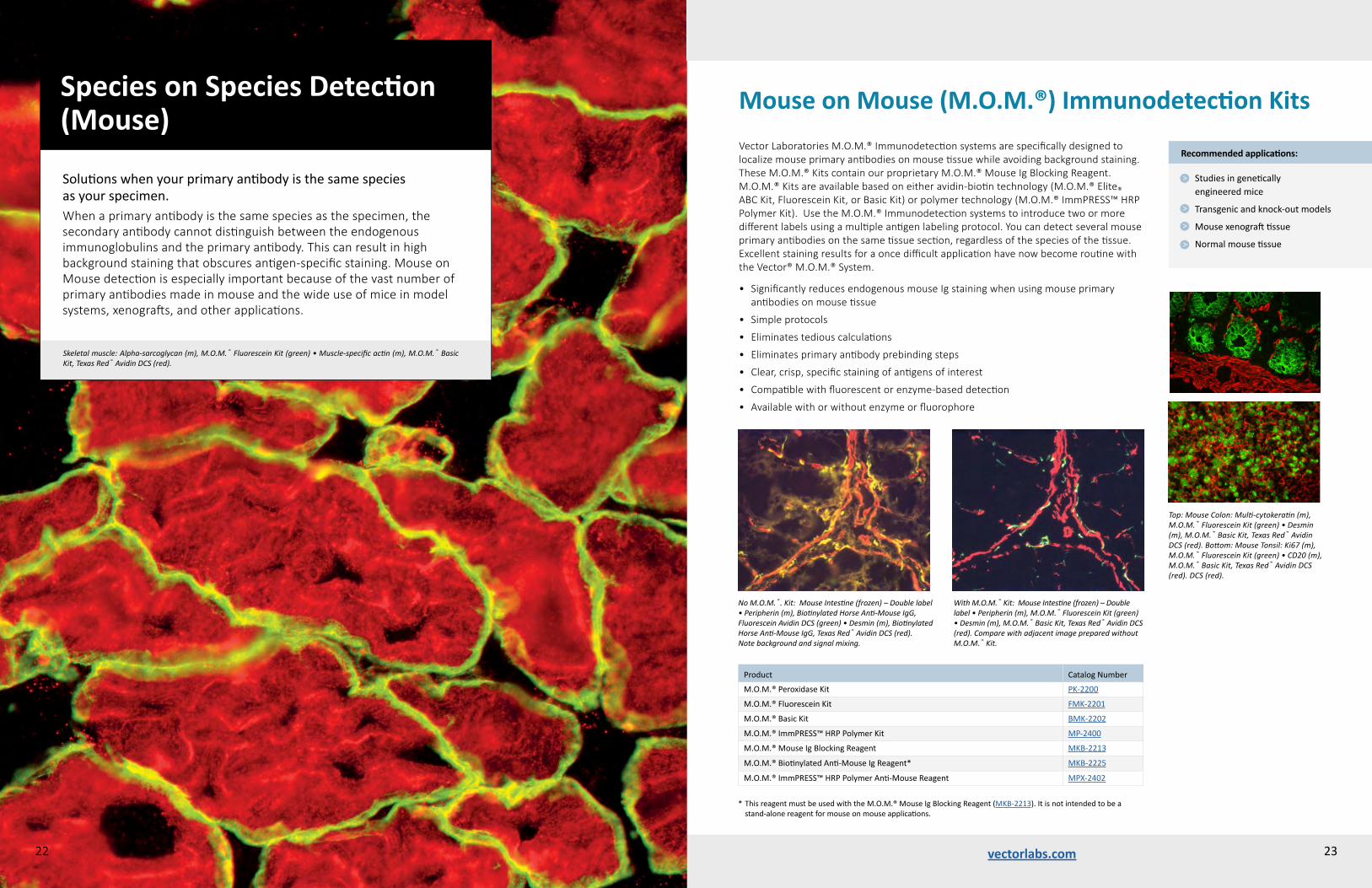
Solutionswhenyourprimaryantibodyisthesamespecies asyourspecimen.When a primary antibody is the same species as the specimen, the secondary antibody cannot distinguish between the endogenous immunoglobulins and the primary antibody. This can result in high background staining that obscures antigen-specific staining. Mouse on Mouse detection is especially important because of the vast number of primary antibodies made in mouse and the wide use of mice in model systems, xenografts, and other applications.
Skeletal muscle: Alpha-sarcoglycan (m), M.O.M.® Fluorescein Kit (green) • Muscle-specific actin (m), M.O.M.® Basic Kit, Texas Red® Avidin DCS (red).
22
Vector Laboratories M.O.M.® Immunodetection systems are specifically designed to localize mouse primary antibodies on mouse tissue while avoiding background staining. These M.O.M.® Kits contain our proprietary M.O.M.® Mouse Ig Blocking Reagent. M.O.M.® Kits are available based on either avidin-biotin technology (M.O.M.® Elite® ABC Kit, Fluorescein Kit, or Basic Kit) or polymer technology (M.O.M.® ImmPRESS™ HRP Polymer Kit). Use the M.O.M.® Immunodetection systems to introduce two or more different labels using a multiple antigen labeling protocol. You can detect several mouse primary antibodies on the same tissue section, regardless of the species of the tissue. Excellent staining results for a once difficult application have now become routine with the Vector® M.O.M.® System.
• Significantly reduces endogenous mouse Ig staining when using mouse primary antibodies on mouse tissue
• Simple protocols
• Eliminates tedious calculations
• Eliminates primary antibody prebinding steps
• Clear, crisp, specific staining of antigens of interest
• Compatible with fluorescent or enzyme-based detection
• Available with or without enzyme or fluorophore
Recommended applications:
Studiesingenetically engineeredmice
Transgenicandknock-outmodels
Mousexenografttissue
Normalmousetissue
Product CatalogNumber
M.O.M.®PeroxidaseKit PK-2200
M.O.M.®FluoresceinKit FMK-2201
M.O.M.®BasicKit BMK-2202
M.O.M.®ImmPRESS™HRPPolymerKit MP-2400
M.O.M.®MouseIgBlockingReagent MKB-2213
M.O.M.®BiotinylatedAnti-MouseIgReagent* MKB-2225
M.O.M.®ImmPRESS™HRPPolymerAnti-MouseReagent MPX-2402
*ThisreagentmustbeusedwiththeM.O.M.®MouseIgBlockingReagent(MKB-2213).Itisnotintendedtobea stand-alonereagentformouseonmouseapplications.
Mouse on Mouse (M.O.M.®) Immunodetection Kits
Top: Mouse Colon: Multi-cytokeratin (m), M.O.M.® Fluorescein Kit (green) • Desmin (m), M.O.M.® Basic Kit, Texas Red® Avidin DCS (red). Bottom: Mouse Tonsil: Ki67 (m), M.O.M.® Fluorescein Kit (green) • CD20 (m), M.O.M.® Basic Kit, Texas Red® Avidin DCS (red). DCS (red).
With M.O.M.® Kit: Mouse Intestine (frozen) – Double label • Peripherin (m), M.O.M.® Fluorescein Kit (green) • Desmin (m), M.O.M.® Basic Kit, Texas Red® Avidin DCS (red). Compare with adjacent image prepared without M.O.M.® Kit.
23
No M.O.M.®. Kit: Mouse Intestine (frozen) – Double label• Peripherin (m), Biotinylated Horse Anti-Mouse IgG, Fluorescein Avidin DCS (green) • Desmin (m), Biotinylated Horse Anti-Mouse IgG, Texas Red® Avidin DCS (red). Note background and signal mixing.

vectorlabs.comvectorlabs.com24
VECTASHIELD® Antifade Mounting Media formulations offer unsurpassed protection against fading and photobleaching. The VECTASHIELD® and VECTASHIELD® HardSet™ Antifade Mounting Media are well-established, market-leading products that complete the workflow and provide excellent signal retention for image acquisition and specimen archiving.
• Inhibits photobleaching of most fluorophores, dyes, fluorescent proteins and stains
• Ideal refractive index
• Ready to use, no warming necessary
• Continues to inhibit photobleaching even after prolonged storage of mounted slides
• Easy-to-use
• With or without nuclear or cytoskeletal counterstain
• Hardening or non-hardening formulations
VECTASHIELD® Antifade Mounting MediumVECTASHIELD® Antifade Mounting Medium is a glycerol-based, aqueous mountant that remains a viscous liquid on the slide rather than solidifying. After mounting, cover-slipped slides will not readily dry out, enabling you to review them for weeks without the need for sealing. For prolonged storage, coverslips can be permanently sealed with nail polish applied on the coverslip perimeter.
VECTASHIELD® HardSet™ Antifade Mounting MediumVECTASHIELD® HardSet™ Antifade Mounting Medium is an aqueous mountant that hardens at room temperature in as little as 20 minutes. This mounting medium provides easy slide handling, eliminates the need to secure the coverslip with nail polish, and is convenient for use with oil immersion microscopy. Available with or without DAPI or TRITC-phalloidin counterstain.
25
Mounting Media
Choosinganeffectivemountingmediumisespeciallyimportantforimmunofluorescenceimaging.Fluorophoresaresusceptibletophotobleachingandfadingfromboththeimagingexcitationlightandduringstorage.Therightmountingmediumwillprotectyoursamplesforshort-andlong-termuse andarchiving.
Rat muscle (FFPE): GFAP (red) and NF200 (green). Counterstained and coverslipped with VECTASHIELD® Mounting Medium with DAPI (blue). The double IF was performed by Dr. Lynn Dong, Dept of Biomedical Sciences, College of Veterinary Medicine, Cornell University, Ithaca, NY, USA.
One optical section of a whole mouse lens stained with phalloidin (F-actin, green) and DAPI (nuclei, red). This image was captured by Dr. Catherine Cheng, Department of Cell and Molecular Biology, The Scripps Research Institute, La Jolla, CA, USA.
Mouse embryonal fibroblasts: Anti-Integrin (m) detected with DyLight® 488 Anti-Mouse IgG, mounted in a 1:1 mixture of VECTASHIELD® HardSet™ Mounting Medium with DAPI and VECTASHIELD® HardSet™ Mounting Medium with TRITC-Phalloidin.
ProductNoCounterstain DAPI PI
TRITC-Phalloidin
VECTASHIELD®MountingMedium (non-hardening)
H-1000 H-1200 H-1300
VECTASHIELD®HardSet™MountingMedium(hardening)
H-1400 H-1500 H-1600
Structured illumination super resolution photomicrograph of a ciliated bovine airway epithelial cell labeled for acetylated alpha tubulin (cilia marker; green), phosphodiesterase 5 (red) and nuclei (blue). Sample prepared and image taken by Michael E. Price, University of Nebraska Medical Center. With assistance of Janice A. Taylor and James R. Talaska, Advanced Microscopy Core Facility, University of Nebraska Medical Center, NE, USA.
VECTASHIELD® Antifade Mounting Media

vectorlabs.com vectorlabs.com26 27
Option B1_B
650600550500450400 750700
DAPI
YFP
AF® 633
AF® 647
AF® 649mCherry
Dy® 594
AF® 594
Texas Red®Rhodamine
RFP
Dy® 549
Cy® 3
AF® 568
AF® 555
AF® 546
YFP
GFP
SYBR® Green
FITC
AF® 488
Dy® 488
AMCA Cy® 7
Cy® 5
Dy® 649
Fluorophore
acridineorange coumarin Fluoro-Jade® NeuroTrace® Quantumdot/Qdot
AlexaFluor®350 dihydroethidium Luciferyellow Nilered SYTOX®Green
AlexaFluor®680 DRAQ5™ LysoTracker® OilredO TAMRA
Atto®dyes Evansblue LysoTracker®Red PacificBlue™ thioflavins
BODIPY® fastblue MitoTracker®Red PicoGreen® TOTO®-3
Lorem ipsum dolor sit amet, consectetuer adipiscing elit, sed diam nonummy nibh euis-mod tincidunt ut laoreet dolore magna aliquam erat volutpat. Ut wisi enim ad minim veniam, quis nostrud exerci tation ullamcorper suscipit lobortis nisl ut aliquip ex ea commodo conse-quat. Duis autem vel eum iriure dolor in hendre-rit in vulputate velit esse molestie consequat, vel illum dolore eu feugiat nulla facilisis at vero eros et accumsan et iusto odio dignissim qui blandit praesent luptatum zzril delenit augue duis dolore te feugait nulla facilisi.
Lorem ipsum dolor sit amet, cons ectetuer adipiscing elit, sed diam nonummy nibh euis-mod tincidunt ut laoreet dolore magna aliquam erat volutpat. Ut wisi enim ad minim veniam, quis nostrud exerci tation ullamcorper suscipit lobortis nisl ut aliquip ex ea commodo conse-quat.Lorem ipsum dolor sit amet, consectetuer adipiscing elit, sed diam nonummy nibh euis-mod tincidunt ut laoreet dolore magna aliquam erat volutpat.
Lorem ipsum
• Fixed frozen sections<10μm
VECTACELL™ Trolox
VECTASHIELD®HardSet™VECTASHIELD®
• Epifluorescence microscopy• Confocal microscopy
• Super-resolution microscopy
• Live cell imaging
• Cytoskeletal counterstain• Lectin imaging
• Whole-mount preps• Gasket slides• Chamber slides• Microtiter plates/wells• Thick sections• Floating sections• 3D imaging
• Nuclear counterstain• Fixed frozen sections• Formalin-fixed paraffin sections • Fixed cultured cells
Option - Original Option B-3
The illustration above features established applications for our antifade mounting media formats. VECTASHIELD® Antifade Mounting Media are widely utilized to protect the inherent fluorescent properties of traditional and contemporary fluorophores in many applications using epifluorescence and confocal microscopy.
The versatility of the original VECTASHIELD® format solves the demands of labs and core facilities using multiple platforms and fluorescent markers. Furthermore, VECTASHIELD® reagents are also recognized as leading media in emerging techniques such as super resolution microscopy (SRM).
Of the SRM techniques currently being performed, the properties of VECTASHIELD® Antifade Mounting Media have been found to be advantageous in stochastic optical reconstruction microscopy (STORM) and structured illumination microscopy (SIM).
*SuperResolution(STORMandSIM)selectreferences:
OlivierN,KellerD,RajanVS,GönczyP,andManleyS“Simplebuffersfor3DSTORMmicroscopy,”Biochemical Optics Express4,885-899(2013)
Wegel,E.,etal.“Imagingcellularstructuresinsuper-resolutionwithSIM,STEDandLocalisationMicroscopy:Apracticalcomparison”,Scientific Reports,6,27290.(2016)
*
The fluorescent compounds listed in the table below are select reagents that are also cited as being successfully used in combination with VECTASHIELD® Antifade Mounting Media. The range of these compounds, from traditional to contemporary, across a broad spectral range, and used in an array of applications, showcase the versatility of VECTASHIELD® reagents. For a comprehensive list of the >130 fluorophores and fluorescent markers that have been used with VECTASHIELD® products please visit our website at: vectorlabs.com/vslist
wavelength (nm)
AF®=AlexaFluor®Cy®=Cyanine®Dy®=DyLight®FITC=Fluoresceinisothiocyanate
VECTASHIELD® Mounting Media and Fluorophore CompatibilityVECTASHIELD® Mounting Media are the most widely referenced antifade mounting media for immunofluorescence applications. Currently over 60,000 published references cite VECTASHIELD® Mounting Media and describe compatibility with over 130 fluorophores and fluorescent markers. This data underscores the prominence of VECTASHIELD® reagents in this application.
The graphic below highlights the most commonly referenced fluorophores used in combination with VECTASHIELD® Antifade Mounting Media.
VECTASHIELD® Mounting Media Formats and Applications
MountingMedia

Accessory ReagentsVECTABOND® Reagent Tissue Section Adhesive VECTABOND® Reagent chemically modifies the surface of glass to form a highly adherent charged surface. This charge significantly increases the adherence of both frozen and paraffin-embedded tissue sections and cell preparations to glass microscope slides and coverslips. Tissue sections will remain attached even when subjected to the most extreme conditions, such as high-temperature antigen retrieval and in situ hybridization. VECTABOND® Reagent-treated slides can be stored indefinitely.
ImmEdge™ Hydrophobic Barrier PenThe ImmEdge™ Pen is a hydrophobic barrier (PAP) pen for immunohistochemistry and in situ hybridization. It provides a water-repellent barrier that keeps reagents localized on tissue specimens and prevents mixing of reagents when multiple sections are mounted on the same slide.
• Heat-stable
• Insoluble in alcohol and acetone
• Stable for use with buffers with and without detergent (Tween 20, Triton X-100, etc.)
• Completely removed by all commonly used xylene and xylene-substitute clearing agents
• Contains no ozone-depleting solvents
• Compatible with both enzyme- and fluorescence-based detection systems
Product CatalogNumber
VECTABOND®Reagent(TissueSectionAdhesive) SP-1800
ImmEdge™HydrophobicBarrierPen H-4000
ImmPrint™HistologyPen H-6100
ControlAntibodies
RabbitIgG I-1000
MouseIgG I-2000
RatIgG I-4000
GoatIgG I-5000
AntigenUnmaskingSolutions
Citrate-based(100X)(pH6.0) H-3300
Tris-based(100X)(pH9.0) H-3301
ImmPrint™ Histology PenThe ImmPrint™ Histology Pen is a permanent marking pen designed for writing on glass microscope slides, tissue cassettes, and most hard surfaces. Unlike other pens commonly used for histology, the ImmPrint™ Pen has a smooth writing tip that resists drying out.
• High-density, fast-drying, black ink
• Resistant to most organic solvents encountered in histological applications
Control AntibodiesThese antibodies are IgG preparations for use as controls for primary antibodies made in rabbit, mouse, rat, or goat. Each antibody has beeW∑n purified from pooled serum of healthy adult animals and contains a spectrum of the IgG subclasses. When applied appropriately, these controls will help determine whether the primary antibody staining signal is specific for the antigen or whether staining is the result of non-specific adsorption of primary antibody to tissue sites.
Antigen Unmasking SolutionsOur Antigen Unmasking Solutions are highly effective at revealing antigens in formalin-fixed, paraffin-embedded tissue sections when used in combination with a high-temperature treatment procedure. We offer two formulations of Antigen Unmasking Solution: Citrate-based solution (pH 6.0) and Tris-based solution (pH 9.0), each supplied as 100X concentrated stocks.
42 vectorlabs.comvectorlabs.com28 29
Whereas immunofluorescence staining gives a snapshot of a cell or tissue at a specific time point, live cell imaging allows the observation of biological processes over a period of time. This is important for studying biological functions, interactions, and structures in various applications (e.g., the effects of drugs and other biomolecules).
VectaCell™ reagents enable and enhance live cell imaging studies. VectaCell™ Trolox Antifade Reagent reduces phototoxicity and photobleaching of reagents to increase cell viability and prolong signal. VectaCell™ Acridine Orange and VectaCell™ Rhodamine 123 reagents offer convenience and ease of use for visualizing different cellular components.
VectaCell™ Trolox Antifade ReagentVectaCell™ Trolox Antifade Reagent is an antifading additive for live cell imaging. VectaCell™ Trolox Antifade Reagent contains both Trolox and its oxidized form Trolox-quinone. This redox system reduces photo-bleaching and blinking during live cell imaging.
Trolox is a water-soluble and cell-permeable analog of vitamin E that efficiently prevents formation of different reactive oxygen species, such as singlet oxygen (1O2), superoxide anion (O2-) or hydrogen peroxide (H2O2). Photo-excitation of a fluorophore generates reactive oxygen species that can lead to photo-bleaching and oxidative damage in cells. Trolox has a cytoprotective effect and low cytotoxicity for different cell lines.
Live Cell Imaging of Organelles VectaCell™ Acridine Orange is a fluorescent dye that stains acidic organelles, such as lysosomes, autosomes or yeast vacuoles. At low pH inside organelles, the dye will emit an orange fluorescence (peak at 590 nm). For optimal endosome visualization, use a blue light excitation (475 nm).
VectaCell™ Rhodamine 123 is a fluorescent dye for staining active mitochondria. This dye accumulates in the mitochondrial membrane based on membrane polarization. Excitation peak at 505 nm, emission peak at 534 nm.
VectaCell™ Products for Live Cell Imaging
Mitochondria stained with VectaCell™ Rhodamine 123 (orange) in MCF-7 cells expressing GFP. Nuclei stained with DAPI (blue).
Acidic endosomes stained with VectaCell™ Acridine Orange in MCF-7 cells expressing GFP.
Product CatalogNumber
VectaCell™Trolox CB-1000
VectaCell™AcridineOrange CB-2000
VectaCell™Rhodamine123 CB-2100

Blocking agents minimize background signal from endogenous enzyme activity, biotin, and non-specific binding of tissue elements (charged particles, macromolecules, Fc receptors) with detection reagents. For IF applications special consideration should be given to the presence of autofluorescence.
Blocking Background Signal
36 vectorlabs.comvectorlabs.com30
BLOXALL® Endogenous Peroxidase and Alkaline Phosphatase Blocking SolutionBLOXALL® is compatible with formalin-fixed, paraffin-embedded tissue sections, frozen tissue sections, and cell preparations. It is supplied ready-to-use in a convenient dropper bottle and only requires a brief 10-minute incubation.
Levamisole SolutionSpecifically inhibits endogenous alkaline phosphatase activity that is added to the alkaline phosphatase substrate solution. It is supplied ready-to-use in a convenient dropper bottle.
Avidin/Biotin and Streptavidin/Biotin Blocking KitsBoth kits block all endogenous biotin and biotin receptors. Due to differing binding affinities and characteristics, kit selection is matched o the specific avidin or streptavidin detection system being used. Supplied ready-to-use in convenient dropper bottles.
Normal Sera and 2.5% Normal SeraAll our sera products are pooled samples collected from healthy adult animals, heat-treated and centrifuged to remove precipitates and then filtered. These sera are intended to be used for blocking non-specific binding or
as an antibody diluent.
Bovine Serum Albumin (BSA)Intended to be used as a diluent or a blocking agent and is free of impurities present in other grades of BSA which can introduce artifacts or increase background staining.
10x Casein SolutionA general blocking agent for IHC, nucleic acid blotting, protein blotting, and other applications.
Carbo-Free™ Blocking SolutionA protein-based agent that is essentially free of glycoproteins making it ideal for applications using lectins. Can be used to block non-specific binding or as an antibody diluent.
R.T.U. Animal-Free Blocker™ and DiluentA plant protein-derived solution intended for cell- and tissue-based IHC and IF applications. Can be used as an alternative to normal sera, BSA, casein and non-fat dry milk. Suitable for use with both HRP and AP enzyme conjugates and detection systems. Supplied as a ready-to -use solution, ideal in multiple antigen labeling IHC to streamline blocking.
Animal-Free Blocker™ (5x concentrate solution)Similar to the R.T.U. format, this plant protein-derived blocking agent and diluent is an alternative to normal sera, BSA, casein and non-fat dry milk, however this concentrate is intended primarily for blotting applications.
Product CatalogNumber
Vector® TrueVIEW™ Autofluorescence Quenching Kit SP-8400
BLOXALL® Endogenous HRP/AP Blocking Solution SP-6000
Levamisole Solution SP-5000
Avidin/Biotin Blocking Kit SP-2001
Streptavidin/Biotin Blocking Kit SP-2002
Normal Goat Serum S-1000
Normal Horse Serum S-2000
Normal Chicken Serum S-3000
Normal Swine Serum S-4000
Normal Rabbit Serum S-5000
2.5% Normal Goat Serum S-1012
2.5% Normal Horse Serum S-2012
Bovine Serum Albumin (BSA) SP-5050
10x Casein Solution SP-5020
Carbo-Free™ Blocking Solution SP-5040
R.T.U. Animal-Free Blocker™ and Diluent SP-5035
Animal-Free Blocker™ SP-5030
31
Vector® TrueVIEW™ Autofluorescence Quenching KitVector® TrueVIEW™ Autofluorescence Quenching Kit provides a novel way to remove unwanted fluorescence in tissue sections due to aldehyde fixation, red blood cells, and structural elements such as collagen and elastin. This unique formulation binds and effectively quenches the autofluorescent elements in even the most problematic tissues, such as kidney, spleen and pancreas.
The use of Vector® TrueVIEW™ Quenching reagent leads to significant enhancement in overall signal-to-noise in most immunofluorescence assays.
Vector® TrueVIEW™ Quenching reagent is a unique approach to diminish unwanted autofluorescence from non-lipofuscin sources, that retains the specific fluorescent antigen staining. The quenching action of the kit reagents therefore, provides the investigator with a clear, unambiguous, “true view” visualization of the intended target.
Patent pending formulation.
Adjacent human spleen sections (FFPE) stained usingmouse anti-CD20 (red) and rabbit anti-Ki67 (green) primary antibodies detected with VectaFluor® Duet Kit (DK-8818). Note significant reduction of autofluorescence in the treated section (right) with retention of well-defined, specific signal in both red and green channels.
Why TrueVIEW™ Quencher?
Specificreductionofautofluorescence fromaldehydefixation
Improvedsignal-to-noiseratio
Effectiveineventhemostchallengingtissues
Easy-to-use,one-stepmethod
Quick5minincubation
Compatiblewithawideselection offluorophores
Compatiblewithstandard epifluorescenceandconfocal lasermicroscopes
WITHOUT Treatment
WITH TrueVIEW™ Quencher
"I would definitely use this reagent in the future – it is quick and reliable on multiple tissue types."
– Dr. K. Sadtler, Postdoctoral Fellow MIT Boston Children's Hospital
Human Pancreas (FFPE): Stained for D 34 (using anti-mouse DyLight® 594, red) and Insulin (using anti-guinea pig, fluorescein, green). Coverslipped with VECTASHIELD® HardSet™ Antifade Mounting Medium. Note signif-icant reduction of autofluorescence in treated section (above) with the retention of specfiic staining.
vectorlabs.com
US OfficeVector Laboratories, Inc.30 Ingold RoadBurlingame, CA 94010, USATel: +1 (650) 697-3600 Tel (Ordering and Technical Support): +1 (800) 227-6666 Fax: +1 (650) 697-0339Customer Service: [email protected] Support: [email protected] International Inquiries: [email protected]
UK Office Vector Laboratories Ltd.,3 Accent Park, Bakewell Road, Orton Southgate,Peterborough, PE2 6XS, United KingdomTel: +44 (0) 1733 237999Customer Service: [email protected]: [email protected] Support: [email protected]
Contact DetailsOrdering InformationOrder online at: www.vectorlabs.com
Orders may also be placed by email, telephone, fax, or mail.Please include the following with each order:
• Product name and catalog number
• Unit size and quantity
• Billing and shipping addresses
• Purchase order number
• Name, phone number, address and email address of person placing order
Orders using VISA, Mastercard, or American Express are accepted and processed immediately. Telephone orders over $2000 may require written confirmation. A confirmation should be boldly marked “Confirming Order. Do Not Duplicate.” Duplicate shipments due to incorrectly marked confirming orders cannot be returned for credit. No returned product will be accepted or credited without prior authorization from Vector Laboratories.
Please contact us to discuss discounts for custom or large orders.
Payment / shipping terms:Payment terms: net 30 days. Prices are FCA Burlingame, California. Shipping charges will be prepaid and added to the invoice. Orders are usually shipped the same day they are received. Unless requested otherwise, all products are shipped 2nd-day air. RCA60 products are shipped in the USA only according to federal transportation regulations requiring additional shipping charges. We require a written confirmation for all RCA60 products.
International OrdersVector products are also available from over 50 distributors worldwide. Please contact us or visit our website for a complete listing of these representatives.
Although provided in a highly purified form, our products are not intended for clinical diagnosis or drug use, nor have they been packaged under sterile conditions. All products listed in this catalog are for research purposes only. The listing of any product in this catalog does not imply the absence of a patent covering its use, does not constitute license under any existing or pending patent, nor is it intended or implied as a recommendation for the use of such products in infringement of any patent. The responsibility for determining the existence of such patents rests solely with the user.
Trademarks of Vector Laboratories: ABC-AmP, Animal-Free Blocker, BLOXALL, DuoLuX, Elite, EndTag, FastTag, Fusion Aid, ImmEdge, ImmPACT, ImmPRESS, ImmPrint, M.O.M., NEUROBIOTIN, NicKit p.s.o., PHOTOPROBE, ProtOn, QuantTag, TrueVIEW, VECTABOND, VectaCell, VectaMount, VECTASHIELD, VECTASHIELD HardSet, VECTASTAIN, Vector, Vector Black, Vector Blue, Vector NovaRED, Vector Red. Atto, Alexa Fluor, BODIPY, DRAQ5, DyLight, Fluoro-Jade, LysoTracker, MitoTracker, NeuroTrace, Pacific Blue, PicoGreen, SYTOX, Texas Red and TOTO are trademarks of Thermo Fisher. Cyanine is a trademark of GE Healthcare.
©Vector Laboratories, Inc. 2018
32HumanmammaryepithelialcellsMCF10A3D-culturedonalow-rigiditysubstrateandstainedwithDAPI(blue)andanti-pY576FAKantibody(red).ImageprovidedbyDr.LaurentFattet,DepartmentofPharmacology,UniversityofCalifornia,SanDiego,CA,USA.USA.
