immunobiology of solid cancers: cellular and molecular pathways...
TRANSCRIPT

BioMed Research International
Immunobiology of Solid Cancers: Cellular and Molecular Pathways as Potential Diagnostic and Therapeutic Targets
Lead Guest Editor: Ilary RuscitoGuest Editors: Elena Ioana Braicu, Maria Luisa Gasparri, and Ilaria Grazia Zizzari

Immunobiology of Solid Cancers:Cellular and Molecular Pathwaysas Potential Diagnostic and Therapeutic Targets

BioMed Research International
Immunobiology of Solid Cancers:Cellular and Molecular Pathwaysas Potential Diagnostic and Therapeutic Targets
Lead Guest Editor: Ilary RuscitoGuest Editors: Elena Ioana Braicu, Maria Luisa Gasparri,and Ilaria Grazia Zizzari

Copyright © 2018 Hindawi. All rights reserved.
This is a special issue published in “BioMed Research International.” All articles are open access articles distributed under the CreativeCommons Attribution License, which permits unrestricted use, distribution, and reproduction in any medium, provided the originalwork is properly cited.

Contents
Immunobiology of Solid Cancers: Cellular andMolecular Pathways as Potential Diagnostic andTherapeutic TargetsIlary Ruscito , Elena Ioana Braicu, Maria Luisa Gasparri, and Ilaria Grazia ZizzariVolume 2018, Article ID 6532019, 2 pages
The Progress of T Cell Immunity Related to Prognosis in Gastric CancerMing Wei, Duo Shen, Sachin Mulmi Shrestha, Juan Liu, Junyi Zhang, and Ying YinVolume 2018, Article ID 3201940, 6 pages
PD-L1 Expression in TNBC: A Predictive Biomarker of Response to Neoadjuvant Chemotherapy?Bruna Cerbelli, Angelina Pernazza, Andrea Botticelli, Lucio Fortunato, Massimo Monti, Paolo Sciattella,Domenico Campagna, Federica Mazzuca, Maria Mauri, Giuseppe Naso, Paolo Marchetti, Giulia d’Amati,and Leopoldo CostarelliVolume 2017, Article ID 1750925, 7 pages
Dynamics of Neutrophils-to-Lymphocyte Ratio Predict Outcomes of PD-1/PD-L1 BlockadeMichele Moschetta, Mario Uccello, Benjamin Kasenda, Gabriel Mak, Anissa McClelland, Stergios Boussios,Martin Forster, and Hendrik-Tobias ArkenauVolume 2017, Article ID 1506824, 5 pages
PD-L1 Promotes Self-Renewal and Tumorigenicity of Malignant Melanoma Initiating CellsFang Zheng, Jianzhong Dang, Hui Zha, Bingyu Zhang, Ming Lin, and Fanjun ChengVolume 2017, Article ID 1293201, 8 pages
Increased EGFR Phosphorylation Correlates with Higher ProgrammedDeath Ligand-1 Expression:Analysis of TKI-Resistant Lung Cancer Cell LinesKenichi Suda, Leslie Rozeboom, Koh Furugaki, Hui Yu, Mary Ann C. Melnick, Kim Ellison,Christopher J. Rivard, Katerina Politi, Tetsuya Mitsudomi, and Fred R. HirschVolume 2017, Article ID 7694202, 7 pages

EditorialImmunobiology of Solid Cancers: Cellular and MolecularPathways as Potential Diagnostic and Therapeutic Targets
Ilary Ruscito ,1,2 Elena Ioana Braicu,2 Maria Luisa Gasparri,3,4,5 and Ilaria Grazia Zizzari1
1UP Cell Therapy and Tumor Immunology, Department of Experimental Medicine, Sapienza University of Rome,Viale Regina Elena 324, 00161 Rome, Italy2Tumorbank Ovarian Cancer Network (TOC), Department of Gynecology, Charite-Universitatsmedizin Berlin,Corporate Member of Freie Universitat Berlin, Humboldt-Universitat zu Berlin, and Berlin Institute of Health,Augustenburger Platz 1, 13353 Berlin, Germany3Department of Obstetrics and Gynecology, University Hospital of Bern and University of Bern,Effingerstrasse 102, 3010 Bern, Switzerland4Department of Gynecology, Obstetrics and Urology, Sapienza University of Rome, Viale del Policlinico 155, 00161 Rome, Italy5Department of Medical and Surgical Sciences and Translational Medicine, Sapienza University of Rome,Via di Grottarossa 1035, 00189 Rome, Italy
Correspondence should be addressed to Ilary Ruscito; [email protected]
Received 18 January 2018; Accepted 21 January 2018; Published 1 March 2018
Copyright © 2018 Ilary Ruscito et al. This is an open access article distributed under the Creative Commons Attribution License,which permits unrestricted use, distribution, and reproduction in any medium, provided the original work is properly cited.
In the last four decades, tumor immunology has shed lighton identity and functions of cells and molecules involvedin tumor rejection through the involvement of the immunesystem [1]. Several groups of immune cells have beendemonstrated to be able to contrast tumor occurrence andtumor progression by killing immunogenic tumor cells, aphenomenon recognized under the definition of “immuno-surveillance” [2]. Unfortunately, cancer may evade immuno-surveillance and progress through the modifications of itsown antigens, which can reduce tumor immunogenicityand/or increase its immunosuppressive action [3]. After yearsof investigations, harnessing the immune system to attackcancer has recently led scientists to gather enough clinicaldata to show what a powerful sword immunotherapy canbe [4]. Data on unexpected clinical recoveries and longprogression-free intervals are increasing regarding patientsaddressed to immunotherapy treatments [5, 6]. Despite itsextraordinary success, only a portion of cancer types andcancer patients benefit from immunotherapy treatments.Understanding the reason why this happens is the big chal-lenge of our time and, in order to answer this question, basicscience is crucial: to elucidate how tumor cells and immunecells interact with each other in cancer patients and clarify the
mechanisms through which tumormutational pattern affectsthe response to therapies is the way to pursue for improvingefficacy of current treatments and promoting new anticancerstrategies. This special issue was conceived with the aim ofcollecting new findings in the field of cancer immunologyand describing novel biological and molecular evidence onthe relationship between cancer and immune system as wellas cancer and immunotherapy.
In response to the aim of the special issue, four originalresearch papers and one review article are presented below.
The study reported by B. Cerbelli et al. from SapienzaUniversity of Rome, Italy, showed that immunohistochemicalPD-L1 expression in ≥25% of triple negative breast cancer(TNBC) chemo-naıve cells, derived from core biopsies, isan independent predictor for pathological complete response(pCR) after neoadjuvant chemotherapy, thus discussingpotentials and limits of PD-L1 future applications as apredictive biomarker for neoadjuvant treatment response inthis subset of patients affected by such a clinically aggressivedisease.
M. Moschetta et al., at Sarah Cannon Research Instituteof London, UK, presented a study assessing the impactof “neutrophil-to-lymphocyte ratio” (NLR) in predicting
HindawiBioMed Research InternationalVolume 2018, Article ID 6532019, 2 pageshttps://doi.org/10.1155/2018/6532019
2 BioMed Research International
PFS among 55 advanced patients enrolled into PD-1/PD-L1inhibitors phase 1 clinical trials. Results showed a significantlonger PFS in patients with a reduction of NLR after twotreatment cycles compared to the median baseline NLR,thus advancing the hypothesis that NLR may be a helpfulpredicting tool in cancer patients treated with anti-PD-1/PD-L1 agents.
An international collaboration between USA (Universityof Colorado and Yale School of Medicine) and Japan (KindaiUniversity and Chugai Pharmaceutical), coordinated by K.Suda et al., obtained evidence concerning molecular mech-anism behind the low expression of PD-1/PD-L1 in NSCLC,associated with reduced efficacy of checkpoint inhibitors(CI) treatments. The study highlighted that EGFR-mutatedlung cancer cell lines do not show high PD-L1 expressionand, furthermore, after acquisition of resistance to EGFR-TKIs, EGFR phosphorylation affects PD-L1 expression, thusidentifying a molecular event able to influence the expressionof biomarkers, which regulate patients’ access to CI agents.
Apart from its immunomodulatory function, F. Zheng etal., from Huazhong University of Science and Technology,China, identify PD-L1 molecule as a potential biomarkerof melanoma cancer stem-like cells, since blocking PD-L1 in melanoma cell lines expressing PD-L1 and ALDH1impaired tumorsphere formation and induced the apoptosisof tumorsphere cells. These findings raise the need to eluci-date the relationship between tumor response to checkpointinhibitors and clonal evolution of cancer stem cells in thefuture.
Finally, the review paper by M. Wei et al., SoutheastUniversity of China, discusses the role of gastric cancerpatients’ T cells immunity and disease prognosis, providing acritical synthesis of recent evidence on this still controversialtopic.
In conclusion, we find this special issue to be a goodopportunity for improving knowledge in the field of cancerimmunobiology and immunotherapy, which is a pivotal stepto respond adequately to the questions of our time in thebattle against cancer.
Ilary RuscitoElena Ioana Braicu
Maria Luisa GasparriIlaria Grazia Zizzari
References
[1] R. D. Schreiber, L. J. Old, and M. J. Smyth, “Cancer immu-noediting: integrating immunity’s roles in cancer suppressionand promotion,” Science, vol. 331, no. 6024, pp. 1565–1570, 2011.
[2] F. M. Burnet, “The concept of immunological surveillance,” inProgress in Experimental Tumor Research, vol. 13, pp. 1–27, 1970.
[3] S. Spranger and T. F. Gajewski, “Impact of oncogenic pathwayson evasion of antitumour immune responses,” Nature ReviewsCancer, 2018.
[4] F. Bellati, C. Napoletano, I. Ruscito et al., “Past, present andfuture strategies of immunotherapy in gynecological malignan-cies,” Current Molecular Medicine, vol. 13, no. 4, pp. 648–669,2013.
[5] F. S. Hodi, S. J. O’Day, D. F. McDermott et al., “Improved sur-vival with ipilimumab in patients with metastatic melanoma,”The New England Journal of Medicine, vol. 363, no. 13, pp. 711–723, 2010.
[6] N. A. Rizvi, J. Mazieres, D. Planchard et al., “Activity and safetyof nivolumab, an anti-PD-1 immune checkpoint inhibitor, forpatients with advanced, refractory squamous non-small-celllung cancer (CheckMate 063): a phase 2, single-arm trial. LancetOncosingle-arm trial. Lancet Oncol,” in Lancet Oncol, vol. 16 of265, p. 257, 2015.

Review ArticleThe Progress of T Cell Immunity Related to Prognosis inGastric Cancer
MingWei,1 Duo Shen,1 Sachin Mulmi Shrestha,1 Juan Liu,1 Junyi Zhang,2 and Ying Yin 1
1Gastroenterology Department, Affiliated Zhongda Hospital of Southeast University, Nanjing, China2Department of Critical Care Medicine, Affiliated Zhongda Hospital of Southeast University, Nanjing, China
Correspondence should be addressed to Ying Yin; [email protected]
Received 25 August 2017; Accepted 6 December 2017; Published 27 February 2018
Academic Editor: Ilaria G. Zizzari
Copyright © 2018 MingWei et al.This is an open access article distributed under the Creative CommonsAttribution License, whichpermits unrestricted use, distribution, and reproduction in any medium, provided the original work is properly cited.
Gastric cancer is the fifth most common malignancy all over the world, and the factors that can affect progress and prognosis ofthe gastric cancer patients are various, such as TNM stages, invasive depth, and lymph node metastasis ratio. T cell immunity isimportant component of human immunity system and immunity responding to tumor and dysfunction or imbalance of T cellimmunity will lead to serious outcomes for body. T cell immunity includes many different types of cells, CD4+ T cell, CD8+ T cell,memory cell, and so on, and each of them has special function on antitumor response or tumor immune escape which is revealedin lung cancer, colorectal cancer, breast cancer, ovarian cancer, and so on. But its correlation with gastric cancer is not clear. Ourreview was preformed to explore the relationship between the progress and prognosis of gastric cancer (GC) and T cell immunity.According to recent researches, T cell immunity may have an important role in the progress and prognosis of GCs, but its functionis affected by location, category, related molecule, and interaction between the cells, and some effects still are controversial. Moreresearches are needed to clarify this correlation.
1. Introduction
Gastric cancer is the fifth most common malignancy all overthe world after lung, breast, colorectal cancers, and prostate.More than 70% of gastric cancer (677,000 cases) happened atdeveloping countries (456,000 in men, 221,000 in women),and half the total located in Eastern Asia, especially in China[1]. Although the lifestyle and smoking play an importantfactor, the main risk factor for advanced gastric canceris infection with the bacterium Helicobacter pylori [2]; Tcell immunity is a hot topic in recent studies. During thedevelopment of cancer, T cells progressively dysfunctionand exhaust; however the T cell responses are necessary tocontrol tumors [3]. And they play important roles in severaltypes of cancers like lung cancer [4], colorectal cancer [5],breast cancer [6], and ovarian cancer [7], but the relationshipbetween the T cell immunity and progression and prognosisof GCs is not clear. And there are many subsets of T cellswhich play different roles in gastric cancer, CD4+ T cell,including regulatory T cells, CD8+ T cell, and CD45RO+memory T cells [8]. The recent researches are more focusedon regulatory T cells.
2. Subsets of T Cell and Molecules Related toPrognosis of Gastric Cancer
T cell immunity is important in tumor response, and there aremany subsets of T cells which played different roles in gastriccancer, CD4+ T cell, including regulatory T cells, CD8+ Tcell, CD45RO+ memory T cells, and other molecules relatedto T cell immunity.
2.1. CD4+ T and CD8+ T Lymphocytes. CD4+ T and CD8+T are two important types of cells in T cell immunity.
CD4+ regulatory T cell is a major cell in self-toleranceand suppresses antitumor immunity [9]. CD4 T cells haveeffector functions by secreting multiple cytokines or activat-ing other immune cells acting on immunity of tumor [3].Among CD4+ T cell, Follicular helper T cells (Tfh cells) arespecial one which are necessary for producing high affinityantibodies. Meanwhile Tfh cells can secrete IL21 and IL4and show high expression of CXCR5, ICOS, PDCD1 (PD-1), and chemokine CXCL13, which also affect gastric cancerprognosis [10]. Cytotoxic CD8 T lymphocytes are present intumors and their functions in recognizing tumor epitopes
HindawiBioMed Research InternationalVolume 2018, Article ID 3201940, 6 pageshttps://doi.org/10.1155/2018/3201940
2 BioMed Research International
are nevertheless generally important in antitumor reaction[11]. And CD8 T cells are an important factor on the initialdevelopment of tumors, especially in existing tumor, and thepresence of CD8 T cells indicates poor prognosis [12].
2.2. Regulatory T Cell. Regulatory T cells (Tregs) are akind of T lymphocytes with an immunoregulatory capacity,which can inhibit the proliferation and cytokine secretion ofeffector T lymphocytes. Giving this function, inappropriateproduction or dysfunction of Tregs could result in severedamage of the host immune system [13]. In recent years,regulatory T cells (Tregs)within tumors, also known as tumorinfiltrating Treg cells, have been considered to play a keyrole in immune evasion [13]. And Tregs are correlated withprogression and poor outcomes in gastric cancer ([2]; [14]),but the relation between tumor infiltrating T cells and gastriccancer is unclear.
2.3. Others. In addition, many other related cells andmolecules also play a role in prognosis of gastric can-cer. Dendritic cells (DC) play the central role in cancerimmunosurveillance as the antigen-presenting cells (APC)are involved in the antitumor immune responses [15]. T cellimmunoglobulin and mucin domain-3 (Tim-3) is negativeregulatory molecules and plays a major role in the tumorimmunological tolerance [16]. And B7-H1 (also known asPDL1) is a member of the B7 superfamily [17]. PD-L1expression has been detected in cancers of the skin [18],lung [19], breast [20], kidney [21], bladder [22], esophagus[23], stomach [24], head, and neck [25], among others. B7-H4 is a coinhibitory molecule which negatively regulates Tcell immunity and is rarely expressed in resting antigen-presenting cells (APCs) [26] but is upregulated in a varietyof cancer tissues including ovary, kidney, stomach, lung, andpancreas [27–29].
3. The Correlation between T Cellsand Gastric Cancer
The factors that can affect progress and prognosis of thegastric cancer patients are various, such as TNM stages [30],invasive depth, lymph node metastasis ratio [31], and tumorimmunity. And there are three molecular carcinogenesismechanismswhichmay be correlatedwithGC: chromosomalinstability [32], microsatellite instability (MSI) [33], andCpG island methylator phenotype [34]. T cell immunityalso plays a role in these mechanisms, and some researcheshave already suggested that beta-catenin/T cell factor- (TCF-) mediated transcription (canonical Wnt signaling) couldresult in chromosomal instability (CIN) [35], andMSI gastriccancers possibly express more PD-L1 and have increasingCD8+ T cells before tumor invasive [36].
Nowadays, since the function of T cell immunity incancer is researched more and more clearly, we found thatit can also influence the progress and prognosis of GCsdirectly or indirectly participating in antitumor responses.For example, DC as the antigen-presenting cells (APC) isinvolved in the antitumor immune responses, while CD8+T cell may dissolve and kill tumor cells, and CD4+ cell
(including Foxp3+ Tregs) impose restrictions on tumorresponse.
Except these cells, many molecule such as Th17 [37],CD133 [38], gastrokine 1 [39], angiogenic factor [40], andLKB1 (Sun J et al., 2016) also have possible impact on diag-nosis, progress, treatment, and prognosis of gastric cancer.
4. The Correlation and Mechanismbetween T Cells Immunity and Prognosisof Gastric Cancer
T cell immunity is important in antitumor response and stud-ied inmany other cancers. Some studies also show there couldbe correlation between gastric cancer prognosis and T cells.Haas et al. introduced a phenomenon in their experimentthat an increasing stromal FoxP3+ TIL infiltration in tumorissues had a negative correlation with UICC- stage (Pearson’scorrelation coefficient, 𝑟 = −0.40; 𝑝 = 0.001), numberof lymph node metastases (𝑟 = −0.36; 𝑝 = 0.009), andN category in general (𝑟 = −0.36; 𝑝 = 0.023). But thisrelationship could not be seen in other cell types [41]. Chenget al. showed that Tim-3 was expressed in CD4+ T cells andCD8+ T cells higher in gastric issues and had a meaningfulrelation with tumor invasion and TNM stage, which couldlead to poorer prognosis [16]. And Qing et al. had confirmedthat PD-L1 could expressmore in highly differentiated gastriccancers, and it had an obvious relationship with the depth ofinvasion (odds ratio [OR] = 3.37; 𝑝 = 0.005), lymph nodemetastasis (OR = 2.68; 𝑝 = 0.020), tumor differentiation(OR = 3.19; 𝑝 = 0.008), pathological type (𝜒2 = 8.676;𝑝 = 0.013), and survival time (OR = 3.39; 𝑝 = 0.003)[42]. And they proposed that targeting the PD-L1 and APE1signaling pathwaysmay be a new treatment for gastric cancer,especially deep invasion and lymph node metastasis [42].Cho et al. indicated that PD-L1 expression was frequentlycorrelated with a lower risk of lymph node metastasis (𝑝 =0.027) and lower tumor stages in intestinal type cancer bythe Lauren classification [43]. But the mechanism betweengastric cancer prognosis and T cells immunity is not verysure.
4.1. CD4+ TCell and CD8+ TCell. There are some researchesfocusing on the relationship between the subsets of CD4+ Tcell and the progress and prognosis of gastric cancer. Shen etal. found that CD4+CD25+CD127low/− Tregs are correlatedwith more advanced stage of gastric cancer through sup-pressed effector T cell proliferation and express Foxp3 [44].In another research, Kindlund et al. suggested that CD4+regulatory T cells can promote tumor growth by inhibiting Tcell mediated tumor cell killing, depending on IL-10 and/orTGF-𝛽, but they also showed that CD4+CD25High expresseshigher IL-10 [2]. As we all know,Helicobacter pylori infectionis related to prognosis of GCs. Zhang et al. investigated thepotential functions of Follicular helper T cells in theGCswithHelicobacter pylori infection. His group found that Th1 andTh17 are themost common subsets of Follicular helper T cellsand can be negatively correlatedwith the disease-free survivalof tumor resection [45].

BioMed Research International 3
And CD8+ T cells have also been studied. Lu et al.indicated that GC patients with high-density CD8+ hadhigher overall survival rates than low-density ones byKaplan-Meier test in MSI-high GCs [11]. But Thompson et al.demonstrated that tumors with high CD8+ T cell densityeither in intratumor or in stromal had worse progression-freesurvival (PFS) and OS compared with the lower ones [17].Tuncel et al. introduced patientswith lower numbers ofCD8+T lymphocytes in the tumor, which has a negative correlationwith HLA-G and had a poorer prognosis [46].
4.2. Foxp3+TregCell. Although the functions of some immu-nity cells have been recognized by studies, some controversiesare still present.
Foxp3+ Tregs are the most concerned cell and theirfunction is still controversial. Hou et al. showed the level ofFoxP3+ Tregs in gastric cancer tissues related to an advancedclinicopathological stage and lymph node metastasis, whichindicted poor prognosis [13]. Yuan et al. found that the level ofFoxP3 is higher in Tregs and it can inhibit the proliferation ofautologous CD4+CD25−T cells in a COX-2-dependent man-ner to lead to poor prognosis which can be reversed by COXinhibitors [14]. In another article, Tuncel et al. got the similarconclusion that the high numbers of Tregs in the primarytumor, positive regulated byHLA-G, are associated with poorprognosis [46]. But there are some researches having oppositeconclusions. Kim et al. showed that GCs with high-densityFoxP3+ TILs had significantly higher overall survival ratesand low density is closely related to a higher TNM stage,invasion depth, and lymphatic and vascular invasion andproved FoxP3+ T cell density in the intraepithelial cells wasan independent predictor for overall survival. But the resultof Kim et al. is confined to microsatellite-unstable gastriccancers [47]. Haas et al. suggested that high level of Treg isassociated with improved outcome probably via inhibitinglocal inflammatory process [41]. And Feichtenbeiner et al.found an interesting conclusion that prognostic effect of TILscells in gastric cancer depends on the distance within cells,and FoxP3+ TILs must be located within 30 and 110 𝜇m farfrom CD8+ T cells to play its positive impact on prognosis[48]. In addition, Ma et al. investigated the expression ofFoxP3 protein in tumor cells and they showed that the highlevel predicts a good prognosis, whereas high-density Treg isopposite [49].
5. Other Cells and Molecule Related T CellImmunity in GCs
Except mainly cells in T cell immunity, there are some othercells and molecule also could have their functions in GCs.
Kashimura et al. suggested that the density of CD83+DCsin negative lymph nodes was an independent prognostic fac-tor bymultivariate analysis for patients withmetastatic lymphnodes [9]. Gao et al. descripted that the overexpression of B7-H1 in carcinomas has been shown to induce apoptosis in theeffector T cells to repress T cell activation and proliferation,which led to lower 5-year OS and DFS [50]. Geng et al. con-firmed that lymph nodemetastasis and B7-H1 overexpressionwere independent prognostic factors which are negative with
gastric cancer through Cox regression multivariate analysis[51]. Shi et al. proposed that soluble B7-H4 (sB7-H4) in cir-culation is a valuable molecule for predicting the progressionand prognosis of GCs and a positive correlation between thetwo things [52]. AndChen et al. suggested that the expressionof T-bet, a keymarker for type 1 immune responses, can serveas a prognostic indicator which has negative effect [53]. Ina recent study, Kim et al. revealed that decreasing NOVA1expression in tumor tissue was related to tumor progressionand poor prognosis via immune dysfunction of T cells andmacrophages [54]. And Th12 [55], Th17 [37, 56], Th1 [57],CX3C chemokines [58], diversity index of mucosal residentT lymphocyte [59], myeloid derived suppressing cells [60],immune activating receptor NKG2D [61], CCR7 [62], and IL-10 [63] are also involved in the progression and prognosis ofGCs.
Another interesting thing is the prognosis and progress ofthe same immunity cells can vary under different locations.Tim-3 is a negative regulatory molecule, only when it over-expresses CD8+ T cell or Tregs can lead to poor prognosis.Another protein is Foxp3+, its higher expression in tumorcells predicts good outcome but in Tregs the function isinverse. It has different prognosis when acting on differentcells. Cheng et al. found that CD4+ and CD8+ T cell can beupregulation in GCs by Tim-3, but CD4+ T cell has poorerprognosis. Tim was also an independent factor for GCs, andthe lower is the better [16]. Shen et al. revealed that thelevel of Tim-3 is up in both H. pylori-infected asymptomaticand gastric cancer patients, which is on Tregs and CD8+ Tcells associated with worse prognosis [64]. And Milasieneet al. observed that higher levels of the absolute numberof lymphocyte had a positive effect on overall survival ofgastrium in stage III, but there is no effect in stage II[65].
6. Forecast
The subsets of immunity cells have their own special rolein response to gastric cancer and lead to different out-comes of patients. But the function is affected by location,category, related molecule, interaction between the cells,and so on. Definite function is still unclear and needsmore studies. More studies are needed to investigate therelationship between the T immunity cell and gastric cancer,especially forcing on Foxp3+ Tregs and the influence oflocation and mutual relations between cells. At the sametime, I think that the role of memory T cell is ignored inthe progress and prognosis of GCs, and more research isessential.
Because of the T cells exhibiting a possible relationship inthe progress and prognosis of GCs, it may provide new theoryand way on diagnosis and treatment of gastric cancer. Morestudies are needed.
Conflicts of Interest
The authors declare that they have no conflicts of interest.
4 BioMed Research International
Acknowledgments
This work was supported by grants from National NaturalScience Foundation of China (no. 81602432) and Programfor Jiangsu Provincial Natural Science Foundation of China(BK20140652).
References
[1] J. Ferlay, I. Soerjomataram, R. Dikshit et al., “Cancer incidenceand mortality worldwide: sources, methods and major patternsin GLOBOCAN 2012,” International Journal of Cancer, vol. 136,no. 5, pp. E359–E386, 2015.
[2] B. Kindlund, A. Sjoling, C. Yakkala et al., “CD4+ regulatory Tcells in gastric cancer mucosa are proliferating and express highlevels of IL-10 but little TGF-𝛽,”Gastric Cancer, vol. 20, no. 1, pp.116–125, 2017.
[3] A. O. Kamphorst and R. Ahmed, “CD4 T-cell immunotherapyfor chronic viral infections and cancer,” Immunotherapy, vol. 5,no. 9, pp. 975–987, 2013.
[4] M.-C. Dieu-Nosjean, M. Antoine, C. Danel et al., “Long-termsurvival for patients with non-small-cell lung cancer withintratumoral lymphoid structures,” Journal of Clinical Oncology,vol. 26, no. 27, pp. 4410–4417, 2008.
[5] J. A. D. Simpson, A. Al-Attar, N. F. S. Watson, J. H. Scholefield,M. Ilyas, and L. G. Durrant, “Intratumoral T cell infiltration,MHC class I and STAT1 as biomarkers of good prognosis incolorectal cancer,” Gut, vol. 59, no. 7, pp. 926–933, 2010.
[6] Z. I. Hu, A. Y. Ho, and H. L. McArthur, “Combined radiationtherapy and immune checkpoint blockade therapy for breastcancer,” International Journal of Radiation Oncology, Biology,Physics, vol. 99, no. 1, pp. 153–164, 2017.
[7] L. Zhang, J. R. Conejo-Garcia, D. Katsaros et al., “IntratumoralT cells, recurrence, and survival in epithelial ovarian cancer,”The New England Journal of Medicine, vol. 348, no. 3, pp. 203–213, 2003.
[8] K. Wakatsuki, M. Sho, I. Yamato et al., “Clinical impact oftumor-infiltrating CD45RO+memory T cells on human gastriccancer,” Oncology Reports, vol. 29, no. 5, pp. 1756–1762, 2013.
[9] S. Kashimura, Z. Saze, M. Terashima et al., “CD83+ dendriticcells and Foxp3+ regulatory T cells in primary lesions andregional lymph nodes are inversely correlated with prognosis ofgastric cancer,” Gastric Cancer, vol. 15, no. 2, pp. 144–153, 2012.
[10] M. J. Ahearne, R. L. Allchin, C. P. Fox, and S. D. Wagner,“Follicular helper T-cells: expanding roles in T-cell lymphomaand targets for treatment,” British Journal of Haematology, vol.166, no. 3, pp. 326–335, 2014.
[11] X. Lu, L. Yang, D. Yao et al., “Tumor antigen-specific CD8+T cells are negatively regulated by PD-1 and Tim-3 in humangastric cancer,” Cellular Immunology, vol. 313, pp. 43–51, 2017.
[12] J. D. Peske, A. B. Woods, and V. H. Engelhard, “Controlof CD8 T-cell infiltration into tumors by vasculature andmicroenvironment,” Advances in Cancer Research, vol. 128, pp.263–307, 2015.
[13] J. Hou, Z. Yu, R. Xiang et al., “Correlation between infiltrationof FOXP3+ regulatory T cells and expression of B7-H1 in thetumor tissues of gastric cancer,” Experimental and MolecularPathology, vol. 96, no. 3, pp. 284–291, 2014.
[14] X.-L. Yuan, L. Chen, M.-X. Li et al., “Elevated expressionof Foxp3 in tumor-infiltrating Treg cells suppresses T-cellproliferation and contributes to gastric cancer progression in a
COX-2-dependent manner,” Clinical Immunology, vol. 134, no.3, pp. 277–288, 2010.
[15] F. Li, J. Huang, S. Li et al., “The subsets of dendritic cells andmemory T cells correspond to indoleamine 2,3-dioxygenase instomach tumor microenvironment,” Tumor Biology, vol. 35, no.9, pp. 8691–8698, 2014.
[16] G. Cheng, M. Li, J. Wu et al., “Expression of Tim-3 in gastriccancer tissue and its relationship with prognosis,” InternationalJournal of Clinical and Experimental Pathology, vol. 8, no. 8, pp.9452–9457, 2015.
[17] E. D. Thompson, M. Zahurak, A. Murphy et al., “Patternsof PD-L1 expression and CD8 T cell infiltration in gastricadenocarcinomas and associated immune stroma,”Gut, vol. 66,pp. 794–801, 2017.
[18] D. Massi, D. Brusa, B. Merelli et al., “The status of PD-L1and tumor-infiltrating immune cells predict resistance andpoor prognosis in BRAFi-treated melanoma patients harboringmutant BRAF𝑉600,”Annals of Oncology, vol. 26, no. 9, Article IDmdv255, pp. 1980–1987, 2015.
[19] J. McLaughlin, G. Han, K. A. Schalper et al., “Quantitativeassessment of the heterogeneity of PD-L1 expression in non-small-cell lung cancer,” JAMAOncology, vol. 2, no. 1, pp. 46–54,2016.
[20] H. R. Ali, S.-E. Glont, F. M. Blows et al., “PD-L1 proteinexpression in breast cancer is rare, enriched in basal-liketumours and associated with infiltrating lymphocytes,” Annalsof Oncology, vol. 26, no. 7, pp. 1488–1493, 2015.
[21] T. K. Choueiri, A. P. Fay, K. P. Gray et al., “PD-L1 expression innonclear-cell renal cell carcinoma,” Annals of Oncology, vol. 25,no. 11, pp. 2178–2184, 2014.
[22] C. Massard, M. S. Gordon, S. Sharma et al., “Safety andefficacy of durvalumab (MEDI4736), an anti–programmedcell death ligand-1 immune checkpoint inhibitor, in patientswith advanced urothelial bladder cancer,” Journal of ClinicalOncology, vol. 34, no. 26, pp. 3119–3125, 2016.
[23] Y. Ohigashi, M. Sho, Y. Yamada et al., “Clinical significance ofprogrammed death-1 ligand-1 and programmed death-1 ligand-2 expression in human esophageal cancer,” Clinical CancerResearch, vol. 11, no. 8, pp. 2947–2953, 2005.
[24] E. D. Thompson, M. Zahurak, A. Murphy et al., “Patternsof PD-L1 expression and CD8 T cell infiltration in gastricadenocarcinomas and associated immune stroma,”Gut, vol. 66,pp. 794–801, 2016.
[25] S. Lyford-Pike, S. Peng, G. D. Young et al., “Evidence for arole of the PD-1:PD-L1 pathway in immune resistance of HPV-associated head and neck squamous cell carcinoma,” CancerResearch, vol. 73, no. 6, pp. 1733–1741, 2013.
[26] Y. K. Jeon, S. G. Park, I. W. Choi, S. W. Lee, S. M. Lee, and I.Choi, “Cancer cell-associated cytoplasmic B7-H4 is induced byhypoxia through hypoxia-inducible factor-1alpha and promotescancer cell proliferation,” Biochemical and Biophysical ResearchCommunications, vol. 459, no. 2, pp. 277–283, 2015.
[27] Y. Chen, H. Zhao, D. Zhu et al., “The coexpression and clinicalsignificance of costimulatory molecules B7-H1, B7-H3, and B7-H4 in human pancreatic cancer,”OncoTargets andTherapy, vol.7, pp. 1465–1472, 2014.
[28] S. Salceda, T. Tang, M. Kmet et al., “The immunomodulatoryprotein B7-H4 is overexpressed in breast and ovarian cancersand promotes epithelial cell transformation,” Experimental CellResearch, vol. 306, no. 1, pp. 128–141, 2005.
[29] Z.-Y. Li, X.-H. Zhang, Y. Chen et al., “Clinical significance ofB7-H4 expression in matched non-small cell lung cancer brain

BioMed Research International 5
metastases and primary tumors,”OncoTargets andTherapy, vol.6, pp. 869–875, 2013.
[30] Z. Shen, Y. Ye, Q. Xie, B. Liang, K. Jiang, and S. Wang, “Effect ofthe number of lymphnodes harvested on the long-term survivalof gastric cancer patients according to tumor stage and location:a 12-year study of 1, 637 cases,”TheAmerican Journal of Surgery,vol. 210, no. 3, pp. 431–440, 2015.
[31] X.-J. Wu, R.-L. Miao, Z.-Y. Li et al., “Prognostic value ofmetastatic lymph node ratio as an additional tool to the TNMstage system in gastric cancer,” European Journal of SurgicalOncology, vol. 41, no. 7, pp. 927–933, 2015.
[32] Q. Huang, “Single disease entity for both chromosomal instablesubtype gastric adenocarcinoma and esophageal adenocarci-noma,” Journal of Digestive Diseases, vol. 18, no. 6, pp. 319–322,2017.
[33] S. Velho, M. S. Fernandes, M. Leite, C. Figueiredo, and R.Seruca, “Causes and consequences of microsatellite instabilityin gastric carcinogenesis,” World Journal of Gastroenterology,vol. 20, no. 44, pp. 16433–16442, 2014.
[34] K. Shigeyasu, T. Nagasaka, Y. Mori et al., “Clinical significanceof MLH1 methylation and CpG island methylator phenotype asprognostic markers in patients with gastric cancer,” PLoS ONE,vol. 10, no. 6, Article ID e0130409, 2015.
[35] K. Aoki, M. Aoki, M. Sugai et al., “Chromosomal instability by𝛽-catenin/TCF transcription inAPC or𝛽-cateninmutant cells,”Oncogene, vol. 26, no. 24, pp. 3511–3520, 2006.
[36] C. Ma, K. Patel, A. D. Singhi et al., “Programmed death-ligand 1expression is common in gastric cancer associatedwith Epstein-Barr virus or microsatellite instability,”The American Journal ofSurgical Pathology, vol. 40, no. 11, pp. 1496–1506, 2016.
[37] Y. Yamada, H. Saito, and M. Ikeguchi, “Prevalence and clinicalrelevance of Th17 cells in patients with gastric cancer,” Journalof Surgical Research, vol. 178, no. 2, pp. 685–691, 2012.
[38] K. Hashimoto, K. Aoyagi, T. Isobe, K. Kouhuji, and K. Shirouzu,“Expression of CD133 in the cytoplasm is associated with cancerprogression and poor prognosis in gastric cancer,” GastricCancer, vol. 17, no. 1, pp. 97–106, 2014.
[39] J. H. Yoon,W. S. Choi, O. Kim et al., “inhibits gastric cancer cellmigration and invasion by downregulating RhoA expression,”Gastric Cancer, vol. 20, no. 2, pp. 274–285, 2017.
[40] H.-H. Yao, B.-J. Wang, Y. Wu, and Q. Huang, “High ExpressionofAngiogenic FactorwithG-Patch andFHADomain1 (AGGF1)Predicts Poor Prognosis in Gastric Cancer,” Medical ScienceMonitor, vol. 23, Article ID 903248, pp. 1286–1294, 2017.
[41] M. Haas, A. Dimmler, W. Hohenberger, G. G. Grabenbauer,G. Niedobitek, and L. V. Distel, “Stromal regulatory T-cells areassociated with a favourable prognosis in gastric cancer of thecardia,” BMC Gastroenterology, vol. 9, 65, pp. 10–1186, 2009.
[42] Y. Qing, Q. Li, T. Ren et al., “Upregulation of PD-L1 and APE1is associated with tumorigenesis and poor prognosis of gastriccancer,”Drug Design, Development andTherapy, vol. 9, pp. 901–909, 2015.
[43] J. Cho, J. Lee, H. Bang et al., “Programmed cell death-ligand 1expression predicts survival in patients with gastric carcinomawith microsatellite instability,” Oncotarget , vol. 8, no. 8, pp.13320–13328, 2017.
[44] L.-S. Shen, J. Wang, D.-F. Shen et al., “CD4+CD25+CD127𝑙𝑜𝑤/−regulatory T cells express Foxp3 and suppress effector T cellproliferation and contribute to gastric cancers progression,”Clinical Immunology, vol. 131, no. 1, pp. 109–118, 2009.
[45] H. Zhang, R. Yue, P. Zhao et al., “Proinflammatory follicularhelper T cells promote immunoglobulin G secretion, suppressregulatory B cell development, and correlate with worse clinicaloutcomes in gastric cancer,”Tumor Biology, vol. 39, no. 6, ArticleID 101042831770574, 2017.
[46] T. Tuncel, B. Karagoz, A. Haholu et al., “Immunoregulatoryfunction of HLA-G in gastric cancer,” Asian Pacific Journal ofCancer Prevention, vol. 14, no. 12, pp. 7681–7684, 2013.
[47] K. J. Kim, K. S. Lee, H. J. Cho et al., “Prognostic implicationsof tumor-infiltrating FoxP3+ regulatory T cells and CD8+cytotoxic T cells in microsatellite-unstable gastric cancers,”Human Pathology, vol. 45, no. 2, pp. 285–293, 2014.
[48] A. Feichtenbeiner, M. Haas, M. Buttner, G. G. Grabenbauer,R. Fietkau, and L. V. Distel, “Critical role of spatial interactionbetween CD8(+) and Foxp3(+) cells in human gastric cancer:the distance matters,” Cancer ImmunolImmunother, vol. 63, no.2, pp. 111–119, 2014.
[49] G.-F. Ma, Q. Miao, Y.-M. Liu et al., “High FoxP3 expression intumour cells predicts better survival in gastric cancer and itsrole in tumour microenvironment,” British Journal of Cancer,vol. 110, no. 6, pp. 1552–1560, 2014.
[50] Y. Gao, S. Li, D. Xu et al., “Prognostic value of programmeddeath-1, programmed death-ligand 1, programmed death-ligand 2 expression, and CD8(+) T cell density in primarytumors and metastatic lymph nodes from patients with stageT1-4N+M0 gastric adenocarcinoma,”Chinese Journal of Cancer,vol. 36, no. 1, 2017.
[51] Y. Geng, H. Wang, C. Lu et al., “Expression of costimulatorymolecules B7-H1, B7-H4 and Foxp3+ Tregs in gastric cancerand its clinical significance,” International Journal of ClinicalOncology, vol. 20, no. 2, pp. 273–281, 2015.
[52] H. Shi, M. Ji, J. Wu et al., “Serum B7-H4 expression is asignificant prognostic indicator for patients with gastric cancer,”World Journal of Surgical Oncology, vol. 12, no. 1, article 188,2014.
[53] L.-J. Chen, X. Zheng, Y.-P. Shen et al., “Higher numbers of T-bet+ intratumoral lymphoid cells correlate with better survivalin gastric cancer,” Cancer Immunology, Immunotherapy, vol. 62,no. 3, pp. 553–561, 2013.
[54] E. K. Kim, S. O. Yoon, W. Y. Jung et al., “Implications of NOVA1suppression within the microenvironment of gastric cancer:association with immune cell dysregulation,” Gastric Cancer,vol. 20, no. 3, pp. 438–447, 2017.
[55] T. Liu, L. Peng, P. Yu et al., “Increased circulatingTh22 andTh17cells are associated with tumor progression and patient survivalin human gastric cancer,” Journal of Clinical Immunology, vol.32, no. 6, pp. 1332–1339, 2012.
[56] Q. Li, Q. Li, J. Chen et al., “Prevalence of Th17 and Tregcells in gastric cancer patients and its correlation with clinicalparameters,” Oncology Reports, vol. 30, no. 3, pp. 1215–1222,2013.
[57] H. Ubukata, G. Motohashi, T. Tabuchi, H. Nagata, S. Konishi,and T. Tabuchi, “Evaluations of interferon-gamma/interleukin-4 ratio and neutrophil/lymphocyte ratio as prognostic indica-tors in gastric cancer patients,” Journal of Surgical Oncology, vol.102, no. 7, pp. 742–747, 2010.
[58] M. Hyakudomi, T. Matsubara, R. Hyakudomi et al., “Increasedexpression of Fractalkine is correlated with a better prognosisand an increased number of both CD8+ T cells and naturalkiller cells in gastric adenocarcinoma,” Annals of SurgicalOncology, vol. 15, no. 6, pp. 1775–1782, 2008.

6 BioMed Research International
[59] Q. Jia, J. Zhou, G. Chen et al., “Diversity index of mucosalresident T lymphocyte repertoire predicts clinical prognosisin gastric cancer,” OncoImmunology, vol. 4, no. 4, Article IDe1001230, 2015.
[60] H. S. Choi, S. Y. Ha, H.-M. Kim et al., “The prognostic effectsof tumor infiltrating regulatory T cells and myeloid derivedsuppressor cells assessed bymulticolor flow cytometry in gastriccancer patients,” Oncotarget , vol. 7, no. 7, pp. 7940–7951, 2016.
[61] F. Lin, C. Dai, X. Ge et al., “Prognostic significance andfunctional implication of immune activating receptor NKG2Din gastric cancer,” Biochemical and Biophysical Research Com-munications, vol. 487, no. 3, pp. 619–624, 2017.
[62] S. Zhou, S. Xu,H. Tao et al., “CCR7 expression and intratumoralFOXP3+ regulatory T cells are correlated with overall survivaland lymph node metastasis in gastric cancer,” PLoS ONE, vol. 8,no. 9, Article ID e74430, 2013.
[63] J. Xi, M. Xu, Z. Song et al., “Stimulatory role of interleukin 10 inCD8,”Tumor Biology, vol. 39, no. 5, Article ID 101042831770620,2017.
[64] P. Shen, R. Yue, J. Tang et al., “Preferential Tim-3 expressionon treg and CD8+ T cells, supported by tumor-associatedmacrophages, is associated with worse prognosis in gastriccancer,” American Journal of Translational Research, vol. 8, no.8, pp. 3419–3428, 2016.
[65] V.Milasiene, E. Stratilatovas, andV. Norkiene, “The importanceof T-lymphocyte subsets on overall survival of colorectal andgastric cancer patients,” Medicina (Kaunas), vol. 43, no. 7, pp.548–554, 2007.
Research ArticlePD-L1 Expression in TNBC: A Predictive Biomarker ofResponse to Neoadjuvant Chemotherapy?
Bruna Cerbelli,1 Angelina Pernazza,1 Andrea Botticelli,2
Lucio Fortunato,3 MassimoMonti,4 Paolo Sciattella,5 Domenico Campagna,6
Federica Mazzuca,2 Maria Mauri,7 Giuseppe Naso,8 Paolo Marchetti,2
Giulia d’Amati,1 and Leopoldo Costarelli6
1Department of Radiological, Oncological and Pathological Sciences, Sapienza, University of Rome, Rome, Italy2Oncology Unit, Sant’Andrea Hospital, Sapienza University of Rome, Rome, Italy3Department of Surgery, San Giovanni-Addolorata, Rome, Italy4Department of Surgical Sciences, Sapienza University of Rome, Rome, Italy5Department of Statistical Sciences, Sapienza University of Rome, Rome, Italy6Department of Pathology, San Giovanni-Addolorata, Rome, Italy7Department of Oncology, San Giovanni-Addolorata, Rome, Italy8Oncology Unit, Sapienza University of Rome, Rome, Italy
Correspondence should be addressed to Giulia d’Amati; [email protected]
Bruna Cerbelli and Angelina Pernazza contributed equally to this work.
Received 8 August 2017; Accepted 19 November 2017; Published 14 December 2017
Academic Editor: Maria L. Gasparri
Copyright © 2017 Bruna Cerbelli et al. This is an open access article distributed under the Creative Commons Attribution License,which permits unrestricted use, distribution, and reproduction in any medium, provided the original work is properly cited.
Triple negative breast cancer (TNBC) has an aggressive clinical behaviour, with a poorer prognosis compared to other subtypes.Recently, tumor-infiltrating lymphocytes (TILs) have been proposed as a predictive biomarker for a better clinical outcome andpathological response (pR) after neoadjuvant chemotherapy (NACT) inTNBC.These data confirm the role of the immune system inthe neoplastic progression and in the response to therapy.We performed a retrospective analysis of 54 pre-NACT biopsies of TNBCand compared both the percentage of stromal TILs and the degree of PD-L1 expression with the extent of pR to standard NACT. Apathological complete response (pCR)was achieved in 35%of cases. Univariate analysis showed (i) a significant association betweenPD-L1 expression in ≥25% of neoplastic cells and the achievement of a pCR (𝑝 = 0.024); (ii) a significantly higher frequency ofpCR in cases showing ≥50% stromal TILs (𝑝 < 0.001). However in the multivariate analysis only PD-L1 expression on tumor cellsremained significantly associated with pCR (OR = 1,13; 95% CI 1,01–1,27), suggesting that the expression of this biomarker couldbe associated with a subpopulation of TNBC more likely to respond to chemotherapy. These data need to be confirmed by largerstudies.
1. Introduction
Triple negative breast cancer (TNBC) accounts for 10–20%of all breast cancers [1]. It is often associated with highhistological grade, presence of lymphocytic infiltration, highrate of distant metastasis, and a poorer prognosis whencompared to other breast cancer subtypes. TNBC is generallytreated with standard chemotherapy regimens, includingboth anthracyclines and taxanes, either in the metastatic,
adjuvant, or neoadjuvant setting. Neoadjuvant chemotherapy[NACT] is increasingly used in the management of thisBC subtype, with pathologic complete response (pCR) rateranging from 30% to 50% [2–4]. These data point to theneed of biomarkers that could be useful to identify the subsetof patients more prone to achieve a pCR. In recent reportsthe presence of tumor-infiltrating lymphocytes (TILs) hasbeen shown to predict the response of TNBC to NACT [5–7]. Moreover, a high number of stromal TILs is predictive
HindawiBioMed Research InternationalVolume 2017, Article ID 1750925, 7 pageshttps://doi.org/10.1155/2017/1750925

2 BioMed Research International
of a more favorable outcome in this BC subset. These dataunderscore the crucial role of the immune system both inthe neoplastic progression and in the response to therapyand support the robustness of biomarkers of tumor-immunesystem interplay in clinical practice [8]. The interactionbetween programmed cell death protein 1 (PD-1) and itsligand (PD-L1) represents a mechanism of immune escapeand a therapeutic target for poor-prognosis malignancies,such as melanoma and non-small-cell lung cancer (NSCLC)[9]. PD-1 is a transmembrane protein of 40 kDa expressed onCD8+ and CD4+ T cells, natural killer (NK) cells, B cells,activated monocytes, and dendritic cells [10]. It is a negativeregulator of the immune system that functions by forming acomplex with its ligands (either PDL1 or PDL2). Only limitedand contrasting data on the role of PD-L1 in breast cancerhave been reported so far. In fact, the expression of thismarker has been correlated with either a worst [11] or a betterprognosis [12].
Apart from their prognostic or predictive value, thepresence of stromal TILs and the expression of PD-L1 arestrong markers of immune activation in breast cancer andcould be involved in the response to preoperative systemictreatment. In this study we aimed to investigate the roleof PD-L1 expression and stromal TILs in predicting thepathological response to NACT in TNBC.We retrospectivelyanalyzed 54 pre-NACT biopsies and compared both thepercentage of stromal TILs and the extent of PD-L1 expres-sion on neoplastic and inflammatory cells with the effect ofneoadjuvant chemotherapy.
2. Materials and Methods
Between January 2011 and December 2016, 54 consecutivepatients with TNBC received standard NACT (4 cycles ofdoxorubicin + cyclophosphamide Q3W followed by 12 cyclesof paclitaxel weekly) at our Institutions. Clinical information,including age, clinical stage at diagnosis, type of surgery,and pathologic response, was extracted from the institutionaldatabases.
2.1. Evaluation of Stromal Tumor-Infiltrating Lymphocytes.Pre-therapy biopsies were retrieved from the PathologyDepartments at the Sapienza University Teaching Hospitaland the San Giovanni-Addolorata Hospital. Hematoxylin-eosin stained slides were blindly re-evaluated for the presenceof stromal tumor-infiltrating lymphocytes (TILs) accordingto a previously published method [13]. Briefly, TILs werequantified as a percentage of the stromal area of the tumorand expressed as a continuous parameter.
2.2. Evaluation of PD-L1 Expression and Immunophenotypingof the Inflammatory Infiltrate. Serial sections were obtainedfrom each paraffin block for (i) immunohistochemical eval-uation of PD-L1 expression on both neoplastic and inflam-matory cells and (ii) immunophenotyping of the inflamma-tory infiltrate. PD-L1 immunostains were performed withone of the antibody clones approved for diagnostic assay(SP142, rabbit IgG, dilution 1 : 200, catalog #M4420; Spring
Bioscience, Pleasanton, CA) [14] at 1 : 100 dilution, using anautomated immunostainer (BenchmarkXT,VentanaMedicalSystem, Tucson, AZ, USA) with the Optiview DAB IHCdetection kit (Ventana Medical Systems, Tucson, Arizona,USA) according tomanufacturer’s instructions. Relevant pos-itive controls (human tonsils and placenta) were used for eachrun of staining. Negative controls were obtained by omittingthe primary antibody.The expression of PD-L1 was evaluatedseparately on all tumor cells and inflammatory infiltrates.A minimum of 200 neoplastic cells were present in eachbiopsy sample. A positive stain was defined as the presenceof membrane staining, either strong or weak, complete orincomplete, in a percentage of cells≥ 1%, that is, the thresholdreported for clinical response to PD-L1 inhibitors in non-small-cell lung carcinoma and has also been reported inbreast carcinoma [15, 16]. For each biopsy, both the intensityof membrane staining (scored as 1+ weak, 2+ moderate, and3+ strong) and the percentage of positive neoplastic cells wererecorded, while only the percentage of positive inflammatorycells was evaluated.
Immunophenotyping of the inflammatory infiltrates wascarried out with the following antibodies: CD3 for T lympho-cytes (Roche, 1 : 100); CD4 (1 : 40) for the helper T subset; CD8for the cytotoxic T subset (1 : 100); CD20 for B lymphocytes(1 : 200) CD68 for macrophages (1 : 100), and N-CAM (1 : 100)(all from Novocastra, Newcastle, UK). Four images at 20xoriginal magnification (accounting for one mm2 of tumorfield) were acquired from the areas of maximum inflamma-tory infiltrate by theNIS ElementsViewermounted on aZeissAxioskop 2 microscope. The number of positive cells/mm2for each antibodywas thenmanually counted on the acquiredimages.
2.3. Evaluation of the Pathologic Response to NACT. Thedegree of pathologic response of each patient to NACT wasobtained from the pathology reports. A complete responsewas defined as the complete disappearance of invasive tumorcells from breast tissue and regional lymph nodes, regardlessof the presence of residual ductal carcinoma in situ (ypT0/is,ypN0) [17, 18].
2.4. Statistical Analysis. In the descriptive analysis, quantita-tive variables were described as mean and range, while qual-itative variables were reported as number and percentage.Univariate associations between clinicopathological featuresand pCR were evaluated using the 𝜒2 test or Fisher’s exacttest, when appropriate. To take into account the effects of allvariables on pCR, multivariate analysis were performed by amultivariate logistic regression to estimate the adjuster OddsRatios (ORs). Statistical significance was set at 𝑝 < 0.05. Allanalyses were performed using SAS 9.4 (SAS Institute Inc.,Cary, NC, USA).
3. Results
Clinicopathological features of the 54 patients are detailedin Table 1. Briefly, the mean age at diagnosis was 50 years(range 28–75). In 87% of cases the pre-NACT tumor diameter
BioMed Research International 3
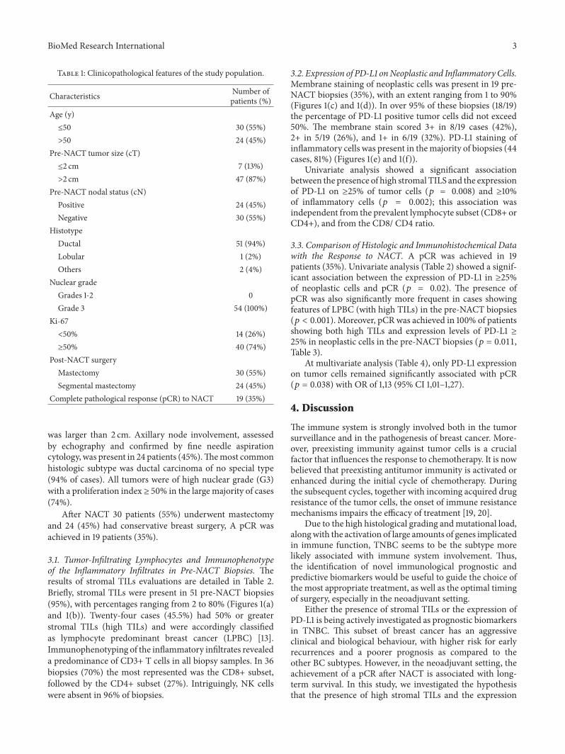
Table 1: Clinicopathological features of the study population.
Characteristics Number ofpatients (%)
Age (y)≤50 30 (55%)>50 24 (45%)
Pre-NACT tumor size (cT)≤2 cm 7 (13%)>2 cm 47 (87%)
Pre-NACT nodal status (cN)Positive 24 (45%)Negative 30 (55%)
HistotypeDuctal 51 (94%)Lobular 1 (2%)Others 2 (4%)
Nuclear gradeGrades 1-2 0Grade 3 54 (100%)
Ki-67<50% 14 (26%)≥50% 40 (74%)
Post-NACT surgeryMastectomy 30 (55%)Segmental mastectomy 24 (45%)
Complete pathological response (pCR) to NACT 19 (35%)
was larger than 2 cm. Axillary node involvement, assessedby echography and confirmed by fine needle aspirationcytology, was present in 24 patients (45%).Themost commonhistologic subtype was ductal carcinoma of no special type(94% of cases). All tumors were of high nuclear grade (G3)with a proliferation index ≥ 50% in the large majority of cases(74%).
After NACT 30 patients (55%) underwent mastectomyand 24 (45%) had conservative breast surgery, A pCR wasachieved in 19 patients (35%).
3.1. Tumor-Infiltrating Lymphocytes and Immunophenotypeof the Inflammatory Infiltrates in Pre-NACT Biopsies. Theresults of stromal TILs evaluations are detailed in Table 2.Briefly, stromal TILs were present in 51 pre-NACT biopsies(95%), with percentages ranging from 2 to 80% (Figures 1(a)and 1(b)). Twenty-four cases (45.5%) had 50% or greaterstromal TILs (high TILs) and were accordingly classifiedas lymphocyte predominant breast cancer (LPBC) [13].Immunophenotyping of the inflammatory infiltrates revealeda predominance of CD3+ T cells in all biopsy samples. In 36biopsies (70%) the most represented was the CD8+ subset,followed by the CD4+ subset (27%). Intriguingly, NK cellswere absent in 96% of biopsies.
3.2. Expression of PD-L1 onNeoplastic and Inflammatory Cells.Membrane staining of neoplastic cells was present in 19 pre-NACT biopsies (35%), with an extent ranging from 1 to 90%(Figures 1(c) and 1(d)). In over 95% of these biopsies (18/19)the percentage of PD-L1 positive tumor cells did not exceed50%. The membrane stain scored 3+ in 8/19 cases (42%),2+ in 5/19 (26%), and 1+ in 6/19 (32%). PD-L1 staining ofinflammatory cells was present in themajority of biopsies (44cases, 81%) (Figures 1(e) and 1(f)).
Univariate analysis showed a significant associationbetween the presence of high stromal TILS and the expressionof PD-L1 on ≥25% of tumor cells (𝑝 = 0.008) and ≥10%of inflammatory cells (𝑝 = 0.002); this association wasindependent from the prevalent lymphocyte subset (CD8+ orCD4+), and from the CD8/ CD4 ratio.
3.3. Comparison of Histologic and Immunohistochemical Datawith the Response to NACT. A pCR was achieved in 19patients (35%). Univariate analysis (Table 2) showed a signif-icant association between the expression of PD-L1 in ≥25%of neoplastic cells and pCR (𝑝 = 0.02). The presence ofpCR was also significantly more frequent in cases showingfeatures of LPBC (with high TILs) in the pre-NACT biopsies(𝑝 < 0.001). Moreover, pCR was achieved in 100% of patientsshowing both high TILs and expression levels of PD-L1 ≥25% in neoplastic cells in the pre-NACT biopsies (𝑝 = 0.011,Table 3).
At multivariate analysis (Table 4), only PD-L1 expressionon tumor cells remained significantly associated with pCR(𝑝 = 0.038) with OR of 1,13 (95% CI 1,01–1,27).
4. Discussion
The immune system is strongly involved both in the tumorsurveillance and in the pathogenesis of breast cancer. More-over, preexisting immunity against tumor cells is a crucialfactor that influences the response to chemotherapy. It is nowbelieved that preexisting antitumor immunity is activated orenhanced during the initial cycle of chemotherapy. Duringthe subsequent cycles, together with incoming acquired drugresistance of the tumor cells, the onset of immune resistancemechanisms impairs the efficacy of treatment [19, 20].
Due to the high histological grading andmutational load,alongwith the activation of large amounts of genes implicatedin immune function, TNBC seems to be the subtype morelikely associated with immune system involvement. Thus,the identification of novel immunological prognostic andpredictive biomarkers would be useful to guide the choice ofthe most appropriate treatment, as well as the optimal timingof surgery, especially in the neoadjuvant setting.
Either the presence of stromal TILs or the expression ofPD-L1 is being actively investigated as prognostic biomarkersin TNBC. This subset of breast cancer has an aggressiveclinical and biological behaviour, with higher risk for earlyrecurrences and a poorer prognosis as compared to theother BC subtypes. However, in the neoadjuvant setting, theachievement of a pCR after NACT is associated with long-term survival. In this study, we investigated the hypothesisthat the presence of high stromal TILs and the expression

4 BioMed Research International
(a) (b)
(c) (d)
(e) (f)
Figure 1: Evaluation of stromal TILs and PD-L1 expression in TNBC core biopsies. (a)-(b): low (a) and high (b) level of stromal tumor-infiltrating lymphocytes (haematoxylin and eosin, original magnification ×10). (c)-(d): membranous PD-L1 stain in scattered (c) and diffuseneoplastic cells (d) (PD-L1 immunohistochemical stain, original magnification ×20). (e)-(f): membranous PD-L1 stain in scattered (e) anddiffuse inflammatory cells (f) (PD-L1 immunohistochemical stain, original magnification ×20).
of PD-L1, both markers of immune activation in the tumormicroenvironment, could be associated with the rate of pCRin TNBC.
We evaluated pre-NACT core biopsies, which proved tobe qualitatively and quantitatively adequate for our analysis.In our study population, 35% of patients achieved a pCR,which is in line with recently published literature [21, 22].
On microscopic evaluation of pre-NACT core biopsies,tumor cell expression of PD-L1 was observed in 35% of cases,although at low levels (≥1% <25% in 15/19 biopsies, 79%).Our observation on a pure sample of TNBC confirms the
results of Dill et al. [16] who analyzed a large number of BCwith various histologic subtypes, showing the highest rate ofPD-L1 expression (32%) in TNBC, with only 5% with diffuseexpression on tumor cells (>50%).
PD-L1 expression, both on neoplastic and inflammatorycells, was significantly associated with high stromal TILs.Our observation extends the results reported by Mori et al.[23], which showed a significant association between PD-L1 expression on tumor cells and percentage of stromalTILs on surgical breast specimens, and confirms the parallelbehaviour of these immune biomarkers in TNBC.The limited

BioMed Research International 5
Table 2: Association between stromal TILs, the expression of PD-L1 on tumor cells and inflammatory infiltrate, Ki-67 value, cT, cN, and pCRin the univariate analysis.
𝑁 (%) pCR𝑁∘ 𝑝 value
Stromal TILsAbsent/low 32 (59%) 8
<0.001High 22 (41%) 11
PD-L1 on tumor cells0% 35 (65%) 11≥1–<25% 15 (28%) 4≥25% 4 (7%) 4 0.024
PD-L1 on inflammatory cellsNegative 10 (19%) 3 nsPositive 44 (81%) 15
Ki-67<50% 14 (26%) 4 ns≥50% 40 (74%) 15
cTT1 7 (13%) 3 nsT2–T4 47 (87%) 16
cNNegative 30 (56%) 13 nsPositive 24 (44%) 6
Table 3:The achievement of pCR according to levels of both stromalTILs and PD-L1 expression on neoplastic cell membranes (lowTILs/low PD-L1; high TILs/low PD-L1; low TILs/high PD-L1; highTILs/high PD-L1).
PD-L1 on neoplastic cellsTILs <25% ≥25%
pt pt pCR pt pCRLow 32 32 25% 0
𝑝 = 0.011High 22 18 39% 4 100%
amount of published reports in pure cohorts of TNBC seemsto suggest a favorable prognostic role of PD-L1, despitesome discrepancies. Mori et al. [23] demonstrated that theinteraction between TILs and PD-L1 correlates with a betterclinical outcome. However, when high PD-L1 expression isassociated with low levels of stromal TILs the prognosisis poor [24]. In the study of Beckers et al. [25] PD-L1,although associated with a better outcome, failed to show anindependent prognostic role in this subset of tumors. Thesepartial discrepancies could be explained by differences in thechoice of clinical outcomes, in the methods of evaluation ofPD-L1 expression on neoplastic cells (membranous versuscytoplasmic) and the cut-off values adopted, and in theantibodies used and the type of sample evaluated (corebiopsies versus surgical samples).
There are only limited data on the predictive value of thesetwo biomarkers in TNBC.We found that in this breast cancer
subtype the concomitant expression of stromal TILs and PD-L1 on tumor cells membranes was significantly associatedwith pCR. According to our results, a cut-off of PD-L1membrane expression on ≥25% of neoplastic cells in pre-neoadjuvant biopsies predicted pCR for TNBC, regardless ofstaining intensity. On the contrary, the predictive role of TILsshowed only a limited power and no statistical associationon multivariate analysis. In light of the preliminary resultsof the KEYNOTE 173 phase II trial [26], reporting a 90%pCR rate in TNBC treated in this setting with the adjunctof pembrolizumab to standard chemotherapy, we hypothesizethat TNBC expressing PD-L1 in less than 25% of tumor cellscould represent the subset most likely to benefit from thisassociation.
Immunophenotyping of tumor inflammatory microenvi-ronment revealed an excess of CD8+with a ratio of CD8/CD4> 1, in line with previous reports [27, 28], although thisobservation did not reach statistical significance probably dueto our limited sample size. Additionally, we found negligibleamounts of NK cells in pre-NACT biopsies, although wecannot exclude the fact that their level could have beenincreased after the first cycle of chemotherapy due to tumorcells death and the release tumor associated antigens.
In conclusion, we showed that a cut-off value of PD-L1in ≥25% of tumor cells predicts pCR in TNBC and to ourknowledge our study is the first dealing with an exclusivepopulation of TNBC cases. A possible explanation for ourobservation is that PD-L1 expression could be associated witha subpopulation of TNBC with a more aggressive behaviour,

6 BioMed Research International
Table 4: Association between the expression of PD-L1 on neoplastic cells and inflammatory infiltrate, stromal TILs, Ki67, clinical T, clinicalN, and pCR in the multivariate analysis.
𝑁 (%) pCR𝑁 (%) 𝑝 value ORR (CI)
Stromal TILsLow 32 (60%) 8 (25%) 0.5 1,61 (0,40–6,52)High 22 (40%) 11 (50%)
PD-L1 on tumor cells0% 35 (65%) 11 (31%)
0.038 1,13 (1,01–1,27)1–25% 15 (28%) 4 (27%)≥25% 4 (7%) 4 (100%)
PD-L1 on inflammatory cellsNegative 10 (18%) 3 (30%) 0.058 0,09 (0,01–1,08)Positive 44 (82%) 15 (34%)
Ki-67<50% 14 (26%) 4 (28%) 0.054 1,05 (1–1,09)≥50% 40 (74%) 15 (37%)
Clinical TT1 7 (13%) 3 (43%) 0.8 0,8 (0,08–8,09)T2–T4 47 (87%) 16 (34%)
Clinical NNegative 30 (55%) 13 (43%) 0.27 0,47 (0,12–1,82)Positive 24 (45%) 6 (25%)
likely to respond to chemotherapy. Further studies with largernumber of cases are warranted to confirm our findings.
Consent
Informed consent was waived from the Ethical Committee.
Disclosure
The preliminary results of this work were presented as aposter at the ESMO 2017 Congress in Madrid, Spain.
Conflicts of Interest
The authors declare no potential conflicts of interest withrespect to the research, authorship, and/or publication of thisarticle.
Acknowledgments
The authors thank Dr. Elisa Concetta Onesti for her help inthe analysis of data and in the preparation of the abstract forESMO.
References
[1] P. Boyle, “Triple-negative breast cancer: epidemiological con-siderations and recommendations,” Annals of Oncology, vol. 23,Supplement 6, Article ID mds187, pp. vi7–vi12, 2012.
[2] P. Rastogi, S. J. Anderson, H. D. Bear et al., “Preoperativechemotherapy: Updates of national surgical adjuvant breast
and bowel project protocols B-18 and B-27,” Journal of ClinicalOncology, vol. 26, no. 5, pp. 778–785, 2008.
[3] V. Guarneri, K. Broglio, S. Kau et al., “Prognostic Value ofPathologic Complete Response After Primary Chemotherapyin Relation to Hormone Receptor Status and Other Factors,”Journal of Clinical Oncology, vol. 24, no. 7, pp. 1037–1044, 2006.
[4] W. D. Foulkes, I. E. Smith, and J. S. Reis-Filho, “Triple-negativebreast cancer,” The New England Journal of Medicine, vol. 363,no. 20, pp. 1938–1948, 2010.
[5] M. Ono, H. Tsuda, C. Shimizu et al., “Tumor infiltratinglymphocytes are correlated with response to neoadjuvantchemotherapy in triple-negative breast cancer,” Breast CancerResearch and Treatment, vol. 132, no. 3, pp. 793–805, 2012.
[6] R. Yamaguchi, M. Tanaka, A. Yano et al., “Tumor-infiltratinglymphocytes are important pathologic predictors for neoad-juvant chemotherapy in patients with breast cancer,” HumanPathology, vol. 43, no. 10, pp. 1688–1694, 2012.
[7] C. Denkert, G. von Minckwitz, J. C. Brase et al., “Tumor-Infiltrating Lymphocytes and Response to NeoadjuvantChemotherapy With or Without Carboplatin in HumanEpidermal Growth Factor Receptor 2–Positive and Triple-Negative Primary Breast Cancers,” Journal of Clinical Oncology,vol. 33, no. 9, pp. 983–991, 2015.
[8] G. Pruneri, A. Vingiani, V. Bagnardi et al., “Clinical validity oftumor-infiltrating lymphocytes analysis in patients with triple-negative breast cancer,” Annals of Oncology, vol. 27, no. 2, pp.249–256, 2016.
[9] M. Ilie, V. Hofman, M. Dietel, J. Soria, and P. Hofman,“Assessment of the PD-L1 status by immunohistochemistry:challenges and perspectives for therapeutic strategies in lungcancer patients,” Virchows Archiv, vol. 468, no. 5, pp. 511–525,2016.

BioMed Research International 7
[10] M. J. Butte, V. Pena-Cruz, M.-J. Kim, G. J. Freeman, and A.H. Sharpe, “Interaction of human PD-L1 and B7-1,” MolecularImmunology, vol. 45, no. 13, pp. 3567–3572, 2008.
[11] S. Muenst, A. R. Schaerli, F. Gao et al., “Expression ofprogrammed death ligand 1 (PD-L1) is associated with poorprognosis in human breast cancer,” Breast Cancer Research andTreatment, vol. 146, no. 1, pp. 15–24, 2014.
[12] K. A. Schalper, V. Velcheti, D. Carvajal et al., “In Situ TumorPD-L1 mRNA Expression Is Associated with Increased TILsand Better Outcome in Breast Carcinomas,” Clinical CancerResearch, vol. 20, no. 10, pp. 2773–2782, 2014.
[13] R. Salgado, C. Denkert, S. Demaria et al., “The evaluationof tumor-infiltrating lymphocytes (TILs) in breast cancer:recommendations by an International TILs Working Group2014,” Annals of Oncology, vol. 26, no. 2, pp. 259–271, 2015.
[14] F. R. Hirsch, A.McElhinny, D. Stanforth et al., “PD-L1 immuno-histochemistry assays for lung cancer: results from phase 1 ofthe blueprint PD-L1 IHC assay comparison project,” Journal ofThoracic Oncology, vol. 12, no. 2, pp. 208–222, 2017.
[15] A. Rittmeyer, F. Barlesi, D. Waterkamp et al., “Atezolizumabversus docetaxel in patients with previously treated non-small-cell lung cancer (OAK): a phase 3, open-label, multicentrerandomised controlled trial,”TheLancet, vol. 389, no. 10066, pp.255–265, 2017.
[16] E. A. Dill, A. A. Gru, K. A. Atkins et al., “PD-L1 Expression andIntratumoral Heterogeneity Across Breast Cancer Subtypes andStages,”The American Journal of Surgical Pathology, vol. 41, no.3, pp. 334–342, 2017.
[17] W. F. Symmans, F. Peintinger, C. Hatzis et al., “Measurementof residual breast cancer burden to predict survival afterneoadjuvant chemotherapy,” Journal of Clinical Oncology, vol.25, no. 28, pp. 4414–4422, 2007.
[18] http://www3.mdanderson.org/app/medcalc/index.cfm?page-name=jsconvert3, 2007.
[19] F. Ghiringhelli and L. Apetoh, “The interplay between theimmune system and chemotherapy: emerging methods foroptimizing therapy,” Expert Review of Clinical Immunology, vol.10, no. 1, pp. 19–30, 2013.
[20] S. Demaria, M. D. Volm, R. L. Shapiro et al., “Development oftumor-infiltrating lymphocytes in breast cancer after neoadju-vant paclitaxel chemotherapy,” Clinical Cancer Research, vol. 7,pp. 3025–3030, 2001.
[21] T. Gamucci, L. Pizzuti, I. Sperduti et al., “Neoadjuvantchemotherapy in triple-negative breast cancer: A multicentricretrospective observational study in real-life setting,” Journal ofCellular Physiology, 2017.
[22] Z. Shao, S. Chaudhri, M. Guo, L. Zhang, and D. Rea, “Neoad-juvant Chemotherapy in Triple Negative Breast Cancer: AnObservational Study,” Oncology Research : Featuring Preclinicaland Clinical Cancer Therapeutics, vol. 23, no. 6, pp. 291–302,2016.
[23] H.Mori,M.Kubo, R. Yamaguchi et al., “The combination of PD-L1 expression and decreased tumor-infiltrating lymphocytesis associated with a poor prognosis in triple-negative breastcancer,” Oncotarget, vol. 8, no. 9, pp. 15584–15592, 2017.
[24] N. Tomioka, M. Azuma, M. Ikarashi et al., “The therapeuticcandidate for immune checkpoint inhibitors elucidated bythe status of tumor-infiltrating lymphocytes (TILs) and pro-grammed death ligand 1 (PD-L1) expression in triple negativebreast cancer (TNBC),” Breast Cancer, 2017.
[25] R. K. Beckers, C. I. Selinger, R. Vilain et al., “Programmed deathligand 1 expression in triple-negative breast cancer is associated
with tumour-infiltrating lymphocytes and improved outcome,”Histopathology, vol. 69, no. 1, pp. 25–34, 2016.
[26] R. Nanda, L. Q. M. Chow, E. C. Dees et al., “Pembrolizumab inpatients with advanced triple-negative breast cancer: phase IbKEYNOTE-012 Study,” Journal of Clinical Oncology, vol. 34, pp.2460–2467, 2016.
[27] S. M. A. Mahmoud, E. C. Paish, D. G. Powe et al., “Tumor-infiltrating CD8+ lymphocytes predict clinical outcome inbreast cancer,” Journal of Clinical Oncology, vol. 29, no. 15, pp.1949–1955, 2011.
[28] A. N. Seo, H. J. Lee, E. J. Kim et al., “Tumour-infiltratingCD8+ lymphocytes as an independent predictive factor forpathological complete response to primary systemic therapy inbreast cancer,” British Journal of Cancer, vol. 109, no. 10, pp.2705–2713, 2013.
Research ArticleDynamics of Neutrophils-to-Lymphocyte Ratio PredictOutcomes of PD-1/PD-L1 Blockade
Michele Moschetta,1 Mario Uccello,1 Benjamin Kasenda,2
Gabriel Mak,1 Anissa McClelland,1,3 Stergios Boussios,4
Martin Forster,3 and Hendrik-Tobias Arkenau1,3
1Drug Development Unit, Sarah Cannon Research Institute, London, UK2University Hospital of Basel, Basel, Switzerland3The University College London Cancer Institute, London, UK4Department of Medical Oncology, University of Ioannina, Ioannina, Greece
Correspondence should be addressed to Michele Moschetta; [email protected]
Received 20 August 2017; Accepted 5 November 2017; Published 28 November 2017
Academic Editor: Ilaria G. Zizzari
Copyright © 2017 Michele Moschetta et al. This is an open access article distributed under the Creative Commons AttributionLicense, which permits unrestricted use, distribution, and reproduction in any medium, provided the original work is properlycited.
Introduction. Baseline neutrophil-to-lymphocyte ratio (NLR) has been repeatedly reported as a significant prognostic factor inadvanced cancer patients. We explored whether changes in NLR may predict outcome of advanced cancer patients enrolled intophase 1 trials and treated with PD-1/PD-L1 inhibitors. Patients and Methods. Advanced cancer patients enrolled into phase 1 trialsbetween September 2013 andMay 2016 and treated with anti-PD-1/PD-L1 agents were included in this retrospective study. NLRwascalculated at baseline and after 2 cycles of treatment. Royal Marsden Hospital (RMH) prognostic score and Eastern CooperativeGroup (ECOG) performance status (PS) were determined at baseline. Kaplan-Meier estimation and Cox regression analyses wereused to assess the impact of NLR dynamics on PFS. Results. Among the 55 patients eligible, 26 (47%) were treated with anti-PD-L1 monotherapy, 22 (40%) received single agent anti-PD-1, and 7 (13%) were given a tyrosine kinase inhibitor (TKI) plus a PD-1inhibitor. Neither ECOG PS nor RMH prognostic score was significantly associated with PFS in our cohort, whereas changes inNLR significantly impacted on PFS. Conclusion. Changes in the NLRmay be a useful predicting factor in advanced cancer patientstreated with anti-PD-1/PD-L1 agents. Further prospective trials are needed to verify these findings.
1. Introduction
Immune checkpoint inhibitors have emerged as potent andeffective treatments for various types of haematological andsolid malignancies [1]. In particular, blockade of the PD-1/PD-L1 axis can result in dramatic and sustained tumourregression in multiple cancer types [2, 3]. Under normalcircumstances, this pathway is necessary tomaintain immunehomeostasis [4]. When PD-L1 binds to PD-1, an inhibitorysignal is transmitted into the T-cell, protecting normal cellsfrom collateral damage. Nevertheless, upregulation of PD-L1 may allow cancer cells to evade immune surveillance[3]. Considering the costs and potential side effects of novelanti-PD-1/PD-L1 agents, it is of vital importance to identify
reliable biomarkers to select the most suitable patients forthese drugs while sparing nonresponders from toxicity.
PD-L1 expression as determined by immunohistochem-istry is considered the most useful biomarker in predictingoutcomes of PD-1/PD-L1 blockade [4]. Several studies haveinvestigated the role of PD-L1 expression in tumour andstromal cells as a potential biomarker of response, but theresults were somewhat contradictory [4, 5]. Indeed, severalfactors can limit the reliability of this biomarker, includingthe use of different monoclonal antibodies for detection ofPD-L1, variable procedures for biopsy collection and storage,lack of defined thresholds to describe PD-L1 expression insamples, and intratumour heterogeneity in PD-L1 expres-sion [5] The presence of microsatellite instability, tumour
HindawiBioMed Research InternationalVolume 2017, Article ID 1506824, 5 pageshttps://doi.org/10.1155/2017/1506824

2 BioMed Research International
mutational load, tumour-infiltrating lymphocytes (TILs),myeloid-derived suppressor cells (MDSCs), indoleamine 2,3-dioxygenase, regulatory T cells, and immune specific signa-tures have been also investigated with promising results [6–8]. Despite the aforementioned methods, there is still a lackof a simple, effective, and definitive biomarker of response toimmune checkpoint inhibitors.
Increased neutrophil-to-lymphocyte ratio (NLR) hasbeen reported as an independent poor prognostic indicatorin several malignancies and its normalisation followingtreatment has been found to predict survival in cancerpatients considered for early phase clinical trials [9]. Here,we investigated the usefulness of NRL changes in predict-ing progression-free survival (PFS) in patients undergoingtreatment with PD-1/PD-L1 inhibitors within phase 1 clinicaltrials.
2. Patients and Methods
Data of metastatic cancer patients enrolled in phase 1 trialsbetween September 2013 andMay 2016 in our institutionwereretrospectively reviewed. Patients treated with PD-1/PD-L1checkpoint-directed therapy were eligible. All the subjectshad a histologically confirmed diagnosis of metastatic solidcancer and were intended to receive treatment with an anti-PD-1/PD-L1 agent given as monotherapy or in combinationwith a tyrosine kinase inhibitor (TKI). Baseline parameters,tumour characteristics, and treatment data were all reviewedand anonymously collected for this study. All the subjectsmet the standard inclusion criteria for phase 1 trials: EasternCooperative Group (ECOG) performance status (PS) 0 or 1;measurable disease based on Response Evaluation Criteria inSolid Tumour (RECIST); adequate bone marrow, liver, andkidney function; life expectancy of at least 3 months. Baselinecharacteristics recorded in the eligible population includeddemographic variables, tumour type, anticancer treatment(anti-PD-1 versus anti-PD-L1 versus anti-PD-L1 plus TKI),number of previous lines for metastatic disease, Royal Mars-den Hospital (RMH) prognostic score [10], white bloodcell (WBC) level, absolute neutrophil count (ANC), abso-lute lymphocyte count (ALC), and neutrophil-to-lymphocyteratio (NLR). The RMH prognostic score (range 0–3) wascalculated at baseline, taking into account albumin level,lactate dehydrogenase (LDH) level, and number ofmetastaticsites [10].TheNLRwas calculated using the standard formula:NLR = ANC/ALC. NLR was calculated at baseline (cycle 1day 1), and after 6 weeks (2 cycles) of treatment. Patientswere treated until disease progression, death, or unacceptabletoxicity. We considered PFS as our main outcome, which wasdefined as the time from treatment start until progression ordeath, whichever occurred first.
To investigate the dynamics in NLR between baselineand after 2 cycles of anti-PD-1/PD-L1 therapy, we used alandmark approach by excluding patients who were not ableto receive at least 2 cycles of treatment to avoid guaranteetime bias. We used multivariate Cox regression analyses withthe relative NLR difference as independent and PFS as thedependent variable. To adjust for possible confounding, we
Table 1: Patients’ characteristics at baseline. NCSLC = non-smallcell lung cancer; ECOG PS = Eastern Cooperative Oncology Groupperformance status; GI = gastrointestinal; TKI = tyrosine kinaseinhibitor; RMH = Royal Marsden Hospital.
Characteristic 𝑛 (%)Sex
Male 19 (35)Female 36 (65)
ECOG PS0 36 (65)1 19 (35)>1 —
Tumour typeNSCLC 18 (33)Upper GI cancer 11 (20)Bladder cancer 8 (15)Renal cell carcinoma 8 (15)Breast cancer 7 (13)Colorectal cancer 2 (4)Ovarian cancer 1 (2)
TherapyAnti-PD-1 22 (40)Anti-PD-L1 26 (47)Anti-PD-L1 plus TKI 7 (13)
RMH prognostic score0 31 (56)1 19 (35)2 3 (5)3 2 (4)
Median (range)Age 61 (40–80)Number of metastatic sites 2 (1–4)Number of previous treatment lines 1 (1–6)
introduced the RMH score into the model and addition-ally added a random effect for tumour entity, in order toaccount for possible heterogeneity between tumour types.We calculated univariate andmultivariate hazard ratios (HR)with accompanied 95% confidence intervals (CI); however,the multivariable analysis is considered as main analysis.To visualize the prognostic effect of the NLR difference,we created Kaplan-Meier plots. All 𝑝 values are exploratoryin nature with a conventional level of significance at 0.05.All analyses were done using the statistical software R(https://www.r-project.org/) and STATA (version 14).
3. Results
A total of 67 potentially eligible patients were identified.Of those, 12 subjects received less than 2 cycles and weretherefore excluded from the analysis. The characteristics ofthe included 55 patients are summarised in Table 1. Medianage of patients included was 61 years (40 to 80 years).The most represented tumour type was non-small cell lungcancer (NSCLC) with 18 (33%) subjects, followed by upper

BioMed Research International 3
Table 2: Distribution of patient population in two groups. GroupA: neutrophil-to-lymphocyte ratio (NLR) after 2 cycles ≤ medianbaseline NLR. Group B: NLR after 2 cycles >median baseline NLR.ECOG PS = Eastern Cooperative Oncology Group performancestatus; RMH = Royal Marsden Hospital; ANC = absolute neutrophilcount; ALC = absolute lymphocyte count; SD = standard deviation;IQR = interquartile range; NLR = neutrophil-to-lymphocyte ratio.
Characteristic Group A(𝑛 = 28)
Group B(𝑛 = 27)
Sex 𝑛 (%)Female 11 (39) 8 (30)Male 17 (61) 19 (70)
ECOG PS 𝑛 (%)0 16 (43) 20 (74)1 12 (57) 7 (26)
RMH prognostic score 𝑛 (%)0-1 25 (89) 25 (93)2-3 3 (11) 2 (7)
Intervention 𝑛 (%)Anti-PD-1 14 (50) 8 (30)Anti-PD-L1 9 (32) 17 (63)Anti-PD-1 plus TKI 5 (18) 2 (7)
Baseline ANCMean (SD) 5.1 (1.8) 4.1 (1.4)Baseline ALCMean (SD) 1.3 (0.6) 1.3 (0.5)Baseline NLRMedian (IQR) 3.9 (2.7–5.6) 3.0 (2.5–4.8)ANC after 2 cyclesMedian (IQR) 3.7 (2.8–5.1) 4.5 (3.5–5.7)ALC after 2 cyclesMean (SD) 1.4 (0.5) 1.2 (0.5)NLR after 2 cyclesMedian (IQR) 2.9 (2.2–3.7) 3.9 (2.8–7)
gastrointestinal (𝑛 = 10; 18%), bladder (𝑛 = 8; 15%), andbreast (𝑛 = 7; 13%) carcinomas. Median number of previouslines of treatment for metastatic disease was 1 (range 1–6)while baseline median number of metastatic sites of diseasewas 2 (range 1–4). RMH prognostic score at baseline was 0in 31 (56%) subjects and 1 or higher in 24 (64%) subjects.Proportion of patients with ECOGPS 0 or 1 was 36 (65%) and19 (35%), respectively. In total, 26 (47%) of the patients weretreated with anti-PD-L1 monotherapy, 22 (40%) receivedsingle agent anti-PD-1, and 7 (13%) were given a TKI incombination with a PD-1 inhibitor.
Onunivariate analysis, baselineNLR, treatmentmodality,RMH score, ECOG PS, and number of metastatic sites didnot have significant impact on PFS. Baseline median NLRwas 3.4 in the overall population. Patient population wasdivided into 2 distinct groups based on decrease (GroupA) orincrease (Group B) of NLR in comparison with median NLRafter treatment with a PD-1/PD-L1 inhibitor. No substantialdifferences in distribution were observed between these 2groups in terms of age, sex, type of treatment, ECOG PS,and RMH prognostic score (Table 2). Increased NLR after 2cycles of anti-PD-1/PD-L1 therapy had a negative effect onPFS (HR 1.14, 95% CI 1.06–1.23, 𝑝 = 0.004), (Figure 1).This effect was also observed in our multivariate analysis,
Progression-free survival by median NLR di�erence
Below or equal to median NLR (N = 28)
Above median NLR (N = 27)
log-rank test p = 0.004
200 400 600 8000Time in days since start of treatment
0.0
0.2
0.4
0.6
0.8
1.0
PFS
prob
abili
ty
Figure 1: Progression-free survival (PFS) stratified by median dif-ferences in neutrophil-to-lymphocyte ratio (NLR) between baselineand after 2 doses of treatment with an anti-PD-1/PD-L1 inhibitor,showing longer PFS in patients with a reduction of NLR comparedto the median baseline NLR.
where increased NLR was associated with decreased PFSafter adjusting for RMH prognostic score (HR 1.03, 95%CI 1.01–1.04, 𝑝 < 0.001). Baseline median ANC level wassignificantly higher in Group A than in Group B (𝑝 = 0.029).In Group A, a reduction in median ANC was shown after 2cycles of treatment when compared with baseline ANC level,whereas this was not observed in Group B. After 2 cyclesof treatment with anti-PD-1/PD-L1 agent, median ANC wassignificantly higher in Group B when compared to Group A(𝑝 = 0.014). Median ALC did not change significantly aftertreatment (𝑝 = 0.222) and no significant differences wereshown between baseline and posttreatment values in bothGroup A and Group B (𝑝 = 0.24) (Table 2).
4. Discussion
Molecular selection in patients undergoing treatment withimmune checkpoint inhibitors is an urgent unmet medicalneed. Ongoing approval of several anti-PD-1/PD-L1 agentsand the emergence of safety concerns from immune-relatedadverse events also highlight the need for biomolecular strat-ification. Several biomarkers have been investigated, someof which have shown potential usefulness in predicting theactivity of these agents. So far, PD-L1 expression in tumourcells remains the most reliable but many technical limitationshave been associated with this biomarker [4, 5]. Furthermore,individuals with negative PD-L1 expression can still respondto PD-1/PD-L1 blockade, further questioning the value ofPD-L1 expression as a universal biomarker [11]. Therefore,alternative markers of response need to be identified.
Clinicopathologic factors have been extensively inves-tigated in several tumour types and anticancer therapies.Among them, ECOG PS has been repeatedly reported as astrong predictor of survival in multiple settings. The RMH

4 BioMed Research International
prognostic score was electively implemented in advancedcancer patients enrolled in phase 1 studies [10]. Unexpect-edly, neither ECOG PS nor RMH prognostic score weresignificantly associated with PFS in our cohort. Our resultsare not consistent with previous studies showing significantprognostic significance of RMH score and ECOGPS in phase1 trial patients [10, 12, 13].
Baseline NLR has been reported to predict overallsurvival in cancer patients undergoing both conventionalchemotherapy and targeted treatments, including immunecheckpoint inhibitors. In previous reports, a correlationbetween baseline NLR and survival was shown in kidneycancer and NSCLC treated with IL-2 and nivolumab, respec-tively [14, 15]. Conversely, in our group of patients, baselineNLRwas not found to correlate with PFS, although this resultmay have been determined by the limited sample size. In ourstudy, we retrospectively analysed phase 1 trial cancer patientswith advanced disease who had received at least 2 cycles oftreatment with anti-PD-1/PD-L1 agent, to assess the signif-icance of NLR as an independent biomarker in predictingclinical benefit in terms of PFS. Interestingly, a decrease inNLR after 2 cycles of treatment with PD-1/PD-L1 blockadewas associated with longer PFS in our cohort. Accordingly,changes in NLR had shown to predict better outcomes incancer patients undergoing conventional chemotherapy ortargeted treatment but had beennever investigated in subjectsreceiving immune checkpoint inhibitors [16–18].
Another important finding in this study was the obser-vation that negative or positive changes in NLR were drivenby a decrease in ANC and not by changes in ALC asone would have expected. Though retrospective in nature,our findings on ANC may be interpreted as hypothesis-generating. Despite the fact the T lymphocyte activity isthe main target of PD-1/PD-L1 blockade, our results maysuggest an important interaction between the neutrophils andtumour microenvironment. We may also speculate that thesystemic effect of anti-PD-1/PD-L1 agents involves a crucialeffect on circulating myeloid populations included in theANCasmeasured by automated full blood cell count analyser.Preclinical evidence shows that MDSCs can impair the effi-cacy of immunotherapy [19]. Under physiological conditions,there is a low level of MDSCs in the bloodstream while thesepopulations rapidly expand during immunological responsesto infections, inflammation, and cancer [20]. MDSCs canadopt multiple mechanisms to induce immunosuppression,including production of arginase 1 and inducible nitric oxidesynthase, leading to T-cell inhibition [19, 20].MDSCs are alsoknown to enhance cancer cell proliferation, confer resistanceto anticancer therapies, and promote angiogenesis andmetas-tasis [20]. Concomitant targeting of MDSCs may thereforeincrease the antitumour activity of PD-1/PD-L1 inhibitorsin nonresponders. Moreover, a decreased mobilisation ofMDSCs from the bone marrow may represent a systemiceffect of anti-PD-1/PD-L1 treatment that needs to be betterinvestigated in preclinical studies. A subsequent decrease oftumour-infiltrating MDSCs may then unleash antitumouractivity of TILs and ultimately contribute to the therapeuticeffect of anti-PD-1/PD-L1 agents.
Our study has several biases, including its retrospectivenature, limited sample size, heterogeneous tumour types, andthe choice of PFS as endpoint instead of overall survival.Nevertheless, we showed that NLR, a simple haematologicalparameter easily obtainable in daily clinical practice, may beused to predict clinical benefit from PD-1/PD-L1 inhibitors.These results are in line with common clinical experiencewith these agents, where a rapid clinical benefit can beobserved despite unusual initial patterns of imaging response.Further studies conducted in larger prospective cohortsof patients undergoing treatment with immune checkpointinhibitors are needed to confirm the predictive role of NLRin this setting.
Conflicts of Interest
The authors declare no conflicts of interest.
Authors’ Contributions
Michele Moschetta, Mario Uccello, and Benjamin Kasendaequally contributed to the article.
Acknowledgments
The authors are grateful to the patients and their families forparticipating in these clinical studies.
References
[1] A. Goodman, S. P. Patel, and R. Kurzrock, “PD-1–PD-L1immune-checkpoint blockade in B-cell lymphomas,” NatureReviews Clinical Oncology, 2016.
[2] J. Hamanishi,M.Mandai, N.Matsumura, K. Abiko, T. Baba, andI. Konishi, “PD-1/PD-L1 blockade in cancer treatment: perspec-tives and issues,” International Journal of Clinical Oncology, vol.21, no. 3, pp. 462–473, 2016.
[3] D. M. Pardoll, “The blockade of immune checkpoints in cancerimmunotherapy,”Nature Reviews Cancer, vol. 12, no. 4, pp. 252–264, 2012.
[4] V. A. Boussiotis, “Molecular and biochemical aspects of the PD-1 checkpoint pathway,” The New England Journal of Medicine,vol. 375, no. 18, pp. 1767–1778, 2016.
[5] S. L. Topalian, J. M. Taube, R. A. Anders, and D. M. Pardoll,“Mechanism-driven biomarkers to guide immune checkpointblockade in cancer therapy,” Nature Reviews Cancer, vol. 16, no.5, pp. 275–287, 2016.
[6] J. Galon, A. Costes, F. Sanchez-Cabo et al., “Type, density,and location of immune cells within human colorectal tumorspredict clinical outcome,” Science, vol. 313, no. 5795, pp. 1960–1964, 2006.
[7] R. B. Holmgaard, D. Zamarin, A. Lesokhin, T. Merghoub, andJ. D. Wolchok, “Targeting myeloid-derived suppressor cellswith colony stimulating factor-1 receptor blockade can reverseimmune resistance to immunotherapy in indoleamine 2,3-dioxygenase-expressing tumors,” EBioMedicine, vol. 6, pp. 50–58, 2016.
[8] W. Joost Lesterhuis, A. Bosco, M. J. Millward, M. Small, A. K.Nowak, and R. A. Lake, “Dynamic versus static biomarkers in

BioMed Research International 5
cancer immune checkpoint blockade: Unravelling complexity,”Nature Reviews Drug Discovery, vol. 16, no. 4, pp. 264–272, 2017.
[9] D. J. Pinato, C. Stavraka, M. J. Flynn et al., “An inflammationbased score can optimize the selection of patientswith advancedcancer considered for early phase clinical trials,” PLoS ONE, vol.9, article e83279, no. 1, 2014.
[10] H.-T. Arkenau, J. Barriuso, D. Olmos et al., “Prospective vali-dation of a prognostic score to improve patient selection foroncology phase I trials,” Journal of Clinical Oncology, vol. 27, no.16, pp. 2692–2696, 2009.
[11] C. Grigg and N. A. Rizvi, “PD-L1 biomarker testing for non-small cell lung cancer: Truth or fiction?” Journal for Immuno-Therapy of Cancer, vol. 4, no. 1, article 48, 2016.
[12] I. Garrido-Laguna, F. Janku, C. Vaklavas et al., “Validation of theRoyal Marsden Hospital prognostic score in patients treated inthe phase i clinical trials program at the MD Anderson CancerCenter,” Cancer, vol. 118, no. 5, pp. 1422–1428, 2012.
[13] T. Kawaguchi, M. Takada, A. Kubo et al., “Performance statusand smoking status are independent favorable prognostic fac-tors for survival in non-small cell lung cancer: A comprehensiveanalysis of 26,957 patients with NSCLC,” Journal of ThoracicOncology, vol. 5, no. 5, pp. 620–630, 2010.
[14] J. A. Kuzman, D. D. Stenehjem, J. Merriman et al., “Neutrophil-lymphocyte ratio as a predictive biomarker for response to highdose interleukin-2 in patients with renal cell carcinoma,” BMCUrology, vol. 17, article 1, no. 1, 2017.
[15] S. J. Bagley, S. Kothari, C. Aggarwal et al., “Pretreatment neutro-phil-to-lymphocyte ratio as a marker of outcomes in nivolu-mab-treated patients with advanced non-small-cell lung can-cer,” Lung Cancer, vol. 106, pp. 1–7, 2017.
[16] W. Chua, K. A. Charles, V. E. Baracos, and S. J. Clarke, “Neu-trophil/lymphocyte ratio predicts chemotherapy outcomes inpatients with advanced colorectal cancer,” British Journal ofCancer, vol. 104, no. 8, pp. 1288–1295, 2011.
[17] P. Zhang, Y. Zong, M. Liu, Y. Tai, Y. Cao, and C. Hu, “Predictionoutcome in breast cancer patients using test parameters fromcomplete blood count,”Molecular and Clinical Oncology, vol. 4,no. 6, pp. 918–924, 2016.
[18] L. Kasmann, L. Bolm, S. E. Schild, S. Janssen, and D. Rades,“Neutrophil-to-Lymphocyte Ratio Predicts Outcome in Lim-ited Disease Small-cell Lung Cancer,” Lung, vol. 195, no. 2, pp.217–224, 2017.
[19] S. L. Highfill, Y. Cui, A. J. Giles et al., “Disruption of CXCR2-mediated MDSC tumor trafficking enhances anti-PD1 efficacy,”Science Translational Medicine, vol. 6, article 237ra67, no. 237,2014.
[20] O. Draghiciu, J. Lubbers, H. W. Nijman, and T. Daemen, “Mye-loid derived suppressor cells—an overview of combat strategiesto increase immunotherapy efficacy,” OncoImmunology, vol. 4,no. 1, Article ID 954829, 11 pages, 2015.

Research ArticlePD-L1 Promotes Self-Renewal and Tumorigenicity of MalignantMelanoma Initiating Cells
Fang Zheng,1 Jianzhong Dang,2 Hui Zha,1 Bingyu Zhang,1 Ming Lin,1 and Fanjun Cheng3
1Department of Pediatrics, Union Hospital, Tongji Medical College, Huazhong University of Science and Technology,Wuhan 430022, China2Department of Geriatrics, Renmin Hospital of Wuhan University, Wuhan 430020, China3Department of Hematology, UnionHospital, TongjiMedical College, HuazhongUniversity of Science and Technology,WuHan, China
Correspondence should be addressed to Fanjun Cheng; [email protected]
Received 22 April 2017; Accepted 10 September 2017; Published 9 November 2017
Academic Editor: Ilary Ruscito
Copyright © 2017 Fang Zheng et al. This is an open access article distributed under the Creative Commons Attribution License,which permits unrestricted use, distribution, and reproduction in any medium, provided the original work is properly cited.
Recent studies have indicated that therapeutic antibodies targeting PD-L1 show remarkable efficacy in clinical trials in multipletumors and that a melanoma cell-intrinsic PD-1: PD-L1 axis promotes tumor growth. However, few studies have shown tumor-intrinsic PD-L1 effects inmalignantmelanoma initiating cells (MMICs). Here, we aim to determine the possible regulatory effects ofPD-L1 onMMICs.TheALDEFLUOR kit was used to identify ALDH+MMICs. Flow cytometry was used to examine the expressionof PD-L1 on ALDH+MMICs. To determine the role of PD-L1 inMMICs self-renewal, we cultured melanoma cells with anti-PD-L1and measured tumorsphere formation and apoptosis. In addition, the effects of anti-PD-L1 on tumorigenicity and residual ALDH+MMICs in tumors were evaluated in vivo. We demonstrated that melanoma cell-intrinsic PD-L1 was expressed in ALDH+MMICs.Blocking PD-L1 in melanoma cell lines impaired tumorsphere formation and induced the apoptosis of sphere cells. In addition,blocking PD-L1 inhibited tumor growth in vivo. We observed residual ALDH+ MMICs within the tumor. The results showed thatblocking PD-L1 also significantly decreased the residual ALDH+ MMICs in the tumors. In conclusion, these results suggest a newmechanism underlying melanoma progression and PD-L1-targeted therapy, which is distinct from the immunomodulatory actionsof PD-L1.
1. Introduction
Metastatic melanoma is an extraordinarily challenging can-cer, with a 16% 5-year survival rate, and it responds poorly tomost standard chemotherapies [1]. It has been established thatmalignant melanoma initiating cells (MMICs) possess notonly the capacity for self-renewal, differentiation, immuneevasion, and multidrug resistance, but also potentially vas-culogenic mimicry and the ability to transition to migratoryand metastasizing derivatives, which are associated withmelanoma progression and metastasis [2, 3]. For this reason,melanoma cure is predicated upon effectively targeting anderadicating the MMICs.
Recently, it has been established that programmed death-1 (PD-1) is a prominent checkpoint receptor that, upon bind-ing its ligands PD-L1 or PD-L2, dampens T effector functionsby inhibiting signaling downstream of the T cell receptor [4].
PD-L2 is predominantly expressed in APCs, whereas PD-L1 is commonly expressed in various cell types, includingtumor cells, immune cells, epithelial cells, and endothelialcells [1, 5]. When PD-1 binds to its ligands in tumors;it leads to T-cell anergy and blocks productive antitumorimmune response [6].Thefirstmonoclonal antibody directedat PD-1, Nivolumab, was approved for treating patients withunresectable melanoma in July 2014.The other PD-1 and PD-L1 directed agents are currently in Phase I–III clinical trialsin multiple tumor types [7].
In contrast to the immunosuppressive effect of PD-L1,it is also known to contribute to the promotion of tumorcell growth and downregulation of quiescent cells [8, 9].Furthermore, it has been found that glioma stem cells expresslower levels of PD-L1 than differentiated glioma cells do[10]. In head and neck squamous cell carcinoma, PD-L1is preferentially expressed in CD44+ tumor-initiating cells
HindawiBioMed Research InternationalVolume 2017, Article ID 1293201, 8 pageshttps://doi.org/10.1155/2017/1293201
2 BioMed Research International
[11]. PD-L1 also has suppressive effects on cancer stem cell-related phenotypes of cholangiocarcinoma [12]. These recentdata highlight the possible involvement of PD-L1 in theregulation of cancer stem cells in various tumors. However,little research has investigated the role of PD-L1 in MMICs.Here, we report on a study to determine the frequency of PD-L1 expression in MMICs, and the possible regulatory effectsof PD-L1 on MMICs.
2. Materials and Methods
2.1. Cells and Cell Culture. B16-F0 and B16-F1 melanoma celllines were maintained in RPMI 1640 (Gibco) containing 10%fetal bovine serum (FBS; ScienceCell), 100U/ml penicillin(Gibco), and 100 𝜇g/mL streptomycin (Gibco). Cells werecultured at 37∘C in 95% humidity and 5% CO
2atmosphere.
All cell lines were routinely screened for mycoplasma con-tamination.
2.2. Flow Cytometry. The ALDEFLUOR kit (StemCell Tech-nologies, British Columbia, Canada) was used to identifythe stem/progenitor cells that expressed high levels of thealdehyde dehydrogenase (ALDH) [13]. Briefly, 1 ∗ 106/mlcells were suspended in Aldefluor Assay Buffer (AAB) andincubated with 5𝜇L ALDH substrate (BAAA) for 45min.5 𝜇L diethylaminobenzaldehyde (DEAB) was added to aseparate sample containing BAAA for an ALDH-inhibitedcontrol. Then, samples were washed and resuspended inAAB. Fluorescence-activated cell gates were established usingthe inhibited control, DEAB, with the fluorescein isothio-cyanate (FITC) channel with excitation and emission wave-lengths of approximately 495 nm and 521 nm, respectively.To evaluate the expression of PD-L1 in ALDH+ cells, PD-L1antibody (10F.9G2, GeneTex) and immunoglobulin G (IgG)isotype-matched control (GeneTex) containing BAAA wereadded to the cells separately. All samples were incubatedfor 30 minutes at 4∘C. Following incubation, the materialwas centrifuged, and pellets were resuspended with 500𝜇lassay buffer prior to data acquisition. Flow cytometry anal-ysis was performed on a BD Biosciences FACSCanto, anddata analysis was conducted using CellQuest Pro (B&DBiosciences).
2.3. Tumorsphere Culture. The B16-F0 and B16-F1 melanomacells were plated as single cells in ultralow attachment six-wellplates (Corning, Lowell, MA, USA) and cultured in RPMI1640 containing 6mg/mL glucose (Sigma-Aldrich), 1mg/mLNaHCO3 (Sigma-Aldrich), 5mM HEPES (Sigma-Aldrich),4 𝜇g/mL heparin (Sigma-Aldrich), 4mg/mL bovine serumalbumin (Sigma-Aldrich), 20 pg/mL insulin (Sigma-Aldrich),N2 supplement (Invitrogen), supplemented with 10 ng/mLbFGF (Peprotech, Neuilly sur Seine, France), and 20 ng/mLEGF (Peprotech), as previously described [14]. On the secondday after seeding, cells were treated with 10 𝜇g anti-PD-L1(10F.9G2, BioXcell) or control rat immunoglobulin G (IgG).Tumorspheres were observed under microscope 14 days later.Individual spheres with diameters larger than 100𝜇m fromeach replicate well were visualized and counted with aninverted microscope.
2.4. Assay for Apoptosis. Cells were double-stained withFITC-annexin V and PI according to the manufacturer’s in-structions (Annexin-V FITC/propidium iodide (PI) Apopto-sis Detection Kit; BD Pharmingen). Analysis was performedby flow cytometry. Early apoptotic cells were stained withAnnexin-V alone, whereas necrotic and late apoptotic cellswere stained with both Annexin-V and PI.
2.5. Animals and Tumor Model. Adult SPF male C57BL/6mice were implanted subcutaneously on the right flank witheither 5 × 10⁵ B16-F0 or 5 × 10⁵ B16-F1 melanoma cells.Then, 100 𝜇g anti-PD-L1 or control rat IgG was administeredintraperitoneally 3, 6, and 9 days following melanoma cellinoculation. All animals were randomly assigned to twogroups of 5 mice each. Tumor size was monitored every twodays. All surgical procedures and care given to the animalswere in accordance with institutional guidelines.
2.6. Statistical Analysis. All data were reported as the mean± standard error. Statistical analysis was performed usingGraphPad Prism 5.0 Software (San Diego). A two-tailedpaired 𝑡-test was used to determine significant differences. 𝑃values < 0.05 were considered statistically significant.
3. Results
3.1. PD-L1 Expression on ALDH+ Melanoma Cells. Previousstudies have described the isolation of MMICs from miceusing ALDEFLUOR/ALDH as a marker [13, 15]. To deter-mine the expression of PD-L1 in MMICs, we detected PD-L1+/ALDH+ subpopulations from these two cell lines. Asshown in Figure 1, ALDH+ cells were identified in melanomacell lines by flow cytometry with the ALDEFLUOR kit. Cellswere then incubated for 30min with mouse monoclonalantibodies specific for PD-L1. The analysis of the percentageof PD-L1+ALDH+ cells was gated by ALDH+ cells. We foundthat approximately 10% to 18% of the culturedmurine B16-F0cells and B16-F1 cells were ALDH+. Approximately, 5% of theALDH+ cells were PD-L1+/ALDH+. These data suggest thatPD-L1 may be involved in regulating MMICs.
3.2. PD-L1 Regulated on MMICs Tumorsphere Formation.To determine whether PD-L1 can mediate MMIC self-renewal, we cultured melanoma cell lines with anti-PD-L1.The results showed that anti-PD-L1 significantly inhibitedtumorsphere formation in B16-F0 and B16-F1 melanomacells compared to the control groups (Figure 2). Cancerstem cell-derived spheres were dissociated and passaged; theyreadily formed secondary spheres [16]. Anti- PD-L1 inhibitedsecondary tumorsphere generation. Anti-PD-L1 induced a 2-fold inhibition of tumorsphere formation in B16-F0 cells andapproximately 1.4-fold inhibition in B16-F1 melanoma cells,in terms of both number and size, compared with controlgroups.
3.3. PD-L1 Affected the Apoptosis of MMICs Enriched Cells.Tumorsphere formation has been reported as a measure ofthe presence of MMICs in enriched cell populations. Wefurther explored the effects of anti-PD-L1 on apoptosis in

BioMed Research International 3
SSC-
H
0
256
512
768
1024
0.00%
100
101
102
103
104
ALDEFLUOR
B16-
F0 ce
lls
11.2%
SSC-
H
0
256
512
768
1024
100
101
102
103
104
ALDEFLUOR
PD-L
1
0.00%
0.00%
99.7%
0.03%
100
101
102
103
104
100
101
102
103
104
ALDEFLUOR
0.00%
0.00%
5.11%
94.89%
PD-L
1
100
101
102
103
104
100
101
102
103
104
ALDEFLUOR(a)
17.79%
SSC-
H
0
256
512
768
1024
100
101
102
103
104
ALDEFLUOR
0.00%
B16-
F1 ce
llsSS
C-H
0
256
512
768
1024
100
101
102
103
104
ALDEFLUOR
0.00% 5.14%
0.00% 94.86%
PD-L
1
100
101
102
103
104
100
101
102
103
104
ALDEFLUOR
0.00% 0.00%
100% 0.00%
PD-L
1
100
101
102
103
104
100
101
102
103
104
ALDEFLUOR(b)
Figure 1:The expression of PD-L1 onMMICs. (a)The left two scatter plots showed the ALDH+ cells identified in the B16-F0 melanoma cellsby flow cytometry using the ALDEFLUOR kit. Only ALDH+ cells were gated for analysis of the percentage of PD-L1+ALDH+ cells. The righttwo scatter plots showed the percentage of PD-L1+ALDH+ cells in B16-F0 melanoma cells. (b) The expression of PD-L1 in ALDH+ B16-F0melanoma cells.
B16-
F0 ce
lls
Control Anti-PD-L1
(a)
B16-
F1 ce
lls
Control Anti-PD-L1
(b)
0
500
1000
1500
Control Anti-PD-L1Control Anti-PD-L1
Num
ber o
f tum
orsp
here
s
B16-F0 cells B16-F1 cells
P = 0.0006P = 0.0302
(c)
Figure 2: PD-L1 promoted tumorsphere formation. After co-culturing with anti-PD-L1, the sphere formation ability of (a) B16-F0 cells and(b) B16-F1 cells was impaired. (c) The chart showed the number of tumorspheres in each group. Each column represents the mean ± SE ofthree independent experiments.

4 BioMed Research International
melanoma tumorspheres. The data illustrated that anti-PD-L1 induced significant apoptosis in melanoma tumorspheres(Figure 3). Anti-PD-L1 increased the rate of apoptosis by 2-fold in both B16-F0 and B16-F1 tumorspheres. Thus, PD-L1inhibited apoptosis of MMIC-enriched cells.
3.4. Blockage of PD-L1 Directly AffectedMMICs In Vivo. Micewere challenged with melanoma cells (B16-F0 and B16-F1)and treated with 100 𝜇g anti-PD-L1 or control rat IgG 3, 6,and 9 days following melanoma cell injection. Other studieshave reported that anti-PD-L1 significantly suppressed tumorgrowth compared with PBS-injected animals in two animalmodels. Anti-PD-L1 promoted tumor rejection in 50% ofB16-F0 melanoma challenged mice (𝑃 = 0.031) and 50%of B16-F1 melanoma challenged mice (𝑃 = 0.031; Figures4(a) and 4(b)). We observed that anti-PD-L1 decreasedresidual ALDH+ MSCs within the tumor. As shown inFigures 4(c)–4(e), anti-PD-L1 promoted the rejection of 1.5-fold residual ALDH+ MMICs in the B16-F0 animal model(𝑃 = 0.016) and 1.4-fold residual ALDH+ MMICs in theB16-F0 animal model (𝑃 = 0.045). These results suggest thatone mechanism for the anti-tumor effects of anti-PD-L1 isrelated to its ability to suppress the tumorigenicity capacityof MMICs.
4. Discussion
Previous studies have shown that PD-L1 expression is acommon phenomenon in immunotherapy-naive melanomas[17–19]. Further studies have indicated that PD-1 expressedbymelanoma cells is a tumor growth-promotingmechanism,and PD-1-driven tumorigenesis requires interaction betweenmelanoma-PD-1 and host or melanoma-expressed PD-L1[18]. Here, we provide several insights into the function ofPD-L1 in MMICs, which is separate from its effects on theimmune response. Our study found that PD-L1was expressedin ALDH+ MMICs and induced tumorsphere formation.PD-L1 further inhibited the apoptosis of MMIC-enrichedcells. Blockage of PD-L1 directly inhibited tumorigenesisin vivo and significantly decreased the residual percentageof MMICs. These results may indicate that melanoma cell-intrinsic PD-L1 promotes self-renewal and the tumorigeniccapacity of MMICs.
Traditionally, PD-1 ligands have been expressed in tumorcells, leading to T-cell exhaustion and tumor cell evadingthe immune response, which was thought to require itsreceptor interaction [20]. Accordingly, several clinical tri-als have focused on using PD-L1-blocking antibodies toenhance immunity in cancers [21–23]. However, a recentstudy found that melanoma-PD-1: host-PD-L1 interactionspromoted murine melanoma growth [8]. In melanoma, asubpopulation of cells, namely, MMICs, is capable of notonly self-renewal, differentiation, plasticity, immune evasion,and multidrug resistance, but also potentially vasculogenicmimicry, and transitioning to migratory and metastasizingderivatives, which are associated with melanoma progressionand metastasis [24]. Thus, we believe that melanoma-PD-L1may contribute tomaintaining the stem cell-like properties ofMMICs.
MMICs are known to have high ALDH. Previous studieshave successfully used ALDH as a marker to isolate MMICsfrommice [13, 15]. Our present flow cytometry results showedthat PD-L1 was expressed in ALDH+ MMICs. Glioma stemcells expressed lower levels of the PD-L1 than those ofdifferentiated glioma cells, which contributed to the highersensitivity of glioma stem cells to the cytotoxicity of theIL-2-activated NK cells [25]. In head and neck squamouscell carcinoma, PD-L1 is preferentially expressed in CD44+tumor-initiating cells and inhibits IFN-𝛾 secretion by tumor-infiltrating lymphocytes (TILs) incubated with CD44+ cells[26].These previous studies focused on the expression of PD-L1 in cancer stem cells, which induced immune evasion incancer. The results presented here demonstrate that anti-PD-L1 inhibited the tumorsphere-forming capacity and inducedapoptosis inmelanoma cancer stem-like cells. Current phase-I studies targeting PD-L1, BMS-936559, and MPDL3280Ahave reported significant responses and survival benefits [27–29]. We showed that anti-PD-L1 inhibited tumor growth invivo, which is in agreement with these published studies.
It has been established that the expression of PD-L1is an indicator of poor prognosis for patients' survival inmany cancers, such as pulmonary adenocarcinoma, gastriccancer, colorectal cancer, and esophageal cancer [30–33]. Incontrast, studies of the prognostic usage of PD-L1 expressionare inconsistent in melanoma [34]. As McLaughlin et al.[35] demonstrated in NSCLC, several factors contributedto false-negative PD-L1 findings, including the fact thattumor samples may be inadequate or not representative ofthe entire tumor mass, different anti-PD-L1-directed anti-bodies perform differently, and a quantitative interpretationof immunohistochemical stains has some deficiencies. Asdescribed above, PD-L1 expression inmelanoma cells showedmarked heterogeneity, which may have implications on thestudy of the prognostic usage of PD-L1 expression analy-sis. The present experimental data showed that anti-PD-L1significantly decreased the residual percentage of MMICs,which indicates that the melanoma-PD-L1 pathway may beone of the many mechanisms involved in PD-L1-mediatedmelanoma progression. According to Tamai et al. [12], PD-L1 can directly affect cancer stem cells, which is distinct fromits immunomodulatory action.
Taken together, our results suggest that melanoma-PD-L1 can enhance tumorigenesis by maintaining the stem cellproperties in MMICs. Future studies are needed to elucidatethe underlying cellular and molecular mechanisms, whichwill be helpful to maximize its clinical benefits.
Conflicts of Interest
No conflicts of interest exit in the submission of this manu-script.
Acknowledgments
The financial supports of this work are received from theNationalNature Sciences Foundation of China (no. 81301954)and Hubei Provincial Health and Family Planning Scientific

BioMed Research International 5
Annexin V
PI
3.93% 14.57%
73.82% 7.68%
B16-
F0 ce
lls
100
101
102
103
104
100
101
102
103
104
5.13% 43.23%
48.40% 3.23%
Annexin V
PI
100
101
102
103
104
100
101
102
103
104
(a)
1.20% 3.54%
74.91% 20.35%
Annexin V
PI
100
101
102
103
104
100
101
102
103
104
1.01% 1.64%
89.10% 8.26%
B16-
F1 ce
lls
Annexin V
PI
100
101
102
103
104
100
101
102
103
104
(b)
B16-F0 cells B16-F1 cells
0
10
20
30
40
50
Con
trol
Ant
i-PD
-L1
Con
trol
Ant
i-PD
-L1
Perc
enta
ge o
f apo
ptos
is (%
)
P = 0.001
P = 0.008
(c)
Figure 3: PD-L1 inhibited the apoptosis of sphere cells. After coculturing with anti-PD-L1 for 14 days, tumorspheres were collected and thendissociated into a single cell suspension.The apoptosis rates of (a) B16-F0 spheres and(b) B16-F1 spheres weremeasured using flow cytometry.(c) The chart shows the apoptosis rate in each group. Each column represents the mean ± SE of three independent experiments.

6 BioMed Research International
0
1
2
3
4
5
Days after tumor inoculation
Anti-PD-L1Control
Tum
or si
ze(=G
2)
0 5 10 15 20 25 30 35
(a)
0
1
2
3
4
5
Days after tumor inoculation
Anti-PD-L1Control
Tum
or si
ze(=G
2)
0 5 10 15 20 25 30 35
(b)
Anti-PD-L1
1.72%0
256
512
768
1024Control
5.22%
B16-
F0 m
odel
100
101
102
103
104
0
256
512
768
1024
100
101
102
103
104
(c)
Anti-PD-L1
2.50%0
256
512
768
1024Control
5.53%
B16-
F1 m
odel
100
101
102
103
104
0
256
512
768
1024
100
101
102
103
104
(d)
Con
trol
Ant
i-PD
-L1
Con
trol
Ant
i-PD
-L1
0
2
4
6
8
B16-F0 model B16-F1 model
Resid
ual A
LD(
+ce
llsin
tum
or (%
)
P = 0.016P = 0.045
(e)
Figure 4: Blockage of PD-L1 affects MMICs in vivo. C57BL/6 mice were separately inoculated with (a) B16-F0 cells and(b) B16-F1 cells.Then,mice were administered anti-PD-L1 3, 6, and 9 days after melanoma cell injection. Tumor growth was monitored. The results are shown asthe mean ± SE of five mice in each group. (c) Images of tumors from representative animals used in (a) and (b). At the end of the experiment,residual ALDH+ MMICs within the tumor were analyzed by flow cytometry in the (c) B16-F0 and B16-F1 (d) cell models. (e) The data forresidual ALDH+ MMICs within two tumor models are shown as the mean ± SE of five mice in each group.

BioMed Research International 7
Research project (no. 2015060101010043). We appreciate theEnglish Language Service (Ameican Journal Experts) whoedit this manuscript.
References
[1] W. Zou, J. D. Wolchok, and L. Chen, “PD-L1 (B7-H1) and PD-1 pathway blockade for cancer therapy: mechanisms, responsebiomarkers, and combinations,” Science Translational Medicine,vol. 8, no. 328, Article ID 328rv4, 2016.
[2] G. F.Murphy, B. J.Wilson, S.D.Girouard,N.Y. Frank, andM.H.Frank, “Stem cells and targeted approaches to melanoma cure,”Molecular Aspects of Medicine, vol. 39, pp. 33–49, 2014.
[3] T. Schatton, U. Schutte, N. Y. Frank et al., “Modulation of T-cell activation by malignant melanoma initiating cells,” CancerResearch, vol. 70, no. 2, pp. 697–708, 2010.
[4] H. Pandha and G. Pawelec, “Immune checkpoint targeting asanti-cancer immunotherapy: promises, questions, challengesand the need for predictive biomarkers at ASCO 2015,” CancerImmunology, Immunotherapy, vol. 64, no. 9, pp. 1071–1074, 2015.
[5] P. Sharma and J. P. Allison, “Immune checkpoint targeting incancer therapy: toward combination strategies with curativepotential,” Cell, vol. 161, no. 2, pp. 205–214, 2015.
[6] J. Duraiswamy, K. M. Kaluza, G. J. Freeman, and G. Coukos,“Dual blockade of PD-1 and CTLA-4 combined with tumorvaccine effectively restores T-cell rejection function in tumors,”Cancer Research, vol. 73, no. 12, pp. 3591–3603, 2013.
[7] M. K. Callahan, M. A. Postow, and J. D.Wolchok, “CTLA-4 andPD-1 pathway blockade: combinations in the clinic,” Frontiers inOncology, vol. 4, p. 385, 2014.
[8] S. Kleffel, C. Posch, S. R. Barthel et al., “Melanoma cell-intrinsicPD-1 receptor functions promote tumor growth,” Cell, vol. 162,no. 6, pp. 1242–1256, 2015.
[9] H. Ghebeh, A. Tulbah, S. Mohammed et al., “Expression ofB7-H1 in breast cancer patients is strongly associated withhigh proliferative Ki-67-expressing tumor cells,” InternationalJournal of Cancer, vol. 121, no. 4, pp. 751–758, 2007.
[10] Y. Yao, R. Tao, X. Wang, Y. Wang, Y. Mao, and L. F. Zhou,“B7-H1 is correlated with malignancy-grade gliomas but isnot expressed exclusively on tumor stem-like cells,” Neuro-Oncology, vol. 11, no. 6, pp. 757–766, 2009.
[11] Y. Lee, J. H. Shin, M. Longmire et al., “Cd44+ cells in headand neck squamous cell carcinoma suppress t-cell-mediatedimmunity by selective constitutive and inducible expression ofPD-L1,” Clinical Cancer Research, vol. 22, no. 14, pp. 3571–3581,2016.
[12] K. Tamai, M. Nakamura, M. Mizuma et al., “Suppressiveexpression of CD274 increases tumorigenesis and cancer stemcell phenotypes in cholangiocarcinoma,” Cancer Science, vol.105, no. 6, pp. 667–674, 2014.
[13] N. Ning, Q. Pan, F. Zheng et al., “Cancer stem cell vaccinationconfers significant antitumor immunity,” Cancer Research, vol.72, no. 7, pp. 1853–1864, 2012.
[14] A.-R. Choi, J.-R. Park, R.-J. Kim et al., “Inhibition of Wnt1expression reduces the enrichment of cancer stem cells in amouse model of breast cancer,” Biochemical and BiophysicalResearch Communications, vol. 425, no. 2, pp. 436–442, 2012.
[15] Y. Luo, K. Dallaglio, Y. Chen et al., “ALDH1A isozymes aremarkers of humanmelanoma stem cells and potential therapeu-tic targets,” Stem Cells, vol. 30, no. 10, pp. 2100–2113, 2012.
[16] M. F. Clarke, J. E. Dick, P. B. Dirks et al., “Cancer stem cells—perspectives on current status and future directions: AACRworkshop on cancer stem cells,” Cancer Research, vol. 66, no.19, pp. 9339–9344, 2006.
[17] J. Gadiot, A. I. Hooijkaas, A. D. M. Kaiser, H. Van Tinteren,H. Van Boven, and C. Blank, “Overall survival and PD-L1expression in metastasized malignant melanoma,” Cancer, vol.117, no. 10, pp. 2192–2201, 2011.
[18] J. M. Taube, R. A. Anders, G. D. Young et al., “Colocalizationof inflammatory response with B7-H1 expression in humanmelanocytic lesions supports an adaptive resistancemechanismof immune escape,” Science Translational Medicine, vol. 4, no.127, Article ID 127ra37, 2012.
[19] R. Hino, K. Kabashima, Y. Kato et al., “Tumor cell expressionof programmed cell death-1 ligand 1 is a prognostic factor formalignant melanoma,” Cancer, vol. 116, no. 7, pp. 1757–1766,2010.
[20] J. M. Taube, A. Klein, J. R. Brahmer et al., “Association ofPD-1, PD-1 ligands, and other features of the tumor immunemicroenvironment with response to anti-PD-1 therapy,”ClinicalCancer Research, vol. 20, no. 19, pp. 5064–5074, 2014.
[21] J. Larkin, V. Chiarion-Sileni, R. Gonzalez et al., “Combinednivolumab and ipilimumab or monotherapy in untreatedMelanoma,”The New England Journal of Medicine, vol. 373, no.1, pp. 23–34, 2015.
[22] M. A. Postow, J. Chesney, A. C. Pavlick et al., “Nivolumab andipilimumab versus ipilimumab in untreated melanoma,” TheNew England Journal of Medicine, vol. 372, pp. 2006–2017, 2015.
[23] P. C. Tumeh, C. L. Harview, J. H. Yearley et al., “PD-1 blockadeinduces responses by inhibiting adaptive immune resistance,”Nature, vol. 515, no. 7528, pp. 568–571, 2014.
[24] G. Parmiani, “Melanoma cancer stem cells: markers and func-tions,” Cancers, vol. 8, no. 3, article no. 34, 2016.
[25] B. Y. Huang, Y. P. Zhan, W. J. Zong et al., “The PD-1/B7-H1pathway modulates the natural killer cells versus mouse gliomastem cells,” PLoS ONE, vol. 10, no. 8, Article ID e0134715, 2015.
[26] Y. Lee and J. Sunwoo, “PD-L1 is preferentially expressed onCD44+ tumor-initiating cells in head and neck squamous cellcarcinoma,” Journal for ImmunoTherapy of Cancer, vol. 2, no. 1,p. 1, 2013.
[27] J. R. Brahmer, S. S. Tykodi, L. Q. M. Chow et al., “Safetyand activity of anti-PD-L1 antibody in patients with advancedcancer,” The New England Journal of Medicine, vol. 366, no. 26,pp. 2455–2465, 2012.
[28] O. Hamid and R. D. Carvajal, “Anti-programmed death-1 andanti-programmed death-ligand 1 antibodies in cancer therapy,”Expert Opinion on BiologicalTherapy, vol. 13, no. 6, pp. 847–861,2013.
[29] O. Hamid, C. Robert, A. Daud et al., “Safety and tumorresponses with lambrolizumab (anti-PD-1) in melanoma,” TheNew England Journal of Medicine, vol. 369, no. 2, pp. 134–144,2013.
[30] C.-Y. Yang, M.-W. Lin, Y.-L. Chang, C.-T. Wu, and P.-C.Yang, “Programmed cell death-ligand 1 expression in surgicallyresected stage i pulmonary adenocarcinoma and its correlationwith driver mutations and clinical outcomes,” European Journalof Cancer, vol. 50, no. 7, pp. 1361–1369, 2014.
[31] R. A. Droeser, C. Hirt, C. T. Viehl et al., “Clinical impact ofprogrammed cell death ligand 1 expression in colorectal cancer,”European Journal of Cancer, vol. 49, no. 9, pp. 2233–2242, 2013.

8 BioMed Research International
[32] L. Chen, H. Deng, M. Lu, B. Xu, Q. Wang, J. Jiang et al., “B7-H1expression associates with tumor invasion and predicts patient’ssurvival in human esophageal cancer,” International Journal ofClinical and Experimental Pathology, vol. 7, pp. 6015–6023, 2014.
[33] Y. Qing, Q. Li, T. Ren et al., “Upregulation of PD-L1 and APE1is associated with tumorigenesis and poor prognosis of gastriccancer,”Drug Design, Development andTherapy, vol. 9, pp. 901–909, 2015.
[34] J. Madore, R. E. Vilain, A. M. Menzies, H. Kakavand, J. S.Wilmott, J. Hyman et al., “PD-L1 expression in melanomashows marked heterogeneity within and between patients:Implications for anti-PD-1/PD-L1 clinical trials,” Pigment Celland Melanoma Research, vol. 28, no. 3, pp. 245–253, 2015.
[35] J. McLaughlin, G. Han, K. A. Schalper et al., “Quantitativeassessment of the heterogeneity of PD-L1 expression in non-small-cell lung cancer,” JAMAOncology, vol. 2, no. 1, pp. 46–54,2016.

Research ArticleIncreased EGFR Phosphorylation Correlates withHigher Programmed Death Ligand-1 Expression: Analysis ofTKI-Resistant Lung Cancer Cell Lines
Kenichi Suda,1,2 Leslie Rozeboom,1 Koh Furugaki,3 Hui Yu,1
Mary Ann C. Melnick,4 Kim Ellison,1 Christopher J. Rivard,1 Katerina Politi,4
Tetsuya Mitsudomi,2 and Fred R. Hirsch1
1Division of Medical Oncology, University of Colorado Anschutz Medical Campus, 12801 E. 17th Ave. RC-1 South, Rm 8402J,Aurora, CO 80045, USA2Division of Thoracic Surgery, Department of Surgery, Kindai University Faculty of Medicine, 377-2 Ohno-higashi,Osaka-Sayama 589-0014, Japan3Product ResearchDepartment, Kamakura Research Laboratories, Chugai Pharmaceutical, 200 Kajiwara, Kamakura 247-8530, Japan4Department of Pathology, Yale University School of Medicine, 310 Cedar St. LH 108, New Haven, CT 06520, USA
Correspondence should be addressed to Kenichi Suda; [email protected] Fred R. Hirsch; [email protected]
Received 5 July 2017; Accepted 27 August 2017; Published 8 October 2017
Academic Editor: Ilary Ruscito
Copyright © 2017 Kenichi Suda et al. This is an open access article distributed under the Creative Commons Attribution License,which permits unrestricted use, distribution, and reproduction in any medium, provided the original work is properly cited.
Despite the recent development of immunotherapies that target programmed death-1 (PD-1) or programmed death ligand-1 (PD-L1) in non-small cell lung cancer (NSCLC) treatment, these therapies are less effective in NSCLC patients with epidermal growthfactor receptor (EGFR)mutations. However, themolecular mechanisms underlying this lower efficacy of immunotherapies in EGFRmutant lung cancers are still unclear. In this study, we analyzed PD-L1 protein expression in lung cancer cell lines with EGFRmutations prior to and after acquisition of resistance to EGFR tyrosine kinase inhibitors (TKIs). We found that parental lungcancer cell lines harboring EGFR mutations showed negative (PC9 and H3255 cells) and positive (HCC827 cells) staining forPD-L1 by immunohistochemistry. Comparing PD-L1 expression between EGFR-TKI resistant cell lines and their parental cells,we found that increased phosphorylation of EGFR was related to increased expression of PD-L1. Increased phosphorylation ofEGFR was accompanied by the T790M secondary mutation. Acquired resistance cells withMET amplification or EGFR loss bothshowed decreased phosphorylation of EGFR and decreased PD-L1 expression. Our results indicate that lung cancer cell lines withEGFR mutations (parental cells) do not harbor high PD-L1 protein expression. In addition, EGFR phosphorylation affects PD-L1expression after acquisition of resistance to EGFR-TKIs.
1. Introduction
Activating mutations in the epidermal growth factor receptor(EGFR) gene define one of the most common molecularsubtypes of non-small cell lung cancers [1]. EGFR tyrosinekinase inhibitor (TKI) monotherapies (gefitinib, erlotinib, orafatinib) are the first choice for these patients [1]; however,acquisition of resistance to these TKIs is almost inevitableafter an average of 1 year [2]. A variety of resistance mech-anisms have been identified including T790M mutation,MET or ERBB2 gene amplification, small cell lung cancer
transformation, and epithelial to mesenchymal transition(EMT) [2].
Osimertinib, a 3rd generation EGFR-TKI, is the appro-priate second-line drug after acquisition of resistance togefitinib, erlotinib, or afatinib if a rebiopsied resistant tumorproves the presence of an EGFR T790M secondary mutation[3]. However, cytotoxic chemotherapies are still the standardof care as second-line drugs for patients who do not havethe EGFR T790M mutation [4]. Recent development ofimmunotherapies that target programmed death ligand-1(PD-L1) or programmed death-1 (PD-1) has shown dramatic
HindawiBioMed Research InternationalVolume 2017, Article ID 7694202, 7 pageshttps://doi.org/10.1155/2017/7694202

2 BioMed Research International
success in some lung cancer patients [5]. However, theseimmune-checkpoints inhibitors have shown poorer responserates and outcomes in patients with EGFR mutations com-pared to those with EGFR wild-type tumors [6, 7].
PD-L1 protein expression has been pursued as a pre-dictive marker for current immunotherapies. To elucidatethe underlying mechanisms of this reduced effectivenessfor immunotherapies in lung cancer patients with EGFRmutations, we performed the current study to analyze PD-L1protein expression status, using the FDA approved detectionkit system, before and after the acquisition of resistanceto EGFR-TKIs in established cell lines harboring EGFRmutations.
2. Materials and Methods
2.1. Cell Lines, Reagents, and Generation of In Vitro Resis-tant Cell Lines. Human lung cancer cell lines used in thisstudy were from the established collections in our labs oras reported in our previous studies [8–10]. PC-9 erlotinibresistant cells were established from PC-9 cells by stepwiseexposure to erlotinib from 0.005 𝜇M to 5 𝜇M for about4 months, and the clone named PC-9 ER clone 5 wasisolatedwith PicoPipet (NepaGene, Chiba, Japan). Cells werecultured in RPMI1640 medium supplemented with 10% fetalbovine serum (FBS) and 1x penicillin/streptomycin solution(Mediatech, Inc., Manassas, VA) at 37∘C in a humidifiedtissue culture incubator with 5% CO
2. All experiments
using acquired resistance cells were performed followingthe removal of drug exposure to avoid the direct effectsof drugs on PD-L1 expression. IFN-gamma (Cell SignalingTechnology, Dancers, MA) stimulation for 24 hours wasperformed to mimic an immune cell interaction.
2.2. Immunohistochemistry (IHC) Analysis. Formalin-fixedparaffin-embedded (FFPE) blocks were prepared from cellpellets to perform IHC analysis as previously described [11].Briefly, cultured cells were gently harvested using Accutase�(Innovative Cell Technologies, Inc., San Diego, CA) andfixed with alcoholic formalin solution for 24 hours. Fixedcells were mixed with melted agarose solution, allowedto solidify, placed in the cassette, and submerged in 70%ethanol. Paraffin-embedding of the agarose cell pellet wasperformed at our pathology core lab. Antibody against PD-L1 was purchased fromDako (22C3 pharmDx, Dako–AgilentTechnologies, Carpinteria, CA). Staining was performed ona Dako Link 48 Auto-Stainer. PD-L1 staining was assessedusing the𝐻-score assessment.
2.3. Western Blot Analysis. All antibodies were purchasedfrom Cell Signaling Technology (Danvers, MA). Total celllysates were prepared, and immunoblotting was conductedas described previously [11]. Briefly, cells were cultured untilsubconfluent, rinsed with phosphate-buffered saline (PBS),lysed in sodium dodecyl sulfate (SDS) sample buffer, andhomogenized. The total cell lysate (10 𝜇g) was subjected toSDS polyacrylamide gel electrophoresis (PAGE) and trans-ferred to Immobilon-P polyvinylidene difluoride (PVDF)membranes (Millipore, Bedford, MA). After blocking with
5%nonfat drymilk,membraneswere incubatedwith primaryantibodies, washed with PBS, and reacted with secondaryantibodies (Cell Signaling Technology), and signals visual-ized using ECL reagent (Clarity, Bio-Rad, Hercules, CA) andfilm or detected by an ImageQuant Imager (GE HealthcareBio-Sciences, Tokyo, Japan).
2.4. Flow Cytometry. PC-9 and PC-9 ER clone 5 cells werestained with the mouse monoclonal BV421-conjugated anti-body to humanPD-L1 (BDBiosciences, San Jose, CA) for flowcytometry analysis with the BD LSRFortessa cell analyzer andthe BD FACSDiva software (BD Biosciences).
3. Results
3.1. PD-L1 Expression in Lung Cancer Cell Lines with EGFRMutation. Initially, we screened for PD-L1 expression inparental lung cancer cell lines by IHC using the Dako 22C3antibody. The efficacy of the 22C3 antibody was recentlydemonstrated in clinical trials [12, 13], and the analyticalperformance seems similar to two other clinically used PD-L1 antibodies (Dako 28-8 and Ventana SP- 263 [14]). Asshown in Figure 1, PC-9 cells (del E746 A750) andH3255 cells(L858R) were negative for PD-L1 IHC, andHCC827 cells (delE746 A750) showedpositive expression for PD-L1membranestaining (H-score: 145) that is weaker than that observed inother non-small cell lung cancer cell lines such as SW900, alung squamous cancer cell line (H-score: 210). These resultsindicate that lung cancer cell lines harboring EGFRmutationdo not have high PD-L1 protein expression prior to EGFR-TKI exposure.
3.2. EGFR Phosphorylation Levels after Acquisition of Resis-tance to TKIs Is Related to PD-L1 Expression Status. Initialevaluation for PD-L1 protein expression changes focusedon comparisons between the parental HCC827 cells anda group of daughter cell lines with acquired resistance toEGFR-TKIs. Each daughter cell line exhibits different typeof resistance mechanisms including MET gene amplifica-tion (HCC827ER), T790M mutation (HCC827EPR), METgene amplification together with T790M mutation (HCC827CNXR S1), and MET gene amplification with EGFR loss(HCC827 CNXR S4). As shown in Figures 2(a)–2(e),HCC827 daughter cells that have acquired resistance toEGFR-TKIs demonstrated various PD-L1 expression patternsincluding slightly decreased PD-L1 expression in HCC827ERand HCC827CNXR S4 cells (𝐻-scores: 125 and 120, resp.)comparedwith parental cells (𝐻-score: 145).Meanwhile, bothof T790M positive lines (HCC827EPR and HCC827 CNXRS1 cells) had higher PD-L1 expression (𝐻-scores: 220 and190, resp.) compared with parental cells. These results wereconfirmed using western blot analysis (Figure 2(f)).
Because previous studies have observed a relationshipbetween PD-L1 expression and EGFR activation in lungcancers (but no data regarding EGFR-TKI resistance and PD-L1 expression) [15–18], we compared EGFR phosphorylationstatus between these cells. We observed that the acquiredresistance cells with PD-L1 downregulation have decreasedphosphorylation of EGFR (Y992 in HCC827ER cells and

BioMed Research International 3
HCC827
(a)
H3255
(b)
PC9
(c)
SW900
(d)
Figure 1: PD-L1 expression in parental lung cancer cell lines with EGFR mutations by IHC (Dako 22C3 antibody). (a) HCC827 showedpositive staining (H-score: 145). (b) and (c) H3255 and PC-9 cells demonstrate negative staining. (d) SW900 lung squamous cells with positivestaining. Images at 20x, captured with a Olympus DP71.
all EGFR phosphorylation sites in HCC827CNXRS4 cells).On the other hand, resistant cells with upregulated PD-L1 have increased phosphorylation of EGFR (Y992 andY845 in HCC827EPR cells and Y1173 in HCC827CNXRS1cells).
To generalize our findings in the HCC827 series, weemployed two other cell lines (PC-9 and H3255 cells) thatdeveloped acquired resistance to EGFR-TKIs via T790 muta-tion (PC-9ER clone 5, BRC1, 853#10, and H3255XLR80 cells).As shown in Figure 3, three out of four cell lines had higherPD-L1 expression together with increased phosphorylationof EGFR compared to their parental cells. These data alsoindicated that no single phosphorylation site of EGFR ismostresponsible for increased PD-L1 expression. Only BRC1 cellsshowed similar EGFRphosphorylation andPD-L1 expressionstatus compared with parental PC-9 cells.
3.3. Effect of IFN-GammaTreatment on Parental and ResistantCells. It has been established that cytokine signaling fromthe surrounding tumor microenvironment regulates PD-L1expression in tumor cells [19]. To mimic an immune cellinteraction, we treated HCC827 and their resistant daughtercell lines with IFN-gamma and measured PD-L1 expression.We observed that IFN-gamma treatment induced significantamount of PD-L1 protein in all cells examined (Figure 2(g)).
4. Discussion
In this study, we observed that cell lines with acquiredEGFR-TKI resistance that harbor increased phosphorylationof EGFR, at any tyrosine residue, showed upregulation ofPD-L1 protein expression. This finding is consistent withprevious reports, which have observed that EGF stimulationupregulates PD-L1 expression or EGFR inhibition downreg-ulates PD-L1 expression [15–18]. Each phosphorylation siteof EGFR provides a binding surface for different substrateproteins, for example, GRB2 adaptor protein – Y1068 [20]or the SH2 domain of PLCgamma – Y992 [21]. However,all major downstream signaling pathways of EGFR, such asAKT serine/threonine kinase (AKT) – mechanistic target ofrapamycin (mTOR) pathway [22], Janus kinase (JAK) – signaltransducer and activator of transcription (STAT) pathway[23], ormitogen-activated protein kinase 1 (MAPK1) pathway[17], are reported to induce PD-L1 expression. Therefore, itwould be reasonable that increased phosphorylation of anytyrosine residue of the EGFR is correlated with increasedexpression of PD-L1.
Our results may also provide for a possible explanationfor lower efficacy of current immunotherapies in lung cancerpatients with an EGFR mutation. We observed that PD-L1protein expression is not high in parental cells with EGFR

4 BioMed Research International
HCC827
(a)
HCC827ER
(b)
HCC827 CNXRS4
(c)
HCC827EPR
(d)
HCC827 CNXRS1
(e)
pEGFR (1173)
pEGFR (992)
PD-L1
Beta-actin
pEGFR (1068)
pEGFR (845)
Total EGFR
HCC
827
HCC
827E
R
HCC
827
CNXR
S4
HCC
827
CNXR
S1
HCC
827E
PR
(f)
− − − − −
PD-L1
Beta-actin
IFN-gamma + + + + +
HCC
827
HCC
827E
R
HCC
827
CNXR
S4
HCC
827
CNXR
S1
HCC
827E
PR
(g)
Figure 2: PD-L1 expression in parentalHCC827 cells and their EGFR-TKI resistant daughter cells. (a)–(e) IHC staining for PD-L1 (Dako 22C3antibody) in each cell line, showing decreased PD-L1 expression in HCC827ER and HCC827 CNXRS4 cells and increased PD-L1 expressioninHCC827EPR and CNXR S1 cells. (f)Western blot analysis for PD-L1 and phosphorylation of EGFR. Beta-actin was used as loading control.(g) The effect of IFN-gamma exposure (100U/ml, 24 hrs) for PD-L1 expression in HCC827 and acquired resistance daughter cell lines.

BioMed Research International 5
pEGFR (1173)
pEGFR (992)
PD-L1
Beta-actin
pEGFR (1068)
pEGFR (845)
Total EGFR
PC9
PC9-
ER cl
one 5
(a)
ND
ND: not determined
No antibody PD-L1Control IgG
PC9PC9-ER clone 5
0
500
1000
1500
2000
Mea
n flu
ores
cenc
e int
ensit
y
(b)
pEGFR (1173)
pEGFR (992)
PD-L1
Beta-actin
pEGFR (1068)
pEGFR (845)
Total EGFR
PC9 BRC1 853#10
(c)
pEGFR (1173)
pEGFR (992)
PD-L1
Beta-actin
pEGFR (1068)
pEGFR (845)
Total EGFR
H3255 H3255XLR80
(d)
Figure 3: PD-L1 expression and EGFR phosphorylation status in H3255 and PC-9 cells that acquired resistance to EGFR-TKIs. (a) and(b) PC-9 ER clone 5 showed moderate increase of p-EGFRs together with slight increase of PD-L1 expression by western blotting (a) andflowcytometry (b). (c)The other series of PC-9 daughter cell lines resistant to EGFR-TKIs. BRC1 cells showed similar phosphorylation statusof EGFR and PD-L1 compared with parental cells, while 853#10 showed dramatic increase of p-EGFR (Y992 and Y1173) and PD-L1. (d)H3255XLR80 also showed dramatic increase of p-EGFR (Y845) and PD-L1.

6 BioMed Research International
mutation, and the PD-L1 expression decreased when cellsdeveloped resistance to EGFR-TKIs by a non-T790M medi-ated resistance mechanism. In our previous study, we alsofound that EMT, another non-T790M mediated resistancemechanism to EGFR-TKIs, decreased PD-L1 expression inlung cancer cells with an EGFR mutation [11]. Althoughacquired resistance cells with increased EGFR phosphoryla-tion (all of them harbored T790M mutation) showed higherPD-L1 expression, osimertinib monotherapy is the currentstandard treatment for these patients.
Oncogenic signaling within tumor cells and stimuli fromthemicroenvironment both affect PD-L1 expression in tumorcells (constitutive and adaptive PD-L1 expression, resp.).Therefore, our current study looked only at half of the picture(constitutive PD-L1 expression only). However, our resultsare consistent with clinical findings that showed poorerresponse rates of PD-1/PD-L1 targeting agents in lung cancerpatients with EGFR mutations, and may explain at least partof the reasons for the lower efficacy of these agents in thesespecific patients.
5. Conclusions
In summary, we showed that lung cancer cell lines with EGFRmutations do not have high PD-L1 protein expression by anFDA approved PD-L1 test. In addition, we demonstrated thatPD-L1 expression changes dramatically after acquisition ofresistance to EGFR-TKIs, and that was correlated with phos-phorylation status of EGFR. Our data implies possible lowPD-L1 expression in TKI-refractory lesions without T790Mmutation, and that can be one of the molecular mechanismsthat attenuates the efficacy of PD-1/PD-L1 targeting agents inlung cancer patients with EGFRmutations.
Conflicts of Interest
The authors declare that they have no conflicts of interestrelated to this study.
Acknowledgments
The study of PC-9 ER clone 5 was funded by Chugai Pharma-ceutical Co., Ltd.The other study was supported by an IASLCYoung Investigator Award 2015–2017 to K. Suda.
References
[1] F. R. Hirsch, K. Suda, J. Wiens, and P. A. Bunn, “New andemerging targeted treatments in advanced non-small-cell lungcancer,”The Lancet, vol. 388, no. 10048, pp. 1012–1024, 2016.
[2] K. Suda, H.Mizuuchi, Y.Maehara, andT.Mitsudomi, “Acquiredresistancemechanisms to tyrosine kinase inhibitors in lung can-cer with activating epidermal growth factor receptor mutation-diversity, ductility, and destiny,” Cancer andMetastasis Reviews,vol. 31, no. 3-4, pp. 807–814, 2012.
[3] T. S. Mok, Y. L. Wu, M. J. Ahn et al., “Osimertinib or plati-num-pemetrexed in EGFR T790M-positive lung cancer,” NewEngland Journal of Medicine, vol. 376, no. 7, pp. 629–640, 2017.
[4] K. Suda and T. Mitsudomi, “Role of EGFR mutations in lungcancers: prognosis and tumor chemosensitivity,” Archives ofToxicology, vol. 89, no. 8, pp. 1227–1240, 2015.
[5] A. G. Sacher and L. Gandhi, “Biomarkers for the clinical use ofPD-1/PD-L1 inhibitors in non-small-cell lung cancer: A review,”JAMA Oncology, vol. 2, no. 9, pp. 1217–1222, 2016.
[6] J. F. Gainor, A. T. Shaw, L. V. Sequist et al., “EGFR mutationsand ALK rearrangements are associated with low response ratesto PD-1 pathway blockade in non-small cell lung cancer: Aretrospective analysis,” Clinical Cancer Research, vol. 22, no. 18,pp. 4585–4593, 2016.
[7] C. K. Lee, J. Man, S. Lord et al., “Checkpoint inhibitors inmetastatic EGFR-mutated non–small cell lung cancer—ameta-analysis,” Journal ofThoracicOncology, vol. 12, pp. 403–407, 2017.
[8] K. Suda, I. Murakami, T. Katayama et al., “Reciprocal andcomplementary role of MET amplification and EGFR T790Mmutation in acquired resistance to kinase inhibitors in lungcancer,” Clinical Cancer Research, vol. 16, no. 22, pp. 5489–5498,2010.
[9] V. Pirazzoli, C. Nebhan, X. Song et al., “Acquired resistanceof EGFR-mutant lung adenocarcinomas to afatinib plus cetux-imab is associatedwith activation ofmTORC1,”Cell Reports, vol.7, no. 4, pp. 999–1008, 2014.
[10] H. Mizuuchi, K. Suda, I. Murakami et al., “Oncogene swap asa novel mechanism of acquired resistance to epidermal growthfactor receptor-tyrosine kinase inhibitor in lung cancer,”CancerScience, vol. 107, no. 4, pp. 461–468, 2016.
[11] K. Suda, L. Rozeboom, C. J. Rivard et al., “Therapy-inducedE-cadherin downregulation alters expression of programmeddeath ligand-1 in lung cancer cells,” Lung Cancer, vol. 109, pp.1–8, 2017.
[12] M. Reck, D. Rodrıguez-Abreu, A. G. Robinson et al., “Pem-brolizumab versus chemotherapy for PD-L1-positive non-small-cell lung cancer,” New England Journal of Medicine, vol.375, no. 19, pp. 1823–1833, 2016.
[13] R. S. Herbst, P. Baas, D.-W. Kim et al., “Pembrolizumab versusdocetaxel for previously treated, PD-L1-positive, advanced non-small-cell lung cancer (KEYNOTE-010): a randomised con-trolled trial,”TheLancet, vol. 387, no. 10027, pp. 1540–1550, 2016.
[14] F. R. Hirsch, A.McElhinny, D. Stanforth et al., “PD-L1 immuno-histochemistry assays for lung cancer: results from phase 1 ofthe blueprint PD-L1 IHC assay comparison project,” Journal ofThoracic Oncology, vol. 12, no. 2, pp. 208–222, 2017.
[15] E. A. Akbay, S. Koyama, J. Carretero et al., “Activation of the PD-1 pathway contributes to immune escape in EGFR-driven lungtumors,” Cancer Discovery, vol. 3, pp. 1355–1363, 2013.
[16] K. Azuma, K. Ota, A. Kawahara et al., “Association of PD-L1overexpression with activating EGFR mutations in surgicallyresected nonsmall-cell lung cancer,”Annals of Oncology, vol. 25,no. 10, pp. 1935–1940, 2014.
[17] N. Chen, W. Fang, J. Zhan et al., “Upregulation of PD-L1 byEGFR activation mediates the immune escape in EGFR-drivenNSCLC: Implication for optional immune targeted therapy forNSCLC patients with EGFR mutation,” Journal of ThoracicOncology, vol. 10, no. 6, pp. 910–923, 2015.
[18] K. Lin, J. Cheng, T. Yang, Y. Li, and B. Zhu, “EGFR-TKI down-regulates PD-L1 in EGFR mutant NSCLC through inhibitingNF-𝜅B,”Biochemical and Biophysical Research Communications,vol. 463, no. 1-2, pp. 95–101, 2015.
[19] N. Rodig, T. Ryan, J. A. Allen et al., “Endothelial expression ofPD-L1 and PD-L2 down-regulates CD8+ T cell activation and

BioMed Research International 7
cytolysis,” European Journal of Immunology, vol. 33, no. 11, pp.3117–3126, 2003.
[20] M. Rojas, S. Yao, and Y.-Z. Lint, “Controlling epidermal growthfactor (EGF)-stimulated Ras activation in intact cells by a cell-permeable peptide mimicking phosphorylated EGF receptor,”Journal of Biological Chemistry, vol. 271, no. 44, pp. 27456–27461, 1996.
[21] D. R. Emlet, D. K. Moscatello, L. B. Ludlow, and A. J. Wong,“Subsets of epidermal growth factor receptors during activationand endocytosis,” Journal of Biological Chemistry, vol. 272, no. 7,pp. 4079–4086, 1997.
[22] K. J. Lastwika, W. Wilson, Q. K. Li et al., “Control of PD-L1expression by oncogenic activation of the AKT-mTOR pathwayin non-small cell lung cancer,” Cancer Research, vol. 76, no. 2,pp. 227–238, 2016.
[23] S. Ikeda, T. Okamoto, S. Okano et al., “PD-L1 is upregulatedby simultaneous amplification of the PD-L1 and JAK2 genes innon-small cell lung cancer,” Journal of Thoracic Oncology, vol.11, no. 1, pp. 62–71, 2016.