immunobiological properties of haemophilus somnus
TRANSCRIPT

Retrospective Theses and Dissertations Iowa State University Capstones, Theses andDissertations
1-1-1984
Immunobiological properties of HaemophilussomnusRandall Duane HubbardIowa State University
Follow this and additional works at: https://lib.dr.iastate.edu/rtd
This Thesis is brought to you for free and open access by the Iowa State University Capstones, Theses and Dissertations at Iowa State University DigitalRepository. It has been accepted for inclusion in Retrospective Theses and Dissertations by an authorized administrator of Iowa State University DigitalRepository. For more information, please contact [email protected].
Recommended CitationHubbard, Randall Duane, "Immunobiological properties of Haemophilus somnus" (1984). Retrospective Theses and Dissertations.18220.https://lib.dr.iastate.edu/rtd/18220

Immunobiological properties of f
Haemophilus somnus
by
Randall Duane Hubbard
A Thesis Submitted to the
Graduate Faculty in Partial Fulfillment of the
Requirements for the Degree of
MASTER OF SCIENCE
Department: Veterinary Microbiology and Preventive Medicine
Major: Veterinary Microbiology and Preventive Medicine
Signatures have been redacted for privacy
Iowa State University Ames, Iowa
1984
118 6584

'
ii
TABLE OF CONTENTS
page
GENERAL INTRODUCTION l
LITERATURE REVIEW 4
Historical Background 4
Characteristics of Haemophilus somnus 7
Immunological Properties of Haemophilus somnus 11
Host Response to Haemophilus somnus 14
Enzyme-labelled Immunosorbent Assay for Detection of Anti-bacterial Antibodies 19
Detection of Anti-lipopolysaccharide Antibodies in the Study of Bacterial Infections 20
Microbial Mechanisms' of Resistance to Phagocytes 22
RESEARCH OBJECTIVES 29
SECTION I. DETECTION OF ANTIBODY TO HAEMOPHILUS SOMNUS LIPOPOLYSACCHARIDE BY ENZYME-LINKE.D IMMUNOSORBANT ASSAY IN SERUMS FROM NORMALLY INFECTED AND VACCINATED CATTLE 31
Summary 31
Introduction 32
Materials and Methods 33
Results 45
Discussion 53
SECTION II. EFFECT OF HAEMOPHILUS SOMNUS FRACTIONS ON BOVINE POLYMORPHONUCLEAR LEUKOCYTE FUNCTIONS 57

Summary
Introduction
Materials and Methods
Results
Discussion
GENERAL SUMMARY
LITERATURE CITED
ACKNOWLEDGMENTS
iii
57
57
59
65
70
76
82
97
1
GENERAL INTRODUCTION
Haemophilus somnus (.!!.:. somnus) is the organism responsible for a
variety of disease syndromes of cattle which have been termed the "~
somnus complex." Among these is the usually fatal, septicemic disease
infectious thromboembolic meningoencephalitis (TEME), pneumonia,
abortion and infertility. The organism is widespread in the cattle
population where it infects dairy cattle and range cattle but is most
prevalent in feedlot cattle.
H. somnus has been identified and studied for over 25 years but
effective methods of prevention, treatment and control of H. somnus
caused disease syndromes have not been developed. The organism is
still a prevalent pathogen and responsible for considerable economic
loss for the cattle industry. H. somnus is considered by some
researchers to be the number one b·acterial pathogen of feedlot cattle
in some parts of the United States.
The bacterial-host interactions in the pathogenesis of disease
and immune response to~ somnus infection are still poorly
understood. Accumulation of neutrophils at the site of infection is a
common characteristic of ~ somnus disease, yet the infection is not
controlled. Experiments were undertaken to determine whether H.
somnus was able to interfere with normal neutrophil killing
mechanisms. This thesis describes the extraction, partial
characterization and effect of two factors from H. somnus which
suppress normal neutrophil function indicating that these factors· may
I

2
play a role in the pathogenesis of the disease.
Serologic studies of antibodies against .!!.:_ somnus have been
accomplished using numerous techniques including gel diffusion,
bacterial agglutination, latex agglutination, passive
hemagglutination, complement fixation and enzyme-linked immunosorbant
assay (ELISA). Recent studies indicate that.!!.:_ somnus antigens cross
react with those from a number of other bovine pathogens. The high
sensitivity of the ELISA procedure and the ability to utilize H.
somnus specific antigens make this test an attractive choice for
detection of anti-H. somnus antibodies. An ELISA test has been
developed and is described in this paper utilizing phenol extracted
lipopolysaccharide (LPS) as a specific .!!.:_ somnus antigen for use in
the test. Specificity of the LPS antigen was tested using rabbit
antiserum to a variety of common bovine pathogens. This ELISA test
was used to survey two herds for ff. somnus antibody before, during and
after a natural infection and the response to vaccination with two
vaccines. These studies were undertaken to examine the specific
antibody response to .!!.:_ somnus that would not be confused by detection
of antibodies to cross-reacting antigens of other microorganisms.
Also, .!!.:. somnus LPS may play a role in the infective process and the
detection of antibody specific for LPS may lead to a better
understanding of the pathogenesis of the disease syndromes.
Examination of both cellular and humeral aspects of host
resistance was conducted to increase our understanding of .!!.:.. somnus-
host interaction. The experiments described in this thesis relate to

3
these two different aspects of the immunobiological response to .!!.:.
somnus. The results of this research will be described in two
separate sections in manuscript form.
4
LITERATURE REVIEW
Historical Background of Haemophilus somnus
Infectious thromboembolic meningoencephalitis in cattle was first
reported in 1956 by Griner et al. (54). Later, in 1960, Kennedy et
al. reproduced the disease in calves with a "Haemophilus-like
organism" (73). They described the organism as a pleomorphic,
microaerophilic, gram-negative bacterium. Bailie et al. (7) reported
TEME in Kansas feedlot cattle and described the presumed pathogen
isolated as a small gram negative rod characterized as Actinobacillus
actinoides-like. 11 In 1969, Bailie (6) proposed the name Haemophilus
somnus on the basis of its morphological, biochemical and cultural
characteristics and the association of the bacterium with sleeper
syndrome (TEME) of cattle. The true genus of this organism has not
been established. H. somnus does not have a strict requirement for
either X or V factor, a requirement for inclusion into the genus
Haemophilus (8,75,139,158), but.!!.:. somnus does have a DNA guanine plus
cytosine ratio within 1% of .!!.:. influenzae b and is therefore
Haemophilus-like; hence the name has persisted. In 1968, Panciera et
al. (104) described the disease state more fully as a septicemia.
They described three separable but frequently overlapping clinical
disease syndromes involving the central nervous system, the
respiratory system, and the joints. The various disease syndromes and

5
their clinical signs were grouped together and referred to by Brown et
al. (14) as the ".!!.!_ somnus complex" in 1970. Included in this complex
of disease syndromes was the peracute infection with clinical signs of
fever, prostration, stiffness, CNS disturbance and sudden death due to
TEME with hemorrhagic lesions in the brain, muscles, respiratory
tract, intestine and kidney and cloudy fluid in the stifle joint. The
acute infection with clinical signs of fever, depression, dyspnea,
excessive lacrimation, nasal discharge, stiffness, soreness and nearly
90% morbidity, showed hemorrhagic lesions in the same organs as the
peracute infection, accompanied by acute pneumonia in the lungs,
laryngitis and peritracheal hemorrhages. The chronic state was
described with the clinical signs of a dry hacking cough, lameness,
stiffness, knuckling at fetlock, poor performance but usually low
mortality. 1!.:_ somnus is also recognized as a major cause of
bronchopneumonia in calves (2,119)° and endometritis in cows (26,28) •
.!!.!_ somnus has been associated with preputial infections of bulls (28),
infertility (27), abortion (25,119,144), weak calf syndrome (147), and
mastitis (57) •
.!!.!_ somnus is distributed world wide but is most prevalent in
temperant climates. It has been isolated in many areas in the United
States (7,24,73,104). H. somnus is a common and very serious pathogen
of cattle in Canada (39,82,85,86,119,144). It has also been isolated
in Germany, Italy, Scotland and Switzerland (139), United Kingdom
(108,109,111) and Japan (156),
H. somnus is primarily a cause of disease in feedlot cattle

6
(73,119). It may infect dairy cattle or range cattle as well
004,119,131) and also both cows and calves in cow-calf herds (30).
The economic loss due to H. somnus infection is very great (li9) •
.!!.!_ somnus ,has been isolated from the nasal cavities
(14,26,30,55,118) and tracheae (29) of healthy cattle. Corstvet et
al. (29) considered .!!.!_ somnus as part of the transient, and possibly
indigenous flora of the bovine respiratory tract, with isolation rates
relatively constant between normal animals and animals with
respiratory disease and between seasons. The organism was more
frequently isolated in animals in which TEME occurred. Crandell et
al. (30) isolated.!!.!_ somnus from the nasal cavities of calves
indicating early infection of calves in a cow-calf herd. The organism
persisted in the nasal cavities of these calves for up to nine weeks.
Horizontal tra~smission to nasal cavities of non-carrier calves
occurred when both the carriers and non-carriers were exposed to
infectious bovine rhinotracheitis (IBR) virus (30). A large cultural
survey has demonstrated a low overall prevalence of the organism in
the nasal cavities of cattle ( 118). After experimentally induced
septicemia and TEME, .!!.!_ somnus could not be isolated from the nasal
cavity (138,153). The organism was not isolated from animals in
contact with other animals with .!!.!_ somnus septicemia (16) •
.!!.!_ somnus has been isolated in high numbers from male
reproductive· tracts or semen of healthy animals (26,52,65,69,147). H.
somnus was isolated from the prepuce in 26 of 31 normal bulls tested
(68). The organism was also isolated, in lower numbers, from the
.I
7
urinary bladder and accessory sex glands of normal animals (67).
Corboz (26) stated that the bull seems to represent one of the most
important reservoirs of.!!..:. somnus. Most isolations from the
reproductive tract .of bulls are from the prepuce. The prepuce has
been shown experimentally to support the growth and survival of
pathogenic strains of H. somnus without loss of virulence (67).
H. somnus has also been isolated from the female reproductive
tract (26,90,147) • .!!..:. somnus had postnatally colonized the genital
tracts of both male and female calves in a closed herd of
asymptomatically infected cows (65). Urinary excretion has been
suggested as a means of transmitting.!!..:. somnus disease (101) and has
been found to be a means of environmental dissemination in .both
natural and experimental cases of TEME (14,119,138,153). The high
prevalence of .!!..:. somnus in the urogenital tract of cattle suggests
this site as an effective reservoir from which spread of H. somnus by
ingestion, inhalation or venereally may occur.
Characteristics of Haemophilus somnus
.!!..:. somnus bacteria are small, pleomorphic coccobacilli. They
occur in both short chains and filamentous forms (27,48,73).
Pleomorphism is reduced by in vitro passage ( 73, 104). H. somnus is
non-motile, non-sporeforming and non-piliated (6). Recent reports
(20,26) suggest .!!..:. somnus consists of a group of genotypically related

,,
8
organisms with variable antigenic and biochemical characteristics.
Staining reactions
H. somnus is a Gram~negative non-acid fast organism which often
demonstrates bipolar staining (6,27,144),
Colony morphology
Under optimal growth conditions typical H. somnus colonies are ~ '
convex, circular, translucent, moist, glisteny and entire. Colonies
are pin-point size after 24 h and l-2mm after 2-3 days of growth.
Older colonies have a granular appearance, become opaque, flatten at
the edges and develop pappillate centers (73,144). Hemolysis on blood
agar has been reported, (48), as have colonial variants. Three
colonial varian.ts have been described as trans lucent, small opaque and
large opaque by one group after egg embryo inoculation ( 98, 99).
Another group described four colonial variants as smooth, mucoid,
intermediate and rough (26,28).
Ultrastructure .!.!!!!_ encapsulation
The cell envelope of .!!.:. somnus has been shown to consist of an
outer membrane, peptidoglycan layer and inner cytoplasmic membrane, as
in other gram-negative organisms (6,28,137).
Encapsulation was reported for one isolate, strain 8025, using
light microscopy (91,153). Other investigators, using light
microscopy, were not able to detect a capsule (6,26,48,101).
Subsequent EM studies to demonstrate a polysaccharide capsule and
negative staining to demonstrate pili have failed (137). EM studies
to demonstrate pili on H. somnus organisms adherent on endothelial

9
cells in tissue culture have also failed (143). Definitive
examination of H. somnus for the presence of a capsule in vivo has not
been done. The fungi Cryptococcus neoformans and the bacterium
Yersinia pestis have been shown to acquire capsules only after they
have infected a host (34). Perhaps.!!.!_ somnus behaves in a similar
manner.
Pigmentation
H. somnus typically exhibits a yellow pigmentation. The color is
most evident when many cells are pelleted together. It can also be
seen when colonies are raised from the agar on a bacteriological loop
(153). Pigmentation differences between colonial variants have been
reported (26). Different quantities of a water soluble yellow pigment
has been demonstrated between strains. Maximum absorption of the
pigment occurred between 430-435 nanometers (136).
Biochemical characteristics
Reported biochemcal reactions of .!!.!_ somnus have been varied.
This is probably due to the various techniques used to study the
reactions and not to variability of the organisms (28,48,134). H.
somnus produces cytochrome oxidase, reduces nitrates (6,48), acidifies
litmus milk (48), produces indole, produces hydrogen sulfide, and
ferments glucose (6,48) • .!!.!_ somnus is negative for gelatin
liquifaction (48)., lecithinase production (123), urease production,
citrate utilization (6,48), arginine dehydrolase production (48),
methyl red/Voges-Proskauer reaction (6,48), lysine decarboxylation,
ornithine decarboxylation (48) and growth on MacConkey agar (136).

10
Most strains ferment maltose, fructose, xylose, mannose, levulose,
trehalose (6,10,48), sorbitol and mannitol (10,48). Most strains
examined by Biberstein (10) fermented galactose and sucrose while all
strains examined by Garcia-Delgado et al. (48) were negative or
doubtful. H. somnus is negative for the utilization of dulcitol, -.-lactose, raffinose, saccharose, rhamnos_e, salicin, arabinose and
inositol (6,10,48).
Growth requirements
H. somnus is a fastidious microaerophile. Optimum growth was
reported with brain heart infusion (BHI) supplemented with 10% bovine
serum and 0.5% yeast extract (52), BHI agar with 10% bovine blood and
0.5% yeast extract (48) and on cystine heart agar with 10% bovine
blood and 0.5% yeast extract (123). !!_ somnus grows well in the yolk
sac of embryonated eggs providing a good medium for culturing and
storing the organism at -70 C (48,104,147). H. somnus will grow
independent of both X and V factors (l0,123) and has been shown to
synthesize porphyrins from amino levulinic acid (10). H. somnus is
therefore different from other established Haemophilus sp. The
en.hanced growth of.!!_ somnus in medium supplemented with "Isovitalex"
(BBL Microbiology Systems, Cockeysville, MD.) led to the discovery
that thiamine mono-phosphate (TMP) or co-carboxylase was a growth
requirement (3). By measuring turbidity of BHI broth supplemented
with TMP it was reported that 7 of 10 strains tested had an absolute
requirement for co-carboxylase (3). Stephens et al. (135) recently
reported the growth of the organism in BHI broth supplemented with

11
soluble starch, L-aspartate, tris(hydroxymethyl)aminomethane (THAM)
and TMP, without serum or blood.
Optimal growt·lt. occurs when ,!!.:a somnus is incubated in air
supplemented with 10% carbon dioxide at 37 C (48) but equal growth at
5%, 10% and 20% carbon dioxide has been reported (123). No growth at
24 C or 47 C, moderate growth at 30 C and 43 C and optimal growth at
pH 7.8 was also reported (123). Recent reports have indicated that
some strains of!!:. somnus grow in ambient air (21). Isolates are
reported to adapt to aerobic conditions after passage in artificial
medium (73,144).
Antibiotic sensitivity
a. somnus is susceptible to most antibiotics (48,73,144). Using
minimal concentrations of antimicrobial agents, 33 isolates were
highly sensitive to penicillin G, ampicillin and novobiocin (140).
Some isolates have been reported to be resistant to sulphonamides,
chlortetracyline, lincomycin, bacitracin, streptomycin, penicillin,
neomycin, polymyxin B, oxacillin, spiramycin and chloramphenicol
(6,48,73,144). Because H. somnus responds well to antibiotic therapy,
isolation of the organism from treated animals is unlikely.
Immunological Properties of Haemophilus somnus
Serological diversity
Originally, most !!:. somnus strains were thought to be
antigenically identical (37,48,109,123). Recently, however,
12
serological diversity among 46 strains of H. somnus has been
demonstrated using cross absorption agglutination tests between Swiss
and American isolates (21). A common antigen associated with all
isolates was reported as well as an American and a Swiss unique
antigen. Utilizing a gelelectrophoresis-derived enzyme linked
immunosorbant assay (GEDELISA) Corboz (26) demonstrated antigenic
differences among colonial variants of the same strain in antigenic
proteins of less than 15,000 MW.
Serological cross-reactivity
A variety of serological techniques have demonstrated antigenic
relationships between.!!.:. somnus and a spectrum of other bacteria.
Using bacterial agglutination tests .!!.:. somnus has been shown to cross-
react with.!!.:. agni (73,91,123), Actinobacillus lignieresii (48,91),
Listeria monocytogenes (91), Campylobacter fetus (91), Streptococcus
agalactiae ( 91), Bruce lla abort us (123), Bordetella bronchiseptica
(123), Yersinia entercolitica and Mycoplasma bovis (48). Using the
same test other authors were not able to demonstrate antigenic cross-
reactivity of.!!.:. somnus with Actinobacillus actinoides (91,123), !.:..
bronchiseptica (48,91), Pasteurella multocida, !:_ haemolytica, A.
equuli, B. abortus, Neiserria catarrhalis (48) 1 Staphylococcus aureus,
Salmonella dublin and!:.. coli (91).
Using a hemagglutination t~st 1 Miller et' 'al. (91) tested the
antigenicity of five H. somnus preparations in rabbits. Whole cells,
sonicate, crude polysaccharide (formalin extract) and protein antigens
were strongly antigenic. The fifth antigen, purified polysaccharide,
13
was weakly antigenic. The antigenic preparations also showed
extensive cross-reactions with antibodies to antigens of ~
actinoides, .!.!_ lignieresii, !.!_ br~nchiseptica, ~ abortus, ..!.:. coli., .!:_
dublin, .§..:_ agalactiae, .!:!_ monocytogenes and Corynebacterium pyogenes.
Using a complement fixation (CF) test Dierks et al. (37) demonstrated
weak relationships between H. somnus and !:!.:_ actinoides, .!!:.. bovis, and
various Haemophilus sp. including.!!!_ agni, ~ aegypticus, .!!..!.
aphrophilus and .!!:.. parainfluenzae. No cross reactions were reported
using an immunodiffusion test (48).
Canto et al. (21) reported cross-reaction only with .!!:.. agni using
whole cell or saline extracts of.!!:.. somnus in an ELISA test, but noted
cross-reactions with .!'..:. multocida 1 .!'..:. haemolytica and .!!:.. agni using an
ELISA test with sonicated or heat extracted H. somnus antigens. Using
anti-H. somnus antiserum th~y found cross-reactions with the previous
three organisms plus .§..:_ dublin, .§. ·agalactiae and.£.:.. pyogenes.
Stephens et al. (136) reported the cross-reaction of anti-H. somnus
antiserum using an immunodiffusion test with antigens from Histophilus
ovis, an Australian sheep pathogen that closely resembles .!!:.. somnus 1
H. agni, .!!:.. haemoglobinophilus, A. seminis and A. lignieresii but not
with.!!.:. influenzae.
Serological tests ~ !.!!_ study H. somnus
The two most commonly used serological tests used to study .!!.:.
somnus have been the bacterial agglutination (BA) and the complement
fixation (CF) tests. These tests have been used to determine the
immune response of animals (14 1 17 1 37 1 60,73 1 82 1 101 1 109), susceptibility

\
14
to .experimental infection (17,37,55,138,153) and cross-reactions with
other bacteria (37,48,91,138)• Other methods have been a plate
agglutination test (73), rapid slide agglutination test ( 109), passive
hemagglutinatiol) ·test (91), immunodiffusion test (48), latex
agglutination test 036) and ELISA tests (21,26).
Results using these techniques have been varied and sometimes
contradictory. Serological surveys of normal cattle have shown 23-
100% to be .!!.:. somnus positive (60,68,138). Seroconversion has been
noted in a.herd of apparently healthy cattle indicating an inapparent
infection (37,73). In herds with cases of TEME, 56-100% of the
survivors were.!!.:. somnus positive (37,60,73,101,108). Cattle with low
titers were considered susceptible to intraveneous challenge while
positive or convalescent cattle were.considered immune to challenge
( 17 ,37). Others have demonstrated an increasing antibody titer during
the acute phase of the disease in all cattle that died while survivors
did not show an increased titer (138).
Host Response to Haemophilus somnus
The host response to.!!.:. somnus has received limited study.
Pennell and Renshaw (106) used whole cell, sonicate and crude
polysaccharide (formalin extract) antigens as immunogens in cattle.
Antigens were mixed with Freunds incomplete adjuvant. No response to
the crude polysaccharide was detected but the sonicate, whole cell and
protein antigens all induced the production of similar levels of

15
bactericidal and opsonic antibody as determined by in vitro tests.
Both of these antibody effects were complement dependent, These
results indicate that serum bactericidal and opsonic antibody, in
conjunction with complement and leukocytes should protect against the
septicemic spread of H. somnus infections. Simonsen and Maheswaran
(127), suspecting animals succumbing to this disease may lack normal
humoral defense factors, tested the in vitro bactericidal activity of
serum from animals of different ages. They immunized four age groups
with a killed vaccine in aluminum hydroxide adjuvant and demonstrated
that the bactericidal activity of serum was dependent on heat-labile
serum components (probably complement). This bactericidal activity
was not passed to newborn calves. 'Calves, from 5 months to 1 year
old, were the most susceptible age group for infection with .!!.:. somnus
and were also the group with the lowest bactericidal activity. The
serum from adult animals had the highest bactericidal activity
inhibiting the growth of.!!.:. somnus by 60.9+-16.9%.
Stephens et al. (138) measured serum antibody titers by seven
serologic tests' but found titers did not correlate with susceptibility
to infection. They could not detect a response to a commercial H.
somnus bacterin using a gel immunodiffusion (GID) test and this
observation disagreed with the findings of Williams et al. (153) and
Hall et al. (55) who detected a response to the bacterin with the
(GID) test. Stephens et al. (138) found that cattle which were sero-
positive when assayed by other tests, did not appear positive on the
GID test. Stephens et al, (138) were able to reproduce disease signs
16
typical of naturally occurring TEME by IV inoculation of live H.
somnus. They reported that previous exposure to.!!.:_ somnus is
necessary for typical TEME to occur, once infection is established.
Inoculation of colostrum-deprived calves did cause septicemia but not
TEME. These findings disagreed with those of Brown et al, (17) and
Dierks et al. (37) who had previously reported that CF negative cattle
were susceptible to IV inoculation of H. somnus and led to the
development of TEME.
While in vitro and controlled studies indicate that vaccination
of cattle with H. somnus bacterins would be beneficial this has not
been apparent in field trials. The occurrence of TEME in vaccinated
cattle is not unusual (86,118). Further field studies of H. somnus
bacterins is needed to establish the efficacy of these products•
Stephens, (134) has isolated two outer membrane antigens that gave good
antibody responses in cattle. One', an anionic antigen, gave good
protection to IV challenge. Whole cell bacterins contain many
antigens which have complex and possibly detrimental effects on the
host immune system. Subcellular vaccines, such as Stephens anionic
antigen, may someday be proved to induce protective immunity in the
field,
Stephens et al. (138) speculated that, "The occurrence of
bacteremia in the face of a high serum antibody titer indicates that
the host phagocytic systems are no longer functioning effectively.
Antibodies may react with the antigen in the blood to form antigen-
antibody complexes that are not phagocytized, resulting in continually

17
increasing smounts of circulating.antigen and immune complexes. This
situation fulfills the requirements for a type III (serum sickness)
hypersensitivity reaction."
Pathogenicity factors
In order for a microbe to be virulent, it must be able to invade
its host and survive the defense mechanisms of the host designed to
stop the infection. Pathogenicity of a microorganism is probably due
to multiple factors including microbial characteristics that enable
the organism to resist the host defense mechanisms (92). The
mechanisms employed by.!!:. somnus to invade the host and avoid the host
defenses and establish a bacteremia are unknown. Simonsen (126) has
shown, using an in vitro test, that only 55% of the organisms from
either a virulent or an avirulent strain of .!!.:_ somnus are phagocytized
by leukocytes. Antiserum to the virulent strain reduced the percent
phagocytized. Stephens et al. (139) suggested that, since H. somnus
is presumably an extracellular pyogenic organism, it must resist
phagocytosis to spread throughout the body and cause disease. There
is a need for more data on the ability of .!!.:_ somnus to withstand
phagocytosis and to define virulence factors of.!!.:_ somnus (66).
Phagocytic cells occupy a central position in host defense
against infection by microorganisms. Neutrophils are primarily
responsible for the host defense against obligate extracellular
pathogens through phagocytosis and destruction of these invading
microorganisms. Many pathogenic microbes have survival mechanisms to
protect against the killing processes of phagocytic cells, thereby

18
increasing the pathogenicity of these organisms. Mechanisms that
protect the microorganisms may include avoidance of recognition,
inhibition of chemotaxis, attachment, ingestion, oxidative metabolism
or degranulation by neutrophils and possibly the elaboration of a
leukocytic factor (35), Any of these inhibitors of neutrophil
function could be considered as factors that increase the
pathogenicity of the microorganism.
Neutrophil infiltration is a common feature in the lesions caused
by.!!.:. somnus. In cases of abortion where H, somnus is implicated,
abundant neutrophils are found in fetal airways (2,25,144), In cases
of pneumonia, Andrews et al, (2) described microscopic lesions of
peribronchiolar filling of alveoli with albuminous fluid and
neutrophils. Neutrophils comprise the prominent inflammatory cell
component of the bronchiolar exudate (2), Gross lesions associated
with TEME caused by .!!.:. somnus occur in the brain and irregularly in
the spinal cord, mucosa of the esophagus, gastrointestinal tract and
urinary bladder, Histological examination of these lesions reveals
severe vasculitis, thrombosis, necrosis of vessel walls and intense
neutrophil accumulation, These observations suggest that the bovine
neutrophil is a likely candidate for an immunosuppressive effect
caused by.!!.:. somnus. Such an effect could explain how H. somnus
induces a disease state.

19
Enzyme-labelled Immunosorbent Assay for Detection of Anti-bacterial
Antibodies
Sensitivity and specificity of serological techniques u•ed to
assay anti-.!!.!_ somnus antibodies in bovine serum have been questioned,
especially in light of the numerous reports of serological cross-
reactions with other bovine pathogens, using common serological assays
(BA and CF) (21). The high sensitivity of the ELISA technique and the
ability to select a specific antigen for high specificity make this
test an attractive procedure for serological studies of H. somnus.
Over the past 15 yea~s, irnmunoassays, utilizing a sensitive
indicator system to detect the specific antigen-antibody reactions,
have gained great popularity. The ELISA technique, originally
developed by·Engvall and Perlmann (42), is an ideal diagnostic test
because it is specific and sensitive. The heterogeneous ELISA··
technique, which employs a solid phase for attachment of specific
antigen or antibody to specifically detect the inverse, has become
very popular. The test has been adopted for the detection of
antibodies to many microorganisms. These include many viruses from
Adenovirus (11) to Rabies (4), parasites (32), fungi (100) and
bacteria. Tests for detection of antibody to bacteria have been
developed for Brucella (23,117), Escherichia (70,129), Mycobacterium
(71,94), Neiserria (15,50), Salmonella (22,128), Staphyloccus (155),
Streptococcus (116), Treponema (145), Vibrio (61), Yersinia (23),
Mycoplasma (62), Rickettsiae (56), Rhodococcus (41) and Haemophilus

20
(21,26). A battery of ELISA tests can be used to screen for
antibodies to a number of agents in a diagnostic setting (100). The
assay can be quantitative and used to detect specific immunoglobulin
classes (4,22). The ELISA assay can be used to detect antibody at one
serum dilution (105) or used to detect endpoint titers from two-fold
dilutions (46, 105). The ELIS.A test has been reported to be 10 times
more sensitive than the CF test and nearly as sensitive as radio
immunoassays (157).
Detection of Anti-lipopolysaccharide Antibodies in the Study of
Bacterial Infections
Bacterial lipopolysaccharide (LPS) has been used as an antigen in
the diagnosis of gram-negative infections. The Widal test is used to
detect an LPS-specific antibody re·sponse to !!_ typhi in cases of
typhoid fever (96). The detection of antibody to LPS has been used in
the diagnosis of other bacterial infections caused by LPS-producing
bacteria including brucellosis, tularemia, salmonellosis, shigellosis
(95), infections due to Pseudomonas CllO) and others. An increase in
LPS-specific antibody titers during and following acute infection has
been used to provide supporting evidence for the role of suspected
·pathogens (49,97). Detection of anti-LPS antibodies has been used to
differentiate between single and mixed infections, relapse or.
reinfection, for the diagnosis of subclinical infection and for the
clarification of epidemiologic aspects of disease (95).

21
The antibody response to LPS from a gram-negative organism can
play a role in the outcome of an infection by that organism. The
anti-LPS antibody can add to the pathogenicity of the LPS. For
instance, it has been reported that antibody to LPS can enhance the
toxicity of the lipid A portion of LPS (47). However, antibody
against LPS is generally beneficial. Antibody against LPS can
function as an antitoxin and as such can diminish the mortality from
gram-negative bacteremias (13). Pseudomonas LPS has been used as an
experimental vaccine for pulmonary protection from infection by that
organism (107). Davis et al. (31) have reported that anti-
meningococcal LPS antibody can neutralize the effects of meningococcal
endotoxins. Experimental immunization of mice and chickens with LPS
from rough !.:_ gonorhoeae protected against infection. Embryonated
eggs from the immunized hens were also protected against infection
(36). Antiserum to core LPS can p·revent death from bacteremia in
neutropenic rabbits when given therapeutically after the onset of
bacteremia due to ~ aeruginosa, !.:. coli and !..:_ pneumoniae ( 13). High
titers of antibody to core LPS at the onset of human bacteremia has
been associated with significantly less frequent shock and death in
patients with bacteremia due to gram-negative bacilli (160).
Other aspects of the humoral immune response to LPS have been
studied. LPS has been reported to be a T-cell independent immunogen
as demonstrated by antibody responses to!.:. coli LPS in thymusless
nude mice (84). It has been reported that young children may respond
less actively to antigenic stimulation by LPS of infecting

22
microorganisms but can mount a specific antibody response to the
organism (49). Antibodies directed against 0-polysaccharide
components of LPS do not regularly elicit a protective response.
Production of such antibodies, even in high titers, by patients with
typhoid fever is frequently encountered and yet the disease continues
and may be fatal (96). Similar observations have been made in
subjects with urinary tract infections. Bacteria in the bladder and
from the kidney may be coated with 0-specific IgG, M, or A antibodies
without termination of infection (96). A variety of tests have been '
used for quantification of antibody to LPS including ELISA (44,53,146)
and passive hemagglutination (146).
Microbial Mechanisms of Resistance to Phagocytes
Two major microbicidal events· occur inside the PMN when a
microorganism is ingested (113). First, the generation of highly
toxic products by the PMNs oxidative metabolism and, second, the
enzymatic destruction and digestion of the ingested microorganism by
the lysosomal enzymes present in the intracellular granules which fuse
with the phagosome releasing these enzymes. Killing of ingested
bacteria by neutrophils can be oxygen dependent or oxygen independent.
The oxygen dependent system consists of myeloperoxidase plus halide
plus hydrogen peroxide, one of the PMNs most potent killing
mechanisms, or myeloperoxidase independent reactions with superoxide
anion, hydroxyl radicals, singlet oxygen and hydrogen peroxide. The

23
oxygen independent mechanisms consist of the acid environment in the
phagolysosome, lysozyme, lactoferrin, cationic proteins, lysosomal
hydrolase, and neutral proteases (78).
The oxygen independent antibacterial systems have a more limited
effect. One of the most abundant enzymes in the neutrophil, lysozyme,
is very effective against se~sitive microbes. Most gram negative
bacteria however, are protected from the action of lysozyme by their
mucopolysaccharide cell wall. Susceptibility to the bactericidal
action of the contents of the primary granule is decreased as the
lipopolysaccharide (LPS) chain of the cell wall increases in length
(34). It is also postulated that LPS sterically hinders the binding
of granule released proteins to the bacterial cell wall.
If the bacteria can be trapped in the phagosome it may die due to
other factors. These include lack of nutrition and exposure to the
acid environment in the vacuole (78). Lactoferrin in the phagosome
can chelate iron that is essential for the growth of some organisms
(103). Hydrolases and cationic proteins found in the granules also
have antibacterial effects (59).
The oxygen dependent but myeloperoxdase independent systems are
activated during the metabolic burst. Singlet oxygen, hydroxyl
radicals, superoxide anion and hydrogen peroxide, the four by-products
of the oxygen burst, have antimicrobial activity which is enhanced by
the addition of myeloperoxidase and halide. Acting alone, the
myeloperoxidase independent antibacterial substances work more slowly
and without as broad an effect (5). Associated with the PMN oxidative
24
me't:abolism is the myeloperoxidase dependent iodination reaction. This
reaction occurs inside the phagocytic vacuole via the action of
hydrogen per~xide and myeloperoxidase from the primary granules in the
PMN.
Interference with neutrophil activity may be an important aspect
of the pathogenicity of a microbe. One possible mechanism would be if
the microbe could circumvent recognition as foreign by the host immune
system. This does occur in the case of adult schistosomes which
acquire red cell determinants in the presence of those cells and· thus
are masked by these host cell antigens and not recognized as foreign.
This mechanism explains why eosinophils are not effective against the
adult worm (51). No examples of bacterial resistance by this
mechanism are known.
Another mechanism of resistance may be to inhibit neutrophil
chemotaxis. Delayed neutrophil arrival at the site of infection may
permit the bacteria to establish an infection. Impairment of
neutrophil migration has been demonstrated in the presence of s. typhi, Neisseria meningitidis, Pseudomonas aeruginosa or Serratia sp.
(34), virulent strains of Brucella but n'ot avirulent strains (40) and
in virulent but not avirulent strains of Mycobacterium tuberculosis.
Cord factor, a surface lipid from !.:_ tuberculosis has been determined
to cause inhibition of chemotaxis (1). A surface mucopeptide from
virulent but not avirulent strains of S. aureus were found to be anti-
chemotactic for mouse leukocytes in vitro (149). The mechanism of
chemotactic impairment may be a failure to stimulate it or direct

25
inhibition of neutrophil motion.
There are some reports of bacterial infections which fail to
stimulate chemotaxis. It has been shown that patients with gonococcal
urethritis activate complement rapidly generating the chemotactic
peptide C5a to stimulate neutrophil chemotaxis. Gonococci isolated
from patients with disseminated g.onococcal infection activate
complement slower and thus stimulate less chemotaxis (34), P.
aerugitiosa uses a diffe.rent tactic to impair phagocyte chemotaxis. It
produces a protease that acts on complement components in fluid phase
or cell bound Cl and C3. The inactivation of these complement
components inhibits neutrophil chemotaxis and phagocytosis (122),
Another anti-chemotactic mechanism is employed by enterotoxigenic
!.:. ~or V. cholera. Both of these organisms produce a. substance
which has a direct toxic effect .on neutrophils. The toxin stimulates-
adenylate cyclase activity, increa·sing the intracellular cAMP levels.
This inhibits neutrophil functions such as chemotaxis, The effect is
enhanced with addition of phosphodiesterase inhibitors or dibutyryl
cAMP (34),
Another method of microbial resistance to neutrophil function is
repression of attachment and ingestion of the microbe by the
phagocyte. Three proteins, the K-88 antigen of!.:. coli, the
polyglutamic acid capsule of.!!..:. anthracis and the.antiphagocytic
fimbriae associated with the M protein of streptococci, inhibit
attachment and ingestion of bacteria by phagocytic cells (34).
Capsular polysaccharides are a common antiphagocytic mechanism of many

26
organisms which results in a need for specific anticapsular antibody
for phagocytosis to proceed. These bacteria include !.:_ gonorrhoeae
(58), !.:_ meningitidis (112), !..:_pneumonia Cl30), ~ aureus (88), B.
anthracis (74), C. fetus (87), Bacteriodes fragilis (102), P.
multocida (83), !.!_coli (64), ~pneumonia and~ pyogenes (45), H.
influenzae group B (141) and.!.!_ pest is (154). Capsular antiphagocytic
mechanisms seem to involve blocking of binding either complement or
antibody to the organfsm or masking bound opsonins and there is
evidence for both hypotheses (34). The antiphagocytic activity is
directly proportional to the amount of surface capsular material (64).
Many bacterial pathogens produce a capsule that enables the microbe to
resist attachment to and ingestion by host phagocytes.
Resistance to ingestion is another bacterial mechanism for
survival. Evidence to support the localized decrease of neutrophil
membrane fluidity inhibiting phago·cytosis caused by virulent organisms
has been published. Virulent but not a virulent !.:_ gonorrhoeae
decreased membrane fluidity but did not interfere with ingestion of a
second organism; however, mycoplasma attached to neutrophils
interfered with killing of a second organism,!.!_ coli (34). Further
study of the gonococci revealed that less'primary granule release was
stimulated by the virulent strains than by avirulent strains. This
may be due to a failure of forming a phagosome containing the
gonococci or a failure of primary granule-phagosome fusion (33).
Sawyer (120) has shown that influenza virus attachment to
neutrophils depressed glycolysis and subsequent ingestion and killing
27
of bacteria. Several viruses depressed chemilluminescence of
neutrophils but did not inhibit ingestion of zymosan. Degranulation
is inhibited by some intracellular pathogens. Macrophages ingesting
live ~ tuberculosis did not undergo normal degranulation into the
phagosome. However, ingestion of killed or opsonized ~ tuberculosis
did not inhibit degranulation, although the live, opsonized microbe
survived despite degranulation. An anionic sulfatide has been
isolated from the cell wall of M. tuberculosis that inhibits
degranulation (18). Degranulation is also inhibited by an increase in
cAMP as seen in the case of macrophages ingesting ~ lepraemurium
(34).
Resistance to microbicidal oxygen products can be achieved by a
number of bacterial mechanisms. Catalase-rich strains of
staphylococci had a better survival rate and decreased myeloperoxidase
dependent iodination, probably due· to destruction of hydrogen
peroxide. The oxidative potential of singlet oxygen is quenched when
in contact with substances with a large number of conjugated double
bonds, e.g. carotenoids (34). Thus, a pigmented bacterium may be able
to quench singlet oxygen produced during the oxidative burst (26).
The ultimate antiphagocytic mechanism is bacterial killing of the
phagocyte. Streptolysins S and o, beta-hemolysins of~ pyogenes and
.[:_ pneumoniae, are toxic to PMNs, macrophages, lymphocytes and
erythrocytes (63). Some staphylococci have a leukocidin which acts
only on phagocytes, increasing membrane permeability and inhibiting
degranulation (63). P. aeruginosa produces a cell bound protein
28
cytotoxic for PMNs but not for erythrocytes (121). This toxin also
disrupts membrane permeability. The action of these cytotoxins can be
neutralized by specific antibody.
29
RESEARCH OBJECTIVES
Disease syndromes caused by ~ somnus are a major problem in the
cattle industry. The objective of this research was to develop a
better understanding of the host immune response to H. somnus. An
enzyme-linked immunosorbent assay (ELISA) test was developed to
measure the humoral iDDDune response of cattle to.!!.!. somnus •.
Neutrophil function assays were employed to assay for pathogenicity
factors associated with H. somnus. Experimentation reported in the
thesis was concerned with these two aspects of the host immune
response. Therefore the findings of this research are presented in
two individual sections in manuscript form.
The study of the humoral immune response to H. somnus is not well
understood. Workers, using various serological methods, have reported
conflicting results. Serological cross-reactivity of.!!.:. somnus
antigens with those of other common bovine pathogens has been
demonstrated (21,37,48,73,91,123,136). The need for a sensitive, yet
specific, serological method for the detection of antibodies to H.
somnus is needed. The ELISA test has become a popular serological
method used to detect antibodies to many microbes. Advantages of the
ELISA include high sensitivity by incorporating a very sensitive
enzyme catalyzed reaction as the indicator system, inexpensive
reagents and equipment, potential for automation and the ability to
use very specific antigens attached to the solid phase for great
·Specificity. This research reports on the development of a specific

30
ELISA test for the detection of antibodies to H. somnus which utilizes
a unique antigenic determinant, LPS of H. somnus. The detection of
antibody to LPS, which may have a role in the pathogenicity of
infection, may lead to a better understanding of the host humeral
response to.!!!_ somnus.
In vitro methods ,to evaluate the functions of bovine neutrophils
have been developed. These methods can be adapted to test for
impairment of neutrophil function by the addition of bacterial
fractions to the reaction mixture. The suppression of neutrophil
function is a common virulence mechanism employed by bacteria to
resist phagocytosis snd killing. Lesions associated with H. somnus
infections are characterized by neutrophil accumulation. The presence
of infection in the face of neutrophil infiltration suggests that the
neutrophils may not be functioning effectively. Humphrey and
Stephens, in their excellent review of H. somnus {66), called for
further studies to establish the nature of the host-parasite
relationship and to define bacteria'! virulence factors of H. somnus.
This research reports on the demonstration of two soluble, heat
extracted surface factors that inhibit separate neutrophil functions.
One factor, of less than 10,000 MW, has a suppressive effect on
iodination of protein in the neutrophil, a key reaction in the potent
myeloperoxidase-halide-hydrogen peroxide killing mechanism of the
neutrophil. The second factor, of greater than 300,000 MW has a
suppressive effect on phagocytosis of opsonized bacteria by the
neutrophil.

31
SECTION I.
DETECTION OF ANTIBODY TO HAEMOPHILUS SOMNUS LIPOPOLYSACCHARIDE BY
ENZYME-LINKED IMMUNOSORBENT ASSAY· IN SERUMS FRO!:f ·NORMALLY INFECTED AND
VACCINATED CATTLE
Summary
A sensitive and specific enzyme-linked immunosorbent assay
(ELISA) has been developed for the detection of immunoglobulins to
Haemophilus somnus lipopolysaccharide (LPS) in bovine serums. Titers
from 0 to 1:10,240 were observed with this assay. This assay was
useful in detecting specific antibody to an.!.!.!_ somnus antigen, LPS,
with little cross-reactivity with antigens of other common bovine
pathogens, as well as providing insight into the humeral response to
natural H. somnus infection and vaccination. Low to moderate antibody
titers were detected in the serum of both naturally infected and
vaccinated cattle. No direct correlation between antibody titer and
disease resistance was observed. Detec·tion of antibody to H. somnus
LPS may lead to a better understanding of the role of LPS in the
pathogenesis of the disease syndromes caused by H. somnus.
Introduction
Haemophilus somnus (H. somnus) is the organism responsible for a

32
variety of disease syndromes of cattle, which have been termed the ".!!.!.
somnus Complex" (14). Among these disease syndromes are infectious
thromboembolic meningoencephalitis (TEME), septicemia, pneumonia,
infertility, weak calf syndrome and abortion
(2,7,14,25,26,27,28,54,73.,l04,l08,ll8,l44,147). The bacterium has
been shown to be an inhabitant of the upper respiratory tract of
healthy cattle (29) and has also been isolated from the normal
reproductive tracts of cattle (27,28,65,69,82,118,l44,l47). Many
healthy cattle have serum antibody against .!!!_ somnus although the
proportion of responders in the cattle population varies with the
serological test used. Hoerlein et al. (60) found 24% of over 2200
cattle had serum bacterial agglutination test titers of 1:25 or
greater. Using the same test Stephens et al. (138) found 91% of 80
cattle had titers of 1:4 or greater. Dierks et al. (37) found 23% of
2000 cattle had complement fixation test titers of 1:8 or greater.
The pathogenic mechanism of infection by.!!!_ somnus, although not
understood, has been thought by some researchers to be due to a lack
of normal humeral defenses. Because of cross-reacting antigens of H.
somnus (21) serological surveys may not reflect the true state of
humeral immunity (139). As one part of an attempt to better
understand the humoral response in calves a specific and sensitive
ELISA test was developed to detect antibody to the lipopolysaccharide
(LPS) of H. somnus. The ELISA test was used to monitor the humeral
response to .!!!_ somnus LPS in a herd of calves undergoing a natural
infection and in ca~ves vaccinated with a comm~rcial ~ somnus

33
bacterin and a ribosomal H. somnus vaccine.
Materials and Methods
Antigen preparation
H. somnus strain 8025, initially isolated from bovine brain with
lesions of TEME, was stored at -70 C in egg yolk suspension (2 ml).
An aliquot of this stock culture of.!!.!._ somnus, in egg yolk, was used
to inoculate 10 ml of brain-heart infusion (BHI) broth (Difeo
Laboratories, Detroit, Mich.) supplemented with 5% normal bovine serum
and 0.5% yeast extract (Difeo), (BHISY), incubated for 12 h at 37 C in
an atmosphere of 5% carbon dioxide and 95% air, then used to inoculate
100 ml of the BHISY medium which was incubated for 24 h. Two ml of
this broth culture were used to inoculate each Roux bottle containing
the BHISY medium and 1.5% Bacto-agar (Difeo). The Roux bottles were
rotated by hand to spread the inoculum over the surface of the agar
and the bottles were incubated for 48 h. The bacteria were harvested
by adding 5 ml of 0.015 M phosphate buffered saline solution (PBS) to
each bottle. The surfaces of the bottles were then scraped with a
sterile wire and the bacterial suspension was harvested by aspiration
and pooled. The cells were washed three times by alternate
centrifugation and resuspension (10,000 x g for 30 min at 5 C) •
.££.!.!!. phenol extraction .!!.!_ LPS Cold phenol extraction of H.
somnus cells was performed by a modification of the method of Westpha_l
34
and Jann (152). Thrice washed cells were resuspended in sterile
distilled water (DW) and mixed for 4-5 min at low speed with a Virtis
homogenizer (Virtis Co., Gardiner, N.Y.), ~hen for 4-5 min at high
speed with a vortex mixer to uniformly suspend the cells. The
homogeneous mixture was then stirred in a Waring blender (Waring
Products, New Hartford, Conn.) for 2 min. To this mixture was then
added 2.6 volumes of phenol and 2 volumes of DW. This suspension was
reblended for 8 min. The mixture was cooled to 20 C and centrifuged
at 3000 x g at 4 C for 10 min.
The upper aqueous phase was siphoned off and saved and the lower
pt enol phase was retreated with one half volume (of the lower phase)
of DW and recentrifuged at 3000 x g at 4 C for 20 min. This second
upper aqueous phase was again aspirated and added to the first aqueous
phase and the mixture was recentrifuged to remove any particulate
material. The supernatant fluid was dialyzed against four changes
(200 x volume) of DW at 4 C for 4 days, clarified by centrifugation at
10,000 x g at 4 C for 20 min and then held at 4 C. One mg NaCl per ml
and two volumes of cold (4 C) acetone were added to the supernatant
material and the mixture was left standing for 1 h at 4 C to allow the
LPS to precipitate with nucleic acids and other materials. The
precipitate was removed by centrifugation at 4000 x g for 20 min at 4
C and then resuspended to the original volume in OW. Cold ( -20 C)
absolute ethanol was then added to a final concentration of 25% (v/v)
and the mixture was allowed to stand overnight at 4 C. Particulate
material (including the precipitated RNA) was removed by

35
centrifugation at 4000 x g for 20 min at 4 C and saved.
Dry NaCl was .added to the supernatant to a final concentration of
0.4 M followed by 8 volumes of cold (-20 C) absolute ethanol, added
dropwise, and the mixture was allowed to stand for 1 h at 4 C. The
precipitate (containing the LPS) was collected by centrifugation at
4000 x g for 20 min at 4 c, allowed to dry for 6-8 h at 4 C and then
redissolved in DW in the same volume as the dialyzed clarified
supernatant. Any insoluble material was removed by centrifugation at
4000 x g for 10 min at 4 C. The supernatant, containing the partially
purified LPS, was dialyzed against three changes of DW (200 x volume)
at 4 C for 72 h and then dispensed in aliquots and stored at -20 C.
An aliquot was evaluated for dry weight, percent protein (2.28%) as
determined by the Bio-Rad method (12) and percent neutral sugars
(20.35%) as measured by the phenol-sulfuric acid method (38) with
glucose used as a standard.
Endotoxic activity was determined by inducing endotoxin mediated
primary skin reactions in rabbits by the method of Larson et al. (BO),
by chicken embryo lethality using the method of Smith and Thomas (133)
and by induction of the local Shwartzman reaction in rabbits by the
method of Kasai and Nowotny.(72).
Preparation of other ELISA antigens A heat extract antigen
(HE) of H. somnus was made by washing cells, grown as for LPS, three
times with PBS. The cells were then diluted with PBS to a standard
concentration containing 420 million cells per.ml. This cell

36
suspension had an optical density of 0.4 O.D. at 600 nm when diluted
1:10 in PBS, This suspension was then heat treated at 60 C for 1 h
with occasional agitation. The killed suspension was then centrifuged
at 3000 x g for 30 min at 4 c, and the supernatant was used as ·an
antigen for the ELISA assay.
An incubated extract (IE) of H. somnus was made by growing and
standardizing the cells as was done for the HE antigen. This
suspension was incubated at 37 C in an atmosphere of 5% carbon dioxide
and 95% air for 18 h, centrifuged at 3000 x g for 20 min at 4 C and
the supernatant used as an antigen for the ELISA assay.
A lysozyme extract (LE) of.!!.:. somnus was also prepared. A
lysozyme buffer (35) containing 60 mM sodium chloride, 30 mM sodium
citrate, 50 mM dipotassium phosphate and ISO ug/ml lysozyme (Sigma) pH
7,5 was used to treat the washed cells. An equal volume of the
lysozyme buffer was mixed with a lOx of 0.4 O.D. (600 nm) suspension
of washed H. somnus cells. The mixture was incubated at 37 C
overnight (18 h). The mixture was then centrifuged at 3000 x g for 30
min at 4 C and the supernatant was used as an antigen for the ELISA
assay.
L A diethylene glycol extract of .!!.:. somnus was also prepared by a
modification of the method of Morgan (93). Washed H. somnus cells
were suspended in ten parts (w/v) of fresh diethylene glycol (Sigma)
and shaken mechanically for three days at room temp. Methanol (5%
v/v) was added to reduce the viscosity, The cells were removed by
centrifugation at 3000 x g for 30 min at 4 C aqd the supernatant was

Live Washed
! .. --------1--------""'H. somju.1 Cells
Extr-with + -· Cold Phenol Standanhze to 10 ports W/W Extraction 10x .4 O.D. (600nml--------...-----~
Dllllhylene Glycol . ~ In PBS
Sh~., .Mjhan'--'ly Cru~~:"'J:lv· l lncub. 60C/1Hr. ... - """ * Centrifuge 3 Days/Rm Temp, I Acetone Pree Ip. . 3000xg/20 Min.
l ~25% Ethnnol Preclp. / " Dial~'" ! Pellet Supernatant • - . DillCllfd Heat Killed
I • ~~-+ B Vol. Ethanol ' Heat Extract IHEI Dllllhylene Glycol I Precip.
Extract (DEGI + i Dialysis
PartialfY Purified Lipopc>lysaceharide
Figure 1, Preparation of ELISA antigens
lncub. 37'C /18Hr. 5%C02
lncub. 37'C/18Hr. in Lysozyme Buffer
Centrifuge 3000xg/20 Min.
1 Supernatant
l Incubated
Extract UEI
Centrifuge 3000xg/20 Min.
1 Supernatant
i Lysozyme
Extract (LEI

38
dialyzed against three changes of DW (200 x volume) at 4 C for 72 h
and then used as an antigen for the ELISA assay.
Catt le
Two groups of mixed breed cattle, A and B, housed together! which
were two months of age and newly weaned at the ·start of the
experimentation were used. Group A consisted of 38 calves which were
randomly assigned to one of three groups, those immunized with
Somnugen (S), 12 calves, those immunized with anexperimental ribosomal
vaccine (R), 13 calves, and a non-vaccinated control group (C), 13
calves. Group B consisted of 63 calves that were not immunized with
H. somnus preparations but were used to follow the humoral response to
H. somnus LPS during natural infection. H. somnus was active in both
groups. Several of the calves in group B died during the
experimentation from confirmed H. somnus pneumonia. A third group of
non-vaccinated, eight month old calves, that had previously shown
clinical signs of .!!.:.. somnus pneumonia, were also tested to compare
,their humoral response to H. somnus LPS with the response in the
younger calves.
Vaccine preparation and vaccination protocol
The two antigen~ used for vaccinating cattle were a commercial
bacterin, Somnugen (S) (Bio-Ceutic, St. Joseph, Mo.) and a r.ibosomal
vaccine (R) prepared by a modification of the method of Smith and
Bigley (132) • .!!.!_ somnus cells were grown and washed by the method for
preparation of LPS except the cells were washe~ in cold (4 C) PBS.

39
After the third wash, the cells were resuspended in a minimal amount
of 0,01 M Tris-HCl buffer (Tris[hydroxymethyl) aminomethane) (Sigma)
containing 5xl0 -3 M magnesium chloride. The cells were disrupted in
a French Press (courtesy of Dr. Williams, ·nept. of Microbiology, ISU)
by running the cells through a cold cylinder twice and collecting them
in a cold slurry of Tris-HCl buffer. Cellular debris arid whole cells
were removed by centrifugation at 39,000 x g for 1 h at 5 c. The
supernatant, containing the RNA protein, was brought to O.l M sodium
concentration by the addition of sodium chloride and extracted with
two volumes of cold (-20 C) 95% ethanol; added dropwise, while
stirring with ~ magnetic stirrer at 4 C. The mixture was then left at
-20 C for 24 h to allow precipitation of nucleic acids and proteins,
followed by centrifugation at 10,000 x g for 10 min at 5 C. The
superna·tant was discarded and the precipitate was allowed to dry by
evaporation for 3 days at -20 C. The precipitate was resuspended in a
minimal amount of Tris-HCl buffer and stored at -70 C. An aliquot of
this material was evaluated for dry weight, protein concentration (6.1
ug/ml) by the Bio-Rad method (12) and for carbohydrate content (10
ug/ml) by the method of Dubois et al, (38),
Group ·s was given Somnugen according to manufacturers
specifications. Two doses were given, the first on day one and the
second at the time of the second bleeding two weeks later. Group R
.was given one dose of the ribosomal vaccine on day one. The control
group, C, was not treated.

40
Serum samples
Serum samples from the vaccinate group and the control group were
collected before immunization and twice more at two week intervals.
Serum samples were obtained at weaning and were continually collected
at two week intervals from an untreated herd in an effort to monitor
serum antibody levels in a natural situation. Serums from a third
group of calves that were recovering from a session of respiratory
disease were collected twice at a three month interval. All serums
were kept frozen at -40 C until assayed for antibody against H. somnus
LPS.
ELISA .f!!!. detection ~ antibodies !.!!. .!!.:. somnus in rabbit serum
An ELISA test for the detection of anti-H. somnus antibodies in
serum from immunized rabbits was developed for the detection of cross-
reactive .!!.:. somnus antigens with those of other common bovine
pathogens using antiserums to eight common pathogens. The common
pathogens used for immunization were strains of§..:.. agalactia, .!:_
bronchiseptica, A. lignieresii, L. monocytogenes, .£:.. fetus, C.
pyogenes, _Mor axe lla_ bovis and .!!.:. somnus.
The following microorganisms were injected as via~le cells: A.
lignieresii, S. agalactia, ~ fetus, B. bronchiseptica, and H.
somnus. For this purpose, these organisms were grown for 18 h on 10%
bovine blood agar (enriched with yeast-extract-BHI base) then washed
off the plates with sterile PBS, washed twice by centrifugation and
resuspension in PBS and standardized to the density of McFarland

41
nephalometer tube #4,
~ monocytogenes, .£.:. pyogenes and~ bovis were grown as above
and then a bacterin was prepared as follows: The· cells were harvested
and washed three times. in formal-saline (0.3% of commercial 40%
formaldehyde) and reconstituted in 10 ml of formal-saline. ·This stock
suspension was incubated for 48 h at 37 C in an atmosphere of 5%
carbon dioxide and 95% air and then stored at 4 C. A portion of this
stock solution was diluted with formal-saline to a density equal to
tube #4 of a McFarland nephalometer and used for immunization, The
immunization schedule and doses for both the viable cell preparations
and the bacterins were to give 1 ml at multiple subcutaneous sites on
the back of rabbits on day 1,4,8 and 11, A 2.0 ml·dose was given on
days 15 and 19. The rabbits were then sacrificed on day 23 and the
serun1 collected,
Antiserum to Pasturella multocida was prepared by Mr. Hyoik Ryu,
Dept. of Veterinary Microbiology and Preventive Medicine, I.S.U. The
organisms were grown in Roux bottles on starch-dextrose agar after an
inoculum was prepared in the BHISY medium. The Roux bottles were
incubated for 24 h at 37 C and the cells harvested in PBS, centrifuged
and an aliquot wa~ used to determine the dry weight of the suspension.
Five mg/ml (dry weight) of 'the whole cell antigen was mixed with an
equal amount of Freunds complete adjuvant. One ml was given at
various sites on the back of rabbits. The rabbits were boosted three
times at one week intervals with 1 ml of the same amount of antigen in
Freunds incomplete adjuvant at various sites OI) the back of rabbits as
,,

42
before. The rabbits were bled out two weeks after the last booster
injection. Antiserum to Pasturella hemolytica was provided by Dr.
Glynn Frank, National Animal Disease Center, Ames, Iowa.
ELISA procedure
Antibody to .!!..:. somnus lipopolysaccharide was quantitated by using
a modification of the enzyme-linked immunosorbant method of Engvall
and Perlmann (42). Polyvinyl chloride (PVC) plates (Dynatech
Laboratories Inc., Alexandria, Va.) were coated with 100 ul (10
ug/well) of phenol extracted LPS in sodium carbonate-bicarbonate
buffer pH 9.6 and were incubated for 3 h at 37 C in a humid incubator
and then overnight at 4 C. The optimum antigen concentration was
determined by a checkerboard assay. Plates stored at 4 C in air-tight
plastic boxes under humid conditions retained stable activity for up
to 1 month• At the time of use the LPS antigen solution was decanted
and the plate was washed six times with wash solution (WS) pH 7.2
containing 0.5 M sodium chloride and 0.5% Tween 80 (w/v) using a
Skatron plate washer (Flow Laboratories Inc. McLean, Va.).
Dilute normal horse serum (100 ul of 3% in WS (v/v)) was added
to each well and incubated at room temperature for 30 min to fill any
open antigen binding sites in the PVC plate wells. Plates were then
washed as before and 100 ul of 3% ovalbumin in WS (w/v) was added to
each well. Bovine antiserum was diluted 1:2 with 0.05M dithiothreitol
(DTT) (Sigma) and incubated for 30 min at 37 C to dissociate the IgM
pentamer which has been reported to non-specifically bind to LPS ( 79).

43
This mixture was then di luted 1 :5 with 3% ovalbumin in WS. The outer
wells of the PVC plate were not used for ELISA because they gave
varying results which left 60 wells in the center of the plate for
use. The plate was divided into 10 columns of six rows allowing for
titration of 9 unknown serums and one known positive or negative
serum. To the first row of each column was added 100 ul of the DTT
treated and diluted antiserums. Two-fold dilutions were then quickly
made d9wn the rows using a multichannel micropipettor (Flow Titertek
12 channel, Flow), and the plate was incubated, covered, at 37 C with
gentle shaking for 20 min, the optimum time having been previously
determined. The plates were then washed as before, followed by
addition of 50 ul of peroxidase conjugated antiserum to each well, the
appropriate dilution having been previously determined by a
checkerboard assay. Affinity purified goat anti-bovine IgG light and
heavy chain specific peroxidase conjugated antiserum, KPI, (Kirkegaard
& Perry Laboratories Inc. Gaithersburg, Maryland), was used for
detection of anti-H. sornnus LPS antibodies in bovine serum while
peroxidase conjugated goat anti-rabbit antiserum (Cappel Laboratories)
was used for detection of .!!.:_ sornnus LPS specific antibodies in rabbit
serums. The plates were incubated for 15 min, washed, and 100 ul of
.the substrate solution were added to each well. The substrate was
prepared by adding O.l ml of 125 rnM ABTS (2-2 Azino-di-(3-ethyl
benzthiazaline-6-sulfonic acid) (Sigma) to 10 ml of 0.05 M citric acid
pH 4 to which had been added 40 ul of a 1:500 dilution (v/v) of 30%
hydrogen peroxide in 0.05 M citric acid. The plate was then

Dilute Antigen To Appropriate Concentration
in Sodium Carb-81-corb Buffar pH 9.6
I Add 100,.1/Well (PVC Pla"81, Dynatech Laba) lncub. 37"c/3 Hr.
Wash&X
l Add 10(\Jll 3% Nonnal Hone Serum in Wash Sain.. to Each Well
Wash6X
50 JJI Antiserum
I Mix w/50jJI 0.05 Dithiothreitol In Tube
Incubate 3/C/30 Min.
Add 0.4ml Ovalbumin in Wash Sain. to Tube to Give a
Final Serum Oil. of 1: 10
Add 100ul of 1: 10 Serum to 18COnd Well in Each Row and Dilute to 1 :640 Transferring 0.1ml
Add 104113% Ovalbumin in Wash Sain . .---------' to Each Well
Figure 2. ELISA procedure
Incubate 20 min. 37'C/Coverad Vibrating
Wash6X Add 50ul Peroxidase Conjugated Goat-anti-bovine lg IKPll at Appropriate Dilution to Each Well
Incubate 15 min. /37"CNibrating/Coverad
Wash6X Add 100ul Substrate (0.1ml of 125mm ABTS/10ml 0.05 M Citric Acid pH 4.0 and 40ul 1:500, 30%H202 in Citric Acid/10mll
Incubate 20 min/37"cNibrating/Covered
Stop Reaction by Adding 50pl 0.5% Hydrofluoric Acid to Each Well
! Read Absorbimce at 405nm

45
incubated for 20 min at 37 C, uncovered, with shaking, in the dark
after which the reaction was stopped by adding 50 ul of 0.5%
hydrofluoric acid in DW (v/v) to each well and incubating the plate.as
before for 5 min. The color reaction was stable for up to 2 h. The
resulting absorbances were read, both visually and on a ELISA reader
(Titertech Multiskan Microelisa Reader, Dynatech) at 405 nm. Serum
antibody titers were determined by comparing the absorbance of the
unknown se-rum p.ilutions with the absorbance of the known negative
serums.
Clinical data
Clinical data consisted of noting clinical signs, rectal
temperature, and .post mortem examination of the animals that died.
Results
Detection~ antibodies .E.£_.!!.!_ somnus antigens in rabbit serums
The results of the rabbit immunoglobulin ELISA using various H.
somnus antigens showed that par.tially purified LPS had the lowest
cross-reactivity with other co.mmon bovine pathogen antiserums. The
incubated extract antigen cross-reacted with A!_ lignieresii, B.
bronchiseptica, L. monocytogenes, !:!.!_ bovis, f!_ multocida, S.
agalactiae, and P. hemolytica as did the lysozyme extract antigen.
The heat-killed supernatant antigen cross-reacted with antiserums to

Figure 3. Comparison of absorbances obtained using the five H. somnus antigens to determine cross-reactivity with other common bovine pathogens

Actlnoboa:llUI llgniorllll
Bordo1lll• bronchlaaptiC11
Cmnpylobac:ter fetus
Ll1tari• monocytogenes
Morualla bovil
Pataurctl• multocil.Ja
Pateurella hemolytica
Streptococcus agalacti•
HllBlllophilus somnu1
Normal Rabbit Serum
Corynebacterium pyogene:s
Heat Killed Supern~I Antigen
Oiethyme Glycol ExtrKtctd Antigen
Ab.orbance . .at 405 nm
Incubation Extracu1d Antigen L y10zyme Extract Antigen Lipopolysaccharide Antigen
48
~ bovis, .E:_ multocida, ~ agalactiae, !..:_ hemolytica, !.!._ lignieresii
and .!!._ bronchiseptica and with 1..:.. monocytogenes and .£..:. pyogenes to a
lesser degree. The LPS antigen was chosen as the most specific
antigen for the ELISA test because, although it showed cross-
reactivity with a number of the antiserums, the absorbance was nearly
that of the negative serum or background absorbance.
Detection of antibodies to H. somnus LPS in bovine serums -- ----The ELISA test for the detection of specific anti-.!!.:_ somnus LPS
immunoglobulins in bovine serum was conducted with all components in
excess except the amount of antibody in the test serum. The LPS
concentration of O.l ug/well was determined to be the optimum amount
that gave little non-specific binding of conjugate, yet detected
minimal amounts of antibody. Binding of the LPS antigen to the I
polyvinyl chloride solid phase in fresh 0.125 M sodium-carbonate
bicarbonate buffer pH 9.6 by incubation for 3 h at 37 C and then
overnight at 4 C gave reproducible results. Optimal conjugate
concentrations for each lot of conjugate was determined by measuring
conjugate binding to positive and negative serum samples in the ELISA
test as a function of concentration. The results showed that a 1:1000
dilution in PBS, of the particular lot used, was in excess for
detection of antibody but did not non-specifically bind to antigen.
The optimum conjugate incubation time of 15 min was chosen because
binding to anti-LPS antibody was accomplished without non-specific
binding to the LPS antigen, as detected in control wells. Twenty min

49
was chosen as the incubation time for substrate giving optimum color
change for positive serums. Time periods longer than 20 min gave non-
specific color changes in wells with negative serums or very dilute
positive serums.
Brown et al. (14) concluded that a significant CF titer was a
four-fold increase of at least 70% complement fixation over the level
of convalescent serum. Because of the low background with the ELISA
assay reported here we believe significant titers are any absorbance
of at least O.l nm over that of the negative control serums at the
same dilution.
Titers to .!!.!_ somnus LPS in calf serum was determined during the
peak of the H.somnus infection season in Iowa, Nov.-Jan. (2). Serum
from 63 control calves at 7 intervals in the three month period were
tested. The titer, as determined by comparison of the absorbance at
various dilutions to that of negative serum, showed a general upward
trend for the entire herd. The response of individual animals varied.
Some showed very little increase in titer, some had little or no
titer, still others showed a marked increase in antibody to LPS.
Several of the animals in the herd died from pneumonia and others
showed obvious signs of respiratory disease. The antibody responses
of those that became ill indicated that several of these animals had a
marked decrease in serum antibody titer after illness. Two animals
that became ill showed an increase in serum antibody during and after
illness. All animals were treated with antibiotics at the first sign
of illness. Of the three that died of respiratory disease, two had

1280 c g ·-= .i 640 le ...I
i 320 } E! s 160 i= :; ...1 80 w 0 .. i 40 ::i:
·s CD 20 i
c:I
50
Newly Infected Herd of Young Calves
0 ...._ _ __,_ __ ..._ _ _.... __ _,__ _ __, 0 2 4 8 8 10
Week of Experimentation
Naturally Infected and Recovered Herd
March June Month
Figure 4. Comparison of the humoral antibody response to~ somnus LPS in two separate calf herds that had been naturally infected with ~ somnus

51
only a marginal (1:20) titer that did not increase before death. The
third showed a drop in serum antibody level just prior to death.
Comparison of'the anti-.!!_:. somnus LPS antibody response between
two cattle herds was conducted. The average serum antibody titer for
the young herd studied above was calculated for each day that the
serums were collected. These values were compared with serum antibody
levels from another herd of older calves that suffered a naturally
occurring respiratory infection in the spring and recovered. The
herd of young calves that became ill never developed the antibody
levels that the older group did. During the disease outbreak, the
mean antibody titer of animals in the young herd was less than 1:8
while the mean antibody titer of animals in the older herd during the
disease outbreak was 1:160. By the time the older herd had recovered,
they had an average serum antibody titer of greater than 1:640.
Serum antibody response .!2, vaccination
Both groups of vaccinates, the Somnugen vaccinated (S) group and
the ribosomal vaccinated (R) group, showed a significant increase in
antibo~y titer over the control group after immunization. All three
groups•developed an increase in titer with the S group showing the
greate~t increase. None of these animals were experimentally
challenged with live.!!_:. somnus; however, in the middle of the second
week individuals in all three groups developed respiratory distress,
ranging from labored breathing to pneumonia. All animals showing
signs of illness were treated with antibiotics. Of those that became
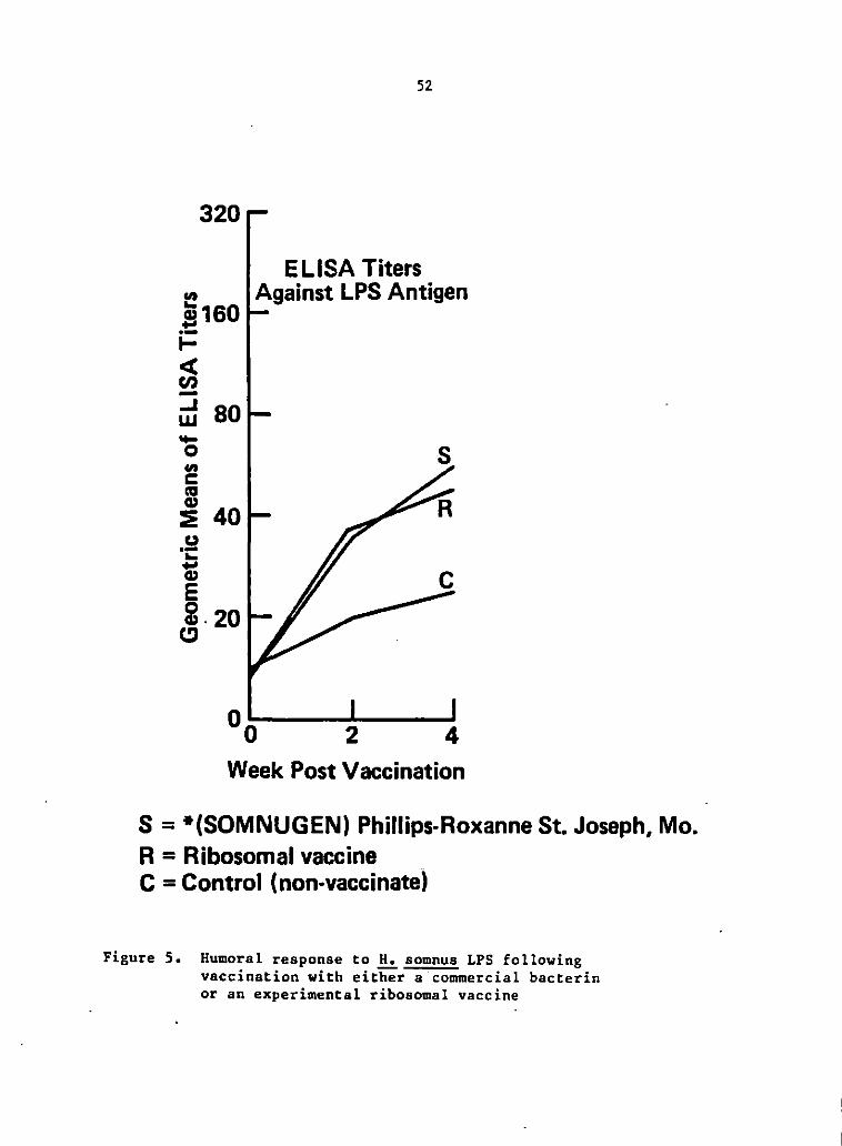
320
"' !160 i= c:s: Cl> -~ 80 -0
"' c ca ~ 40 u ·-... ~ E 2.20
CJ
52
EL lSA Titers Against LPS Antigen
s
2 4 Week Post Vaccination
S = *(SOMNUGEN) Phillips-Roxanne St. Joseph, Mo. R = Ribosomal vaccine C =Control (non-vaccinate)
Figure 5. Humoral response to H. somnus LPS following vaccination with either a'commercial bacterin or an experimental ribosomal vaccine

53
clinically ill four were in the S group, five were in the R group and
one was in the C group. All ten animals recovered after antibiotic
treatment.
Discussion
The presence of H. sornnus anti-LPS antibody in bovine serum can
be measured by using an ELISA technique which employs phenol-water
extracted LPS as the antigen. The specificity of the ELISA relies in
part on the purity of the antigen prepared by the phenol-water
extraction method and subsequent alcohol precipitation to remove
nucleic acids and proteins from the preparation. With the results
presented here the LPS antigen from.!!.!. somnus strain 8025 appears to
be specific for .!!.!. somnus antibodies as indicated by the absence of
cr~ss-reactivity with antiserums to various bovine pathogens. In
contrast a diethylene glycol extract, lysozyme extract, heat extract
and an incubation extract showed cross-reactions with a number of the
antiserums against common bovine pathogens. These pathogens include
!::.:_ lignieresii, ~ bronchiseptica, f.!_ fetus, ..h!_ monocytogenes, ~
bovis, ~ multocida, !..!_ haemolytica, ~ agalactiae, and~ pyogenes,
which is consistent with results of other workers which have reported
common antigens associated with .!!...!. sOmnus and other bovine pathogens
(37,48,73,91 1 123 1 136). However, contradictory reports of cross-
reactions of H. S_omnus antigens with other organisms have been
reported.

54
Canto et al. (21) reported that cross-reactivity of.!!.:. somnus
antibodies depended greatly on the type of serological procedure and
method of antigen preparation employed. They noted the greatest
cross-reactivity in agglutination tests using whole cell antigens.
They developed an ELISA test using a supernatant of a saline extract
produced by incubating a standardized number of unwashed cells in PBS
at 4 C for 24 h, as their ELISA antigen which they concluded consisted
largely of an H. somnus common antigen and which showed cross-
reactivity on the ELISA test with antigens from.!!.:. agni only. This
saline extracted antigen would be similar to the incubation extract
antigen described in this paper which cross-reacted with P. multocida
antisera in our ELISA test.
Partially purified LPS showed the least cross-reactivity of any
antigen we tested, but had good reactivity with anti-!!:. somnus serums
and was chosen as the specific antigen for the ELISA test overcoming
the common problem of cross-reactivity associated with other
serological methods. This ELISA test, using.!!.:. somnus LPS as the
antigen, showed high sensitivity with positive titers in hyperimmune
rabbit serum to 1:10 1 240 and to 1:5120 in immunized bovine serum, and
satisfact0ry precision between replicate measurements of the same and
different serum samples from day to day and from worker to worker.
All serums from animals in group A, injected with one of the two .!!.:.
somnus vaccines showed a five-fold increase in antibody titer while
control non-vaccinates of the same herd showed a 2.5-fold increase in
antibody titer during the one month testing period. During this time,

55
the animals became ill with respiratory disease of undetermined
etiology, but probably due to .!!.:_ somnus, which may account for the
rise in antibody levels in .the control group. The fact that animals
in all three groups became clinically ill may indicate that humeral
immunity alone is not sufficient to completely protect the animal from
infect ion by .!!.:_ somnus.
Group B, consisting of 63 cattle, was followed for two months
during which serum samples were collected for e·valuation of serum
antibody to .!!.:_ somnus LPS. During the time of testing, this herd
suffered an attack of respiratory disease. H. somnus was recovered
from the animals that died. The herd showed an overall increase in
anti-II. somnus LPS antibody level up to a mean titer of 1 :160,
although individual antibody levels varied greatly. Of particular
interest were the serum antibody levels of those animals that became
severely ill but survived and those that died. Several of the sick
animals showed a decrease in antibody levels during illness, two
however showed a moderate increase in antibody levels. Of those that
died, two had a low (1:20) level of antibody to.!!.:_ somnus LPS and the
third animal had a decrease in antibody titer prior to death. Animals
that died were confirmed to have had pneumonia caused by H. somnus. . ~
In another herd of naturally infected cattle, that had been clinically
infected by .!!..!. somnus and had recovered, a much higher average serum
antibody level to .!!.:_ somnus LPS ( 1 :640) was found. The importance of
these findings is speculative but indicates a role for humeral
immunity in recovery from H. somnus infections. Antibody specific for

56
.!!.!_ somnus LPS may help neutralize the pathogenic activities of the LPS
antigen which may occur during infection; however, these results
indicate that.vaccination does not provide sufficient protection
against disease caused by H. somnus.
Antibody against LPS can function as an antitoxin and as such can
diminish the mortality from gram-negative bacteremias (13). It has
been reported that anti-LPS antibody can neutralize the effects of
meningococcal endotoxins (31). High titers of antibody to core LPS at
the onset of human bacteremia has been associated with significantly
less frequent shock and death in patients with bacteremia due to gram-
negative bacilli (160),
To reach any final conclusions about the clinical significance of
the results obtained by this ELISA assay will require more extensive
and controlled studies. This assay will be employed for an ongoing
evaluation of the humeral response to .!!.!_ somnus infections and
vaccination to assess whether the rate and amount of antibody increase
correlates with certain respiratory d'isease syndromes and recovery
from disease, If possible, we also hope to evaluate the feasibility
of using this ELISA assay as an aid in the diagnosis of H. somnus
infection in cattle and the assessment of response to therapy instead
of the commonly applied complement fixation test (CFT) because of the
increased specificity of the ELISA test over the CFT.

57
SECTION II.
EFFECT OF HAEMOPHILUS SOMNUS FRACTIONS
ON BOVINE POLYMORPHONUCLEAR LEUKOCYTE FUNCTIONS
Summary
The effect of Haemophilus somnus on bovine polymorphonuclear
leukocyte (PMN) function was examined in vitro with whole cells and
fractions extracted from the surfac~ of this bacterium. The ability
of PMNs to iodinate protein and ingest Staphylococcus aureus was
significantly inhibited in the presence of live cells, heat-killed
whole cells or supernatant fluid from heat-killed cells, but not in
the presence of washed, heat-killed cells. None of the fractions
inhibited nitroblue tetrazolium (NBT) reduction by PMNs. The PMN
inhibitory factors were further characterized. The material that
inhibited S. aureus ingestion was found to be a heat-stable cell
surface material of greater than 300,000 MW. The fraction inhibiting
iodination of protein was found to be less than 10,000 MW.
Introduction
Haemophilus 'somnus (H. somnus) is recognized as a cause of
several disease syndromes of cattle. H. somnus was first recognized
58
as the bacterium that caused infectious thromboembolic
meningoencephalitis (TEME) in cattle (7,14,73,81,101,104,144). The
bacterium has since been discovered to be a major etiological factor
in a variety of bovine disease syndromes, including respiratory
infection, septicemia, and resulting thrombosis (14). Therefore, H,
somnus is considered an important pathogen which causes great economic
loss to the cattle industry (118).
Lesions produced in cattle by infection with H. somnus are
characterized by the presence of large numbers of polymorphonuclear
leukocytes (PMNs). Cattle experimentally infected with H. somnus
exhibit .a neutrophilic leukocytosis within 8 hours of challenge (153).
Microscopically, vasculitis and neutrophilic cuffing has been observed
in the brain along with neutrophil infiltration of the submucosa of
the trachea (37). Vasculitis, thrombosis, microabscessation and focal
necrosis were also observed in spinal cord, kidney, bladder, mucosa of
the gastrointestinal tract, myocardium, skeletal muscles and retina
(138). These areas of thrombosis and vasculitis are often infiltrated
by neutrophils (73,104,138).
PMNs are one of the major cellular defense mechanisms in
protecting animals from microbial infection. The nature of the
interaction of the bacterium with the PMN may determine whether or not
infection occurs. The nature of the infectious process suggests that
.!!.:. somnus may escape host defense mechanisms. Therefore the in vitro
interaction of ~ somnus cells and fractions derived from those cells
with PMNs were evaluated. Major suppressive effects were observed on
59
PMN functional activities. It has been postulated that the occurrence
of bacteremia in the face of a high serum antibody titer indicates
that the host phagocytic system is no._.longer functioning effectively.
Evidence for this hypothesis has be·en reported. Only 55% of the
organisms of either a pathogenic or non-pathogenic strain of H. somnus
were phagocytized in vitro, while antiserum to the pathogenic strain
decreased the percentage of the bacteria phagocytized (126). In most
infectious diseases pathogenicity is related to more than one
microbial characteristic (93). A correlation can often be made
between cell surface factors and pathogenicity. If these factors that
increase the pathogenicity of an organism were identified it could be
important in understanding the mechanism by which the microorganism
invades a host and the possible prophylactic measures that can be used
against the pathogen.
Materials and Methods
Bacterial propagation
H. somnus strain 8025 was used throughout these experiments.
This strain was initially isolated at _the Veterinary Diagnostic
Laboratory, Iowa State University, Ames, Iowa, from a bovine brain
with lesions of infectious thromboembolic meningoencephalomyelitis.
The bacterium, which had been stored at -70 C in egg yolk culture (2
ml), was inoculated into 10 ml of brain heat infusion broth (Difeo
Laboratories, Detroit, Mich.) supplemented with 5% normal bovine serum

60
and 5% yeast extract (Difeo) (BHISY). This broth culture was
incubated for 12 h at 37 C in an atmosphere of 5% co2 and 95% air and
then used to inoculate Roux flasks containing the BHISY medium and
1.5% Bactoagar (Difeo). The bottles were rotated by hand to spread
the inoculum over the agar surface and the bottles were incubated for
48 h. The bacteria were harvested by adding 5 ml of .015 M phosphate
buffered saline solution (pH 7.2 ,PBS) to each bottle, scraping the
agar surface with a sterile wire and aspirating the suspended bacteria
from the bottle. The harvested cells were pooled and washed 3 ti'\1es
in PBS by alternate centrifugation at 10,000 x g for 30 min at 5 C and
resuspension. After the last wash, the bacteria were resuspended in
PBS to a lOx concentration of a suspension with an optical density
(O.D.) of 0.4 at 600 nm which was equivalent to 4.2 x 10⁸ cells/ml.
Bacterial fractions
Bacteria suspended in PBS were incubated in a water bath at 60 C
for 1 h. After this heat extraction an aliquot was saved for the
"heat-killed cells and supernatant" (HKC+S). The remainder was
centrifuged at 10,000 x g for 20 min at 5 C. The pellet was
resuspended in PBS to the original volume ("heat-killed cells" or
HKC). The supernatant fluid was termed heat extract (HE).
An aliquot of HE was saved and the remainder was filtered through
a 300,000 molecular weight cutoff filter (Amicon Corporation,
Lexington, MA). The retentate was resuspended to the original volume
in PBS and considered the heat extract fraction of greater than
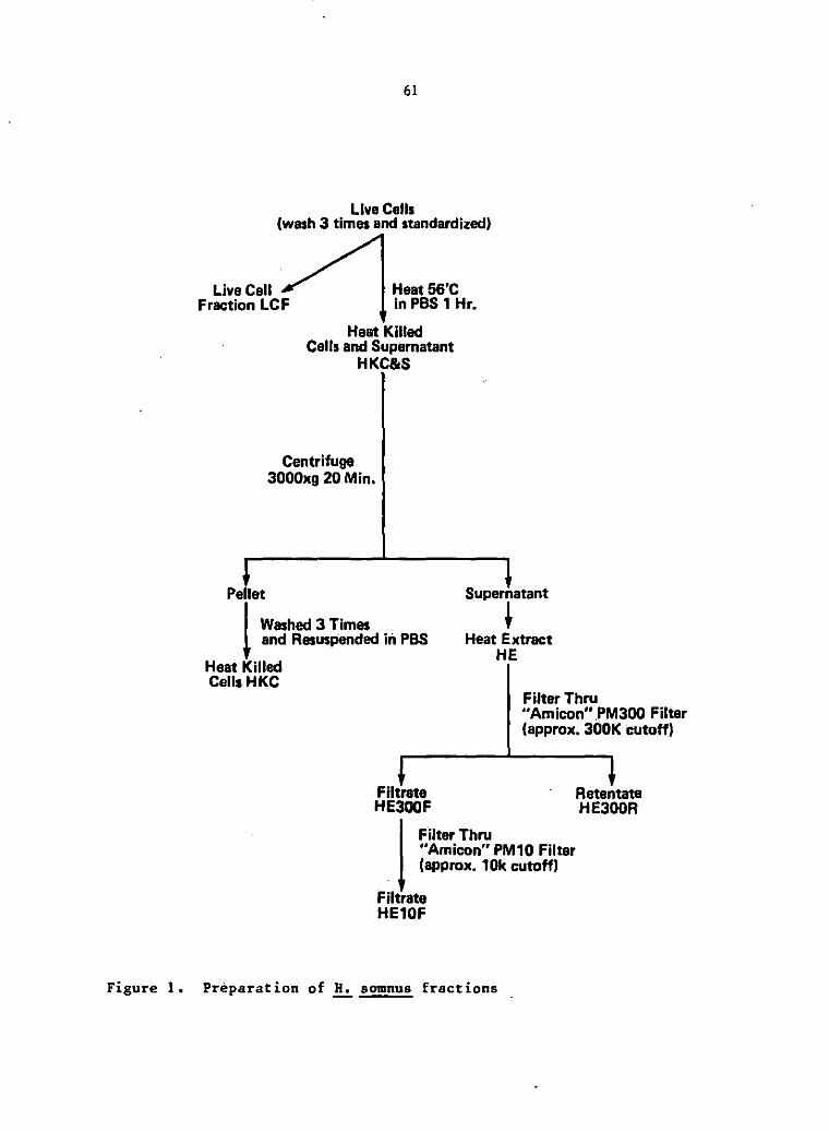
61
Live Cells (wash 3 times and standardized)
Live Cell Fraction LCF
Heat56'C in PBS 1 Hr.
Heat Killed Cells and Supernatant
HKC&S
Centrifuge 3000xg 20 Min.
Pellet Supernatant
~ 1 Washed 3 Times and Resuspended iii PBS Heat Extract
HE Heat Killed Cells HKC
Filter Thru "Amicon" PM300 Filter (approx. 300K cutoff)
Filtrate Retentate HE300F HE300R
I Filter Thru "Amicon" PM10 Filter (approx. 10k cutoff)
Filtrate HE10F
Figure 1. Preparation of H, somnus fractions

62
300,000 MW (HE300R). The filtrate was divided into two aliquots, one
of which was saved and called the heat extract of less than 300,000 MW
(HE300F). The other aliquot was passed through a 10,000 MW cutoff
filter (Amicon) and called the heat extract of less than 10,000 MW
(HE!OF).
~ preparation
PMNs were prepared by the method of Roth and Kaeberle (1980).
Peripheral blood was collected from apparently healthy cattle into ACD
solution. After the blood was centrifuged at 1000 x g for 20 min, the
plasma and buffy coat layers were aspirated and discarded. The RBCs
were lysed by the addition of 2 volumes of cold phosphate buffered pH
7 .4 deionized water for 50 seconds at which time the isotoni'city was
restored by adding I volume of cold phosphate buffered pH 7.4, 2.7%
NaCl.
The PMNs present in the RBC lysate were pelleted by
centrifugation and washed once with PBS. The cell suspension was
adjusted to a final concentration of 50 x 106 PMNs per ml in PBS and
used in all three PMN function tests. Differential stains indicated
that these preparations contained an average of 96% neutrophils and 3-
4% eosinophi ls.
PMN function tests
The procedures for evaluating PMN function have been described in
detail previously (115). All PMN function tests were conducted in
63
duplicate and the average of duplicate values were used for
calculations.
Iodination The iodination test was conducted in 12 x 75 mm
polyethylene' snap cap tubes (#2050 1 Falcon, Oxnard, Ca.). The
standard reaction mixture contained 2.5 x 10⁶ PMNs, 0.05 mCi of 125
Iodine (Carrier free in O.l M NaOH, New England Nuclear, Boston MA),
40 nmole of NaI, 0.05 ml opsonized zymosan (10 mg/ml) and 0.3 ml
Earles balanced salt solution (EBSS, GIBCO, Grand Island, N.Y.). To
determine the effect of bacterial fractions on iodination by PMNs,
0.05 ml of H. somnus fractions (4.8 x 10⁶ cells or equivalent soln.)
or 0.05 ml of PBS for controls were added to the standard reaction
mixture. The reaction was allowed to proceed for 20 min at 37 C as
the tubes were tumbled end over end. The amount of trichloroacetic
acid (TCA)-precipitable radioactivity was determined. The results are
expressed as a percent of control values and plotted as bar graphs.
Nitroblue tetrazolium (NBT) reduction The NBT test was
conducted in duplicate in 16 x 100 mm siliconized glass tubes. The
standard reaction mixture contained 0.2 ml of NBT solution (2 mg/ml),
5 x 10⁶ PMNs, 0.1 ml preopsonized zymosan preparation (10 mg/ml), 0.1
ml PBS in control tubes or 0.1 ml of the bacterial fractions diluted
in PBS and sufficient EBSS to bring the total volume of the reaction
mixture to 1.0 ml. All of the reactants except the PMNs were added to
the tubes and allowed to equilibrate in a 37 C waterbath. The tubes
were left in the waterbath and the reaction was initiated by adding
the PMN suspension to each tube. The tubes were incubated in the

64
shaking water bath for 10 min at 37 C after which the reaction was
stopped by adding 5 ml of cold 1 mM n-ethylmaleimide diluted in
saline. The cells and the precipitated formazan were pelleted by
centrifugation at· .500 x g for 10 min, the supernatant fluid was
discarded and the pellet was resuspended in 5.0 ml of pyridine.
Formazan was extracted by brief sonification of each tube followed by
heating in a boiling waterbath for 10 min. The pyridine-formazan was
clarified by centrifugation at 500 x g for 10 min and the O.D. was
immediately determined at 580 nm in a spectrophotometer using a
pyridine blank.
Staphylococcus aureus ingest·ion Heat-killed (1251)
iododeoxyuridine ([1251] UdR, New England Nuclear, Boston, MA)-
labeled S. aureus was used to evaluate ingestion by PMNs. The test
was conducted in 12 x 75 mm plastic tubes and the standard reaction
mixture contained 0.1 ml of ([1251] UdR labeled) E.:_ aureus, 0.05 ml of
PMNs (25 x 106), (bacteria to PMN ratio= 60:1), 0.05 ml of a 1:10
dilution of bovine anti-S. aureus serum and 0.3 ml of EBSS. To
determine the effect of bacterial fractions on ingestion by PMNs, 0.05
ml of.!!:. somnus fraction (6 x 10⁶ cells or equivalent soln.) or 0.05
ml of PBS as a control were. added to the standard reaction mixture.
The reaction was allowed to proceed for 10 min at 37 C in a shaker
waterbath after which the extra-cellular, non-phagocytized E.:_ aureus
was removed by adding Lysostaphin (Sigma Chemical Co., St. Louis, Mo.)
to the reaction mixture followed by incubation for an additional 20
min. The tubes were then filled with PBS and the PMNs were washed

65
twice by alternate centrifugation and resuspension. The amount of
PMN-associated radioactivity was determined and the results were
expressed as percentages of (125I) UdR-labeled i.:_ aureus that was
ingested. The values for the tubes containing bacterial fractions.
were then calculated as a percent of the control values.
Data analysis To determine the effect of bacterial fractions
on each PMN function the value obtained when a bacterial fraction was
added to the PMNs was compared to the value obtained in control
reactions (with PBS only). An analysis of variance procedure was used
to determine significance of the differences in PMN function due to
addition of the bacterial fractions. For the graphic presentation of
the data, all treatment values were expressed as a percentage of the
untreated controls.
Results
Effect .!!.f ~ somnus fractions on iodination The ability of
PMNs to iodinate protein in the presence of ~ somnus fractions was
determined to study the effect of these fractions on the
myeloperoxidase-hydrogen peroxide-halide antibacterial system of the
PMN. The value for iodination by PMNs in control reactions was 37.99
_! 12.02 (mean_! S.E.M.) (n = 16) nmole NaI per 107 PMNs per hour.
The ability of PMNs to iodinate proteins in the presence of live
cells, HKC+S, HE and HKC was determined. HKC+S and HE suppressed the
iodination reaction. HKC had no effect on iodination by PMNs. The HE
was proces,sed for further characterization of the inhibitory factor. by

66
filtration through two filters, one with MW cutoff of approximately
300,000 (300K) and the other with MW cutoff of approximately 10,000
(IOK). Both HE and the HE filtrate from the IOK filter (HEIOF)
inhibited iodination by 25%. The He retentate from the 300K filter
(HE300R) did inhibit iodination but only by 9%.
Effect .!!.£ .!!.!_ somnus fractions .!!!!. ~ aureus ingestion To
determine the effect of H. somnus fractions on phagocytic activity,
PMNs were added to a standard suspension of opsonized ~ aureus in the
presence or absence of.!!.!_ somnus fractions. The PMNs in control
reactions ingested 26.01.!.11.75 (mean.!. S.E.M.) (n = 13) percent of
the s. aureus in the reaction mixture. !.:_ aureus ing~stion was
inhibited in the presence of live cells, HKC+S7 HKC and HE. To
further characterize the inhibitory factor, HE was filtered through
the two filters as described previously. HE300R inhibited S. aureus
ingestion by 50%. Neither HE300F or HElOF had an effect on S. aureus
ingestion by PMNs.
Effect of .!!.!_ somnus fractions .!!!!. .fil reduction To study the
effect of.!!.!_ somnus fractions on the oxidative metabolism of.PMNs, the
ability of PMNs to reduce NBT by the production of superoxide anion in
the presence of the bacterial fractions was determined. The value for
NBT reduction by PMNs in control reactions was 0.499 .!. 0.144 (Mean +
SEF) (n = 18). None of the H. somnus fractions had a significant
effect on NBT reduction (p 0.05).

100
90
80
2 .. 5 10 (J -0
'C B 60 .. :.
50
67
LCF HKC+S HE HKC HE300R HE300F HE10F
Figure 2. Effect of.!!..:. somnus fractions on iodination by bovine PMNs stimulated with opaonized zymosan. Values represent mean percentages (+- standard deviation) of the control value. Statistically significant differences from control values are indicated as **• p < 0.05; nal6

100
90
80
g c 8 70 0 i ~ 60
CL
50
40
0
Figure 3.
68
LCF HKC+S HE HKC HE300R HE300F HE10F
Effect of H. somnua fractions on S. aureus ingestion by bovine PMNs. Values-;epresent mean per.cent ages ( +- standard deviation) of the control value. Statistically significant differences from control values are indicated as **, p < O .05; n~IJ

Figure 4.
69
LCF HKC+S HE HKC HE300R HE300F HE10F
Effect of H. somnus fractions on NBT reduction by bovine PMN;-stimulated with opsonized zymosan. Values represent mean percentages (+- standard deviation) of the control value. None of the differences were statistically significant, p < 0.05; n•lB

70
Discussion
Phagocytic cells occupy a central position in host defense
against infection by microorganisms and neutrophils are primarily
responsible for the host defense against obligate extracellular
pathogens. A major function of the PMN is the phagocytosis and
destruction of these invading microorganisms. Some microbial pathogens
have survival properties to protect against the killing mechanisms of
phagocytic cells. These properties increase the pathogenicity of the
organisms. These anti-phagocytic mechanisms include avoidance of
recognition, inhibition of chemotaxis, attachment, ingestion,
oxidative metabolism or degranulation, and possibly the elaboration of
a leukocidic factor (113). Any of these inhibitory actions on PMN'
function should be C()nsidered as virulence factors of the pathogenic'
microorganism (34).
The ingestion of bacteria is the first step in the bactericidal
activity of the PMN. The results of the S. aureus ingestion assay
indicate that cell components of .!!!_ somnus inhibit the ability of PMNs
to ingest partic!es. When bacterial fractions containing whole
organisms were used, the inhibition of ingestion of ~ aureus may have
been due to competition between the two bacterial species. Since
soluble bacterial fractions would not be phagocytized, they should not
compete with particulate materials in the ~ aureus ingestion assay.
The factor associated with H. somnus which inhibited S. aureus
ingestion is soluble and greater than 300,000 MW· This factor may be

71
heterogeneous in composition and may be comparable to a fraction
extracted by Stephens et al. (135). These researchers isolated two
antigens from the outer membrane complex of H. somnus using a· saline
extraction of whole .!!:_ somnus cells for use as possible vaccines, and
showed these fractions to be greater than 150,000 MW.
Oxidative metabolism of the PMN is an important aspect of its
bactericidal activity (5,113). When a PMN receives the proper
stimulus, an·oxidase enzyme on the surface of the plasma membrane or
.phagosomal m<:<mbrane will catalyze the .conversion of oxygen to
superoxide anion. This reaction is known as the respiratory burst.
Superoxide anion spontaneously dismutates to hydrogen peroxide. NBT
is directly reduced by the superoxide anion to an insoluble purple
formazan (158). NBT reduction is therefore a measure of superoxide
anion.generation by the PMN. Since NBT reduction was not inhibited by
whole bacteria or .bacterial fractions, .!!:_ somnus apparently does ,{ot
inhibit the production of superoxide anion by PMNs.
Associated with the PMN oxidative metabolism is the iodination
reaction. This reaction is a measure of the ability of PMNs to
convert inorganic iodide to a.trichloroacetic acid-precipitable
(protein-bound) form and occurs inside the phagocytic vacuole via the
action of hydrogen peroxide plus myeloperoxidase from the primary
granules in the PMN. This reaction is dependent upon the generation
of hydrogen peroxide during the respiratory burst, degranu!ation to
release myeloperoxidase, the presence of iodine, the unimpaired
ability of myeloperoxidase to catalyze the rea~tion, and the presence

of tyrosine to bind iodine. This system has been reported to kill
bacteria, fungi and viruses (9,125). Hydrogen peroxide is formed from
superoxide anion. NBT reduction was not inhibited by the H. somnus
fractions indicating that the formation of superoxide anion and
presumably hydrogen peroxide was not impaired. It is possible that
the bacterial fractions inhibited degranulation of the primary granule
into the phagosome or that myeloperoxidase activity was inhibited.
Little is known about the mechanisms that allow .!!.:. somnus to
survive in the bovine ·respiratory tract or to develop a septicemia •
.!!.:. somnus is commonly isolated from healthy cattle without causing
signs of disease. During conditions of stress, the bovine host
apparently becomes more susceptible to infection by .!!.:. somnus. In
vitro studies have shown that H. somnus can be opsonized. and killed by
bactericidal antibodies from vaccinated cattle in conjunction with
complement and neutrophils. It does not appear that all.!!.:. somnus
organisms are normally killed however (126). The neutrophil
suppressive factors associated with.!!.:. somnus, reported in this paper,
may act in conjunction with other immunosuppressive events to hinder
the host defense mechanisms enough for an H. somnus infection to
become established. Immunosuppressive effects that could help
establish an .!!.:. somnus infection may include increased corticosteroid
levels associated with stress. High plasma cortisol has been reported
to inhibit both the cellular and humoral immune response. Also, high
serum cortisol levels inhibit the hydrogen peroxide-halide-
myeloperoxidase bacterial killing system (114), The combined effects

73
of stress and .!!..!_ somnus factors may suppress neutrophil function to
the point that infection leads to a disease process.
Some bacterial pathogens circumvent recognition as foreign by the
host surveillance mechanisms (51) or inhibit chemotaxis of neutrophils
(34,64). It does not appear that inhibition of recognition or
impairment of Chemotaxis o'f neutrophils occurs in .!!.!_ somnus
infections. An abundance of neutrophils at the site of infection is a
common pathologic sign in .!!..:. somnus disease syndromes, indicating
intact and even extreme chemotaxis of neutrophils to the site of
'infection. In cases of abortion, where .!!:.. somnus is implicated,
abundant neutrophils are found in fetal airways (2,25,144).
Neutrophil infiltration of synovia is noted in cases of weak calf
syndrome when.!!..:. somnus is an expected cause (147). In cases of
pneumonia, Andrews et al. (2) described microscopic lesions of
peribronchiolar filling of alveoli with albuminous fluid and
neutrophils. Gross lesions associated with TEME caused by .!!..:. somnus
occur in the brain and irregularly in the spinal cord, mucosa of the
esophagus, gastrointestinal tract and urinary bladder. Histological
examinat-ion reveals severe vasculitis, thrombosis, ne<!;"osis ·Of vessel
walls and intense neutrophil accumulation associated with the lesions
(73,104,138). Many bacterial pathogens produce a capsule that enables
the microbe to resist attachment to and ingestion by host phagocytes •
.!.:. pestis is reported to acquire a capsule only after infecting a host
(154) • .!!..:. somnus capsulization has been reported by Miller et al.
(91) and Williams et al. (153) but refuted by stephens and Little

74
(137) who examined the cell with electron microscopy. Stephens and
Little have shown that the cell envelope is composed of an outer
membrane, peptidoglycan layer and an inner cytoplasmic membrane.
Also, EM studies of .!!:. somnus adherent to endothelial cells in tissue
culture did not demonstrate pili (143). It remains possible that
certain Strains of .!!..:_ somnus may possess a capsule or may acquire a
capsule only in the host, enabling .!!:. somnus to resist phagocytosis.
The H. somnus strain used in this study, strain 8025, was not examined
for capsule formation.
Viral induced suppression of neutrophil function may play a role
in the pathogenesis of secondary bacterial infections (43). H. somnus
fractions did not inhibit the oxidative metabolism of bovine
neutrophils phagocytizing particles of opsonized zymosan. However,
primary or simultaneous viral infection and subsequent depression of
neutrophil oxidative metabolism may be involved in the pathogenesis of
.!!:. somnus infections. Pennell and Renshaw (106) speculated that
primary viral infection inhibiting bactericidal activity in the lung
may be a predisposing factor in.!!:. somnus caused disease syndromes.
Resistance to microbicidal oxygen products can be achieved by a
number of bacterial mechanisms. Catalase-rich strains of
staphylococci had a better survival rate and decreased myeloperoxidase
dependent iodination by neutrophils, probably due to destruction of
hydrogen peroxide (34). Although there have been some reports of
catalase positive isolates, ..!!.!_ somnus is cpnsidered catalase negative
and hence would not destroy hydrogen peroxide •. The oxidative

75
potential of singlet oxygen is quenched when in contact with
substances with a large number of conjugated double bonds, e.g.
carotenoids (34) • .!!.!_ somnus characteristically._.has a bright yellow .·
color. This pigmentation varies between.!!.:. s·omnus strains but may
serve to quench singlet oxygen produced during the oxidative burst
(26). It is apparent that the virulence of a microorganism is
associated with its ability to interfere with the normal immune
response of the host • .!!.!_ somnus apparently has one or several
virulence factors associated with it. These factors enable this
organism to produce the di~ease syndromes known as the "H. somnus
complex". The research reported in this paper has shown that soluble
surface bacterial fractions from H. somnus, isolated by heating the
orga,nisms in saline, inhibit PMN ingestion of opsonized ~ aureus and
iodination of protein. The mechanisms of these two effects are still
under investigation. These fractions may be virulence factors acting
to induce the disease state. Whether specific antibody to these
factors will neutralize their effect and reduce virulence remains to
be seen.

76
GENERAL SUMMARY
The experimentation reported in this thesis includes two
subjects: (1) development of a specific and sensitive ELISA test to
evaluate the humoral immune response of cattle to .!!.!_ somnus LPS during
infections and vaccination, (2) demonstration of H. somnus virulence
factors that suppress two neutrophil functions.
Five antigen preparations were isolated from H. somnus. These
five antigens were used in the ELISA assay. Rabbit antiserums to ten
common bovine pathogens were made and used to determine the
specificity of the five H. somnus antigens for use in the ELISA
procedure. It was found that phenol-water extracted LPS of H. somnus
was· the most specific antigen. This antigen was used to monitor anti-
.!!!_ somnus LPS antibody levels in a herd of calves during the months of
highest incidence of H.somnus infections. During the test period, a
number of the calves developed respiratory disease. H. somnus was
isolated from those animals that died from pneumonia. Variable
responses to .!!.!_ somnus LPS were seen among individual animals. Of the
animals that became clinically ill, several showed a decrease in serum
antibody titer after illness, two had an increase in titer after
illness. All animals were treated with antibiotics at the first sign
of illness. Of the three animals that died, two had low titers at the
time of death while the third showed a drop in antibody level prior to
death. Animals immunized with one of tWo vaccines developed an
increase in antibody titers over a control gro~p. The ELISA test

77
presented here is. specific and sensitive. It can be used for further
studies on the humeral immune response of cattle to H. somnus and
possibly as a diagnostic aid.
The role of the humoral immune response in protection from
infection and recovery from infection by.!!!_ somnus in cattle remains a
mystery. Serological surveys have shown that almost all cattle have
antibody to H. somnus (14 1 82) 1 but in vitro studies suggest that
animals succumbing to ~ somnus infection may lack normal humoral
defense factors (127). The most susceptible age group, with the
highest incidence of H. somnus infection, were young calves between
five months to one year of age. This group had lower bactericidal
activity than adults and it appears that the anti-bacterial antibodies
were not maternal in origin but eventually appear in serum due to
increased exposure to .!!.:.. somnus or due to maturation of the calves
immune system. Simonsen (126) has reported that in vitro serum
bactericidal activity varies with the age of the animal, and is
complement dependent with the concentration of anti-H. somnus antibody
being the limiting factor. Pennell and Renshaw (106) also reported
substantial levels of bactericidal activity with a combination of
complement, leukocytes and specific opsonin, anti-H. somnus antibody.
Stephens (134) reported that serum titers do not determine
susceptibility to challenge or the outcome of infection with H.
somnus. They found increasing titers in cattle 2-3 days post
challenge indicating an anamestic response in cattle acutely ill with
TEME. They proposed that high antibody titers .in cattle with

78
septicemia may contribute to the pathogenesis of disease. They also
reported evidence. that indicates the host's phagocytic systems are not
functioning effectively and that antigen-antibody complexes are
probably not phagocytized. Stephens (134) also found that calves
without antibody did develop septicemia but not TEME after IV
inoculation of live H. somnus. The results reported in this thesis
support the idea that antibody contributes to the severity of disease
in some cases. However, the loW titers to.!!.!.. somnus LPS obtained
during immunization and natural infection indicate that H. somnus is
not highly immunogenic. The limited hunioral response to.!!.!_ somnus may
therefore also contribute to the establishment of disease •.
These results reflect our present understanding of the role of
humeral immunity in .!!.!_ somnus disease which is conflicting and
complicated. This confusion probably reflects the complexity of the
overall immune response to .!!.:.. somnus infection which is often preceded
by stress, viral infection, cold weather or other factors which may
each elicit a different effect on the host immune system.
The neutrophil function results indicate the heat killing o'f H.
somnus releases at least two virulence factors into the medium. Two
separate neutrophil functions were suppressed by the bacterial
fractions of differing molecular weight. One fraction, of less that
10 1000 MW was found to inhibit iodination of protein, an important
step in the potent myeloperoxidase - halide - hydrogen peroxide
killing mechanism of neutrophils. The second fraction, of greater
than 300,000 MW was found to inhibit the ingestion of radiolabelled S.

79
aureus. Inhibition of these two neutrophil functions corresonds with
the pathologic findings of H. somnus infections which occurs in the
presence of an accumulation of neutrophils. Virulence factors of H.
somnus had been suspected but not previously demonstrated. These two
fractions from H. somnus can be considered as probable virulence
factors that are involved in the pathogenesis of H. somnus infections.
The overall picture of .!!.:. somnus disease presents a combination
of bacterial and host responses and it is this joint action that is
probably responsible for the disease syndromes and lesions found in H.
somnus infections. It is probable that.!!:_ somnus, properly opsonized
with both antibody and complement, are phagocytized and killed by
neutrophils. However, it is possible that not all of the.!!:_ somnus
organisms are phagocytized. EM studies of neutrophils incubated with
opsonized .!!:_ somnus should be undertaken to resolve whether the
bacteria are ingested, are enclosed in a proper phagolysosome and
survive ingestion. Two other aspects that should be investigated are
whether H. somnus can produce polyanionic degranulation inhibitors or
whether H. somnus can effect the cAMP levels in the neutrophil which
are two mechanisms that enable some mycobacteria to resist
phagocytosis (18,34).
I would like to present a possible pathogenic mechanism for .!!:_
somnus to stimulate further work in order to understand and control
this important pathogen. First, .!!:_ somnus is common in the
environment of its host, cattle. The bacterium has been commonly
isolated from the tracheas of normal cattle (29), norm~l reproductive

80
tracts of cattle, and in high numbers from the prepuce of bulls (26).
These sites could serve to easily spread the organism by inhalation,
by ingestion, venereally or in the urine. Normally, the organism is
controlled by host resistance mechanisms such as the mucocilliary
blanket of the trachea, neutrophils, etc. However, during stress from
any number of causes, including viral infection, extreme cold
temperatures, handling, weaning, castration, vaccination or
transportation, suppression of the normal immune functions may occur
in the animal giving.!!.!_ somnus a chance to infect the host. After
infection the first line of defense, the neutrophils along with
circulating antibody to .!!.!_ somnus, attempts to control the spread of
the organism. The presence .of large numbers of neutrophils in lesions
associated with H. somnus suggests neutrophil chemotaxis is working
well, which is probably associated with complement fixation to
opsonized bacteria and release of C3a and C5a, potent chemotactic
attractants. The cell wall of H. somnus, which may or may not have a
capsular layer in vivo, in either case contains virulence factors that
suppress bacterial ingestion and killing by the phagocyte. The cell
wall also contains LPS which may activate the complement pathway,
degrading complement components and interfering with the fixation of
complement components to the bacterial cell surface further hindering
phagocytosis. Soluble antigens from the bacterial surface or the
bacterial cell itself may bind with circulating antibody resulting in
a population of immune complexes.
LPS, C5a, and immune complexes can all in~uce toxic effects on
81
endothelial cells, exposing the basement membrane which may lead to
typical lesions associated with .!!.!._ somnus infections (18). Th.ese
lesions include disseminated intravascular coagulation, vasculitis,
thrombus formation, hemorrhagic necrosis, fibrinopurulent
polyarthritis, synovitis, necrotic laryngitis, neutrophil accumulation
within the thrombus, and glomerulonephritis (66). Kidneys and joints
are also target organs of immune complex disease (18). Antibody thus
may contribute to tissue damage. This scenario incorporates the
classical signs associated with H. somnus infections. Whether it is
c;orrect remains to be -seen. It is hoped that this research and future
work will lead to protective control measures against this pathogen.

. ·
1.
82
LITERATURE CITED
Allgoweg, M., and H. Block. bacilli on the migration Tuberc. 59:562-566 •
1949. The effect of tubercule of phagocytes in vitro. Am. Rev.
2. Andrews, J. J., L. N. Slife, and G. W. Stevenson. 1981.
3.
Microscopic lesions associated with isolation of Haemophilus somnus from pneumonic bovine lungs. Abstr. Annu. Me~t. Am. Coll. Vet. Pathol. 32:130. '
Asmussen, M. D., and C. L. Baugh. 1981. Thiamine pyrophosphate (Cocarboxylase) as a growth factor for Haemophilus somnus. J. Clin. Microbial. 14:178-183.
4. Atanasiu P., V. Savy, and C. Gilbert. 1978. Rapid immunoenzymatic technique for titration of rabies antibodies IgG and IgM results. Med. Microbial. Immunol. 166:201-206
s. Babier, B. M. 1978. phagocytes. N.
Oxygen dependent microbial Engl. J. ~ed. 298:659-668.
killing by
6. Bailie, W. G. 1969. Characterization of Haemophilus somnus (n. sp.), a microorganism isolated from infectious thromboembolic meningoencephalitis of cattle. Ph.D. Thesis. Kansas State University, Manhattan, Kansas.
7. Bailie, W. E., H. D. Anthony_, and K. D. Weide. 1966. Infectious Thromboembo lie meningoencephalomye lit is (sleeper syndrome) in feedlot cattle. J. Am. Vet. Med. Assoc. 148:162-166.
8. Bailie, W. E., E. H. Coles, and K. D. Weide. 1973. Deoxyribonucleic acid characterization of a microorganism isolated from infectious thromboembolic meningoencephalomyelitis of cattle. Int. J. Syst. Bacterial. 23:231-237.
9. Belding, M. E., and S. J. Klebanoff. 1970. Peroxidase-mediated virucidal systems. Science 167:195-196.
10 • Biberstein, E. L. 1981. "Haemophi lus somnus" and ""Haemophilus agni". p • .125-132. In M. Kilian, W. Frederiksen, and E. L. Biberstein (eds.). Haemophilus, Pasteurella, and Actinobacillus. Academic Press, London.
11. Bidwell, D. E., A. Bartlett., and A. Volter. 1977. Enzyme immunoassays for viral diseases. J. Infect. Dis. (Supplement) 136 :S274-S278.

83
12. Bio-Rad Protein Assay. 1977. Tech. Bull. 1051. Bio-Rad Laboratories. Richmond, Calif.
13. Braude, A. I., and E. J .• Ziegler. 1983. Protection against gram-negative bacteremia with antiserum to endotoxins. P• 111-126. In A. Nowotny (ed.), Beneficial effects of endotoxins-:- Plenum Press, New York.
14, Brown, L. N., R. C. Dillman, and R. E. Dierks, 1970. The Haemophilus somnus complex. Proc. Annu. Meet. U. S. Anim. Health Assoc. 74:94-108.
15. Brodeur, B. R., R. E, Ashton, and B. B. Diena. 1978. Enzyme-linked i11D11unosorbant assay for the detection of Neisseria gonorrhaeae specific antibodies. Can. J, Microbial, 24:1300-1305.
16. Brown, L. N., R. E. Dierks, and R. C. Dillman. 1970. Problems in differential diagnosis of Haemophilus somnus infections ("Thrombo") of feedlot cattle. Bovine Pract. 5:36-37.
17, Brown, L. N., C, Reggiardo, M. W. Khai, P. G. Eness, and .H, L. Self, 1972. Application of the complement fixation test to the study of Haemophilus somnus infection in cattle, Proc. Annu. Meet, U, S. Anim. Health Assoc. 76:502-508.
18. Brunham, R. c., and K. K. Holmes, 1983. Immune mechanisms involved in bacterial infections. p. 69-81. In J, I. Galin, and A. S. Franci (eds,). Advances in host defense mechanisms. Vol. 2. Raven Press, New York,
19. Bulmer, G. s., and M. D, Sans, 1968. Cryptococcus neoformans. III. Inhibition of phagocytosis. J, Bacterial. 95:5-8.
20. Canto, G. J., and E, L. Biberstein. 1982. Serological diversity in Haemophilus somnus. J. Clin, Microbial. 15:1009-1015.
21. Canto, J., E, L. Biberstein, T. A, Schulte, and D. E. Behymer. 1983. Cross-reactivity of Haemophilus somnus antibody in agglutination and complement fixation tests and in the enzyme-linked immunosorbent assay. J. Clin. Microbial. 17:500-506.
22. Carlsson, H. E., A. A. Lindberg, and S. Hammarstrom. 1972. Titration of antibodies to salmonella 0 antigens by enzyme-linked immunosorbent assay. Infect, Immun. 6:703-708.

84
23. Carlsson, H. E., B. Hurvell, and A. A. Lindberg. 1976, ELISA for titration of antibodies against Brucella abortus and Yersinia enterocolitica. Acta Pathol. Microbial. Scand. 84 :168-174.
24. Case, M. T., w. G. Raudabaugh, and J. H. Finnell. of embolic meningo-encephalitis in Illinois • .!!. 2:31-32.
1965. Report Illinois Vet.
25. Chaldek, D. W. 1975, Bovine abortion associated with Haemophilus somnus, Am. J. Vet. Res. 36:1041.
26. Corboz, L. 1981. Epidemiology of Haemophilus somnus infection in cattle: Colonial variants of strains isolated from various sources, p. 133-142. In M, Kilian, W. Frederiksen and E. L. Biberstein (eds.). Hae;;iophilus, Pasteurella, and Actinobacillus. Academic Press, New York.
27. Corboz, L., and J. Nicolet. 1975. Infektionen mit sogenannten Haemopphilus somnus beim Rind: Isolierung und charakterisierung von Stammen aud Rewspirations-und eschlechtsorgen. Schweizer Arch, Tierheilkd. 117:493-502.
28. Corboz, L., and P. Wild. 1981. Epidemiologie der Haemophilus somnus - Infektion beim Rind: Vergleich von Stammen in der Polyacrylamidgel-Elektrophorese (PAGE). Schweizer Arch. Tierheilkd. 123:79-88.
29. Corstvet, R. E., c. Howard, Haemophilus Med. Assoc.
R. J, Panciera, H. B, Rinker, B. L. Starks, and 1973. Survey of tracheas of feedlot cattle for somnus and other selected bacteria. J. Am. Vet. 163 :9570-873.
30. Crandell, R. A., A. R. Smith, and M. Kissil. 1977. Colonization and transmission of Haemophilus somnus in cattle. Am. J. Vet. Res. 38:1749-1751.
31. Davis, C., E. Zeigler, and K. Arnold. 1978. Neutralization of meningococcal endotoxin by antibody to core glycolipid. J. Exp. Med. 147:1007-1012.
32. Deelder, A. M., E. J. Ruitenberg, D. Kornelis, and P. A. Steerenber. 1977. Schistosoma mansoni: Comparison of the immunoperoxidase techniques DASS and ELISA for human diagnosis. Exp. Parasitol, 41:133-140.
33. Densen, P., and G. L. Mandell. 1978. Go·nococcal interactions with polymorphonuclear neutrophils: Importance of the phagosome for bactericidal activity •. J. Clin. Invest. 62 :1161-1171.

85
34. Densen, P., and G. L. Mandell. 1980. Phagocyte strategy vs. microbial tactics. Rev. Infect. Dis. 2:817-838.
35. Devoe, I. W. 1976. Egestion of degraded meningococci by polymorphonuclear leukocytes. J. Bacteriol. 125:258-266.
36. Diena, B. B., F. E. Ashton, A. Ryan, and R. Wallace. 1978. The lipopolysaccharide (R type) as a common antigen of Neisseria gonorrhoeae. I. Immunizing properties. Can. J. Microbial. 24 :117-122.
37. Dierks, R. w., s. A. Hanna, and R. c. Dillman. 1973. Epizootiology and pathogenesis of Hemophilus somnus infection. J. Am. Vet. Assoc. 163:866-869.
38. Dubois, M., K. A. Gilles, H.K. Hamilton, P. A. Rebers, and F. Smith. 1956. Colorimetric method for determination of heptoses. J. Biol. Chem. 204:983-997.
! 39. Dukes, T. W. 1971. The ocular lesions in thromboembolic
meningoencephalitis (TEME) of cattle. Can. Vet. J. 12:180-182.
40. Elberg, s. S.; and P. Schneider. 1953. Directed leukocyte migration in response to infection and other stimuli. J. Infect. Dis. 93:36-42.
41. Ellenberger, M., M. L. Kaeberle, and J. A. Roth·. humeral immune response to Rhodococcus equi. J. Vet. Res.
1984. Equine In press. Am.
42. Engvall, E., and P. Perlmann. 1971. Enzyme-linked immunosorbant assay (ELISA): quantitative assay of immunoglobulin G. Immunochemistry. 8:871-874.
43.- Faden, H., P. Sutyla, and P. L. Ogra. 1979. Effect of viruses on luminol-dependent chemiluminescence of human neutrophils. Infect. Immun. 24:673-678.
44. Fink, P. C. and C. Galanos. 1981. Determination of anti-lipid A and lipid A by enzyme immunoassay. Immunobiology. 158:380-389.
45. Foley, M. J., and W. B. Wood, Jr. 1959. Studies on the pathogenicity of group A streptococci. II. The anti-phagocytic effects of the M protein and the capsular gel. J. Exp. Med. 110:617-628.

86
46. Forghani, R., N. J. Schmidt, and J. Dennis. 1978. Antibody assays for varicella-zoster virus: Comparison of enzyme iiranunoasSay with neutralization, immune adherence, hemagglutination and complement fixation. J. Clin. Microbiol. 8:545-555.
47. Galanos, C., M. Freudenberg, S. Hase, F. Jay, and E. Ruschmann. 1977. P• 269-276. In D. Schlessinger (ed.). Microbiology 1977. American Society of Microbiology, Washington, D.C.
48. Garcia-Delgado, G. A., P. B. Little, and D. A. Barnum. 1977. A comparison of various Haemophilus somnus strains. Can. J. Comp. Med. 41:380-388.
49. Giacoia, G. P., E. Neter, and P. Ogra. 1981. Respiratory infections in infants on mechanical ventilation: The immune response as a diagnostic aid. J. Pediatr. 98:691-702.
50. Glynn, A. A., c. Ison. 1978. Serological diagnosis of gonorrhea by an enzyme-linked immunosorbent assay (ELISA). Br. J. Vener. Dis. 54:97-102.
51. Goldring, O. L., J. A. Glegg, S. R. Smithers, and R. J. Terry. 1976. Acquisition of human blood group antigens by Shistosoma mansoni. Clin. Exp. Immunol. 26:181-187.
52. Gossling, J. 1966. The bacteria isolated from lesions of embolic meningoencephalitis of cattle. Illinois Vet. 9:14-18.
53. Granfors, K., M. K. Viljanen, A. Toivanen. 1981. Measurement of immunoglobulin M, immunoglobulin G, and immunoglobulin A antibodies against Yersinia enterocolitica by enzyme-linked immunosorbant assay: Comparison of lipopolysaccharide and whole bacteria as antigen. J. Clin. Microbiol. 14:6-13.
54.
55.
Griner, L. A., R. Jensen, and W. W. Brown. embolic meningoencephalitis in cattle. Assoc. 129:417-421.
1956. Infectious J. Am. Vet. Med.
Hall, R. E., J. M. Williams, and G. L. Smith. 1977. Field evaluation of Haemophilus somnus bacterin. Vet. Med. Small ·Anim. Clin. 12·: 1368-13 70.
56. Halle, S., G. A. Dasch, and E. Weiss. 1977. Sensitive enzyme linked immunosorbent assay for detection of antibodies against Typhus Rickettsiae, Rickettsiae prowazekii and Rickettsia typhi. J. Clin. Microbiol. 6:101-110.

87
57. Hazlett, M. J., P. B. Little, and D. A. Barnum. 1983. Experimental production of mastitis with Haemophilus somnus in the lactating bovine mammary gland. Can. Vet. J. 24:135-136.
58. Hendley, J. o., K. R. Powell, R. Rodewald, H. H. Holzgrefe, and R. Lyles. 1977. Demonstration of a capsule on Neiserria gonorrhoeae. Infect. Immun. 15:663-664.
59. Hirsch, J. G. 1960. Anti-microbial factors in tissues and phagocytic· cells. Bacteriol. Rev. 24:133-140.
60. Hoerlein, A. B., K. Goto, and S. Young. 1973. Haemophilus somnus agglutinins in cattle. J. Am. Vet. Med. Assoc. 163 :1375-1377.
61. Holmgren, J., and A. M. Svennerholm. immunosorbent assays for cholera 7:759-763.
1973. Enzyme linked serology. Infect. Immun.
62. Horowitz, S. A., and O. H. Cassell. 1978. Detection of antibodies to Mycoplasma pulmonis by enzyme-linked immunosorbent assay. Infect. Immun. 22:161-170.
63. Horwitz, M. A. 1982. Phagocytosis of microorganisms. Rev. Infect. Dis. 4:104-123.
64. Howard, C. J., and A. A. Glynn. 1971. The virulence for mice of strains of Escherichia coli related to the effects of K antigens on their resistance to phagocytosis and killing by complement• Immunology. 20:767-777.
65. Humphrey, J. D. 1982. Haemophilus somnus: Colonization of the bovine reproductive t-ract, .strain variation and pathogenicity. Thesis. Univ. Guelph, Guelph, Ontario.
66. Humphrey, J. D., and L. R. Stephens. 1983. Haemophilus somnus: A review. Vet. Bull. 53:987-1004.
67. Humphrey, J. D., P. B. Little, L. R. Stephens, D. A. Barnum, P. A. Doig, and J. Thorsen. 1982. Occurrence of Haemophilus somnus in bovine semen and in the prepuce of bulls and steers. Can. J. Comp. Med. 46:215-217.
68. Humphrey, J. D., P. B. Little, L. R. Stephens, D. A. Barnum, O. A. Doig, and J. Thorsen. 1982. Prevalence and distribution· of Haemophilus somnus in the male bovine reproductive tract. Am. J. Vet. Res. 43:791-795.

88
69, Janzen, E. D., W. F. Cates, A. Barth, L, Nechala, V. Pawlyshyn, J, R. Saunders, and A. D. Osborne. 1981, Prevalence of Haemophilus somnus in the semen of bulls in Saskatchewan. Can. Vet. J, 22:361-362.
70. Jodal, U,, S, Ahlstedt, B. Carlsson, L.A. Hanson, u;·Lindberg, and A. Sohl. 1974, Local antibodies in childhood urinary tract infection. Int, Arch, Allergy Appl. Immunol. 47:537-542,
71. Jorgensen, J, B., and P. T. Jensen. 1978. Enzyme-linked immunosorbant assay (ELISA) for detection of antibodies to Mycobacterium paratuberculosis in cattle. Acta Vet. Scand, 19 :310-314.
72. Kasai, N., and A. Nowotny, 1967. Endotoxic glycolipid from a heptoseless mutant of Salmonella minnesota. J, Bacterial. 94:1824-1836.
73, Kennedy, P. c., E. L, Biberstein, J, A. Howarth, L, M. Frazier, and D, L• Dungworth. 1960, Infectious meningo-encephalitis in cattle caused by a Hemophilus-like organism. Am. J, Vet, Res. 21:403-409.
74. Keppie, J, P., W. Harris-Smith, and H. Smith. 1963, The chemical basis of the virulence of Bacillus anthracis. IX. Its aggressins and their mode of action, Br. J, Exp. Pathol, 44:446-453.
75. Kilian, M. 1976. A taxonomic study of the genus Haemophilus, with the proposal of· a new species. J, Gen. Microbial, 93 :9-62.
76. Klebanoff, $, J. 1968, Myeloperoxidase - halide - hydrogen peroxidase antibacterial system, J, Bacterial. 95:2131-2138.
77, Klebanoff, S, J,, and R. A, Clark. 1977. Iodination.by human polymorphonuclear leukocytes: A re-evaluation. J, Lab. Clin, Med. 89:675-686.
78. Klebanoff, S. J,, and R. A. Clark. 1978. The Neutrophil: Function and Clinical Disorders. North-Holland Pub, Co., New York.
79. Lamb, V. L., L, M, Jones, G. G. Schurig, and D. T. Berman, 1979, Enzyme-linked immunosorbant assay for bovine immunoglobulin subclass-specific response to Brucella abortus · lipopolysaccharide, Infect. Immun. 26:240-247.
\

89
80. Larson, C. L. , E. Ribi, K. C. Milner, and J. E. Lieberman. 1960 •. A method for titrating endotoxic activity in the skin of rabbits. J. Exp. Med. 111:1-20.
81. MacDonald, D. w., R. G. Christian, and G. A. Chalmers. 1973.
82.
Infectious thromboembolic meningoencephalitis: Literature review and occurrence in Alberta. Can. Vet. J. 14:57-61.
MacDonald, D, w., and P. B. Little. 1976. titers in cattle following intranasal Hemophilus somnus. Can. J, Comp. Med.
Complement fixation inoculation of 40 :442-444.
83, Maheswaran, S. K •. , and E. S. Theis. 1979. Influence of encapsulation on phagocytosis of Pasteurella multocida by .bovine neutrophi ls. Infect. Immun. 26: 76-81.
84. Manning, J. K., N. D. Reed, and J .• w. Jutila. 1977. Antibody response to Eschericia coli lipopolysaccharide and type III pneumococcal polysaccharide by congenitally thymusless (nude) mice. J. Immunol. 108:1470-1482.
85. Martin, S. W. 1979. The Bruce County beef cattle project, Factors relating to mortality. Can. Vet. J. 20:336-337.
86. Martin, S. W., A. H. Meek, D. G. Davis, R. G. Thomson, J. A. Johnson, A. Lopez, L. Stephens, R. A. Curtis, J. F. Prescott, s. Rosendal, M. Savin, A. J. Zubaidy, and M• R. Bolton. 1980. Factors. associated with mortality in feedlot cattle: The Bruce County beef cattle project. Can. J. Comp. Med. 44:1-10.
87. McCoy, E. C., D. Doyle, K. Burps, L. B. Corbeil, and .A. J. Winter. 1975. Superficial antigens of Campylobacter (vibrio) fetus: Characterization of an anti phagocytic component. Infect. Immun. 11:517-525.
88. Melly, M. A., L. J. Duke, P. Liau, and J. H. Hash. 1974. Biological properties of the encapsulated Staphylococcus aureus. Infect. Immun. 10:389-397~
89. Miesner, A. w. 1978. Infectious thromboembolic meningoencephalitis (TEME) in an Iowa feedlot. Vet. Med. Small Anim. Clin. 73:1189-1190.
90. Miller, R. J, 1980. An investigation into the prevalence, pathogenesis and pathology of Haemophilus somnus infections of the reproductive tract in cows. Diss. Abstr. Int. 41B :84 7-848.

90
91. Miller, R. J., H. w. Renshaw, and J. A. Evans. 1975. Haemophilus somnus complex: Antigenicity and specificity of fractions of Haemophilus somnus. Am. J. Vet. Res. 36:1123-1128.
92. Mims, C. A. 1977. The pathogenesis of infectious disease. Academic Press, New York.
93. Morgan, W. T. J. 1965. Bacterial polysaccharide extraction with diethylene glycol. p. 80-91. In Methods in Carbohydrate Chemistry Vol. 5. General Polysaccharides. Academic Press, New York.
94. Nassau, E., E. R: Parsons, and G. D. Johnson. 1976. The detection of antibodies to Mycobacterium tuberculosis by microplate enzyme-linked immunosorbent assay (ELISA). Tubercle 57:67-72.
95. Neter, E. 1979. Differentia1 diagnosis of urinary tract infection by immunological methods. Contrib. Nephrol. 16 :22-28.
96. Neter, E. 1983. Immunogenicity of endotoxin. P• 91-109. In A. Nowotny (ed.). Beneficial effects of endotoxins. Plenum Press, New York.
97. Neter, E. 1971. The immune response of the host: An aid to etiology, pathogenesis,_ diagnosis, and epidemiology of bacterial infections. Yale J. Biol. Med. 44:241-252.
98. Nivard, J, L. 1980. chicken embryo.
Pathogenesis of Haemophilus somnus in the Thesis. Univ. Minn., St Paul, Minn.
99. Nivard J. L., G. E. Ward, J. B. Stephens, and s. K. Maheswaran. 1982. Model infection of the chicken embryo with Haemophilus somnus. Am. J. Vet. Res. 43 :1790-1792.
100. O'beirne A. J., and H. R. Cooper. 1979. Heterogeneous enzyme immunoassay. J. Histochem. Cytochem. 27:1148-1162.
101. Olander, H. J., A. M. Gallina, D. Beckwith, and M. Morrow. 1970. Observations of thromboembolic meningoencephalitis (TEME) in cattle in Indiana feedlots. Proc. U.S. Anim. Hlth. Assoc. 74: 589-600.
102. Onderdonk, A. B., D. L. Kasper, R. L. Cisneros, and J. G. Bartlett. 1977. The capsular polysaccharide of Bacteroides fragilis as a virulence factor: Comparison of the pathogenic potential of encapsulated and unencapsulated strains.. J. Infect. Dis. 136:82-89.

103.
91
Oram, J. D·., and B. Reiter. 1968. Inhibit ion of lactoferrin and other iron-chelating agents. Biophys. Acta (Amst.) 170:351-365.
bacteria by Biochim.
104. Panciera, R. J., R. B. Dahlgren, and H. B. Rinher. 1968. Observations on septicemia of cattle caused by a Haemophilus-like organism. Pathol. Vet. 5 :212-226.
105. Parker., J. C., A. J. O'beirne, and M. J. Collins. 1979. Sensitivity of enzyme-linked immunosorbent assay, complement fixation, hema:gglutination-inhibition serological tests for detection of Sendai virus antibody in laboratory mice. J. Clin. Microbial. 9:444-447.
106. Penne)l, J. R., and H. W. Renshaw. 1977. Haemophilus somnus complex: in vitro interactio~s of Haemophilus somnus, leukocytes, complement, and antiserums produced from vaccination of cattle with fractions of the organism. Am. J. Vet. Res. 38:759-769.
107. Pennington, J. E., and D. Kuchmy. 1980. Mechanisms for pulmonary protection by lipopolysaccharide Pseudomonas vaccine. J. Infect. Dis. 142 :191-205.
108. Pritchard, D. G., and N. S. Macleod. 1977. The isolation of Haemophilus somnus following sudden death in suckler calves in Scotland. Vet. Rec. 100:126-127.
109. Pritchard, D. G., J. Shreeve, and. R. Bradley. 1979. The experimental infection of calves with a British strain of Haemophilus somnus. Res. Vet. Sci. 26:7-11.
110. Reynolds, H. Y., R. E. Thompson, and H. B. Delvin. 1974. Development of cellular and humoral immunity in the respiratory tract of rabbits to Pseudomonas lipopolysaccharide. J. Clin. Invest. 53:1351-1360.
111. Roberts, L., D. A. Wood, A. R. Hunter, R. Munro, and W. s .• Imray. 1979. Thromboembo lie meningoencepha lit is associated with Haemophilus somnus infection. Vet. Rec. 104:605-607.
112. Roberts, R. B. 1967. The relationship between group A and group C meningococcal polysaccharide and serum opsonins in man. J. Exp. Med. 126:795-818.
113. Root, K. R., and M. s. Cohen. 1981. The microbicidal mechanisms of human neutrophils and eosinophils. Rev. Infect. Dis. 3:565-598.

92
114. Roth, J. A. 1984. Immunosuppresion and immunomodulation in bovine respiratory disease. In R. w. Loan (ed.). Bovine Respiratory Disease. In presS: Texas A & M Univ. Press.
115. Roth, J. A., and M. L. Kaeberle. 1981. Evaluation of bovine polymorphonuclear leukocyte function. Vet. Immunol. Immunopathol. 2:157-174.
116. Russell, H. R. R. Facklam, and L. R. Edwards. 1976. Enzyme-linked immunosorbent assay for streptococcal M protien antibodies. J. Clin. Microbiol. 3:501-505.
117. Saunders, G. c., E. H. Clinard, M. L. Bartlett, and w. M. Sanders. 1977. Application of the indirect enzyme labelled antibody microtest to the detection and surveillance of animal disease. J. Infect. Dis. 136(Suppl.):S258-S226.
118. Saunders, J. R., and E. D. Janzen. 1980. Haemophilus somnus infections. II. A Canadian field trial of a commercial bacterin: Clinical and serological results. Can. Vet. J. 21 :219-224.
119. Saunders, J. R., w. A. Thiessen,c and E. D. Janzen. 1980. Haemophilus somnus infections. I. A ten year (1969-1978) retrospective study of losses in cattle herds in Western Canada. Can. Vet. J. 21:119-123.
120. Sawyer, w. D. 1969. Inter.action of influenza virus with leukocytes and its effect on phagocytosis. J. Infect. Dis. 119:541-556.
121. Scharmann, W. 1976. Cytotoxic effects of leukocidin from Pseudomonas aeruginosa on polymorphonuclear leukocytes from cattle. Infect. Immun. 13 :836-843.
122. Schultz, D. R., and K. D. Miller. 1974. Elastase of Pseudomonas aeruginosa: Inactivation of complement components and complement derived chemotactic and phagocytic factors. Infect. Immun. 10:128-135.
123. Shigidi, M. A., and A. B. Hoerlein. 1970. Characterization of the Haemophilus-like organism of infectious thromboembolic meningoencephalitis of cattle. Am. J. Vet. Res. 31:1017-1022.
124. Simberkoff, M. S., and P. Elsbach. vitro between polymorphonuclear J. Exp. Med. 134:1417-1430.
1971. The interaction in leukocytes and mycoplasma.

93
125. Siminons, S. R., and M. L. Karnovsky. 1973. Iodination ability of various leukocytes and their bactericidal activity. J. Exp. Med. 138:44-63.
126. Simonsen, R. R. 1980. Haemophilus somnus disease in cattle: Studies on chemotyping and on the interaction between the host humeral factors and surface virulent factors of the
-organism. Thesis. Univ. of Minn., St. Paul, Minn.
127. Simonsen, R. R., and S. K. Maheswaran. 1982. Host humoral factors in natural resistance to Haemophilus somnus. Am. J. Vet. Res. 43:1160-1164.
128. Sippel, J. E., H. K. Mamay, E. Weiss, S. W. Joseph, and W. J. Beasley. 1978. Outer membrane protein antigens in an enzyme-linked immunosorbant assay for Salmonella enteric fever and meningococcal meningitis. J. Clin. Microbial. 7:372-378.
129. Smith, J., J. Holmgren, S. Ahlsted, and L. A. Hanson. 1974. Local antibody production in experimental pyelonephritis. Infect. Immun. 10:411-415.
130. Smith, M. R., and w. B. Wood, Jr. 1947. Studies on the mechan'ism of recovery in pneumonia due to Friedlander 0 s bacillus. III. The role of "surface phagocytosis" in the destruction of the microorganism in the lung. J. Exp. Med. 86 :257-265.
131. Smith, P. B., and E. L. Biberstein. 1977. Septicemia and meningoencephalitis in pastured cattle caused by a Haemophi lus-Hke organisms ( "Haemophi lus somnus"). Cornell Vet. 67:300-305.
132. Smith, R. A., and N. J. Bigley. 1972. Ribonucleic acid-protein fractions of virulent Salmonella typhimurium as protective
133.
immunogens. Infect. Immun. 6:377-383. '>
Smith, R. T., and L. Thomas. 1956. endotoxin on the chick embryo.
The lethal effect of J. Exp. Med. 104:217-231.
134. Stephens, L. R. 1981. Isolation of Haemophilus somnus antigens and their use as vaccines for prevention of bovine infectious thromboembolic meningoencephalitis. Thesis. Univ. Guelph, Guelph, Ontario.
135. Stephens, L. R., P. B. Little, B. N. Wilkie, and D. A. Barnum. 1984. Isolation of Haemophilus somnus antigens and their use. as vaccines fqr preventic>11 of b.oyine thromboembolic meningoencephalitis. · 'i\m; J. Vet. Res. 45:234-239.

94
136. Stephens, L. R., J. D. Humphrey, P. B. Little, and.D. A~ Barnum. 1983. Morphological, biochemical, antigenic, and cytohemical relationships among Haemophilus somnus, Haemophilus agni, Haemophilus haemoglobinophilus, Histophilus ovis and Actinobacillus seminis. J. Clin. Microbiol. 17:728-737.
137. Stephens, L. R., and P. B. Little. 1981. Ultrastructure of, Haemophiius somnus, causative agent of bovine infectious thromboembolic meningoencephalitis. Am. J. Vet. Res. 42 :1638-1640.
138. Stephens, L. R., P. B. Little, B. N. Wilkie, and D. A. Barnum. 1981. Humeral immunity in experimental thromboembolic meningoencephalitis in cattle caused by Haemophilus somnus. Am. J. Vet. Res. 42:468-473.
139. Stephens, L. R., P. B. Little, B. N. Wilkie, and D. A. Barnum. 1981. Infectious thromboembolic meningoencephalitis in cattle: A review. J. Am. Vet. Med. Assoc. 178:378-384.
140. Sugimoto, c., K. Mitani, M. Nakazawa, T. Sekizaki, N. Terakado, and Y. Isayama. 1983. In vitro susceptibility of Haemophilus somnus to 33-;ntimicrobial agents. Antimicrob. Agents Chemother. 23:163-165.
141. Suter, E. 1956. Interaction between phagocytes and pathogenic microorganisms. Bacteriol. Rev. 20:94-132.
142. Tauber, H., and H. Russell. 1961. General method for the isolation of endotoxin: study of lipid and polysaccharide moieties. Exp. Med. Surg. 19:161-170.
143. Thompson, K. G., and P. B. Little. 1981. somnus on bovine endothelial cells in Vet. Res. 42:748-754.
Effect of Haemophilus organ culture. Am. J.
144. VanDreumel, A. A., and M. Kierstead. 1975. Abortion associated with Hemophilus somnus infection in a bovine fetus. Can. Vet. J. 16:367-370.
145. Veldkamp, J., and A. M. Visser. 1975. Application of the enzyme linked immunosorbent assay (ELISA) in serodiagnosis of syphilis. Br. J. Vener. Dis. 51:227-232.
146. Vos, J. c·., J. Buys, J. G. Hanstede, and A. M. Hagenaars. 1979. Comparison of enzyme-linked immunosorbant assay and passive hemagglutination method for quantification of antibodies to lipolysaccharides and. tetanus toxoid .in rats. Infect. Immun. 24:798-808.

95
147. Waldham, D. G., R. F. Hall, W. A. Meinershagen, c. S. Card, and F. W. Frank. 1974. Haemophilus somnus infection in the cow as a possible contributing factor to weak calf syndrome: isolation and animal inoculation studies. Am. J. Vet. Res. 35:1401-1403 • . ·
148. Weide, K. D., C. M. Hibbs, and H. D. Anthony. 1964. Diagnosis of bovine feedlot encephalitides. Proc. U.S. Livestock San it. Assoc. "68 :469-4 78.
149. Weksler, B. B., and M. J. Hill. 1969. Inhibition of leukocyte migration by a staphylococcal factor. J. Bacteriol. 98 :1030-1035.
150. Welch, D. F •. , C. P. Sword, S. Brehm, and D. Dusanic. 1979. Relationship between superoxide dismutase and pathogenic mechnisms of Listeria monocytogenes. Infect. Immun. 23:863-872.
151. Westenfelder, M., C, Galanos, P.O. Madsen, and W. Marget. 1977. Pathological activities of lipid A: Experimental studies in relation to chronic pyelonephritis. p. 277-279. In D. Schlessinger (ed,), Microbiology 1977. American Society· of Microbiology, Washington, D.C.
152. Westphal, o. and K, Jann. 1965. Bacterial lipopolysaccharides. Extraction with phenol-water and further applications of the procedure, Methods Car.bohydr, Chem. 5 :83-91.
153. Williams, J, M., G, L. Smith, F. M. Murdock. 1978. Immunogenicity of a Haemophilus somnus bacterin in cattle. Am. J, Vet, Res. 39:1756-1762.
154. Williams, R, c., Jr., H. Gewurz, and P. G. Quie. 1972. Effects of fraction I from Yersinia pestis on phagocytosis in vitro, J. Infect. Dis. 126:235-241.
155. Yamada, J, K., C. B. Indertied, and R. K. Porschen. 1983.
156 .•
Detection of antibody to Staphylococcus aureus teichoic acid by enzyme-linked immunosorbent assay. J, Clin. Microbiol. 17:898-905.
Yamamoto, A •. cattle.
1981. Haemophilus somnus infection in fattening Jpn. J. Vet, Sci. 43:143-147.
157. Yolken, R. H. 1978. ELISA: Enzyme-linked immunosorbent assay. Hosp. Pract. 12:121-1'27.
158. Yost, F. J., and I. Fridovich, 1974, S~peroxide radicals and phagocytosis. Arch. Biochem, Biophys. 161:395-401.

96
159. Zinneman, K., and E. L. Biberstein. 1974. Haemophilus. .!.!!. R. E. Buchanan, N. E. Gibbons (eds.). Bergey s manual of determinative bacteriology. 8th ed. Williams and Wilkins, Co., Baltimore.
160. Zinner, s., and W. McCabe. 1976. Effect" of lgM and lgG antibody in patients with bacteremia due to gram-negative bacilli. J. Infect. Dis. 133:32-40.

97
ACKNOWLEDGMENTS
I wish to sincerely thank everyone who has helped in this
experimentation and thesis preparation. First I would especially like
to thank Dr. Merlin L. Kaeberle for his knowledge, patience and
guidance and for providing the opportunity to accomplish this work.
I would also like to thank Dr. James A. Roth who provided
assistance in performing the neutrophil assays and who computed the
statistical analysis of the ·results. He also was a source of ideas
and encouragement throughout this research.
I am grateful to Mr. Ervin Johnson for his friendship and
support, Mr. Yu Wei Chiang who initiated the studies of H. somnus and
the ELISA procedure and to Mr. Hyoik Ryu who assisted me in the
biochemical determinations.
I am also grateful to Drs. P. A. Gough and J. Robyt who served on
my graduate commitee.
I would like to express my appreciation to my wonderful wife,
Robin, who continually encouraged and supported me throughout the
course of my graduate study.
Finally, and most of all I would like to thank and acknowledge my
Lord and Savior Jesus Christ whose grace enabled the completion of
this project.
This research was supported by grant number 400-23-80 from the
State of Iowa.