immuno-cyto/histo-chemistry : overview
TRANSCRIPT
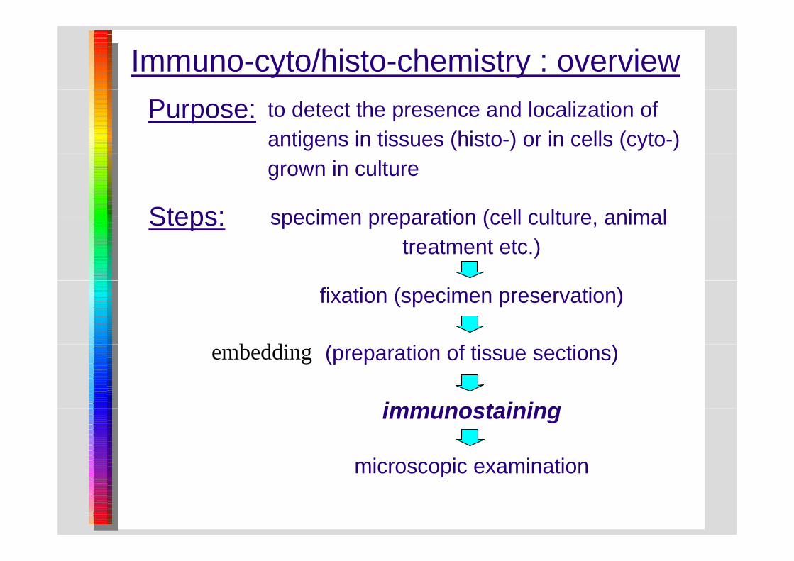
Immuno-cyto/histo-chemistry : overviewPurpose: to detect the presence and localization of
antigens in tissues (histo-) or in cells (cyto-)grown in culture
Steps: specimen preparation (cell culture animalSteps: specimen preparation (cell culture, animal treatment etc.)
fixation (specimen preservation)
b ddi (preparation of tissue sections)
immunostaining
embedding
immunostaining
microscopic examinationmicroscopic examination

Fixation: a matter of trial and errorObjectives: prevent antigen leakage
permeabilize cells to make antigens accessiblemaintain cell & tissue structure
preserve antigenicity
⇔
!preserve antigenicity
Methods: Precipitating agents
Organic solvents Strong acidsti id i i idt th l
Cross-linking agents
acetic acid, picric acid acetone, methanol
paraformaldehyde, glutaraldehyde
Remarks: take small biopsies (one dimension < 4 mm)Remarks: take small biopsies (one dimension < 4 mm)if possible, perfuse the tissue

Making tissue sections
Pro Con
Paraffin
Pro Con
Thin sections (down to 4 μm) Harsh embedding protocolssections
μExcellent histological quality Time consuming
Fixation is needed
Frozen sections
FastExcellent antigen preservation(Pre)fixation not necessary
Freezing artifactsCryostat needed
Vibratome sections
( ) y
Floating sectionsThicker sections (>30 m)
FastExcellent antigen preservationsections Thicker sections (>30 μm) Excellent antigen preservationGood histological qualityGood sensitivity

Unembedded tissue can be sectioned with a Vibratome modified with a 1000 W rheostat in the cutting blade advance. This allowed a slow gforward motion of the oscillating blade through the specimen. This rheostat modification was especially useful for specimens such as airways, which are tough and difficult to cut. A faster forward blade movement for these specimens resulted in deformation of the block and uneven section thickness Without the rheostat modification theand uneven section thickness. Without the rheostat modification, the forward motion of the razor blade carriage had to be stopped for short time periods with the speed control knob while the vibration of the p pblade continued. A blade clearance angle of 15-17° worked best for dry and ethanol dehydrated specimens while 17-20° seemed better for
l i b dd d igelatin-embedded specimens.

Microtomes
VibratomeVibratome
ParaffinParaffin
Cryostaty

I t i i tib d h iImmunostaining : antibody choice

Antibody binding and detection
Essentially the same principles apply as in immunoblotting, but :
Only enzymes fluorochromes and colloidal gold (espOnly enzymes, fluorochromes and colloidal gold (esp. in EM) are used as labels (no radioactivity).
The enzyme system of choice is :peroxidase + diaminobenzidine (DAB) + H2O2 . peroxidase diaminobenzidine (DAB) H2O2 .
Alkaline phosphatase is second choice.
Direct detection is seldom used, only for dual labeling purposes, if there is no alternative.

?Fluorescent or enzymatic staining?
F di C f l iFadingMicroscopy is tiresome
Confocal microscopy

The principle of epifluorescence microscopyThe principle of epifluorescence microscopy
Blue excitation and green emission light are typical for fluorescein.

Immunochemical detection systemsDIRECT METHOD INDIRECT METHODGlass slide Glass slide
Ag
E
fixed antigen molecule in tissue section
labeled primary antibody
Ag fixed antigen molecule in tissue section
(unlabeled) primary antibodyE
(cGH)
(RacGH)
signal: coloured precipitate
signal:coloured precipitate
E
labeled secundary antibody (GaRIg-PO)
substrate
E Esubstrate
E
E E
E
Ag Ag Ag Ag Ag AgAg
1 2 3 1 2 3 41. 2. 3. 1. 2. 3. 4.

Enhanced detection systemsGlass slidefixed antigen molecule in tissue section
(cGH)Ag
tissue section(unlabeled) primary antibody (RacGH)
biotinylated secundary antibody (biot. GaRIg)
substrate
y y y ( g)
B
E
labeled (strept)avidin (PO-conjugated (strept)avidin)
E E
substratesignal : coloured precipitate
B BB
Ag Ag
1. 2. 3. 4. 5. Ag Ag Ag

Glass slide
The Peroxidase-Anti-Peroxidase (PAP) technique
fixed antigen molecule in tissue section
(cGH)Ag
(unlabeled) primary antibody (RacGH)
bridging secundary antibody (excess) (GaRIg)
Rabbit PAP complex EEE Substrate: peroxide + DAB
signal : coloured precipitate
p
EEE E
EEsubstratesubstrate
Ag Ag
1. 2. 3. 4. 5. Ag Ag Ag

Fighting background problemsTitration : use the lowest effective concentration
of any antiserum.
Use of detergents (Triton X-100, 0.1% v/v)and salts (0 9% w/v NaCl) and salts (0.9% w/v NaCl).
Blocking with 1-5 % (v/v) normal serum from theBlocking with 1 5 % (v/v) normal serum from the secundary antibody species or with excess protein (3 % BSA).p ( )
Add the blocking agents to the antibody dilution buffers.
Adsorb detection reagents with appropriate acetone powders.

Some essential controlsNegative antibodies : preimmune serum or an irrelevant antibody.antibody.
Antigen blocking : preferably by solid-phase antigen (not immunogen!), otherwise by liquid-phase blocking.
Immunoblot : although not perfect the best comparisonImmunoblot : although not perfect, the best comparison.
Multiple distinct antibodies : two or three identical plocalization patterns are ideal.
A ti ti ll / tiAntigen negative cells / tissue : "knock-out".
Green Fluorescent Protein fusions : similar stainingGreen Fluorescent Protein fusions : similar staining patterns strengthen your case.