immunity of the gramineae to violet root rot caused …experiment-2 (1947) in the growing season of...
TRANSCRIPT

Immunity of the Gramineae to Violet Root Rot
Caused by Helicobasidium mompa TANAKA
By
Kazuo ITom
Introduction
The violet root rot caused by Helicobasidium mompa TANAKA which has been well known
under the name of "Murasaki-monpa" disease among Japanese phytopathologist and growers is
one of the most important soil-borne diseases in Japan, Taiwan (Formosa) and Korea.
Helicobasidium mompa TANAKA was first described as a fungus affecting mulberry trees
(Morus alba L.) by N. TANAKA1o> in 1891.
Helicobasidium mompa is an aggressively parasitic fungus which causes a destructiv'e root
rot in woody and herbaceous plants, and it has now a known host range of more than 100
wild and cultivated species (ITo 1949) 4>.
Although the violet root rot has been investigated since 1891, comparatively few resistant
plants have been found. As early as 1895, FuNAzu2> advocated the rotation of crops on diseased
soil, and he said: "Maize, Italian millet and other members of the grass family do not die
from this cause, and consequently should be used in soil infested by the disease." Then,
MIYAKE. (1917)6>, YAsu (1927)12>, BoKURA (1934)1>, TocHINAI (1938)11>, HIURA (1939)3> and others
noted, in their handbooks, that gramineous plants were immune or highly resistant to the
disease. On the contrary, Saccharum o[[icinarum L., sugar cane (SAWADA 1919)7>, and Pleioblastus
(Arundinaria) simoni NAKAI, a bamboo grass (SuEMATsu 1930)8>, belonging to the Gramineae,
have been listed as hosts of Helicobasidium mompa.
Many species of the Gramineae have been generally considered to be immune or completely
resistant to the disease, but the nature of resistance still remains· obscure. Since 1946, the
author has made some efforts to study the nature of immunity of the gramineous plants to
the disease, and a brief note on this study has already been published in a preliminary report
(ITo 1952)5>. While the results of this study are not wholly conclusive at this time, ·the
purpose of this paper is to present data accumulated to date as an aid to other investigators
who may be studying this disease.
The author wishes to express his special appreciation to the late Emeritus • Professor Dr.
Hazime Y osHII, of Kyushu University, under whose direction this study was made, for advice
and criticism so willingly given. Thanks are due to Mr. Haruzo 0GANUKA, of Tohoku Branch
of the Government Forest Experiment Station, for assistance in field works. · ·
Received April 23, 1969.
(1) Chief, Forest Protection Division, Government Forest Experiment. ,Station, Meguro, Toky(/.
Japan.

-112-
l<'ield observations on Arundinaria chino (FR. et SAv.) MAKINO
As pointed out by SuEMATsu (1930)8>, sporophores of Helicobasidium mompa are frequently
formed on culms and leaf sheaths of bamboo grasses.
In November 1943, the author first found well·developecl sporophores on Arundinaria chino
(FR. et SAv.)MAKINo (Pleioblastus chino MAKINo), a bamboo grass, in the field of the Government
Forest Experiment Station, Meguro, Tokyo (Plate 1, A). A number of sporophores of the
fungus were produced on culms and leaf sheaths of the same species in the forest of the
Station under moist conditions in 1948 (Plate 1, B). Typical purplish brown strands of the
fungus frequently got entangled on rhizomes of the bamboo grass, but that did not necessarily
prove that the fungus got nourishment from such rhizomes. Some parts of the root system,
especially fibrous roots, were blackened and killed by the fungus, but no visible symptoms of
the disease above ground or no remarkable retardation in growth were found in such plants
(Plate 1, C, D). However, roots of Sambucus racemosa L. subsp. sieboldiana HARA, Mallotus
japonica MULLER, ARG., and fleshy roots of snake-gourd, Trichosanthes cucunzeroides (SER). MAix ,
which were very susceptible to the disease were so completely rotted by the fungus that the
bark or skin could be easily removed from the central part (Plate 1, E).
From the field observations it may be said that Arundinaria chino, a bamboo grass, is
highly resistant to the violet root rot.
Inoculations in the field
In 1946 and 1947, the inoculation experiments were conducted in the field at Koma, Iwate
Prefecture. Precipitation, air and soil temperatures in the plots were recorded throughout
the experimental period.
Experiment·! (1946)
Wooden-boxes (32X32X30cm) were filled with the field soil and steamed for one hour.
After cooling down, on May 20th, the fungus was inoculated to the soil in the boxes. The
inoculum was a pure culture of Helicobasidium mompa (Strain M·1) (ITo 1949)4> grown on
steamed barks of paper mulberry (Broussonetia kazinoki SIEs.) in 300 cc Erlenmayer flasks for
about 60 days at 20~2~° C. The inoculum was removed from the flask and placed in the soil
approximately 1 em deep from the surface. As checks, steamed ,barks of paper mulberry
which were not cultured with' the fungus were used.
On May 27th, seeds of the following plant species were sown in the boxes: Maize (Zea
mays L.), rice (an upland rice) (Oryza sativa L.), Italian millet (Setaria italica BEAUR.), wheat
(Triticum sativum LAM.), oa_ts (Avena sativa L.), Japanese barnyard millet (Echinochloa crus·galli
var. frumentaceae W. F. WIGHT), and soy bean (Glycine max MERRILL). According to the macro
scopic observation all of the gramineous plant species tested which had been inoculated with
the fungus were normal· in growth and fructification, and, there were no remarkable differ·
ences between the inoculated plant and the uninoculatd one (Plates 2~3). In soy bean, the
inoculated plant bearing yellowish green leaves was inferior to the uninoculated one in growth
and fructification (Plate 4; A; B).
In the middle part of October, all plants tested were carefully excavated and examined.

-113-
The gramineous plant species which bore no visible symptoms of the disease above the ground
were found to have purplish brown strands of the fungus on their roots, but showed no
distinct pathological changes. However, on the contrary, roots of soy bean inoculated were
heavily damaged, and a number of sclerotia of the causal fungus were produced on the surface
of the infected roots.
Experiment-2 (1947)
In the growing season of 1947, another inoculation test was made by the same procedure
as in the previous experiment. Plant species used in this experiment were as follows: Maize,
Italian millet, perso-millet (Panicum miliaceum L.), oats, Japanese barnyard millet, soy bean,
and sweet potato.
Results obtained in the cases of the gramineous plants and soy bean are very accordant
with those in Experiment-! (Plate 3, E, F). Some of the roots and the fleshy roots of sweet
potato inoculated were covered with networks of the rhizomorphs of the fungus and were
completely ·rotted.
Results of the field inoculations made in 1946 and 1947 showed that the gramineous plants
were immune from the violet root rot, at least, at maturity under field conditions, whereas
soy bean and sweet potato which had been recorded as being very susceptible were heavily
affected by the disease.
Inoculation in vitro
Artificial inoculations with the fungus were made to very young seedlings growing under
aseptic conditions in vitro. The gramineous plant species used in the experiments were as
follows: Maize, rice, Italian millet, perso millet and Japanese barnyard millet.
Methods Seeds of the gramineous species were surface-sterilized by treatment with 80
per cent alcohol, mercuric chloride solution (0.1 per cent), washed several times in sterile
water, and then placed on sterile 2 per cent glucose agar in Petri dish. As soon as the
majority of seeds in any given lot had produced seedlings with primary roots (radicles) from
several mm long, all dishes were examined carefully for the presence of possible contami
nating organisms. Contaminated plates were discarded; the remainder were inoculated by
placing near or on the roots of each seedling with a small piece of mycelium, taken from a
pure culture of the fungus (Strain M-1). The cultures were theri placed on the desk iri the
laboratory at room temperature (18~25° C).
Results From the inoculated parts the mycelia of the fungus crept clown or up on the
surface of root of the gramineous seedlings. Young primary roots inoculated became purplish
brown in color, being more or less withered and necrotic. Apical parts, especially root caps
and fibrous roots, were heavily affected by the fungus and became dark purplish brown in
color, slender and necrotic. The basal parts of the main roots became slightly discolored,
but they were not withered and necrotic at all (Plates 4~6, Figs. 1~2). Though mycelial
strands of the fungus were frequently formed on the surface of adult main roots of the
gramineous plants, no remarkable pathological change was observed in plant tissue beneath
the fungus mycelium.
From infected roots of the gramineous seedlings adequate material was preserved for
anatomical study.

-114-
\.
b
',' . ' ','
Fig. 1 Primary roots of maize seedlings inoculated with Helicobasidium mompa. A week after inoculation at room temperature (18~25°C). x 2
a, main root b, lateral root h, hyphae of the fungus
h
a b
c
Fig. 2 Young seedlings of rice, perso millet and Japanese barnyard millet inoculated with Helicobasidium mompa. A week after inoculation at room temperature (18 ~25°C). x 2
a, rice b, perso millet c, Japanese barnyard millet h, hyphae of. the fungus
Patho-anatomical observations
Materials and methods Variously infected primary roots of the young gramineous seed
lings cultured under aseptic conditions in vitro were removed and fixed in chromo-acetic acid
solution (chromic add 1 g., glacial acetic acid 1 cc., sterile water 100 cc.). All material was
embedded in paraffin by ZIRKLE's (1930)14l n-butyl alcohol method and sectioned serially at s~
10 fl. Two staining techniques were employed; the Flemming's triple stain, and Yosau's
(1933)18> Sudan Ill-methyl green method for microchemical test.
Results In patho-histological characters, there were observed no remarkable differences

-115-
among five species of the Gramineae examined, and maize, one of them, was chosen for
detailed observation.
The hypha of the fungus penetrates usually the suture of epidermal cell of the root. It
is not rare to o)Jserve the hypha actually piercing the epidermal wall. The original penetra
tion from the outside may or may not be attended with constrictions of the hyphae at the
point of entrance. Fragments of the fungus hypha are frequently observed in the root hairs
developed from the epidermal cells (Fig. 3).
The hyphae which penetrated the epidermal cell develop intercellularly as well as from
cell to cell in the cortex. As penetration of the cortical cell wall is sometimes effected, a
a
d
e f g
rd~,~' llf('?_ ~ f)r-~~ ~ ~' ~ Fig. 3 Penetration of hyphae of Helicobasidium mompa through
the epidermal cells of primary or lateral roots of maize seedling (1--1 = 10 f').
k, root hair

-116-
a b c
d e f
Fig. 4 Hypae of Helicobasidium mompa in or near the cortical cells of primary or lateral roots of maize seedling (1--1 = 10 ,..).
marked constriction occurs in the penetrating hypha.
Shortly after or even before the entry of hypha into a cortical cell its wall becomes
thickened. In some cells the hyphae become aggregated much more than in others. The wall
of a cell thus filled with hyphae becomes greatly thickened, making it difficult for the hyphae
to pass from it to the next cell. In main and lateral young roots, the author has never seen
a case in which the hyphae have penetrated the other cortical cells beyond the thickened cell
wall even at the end of 18 days from the original penetration. In fibrous roots, however, the
fungus penetrates the epidermis, passes through the cortex and its progress is hindered by
the endodermis (Fig. 4).
The thickened cell wall of the cortex stained strongly with safranin shows wound gummy
reaction by Sudan III-methyl green staining. Plasmolysis, degeneration and wound-gummi-

-117-
fication occur in the cells infected as well as in those beyond the fungus hyphae.
In the gramineous plants, the hyphae of the fungus infect only young roots, and they are
unable to penetrate the well developed exodermis of adult roots.
Consideration and conclusion
Without any experimental proof it has been generally considered from field observations
that the Gramineae are immune or highly resistant to the violet root rot caused by Helico
basidium mompa TANAKA, a polyxenic pathogen. Results of the field inoculations made by the
author showed that the gramineous plant species were immune from the disease at maturity
under field conditions, although mycelial strands of the fungus were observed on the surface of
their underground parts. As to the artificial inoculation in field and in vitro, the secondary
or permanent roots were completely free from the disease, but the primary roots were infected
by the fungus. Fibrous roots in. seedling stages were very readily affected by the disease.
These facts show that the causal fungus is unable to attack adult root systems, except fibrous
roots.
Hyphae of the fungus penetrated the epidermal cells of the primary root of the gramineous
plants, developed intercellularly, and penetrated adjacent cortical cells in the same manner as
reported in sweet potatoes and soy beans by ITo (1949) 4'. The wall of the cortical cell thus
filled with hyphae became greatly thickened and wound-gummified or lignified, making it very
difficult for the hyphae to pass from it to the next cell. In sweet potatoes, it seemed likely
that the advance of the parasite was checked, for no further development of the hyphae beyond
the barrier was noted (ITo 19494 ', SuzuKI 19579'). But, in the gramineous plants, the hyphae
in the cortical cell which had been completely enclosed by the thickened and wound-gummified
wall were probably unable to pass out of the cell. This may be due to the difference between
sweet potato and gramineous plant in quantity as well as in quality in the thickening of the
cell wall. Hyphae enclosed in the cell are probably pushed out and leave the diseased host
tissue.
The general immunity of the gramineous plants to the violet root rot is considered to be
due at least in part to the local necrosis by a hypersensitive reaction in the cortical cell of
primary roots of these plants, and, possibly, this restricts the infection to a limited part.
Literature cited
1) BoKURA, U.: Jitsuy6 N6sakumotsu By6gai Y6setsu (An outline of diseases of cultivated
plants) (in Japanese). Tokyo, pp. 574~577, (1934)
2) FuNAzu, D.: (The cause of death of hemp palm (Trachycarpus fortunei (HooK) H. WENDL.)
(in Japanese). Dainihon N6gakukaih6 167, pp. 26~27, (1895)
3) HmRA, M.: Kaisetsu Shokubutsu By6genkinrui (Phytopathogenic fungi) (in Japanese).
Tokyo, pp. 230~232, (1939)
4) ITo, K.: Studies on "Murasaki-monpa'' disease caused by Helicobasidium mompa TANAKA.
Bull. Gov. For. Exp. Sta., 43, pp. 1~126, (1949)
5) ITo, K.: (Immunity of the gramineous plants to the violet root rot) (in Japanese). Agr.
& Hortic., (Tokyo), 27, pp. 85~86, (1952)

-i18-
6) MIYAKE, I.: (On the control of the violet root rot (Preliminary report) (1)) (In Japanese).
Jour. Plant Protection (Tokyo), 4, pp. 409~414, (1917)
7) SAWADA, K.: Descriptive catalo15ue of the Formosan fungi. Part 1 (in Japanese). Agr. Exp.
Sta. Form. Spec. Bull., 19, pp. 402~311, (1919)
8) SuEMATsu, N. and K. YoKoYAMA: Soju B:yochugai Ron (Diseases and insect pests of the
mulberry tree) (in Japanese). Tokyo, pp. 65~67, (1930)
9) SuzuKI, N.: Studies on the violet root rot of sweet potatoes caused by Helicobasidium mompa
TANAKA. VI. Histochemical studies of the infected tissues (In J<!panese with English resume).
Bull. National Inst. Agr. Sci., Ser, C. 8, pp. 69~130, (1957)
10) TANAKA, N.: A new species of hymenomycetous fungus injurious to the mulberry tree.
Jour. Coll. Sci. Imp. Univ. Jap., 4, pp. 193~204, (1891)
11) TocHINAI, Y.: Shokubutsu Byorigaku Tsuron (An outline of plant pathology) (in Japanese).
Tokyo, p. 361, (1938)
12) YAsu, T.: (Diseases of the mulberry tree in Japan (2)) (in Japanese). Jour. Plant Protec
tion (Tokyo), 14, pp. 525~529, (1927)
13) YosHII, H.: Jikken-shitsu no Mado Yori (Phytopathological note) (in Japanese). Jour.
Plant Protection (Tokyo), 20, pp. 225~226, (1933)
14) ZIRKLE, C.: The use of n-butyl alcohol in dehydrating woody tissue for paraffin embed
ding. Science, N. S., 71, pp. 103~ 104, (1930)
-119-
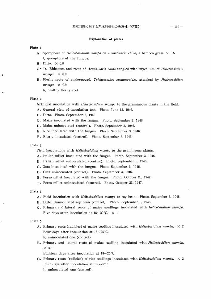
Explanation of plates
Plate 1
A. Sporophore of Helicobasidium mompa on Arundinaria chino, a bamboo grass. X 0.5
f, sporophore of the fungus.
B. Ditto. X 0.8
C~D. Rhizomes and roots of Arundinaria chino tangled with mycelium of Helicobasidium
mompa. X 0.8
E. Fleshy roots of snake-gourd, Trichosanthes cucumeroides, attacked by Helicobasidium
mompa. X 0.9
h, healthy fleshy root.
Plate 2
Artificial inoculation with Helicobasidium mompa to the gramineous plants in the field.
A. General view of inoculation test. Photo. June 13, 1946.
B. Ditto. Photo. September 3, 1946.
C. Maize inoculated with the fungus. Photo. September 3, 1946.
D. Maize uninoculated (control). Photo. September 3, 1946.
E. Rice inoculated with the fungus. Photo. September 3, 1946.
F. Rice uninoculated (control). Photo. September 3, 1946.
Plate 3
Field inoculations with Helicobasidium mompa to the gramineous plants.
A. Italian millet inoculated with the fungus. Photo. September 3, 1946.
B. Italian millet uninoculated (control). Photo. September 3, 1946.
C. Oatsinoculated with the fungus. Photo. September 3, 1946.
D. Oats uninoculated (control). Photo. September 3, 1946.
E. Perso millet inoculated with the fungus. Photo. October 23, 1947.
F. Perso millet uninoculated (control). Photo. October 23, 1947.
Plate 4
A. Field inoculation with Helicobasidium mompa to soy bean. Photo. September 3, 1946.
B. Ditto. Uninoculated soy bean (control). Photo. September 3, 1946.
C. Primary and lateral roots of maize seedlings inoculated with Helicobasidium mompa.
Five days after inoculation at 18~20"C. X 1
Plate 5
A. Primary roots (radicles) of maize seedling inoculated with Helicobasidium mompa. X 2
Four days after inoculation at 18~25"C.
h, uninoculated one (control)
B. Primary and lateral roots of maize seedling inoculated with Helicobasidium mompa.
X 3.5
Eighteen days after inoculation at 18~25"C.
C. Primary roots (radicles) of rice seedlings inoculated with Helicobasidium mompa. X 2
Four days after inoculation at 18~25"C.
h, uninoculated one (control).

-120-
Plate 6
A. Primary roots (radicles) of perso millet seedling inoculated with Helicobasidium mompa.
X 1.2
Five days after inoculation at 18~25"C.
B. Primary roots (radicles) of perso millet seedling inoculated with Helicobasidium mompa.
X 2
h, uninoculated one (control)
C. Primary roots (radicles) of Japanese barnyard millet seedling inoculated. with Helico
basidium mompa. X 2
Four days after inoculation at 18~25"C.
h, uninoculated one (control).

4・
一 121 ←
紫紋羽病に対する禾本科植物の免疫性
(摘要)
伊藤一雄山
紫紋羽病菌 Helicobasidium mompa T ANAKA は代表的な多犯性土壌伝染病菌のーっとして知られている
もので,多くの科,属にわたる木本性および草木性植物を侵し,その寄主の数は 100 以上K 及んでいる
(伊藤 1949) 。
本菌は禾本科純物を伎しがたいことはすでに古くから知られており,本病被害跡地にはとれらを輪作す
ることによって,その後の被害をまぬがれると述べられていた(船津 1895)。そして, その後も同様の
記述が散見されるのであるが(三宅 1917,保 1927, ト蔵 1934,栃内 1938, 樋浦 1939) , これら
はもとより圃場観察によるいわば経験的な記述で,実験的1<::確認されたものではない仇一方,禾木科!C:属
すサトウキピ〔沢[日 1919) およびメダケ(末松 1930) が本菌の寄主植物としてあげられている記録も
ある。
著者は本病に対する禾本科植物の免疫性(強抵抗性)を実験的lと明らかにする目的で野外観察,国場接
種試験および~商状態における人工接種試験を行ない,さらに病態解剖観察によって細胞・組織の病変経
過を追跡した。
ニワトコ,アカメガシワ,カラスウリなどとともに群生しているアズ7ネザサの得および葉輸に本菌の
子実体がおびただしく形成され,夏季には担子胞子の生成をみる乙とはしばしば認められる (Plate 1 A,
B)。そして一見するところ, ササははなはだしく挺病しているように思われるが, いちじるしい成長阻
害も枯死も起こらず,土を掘ってよく観察すると,近接するニワトコ,アカメガシワおよびカラスウリの
地下部は本菌によってはなはだしく侵されて廃敗しているにもかかわらず,ササの地下茎・根部には菌糸
束はからまりついてはいるが,細根(毛根)をのぞき,ほとんど全く何らの病変も認められない。ただし
細根は黒色壊死状を呈し,本菌の侵害をうけていることは明らかに察知された (Plate 1 C, D, E) 。
圃場において 2 か年間本菌の培養菌糸を接種源として人工接種試験を行なった。供試禾本科植物として
はトウモロコジ,アワ,ヒエ, リクトウ,エンパク,コムギおよびキピの 7 種,それに対照として本病に
権病しやすい(伊藤 1949) サツマイモとダイズを用いた c 2 か年間の結果を総括すると,供試末本類は
いずれも,接種区,無接種区を問わず,正常な成長をとげて開花結実し,その聞に差異は認められなかっ
た (Plate 2~3)。ただし接種区では地下部lと本菌の菌糸束がからまりついている乙とは明らかに認められ
た。一方,本病にきわめて感受性のサツマイモおよびダイズの地下部は,はなはだしく侵されて腐敗し,
菌糸束および菌核が根の表面に多量に形成されていた。ダイズの接種区では成長が不良になり,葉は黄変
し,結実も不良になり,無接種区と明らかな対照を示した (Plate 4 A, B) 。
トウモロコシ,アワ,キピ, リクトウおよびヒエの 5 種について,これらの無菌培養子苗に本菌を人工
接種すると,接種点、を中心lとして菌糸は蔓延し,発芽後間もない幼根は紫褐色lζ変じて萎狗壊死し,とれ
1969年 4 月 23 日受理(1 ) 保護部長・農学博土

-122 ー 林業試験場研究報告第 224 号
は根端およびこれに近い部分ほどはなはだしく,基部は軽微である。すなわち,根冠付近および細根(毛
根)は濃紫褐色を呈して軟化萎縮壊死する。しかし幼根基部では変色は認められるが,はなはだしく陥凹
軟化することはほとんどなく,したがって幼苗は発育をつづける。老成根には菌糸束がその表面にほふく
することはあるが,顕著な病変は認められない (Plate 4 C, Plate 5, 6, Fig. 1~2) 。
本菌の禾本類幼根への侵入方法およびその後の細胞・組織病変経過は各種とも大同小異なのでトウモロ
コシについてくわしく調べた。菌糸は幼根の表皮細胞縫合部から侵入することが多く,また表皮細胞膜を
直接貫通することもある。貫入にさいして菌糸はややその太さを減じ,貫通後ふたたび復元し,はなはだ
しく細くなることはほとんどない。菌糸はさらに表皮直下の皮層細胞に侵入し,侵入と同時に,あるいは
侵入K先だってこの部分の細胞,嘆は急激に肥厚する。この場合!C は簡糸は貫通にあたりその太さをいちじ
るしく減ずる。菌糸の侵入をうけた皮層細胞膜ははなはだしく肥厚し,サフラニン濃染性で, Sudan illュ
メチルグリン反応からみると傷演ゴム化(木質化)したものである。幼主根および幼側根においては肥厚
した細胞の内陸に菌糸は繊封され,侵入後18日経過したものでも菌糸が乙の細胞膜を通過してさらに内方
に蔓延したものは全くなかった。しかし毛根ではやや趣きを異にし,さらに内方まで侵され,同様に皮層
細胞膜も傷療ゴム化して肥厚するが,乙れも内皮まででとどまり, rþ心柱は変化がない。侵入をうけた細
胞および乙れに近接する細胞は原形質分離を起乙し,なお傷演プム質が小塊状IC生成される乙ともある
(Fi耳. 3~4)。本繍菌糸の侵入は幼組織に限られ,すでに外皮が完成された老成根を貫通して侵入する ζ
とは全くない。
以上のととがらから,禾本科植物は木病lと全然擢病しないのではなく,幼根は木病病原菌の侵入の対象
となり,細胞・組織の病変をもたらすととは明らかである。本菌の繭糸が皮膚細胞に侵入するとその細胞
膜は急速に肥厚し,かっ木化(傷演コeム化)し,菌糸は肥厚細胞腔内 !C械封されて脱出蔓延することがで
きない。肥厚細胞腔内にとじこめられた菌糸はその細胞とともに押し出されて離脱するか,あるいはその
まま死の運命をたどるものと恩われる。すなわち,木菌の侵入にあった禾本類の幼根細胞は一種の過敏性
壊死を起こし,侵害を局所的にとどめるとも解され,この現象が禾本科植物の木病lζ対する免疫性(強抵
抗性)の少なくとも一部にはなっているものと考えられる。

紫紋羽1丙 lζ対する禾木科植物の免疫性(伊藤) -Plate 1-

-Plate 2- 林業試験場研究報告第 224 な

紫紋羽病lζ対する禾本科植物の免疫↑生(伊藤) -Plate 3-

-Plate 4- 林業試験場研究報告第 224 号

1"
紫紋羽病lζ対する禾本科植物の免疫性(伊藤) -123 ー
Appendex
Supplement to host plants of Helicobasidium mompa TANAKA
Host plant*
Scientific name Japanese name
Pinaceae
1. Larix leptolepis GORDON I Karamatsu 2. Pinus taeda L. I T鹽a-matsu
Palmae
3. Trachyc的us fo仰llei (HOOK) I Shuro 日. WENDL.
Crommelinaceae
4. Commelira communis L.
Liliaceae
5. Asparagus offici叫alis L.
Betulaceae
6ふ. Al初n側包仰z
miたcroρhylla NAKAl
Polygonac巴ae
7. Rumex jaρonicus HOUTTUYN
Leguminosa巴
8. Lespedeza cuneata G. DON.
9. Medicago sativa L.
10. Trifolium repens L.
Rutaceae
11. Phellodendron amurense LUPR.
Cucurbitaceae
12. Trichosanthes cucumeroides
(SER.) MAXIM.
Compositae
13. Sonchus bra日;hyotis DC.
14. Sonchus oleraceus L.
15. Paraxacum officinale WEBER
Tsuyukusa
Oranda-kijikakushi (Asuparagasu)
Gishigishi
Medohagi
R皛an
Shiro-tsumekusa
Kihada
Karasuuri
Hachij�a
Nogeshi
Seiyô・tanpopo
Author
lTo (1959)3)
lTo (1961)剖
FUNAZU (1895)1)
SUZUKl & ABUMIYA (1963)8)
SUZ山1 & ABU~川A (1963)8)
SUZUKl & ABUMIYA (1963)8)
lTo (1962)6)
SUZUKl & ABUMIYA (1963)8)
SUZUKl & ABUMIYA (1963)8)
I lTo (1960)4)
I lTo (1952)2)
SUZUKl & ABUMIYA (1963)8)
SUZUKl & ABUMIYA (1963)8)
SUZUKl & ABUMIYA (1963)8)
* In using the scientific and Japanese names of the host plants the author chiefly followed
OHWl , J. (1965). Flora of Japan. Tokyo.
Literature cited
1) FUNAZU, D.: (The cause of death of hemp palm (Trachycarpus fortunei CHOOK) H. WENDL.))
(in Japaneses). Dainihon Nôgakukaihô, 167, pp. 26~27. (1895)
2) lTô, K.: (Immunity of th巴 gramineous plants to the violet root rot (in Japanese)). Agr. &
Hortic. (Tokyo) , 27, pp. 85~86, (1952)
3) lTô, K.: Zusetsu Naehata-By�ai Shindan-H� (Illustrated diagnosis of tree diseases in
nursery. Part 1). (in Japanese). Tokyo, pp. 42~44. (1959)

dせ
っ白 林業試験場研究報告第 224 号
4) lTô, K.: Zusetsu Tokuy�u-By�ai Shindan・Hô (IIlustrated diagnosis of diseases of trees
for special us巴) (in Japanese). Tokyo, pp. 9~10, (1960)
5) lTô, K.: Zusetsu Rinboku-By�ai Shindan-H� (IIlustrat巴d diagnosis of forest tr巴巴s) (in
Japanes巴). Tokyo, pp. 99~100, (1961)
6) lTô, K.: Zusetsu Juby� Shink� (IIlustrated text-book of forest trees) (in Japan巴se). Tokyo, pp. 81~86, (1962)
7) SATÔ, K.: (Important diseas巴s of Alnus llatoana TANAGI'!'A va1'.削icroρhylla NAKAI) (in Japanュ
ese). (Mimeographed). pp. 1~5 , (1962)
8) SUZUKI , T. and H. ABUMIYA: On th巴 1'oot observation of w巴巴ds for th巴 d巴tection of violet
root rot fungus in soil (in Japanese with English r駸um�). Research Bull. Hokkaido National
Agr. Exp. Sta., 82, pp. 55~59, (1963)