immune response to recombinant capsid proteins of …jvi.asm.org/content/72/3/2388.full.pdf ·...
TRANSCRIPT
JOURNAL OF VIROLOGY,0022-538X/98/$04.0010
Mar. 1998, p. 2388–2397 Vol. 72, No. 3
Copyright © 1998, American Society for Microbiology
Immune Response to Recombinant Capsid Proteins of Adenovirus inHumans: Antifiber and Anti-Penton Base Antibodies Have
a Synergistic Effect on Neutralizing ActivityHANNE GAHERY-SEGARD,1* FRANCOISE FARACE,2 DOMINIQUE GODFRIN,1 JESINTHA GASTON,1
RENEE LENGAGNE,1 THOMAS TURSZ,2 PIERRE BOULANGER,3 AND JEAN-GERARD GUILLET1
Laboratoire d’Immunologie des Pathologies Infectieuses et Tumorales, INSERM Unite 445, Institut Cochin de GenetiqueMoleculaire, Universite R. Descartes, Hopital Cochin, 75014 Paris,1 Departements de Biologie Clinique ou de
Medecine, Institut Gustave Roussy, 94805 Villejuif,2 and Laboratoire de Virologie Moleculaire etPathogenese Virale, CNRS-UMR 5812, Faculte de Medecine, 34060 Montpellier,3 France
Received 14 August 1997/Accepted 1 December 1997
Replication-deficient adenovirus used in humans for gene therapy induces a strong immune response to thevector, resulting in transient recombinant protein expression and the blocking of gene transfer upon a secondadministration. Therefore, in this study we examined in detail the capsid-specific humoral immune responsein sera of patients with lung cancer who had been given one dose of a replication-defective adenovirus. Weanalyzed the immune response to the three major components of the viral capsid, hexon (Hx), penton base(Pb), and fiber (Fi). A longitudinal study of the humoral response assayed on adenovirus particle-coatedenzyme-linked immunosorbent assay plates showed that patients had preexisting immunity to adenovirus priorto the administration of adenovirus–b-gal. The level of the response increased in three patients after adeno-virus administration and remained at a maximum after three months. One patient had a strong immuneresponse to adenovirus prior to treatment, and this response was unaffected by adenovirus administration.Sera collected from the patients were assayed for recognition of each individual viral capsid protein todetermine more precisely the molecular basis of the humoral immune response. Clear differences existed in thehumoral response to the three major components of the viral capsid in serum from humans. Sequentialappearance of these antibodies was observed: anti-Fi antibodies appeared first, followed by anti-Pb antibodiesand then by anti-Hx antibodies. Moreover, anti-Fi antibodies preferentially recognized the native trimeric formof Fi protein, suggesting that they recognized conformational epitopes. Our results showed that sera with noneutralizing activity contained only anti-Fi antibodies. In contrast, neutralizing activity was only obtained withsera containing anti-Fi and anti-Pb antibodies. More importantly, we showed that anti-native Fi and anti-Pbantibodies had a synergistic effect on neutralization. The application of these conclusions to human genetherapy with recombinant adenovirus should lead to the development of strategies to overcome the formationof such neutralization antibodies, which have been shown to limit the efficacy of gene transfer in humans.
Many studies with animal models have indicated that thereplication-defective recombinant adenovirus (Rec-Ad) is use-ful for in vivo gene transfer because it allows expression ofrecombinant proteins in dividing and resting cells (17, 22, 30).Rec-Ad has been used in phase I gene therapy clinical trials inpatients with cystic fibrosis and lung cancer (19, 27). However,studies have shown that cellular and humoral immune re-sponses to the vector and transgene product were involved inthe transient recombinant gene expression observed in Rec-Ad-injected hosts (11, 26, 32, 33). In phase I gene therapyclinical trials in patients with lung cancer, we showed that onepatient who already had high levels of neutralizing antibodiesprior to Rec-Ad administration did not develop an immuneresponse to the transgene product (4). This is of interest be-cause Rosenecker et al. showed that cystic fibrosis patientshave high levels of specific anti-Ad antibodies, suggesting ahigh prevalence of Ad infection in these patients (18). Thus,preexisting neutralizing antibodies reactive to surface epitopes
of the virion may affect expression of the transgene product ingene therapy. Moreover, other groups have also reported thatthe formation of neutralizing antibodies may prevent genetransfer when a second injection of Rec-Ad is given (34, 35).The mechanism by which antibodies neutralize adenovirus isstill poorly understood. Therefore, analysis of neutralizing an-tibodies that recognize viral proteins is necessary to shed somelight on the functional basis of neutralization.
The Ad viral capsid is composed of three major types ofproteins: hexons (Hx) (130 kDa), penton bases (Pb) (82 kDa),and fibers (Fi) (62 kDa). Five Pb subunits (82 kDa) form a Pbcapsomere, which is linked to the trimeric Fi by noncovalentbonds (9). Three minor proteins, IIIa, VIII, and IX, are thoughtto stabilize the particle. The entry of human Ad into host cellsinvolves the interaction of virus particles with two separate cellreceptors. The initial binding of the virus to recently identifiedcell receptors is mediated by Fi protein (1, 8, 24). The subse-quent event of virus infection is mediated by Pb protein bind-ing to integrins, promoting virus internalization and/or pene-tration (10, 14, 29).
In this study, we examine the temporal recognition of thethree major molecular components of the adenovirus capsid(Hx, Pb, and Fi) in sera of patients with lung cancer who weregiven a single intratumoral administration of Rec-Ad. We alsoshow that the recognition of the three major capsid proteins
* Corresponding author. Mailing address: Laboratoire d’Immunol-ogie des Pathologies Infectieuses et Tumorales, INSERM Unite 445,Institut Cochin de Genetique Moleculaire, Hopital Cochin, 27 rue duFg Saint Jacques, 75014 Paris, France. Phone: 33 1 46334395. Fax: 331 44071425. E-mail: [email protected].
2388
on June 20, 2018 by guesthttp://jvi.asm
.org/D
ownloaded from

differed among patients. Synergistic recognition of viral capsidproteins led to the emergence of neutralizing antibodies.
MATERIALS AND METHODS
Patients and clinical protocol. The gene therapy approach has been describedin detail elsewhere (4, 27). Briefly, Rec-Ad containing lacZ (Ad–b-gal) wasadministered locally by fiberoptic bronchoscopy to patients with advanced lungcancer. The patients received no chemotherapy before the administration ofAd–b-gal, but standard chemotherapy began 3 days after administration. Co-horts of patients received a single dose of 109 PFU (patients 1 through 4 [Pt-1through Pt-4]), 108 PFU (Pt-5 through Pt-7), or 107 PFU (Pt-8 through Pt-10) ofAd–b-gal. Sera were collected on day 0 (before treatment) and on days 8, 30, 60,and 90 after the administration of Ad–b-gal.
Virus. Rec-Ad was constructed from a modified adenovirus type 5 (Ad5)genome with the entire E1 and E3 regions deleted (23). The proteins of 19, 11.6,10.4, 14.5, and 14.7 kDa are not synthesized, because the whole E3 region hasbeen deleted. Almost all of these proteins are involved in modulation of theimmune system (20). The Rous sarcoma virus promoter was used to drivetranscription of the lacZ transgene. lacZ encodes the bacterial b-galactosidase(b-gal). Rec-Ad was prepared by infecting the 293 cell line and was purified twiceby centrifugation through a CsCl gradient. The virus preparation was subjectedto titer determination by a limiting-dilution plaque assay on 293 cells. The virusstock was supplied by Transgene (Strasbourg, France).
Anti-Ad antibodies detected by enzyme-linked immunosorbent assay. Poly-styrene plates (Nunc, Roskilde, Denmark) were coated by incubation with 107
PFU of Rec-Ad per well at 4°C overnight. Saturation was performed with asolution of phosphate-buffered saline (PBS) containing 0.1% Tween 20 and 3%bovine serum albumin (3). Dilutions of sera were incubated in the coated wellsat 4°C overnight, and bound antibodies were detected with alkaline phosphatase-conjugated goat anti-human immunoglobulin G (IgG) (1/5,000; Sigma-AldrichChimie). The phosphatase activity was measured with 4-methylumbelliferylphosphate as the substrate (Sigma-Aldrich Chimie) and with fluorescence read at360/460 nm in a Cytofluor 2300 apparatus (Millipore, Saint Quentin, France). Tocompare specific anti-Ad antibodies between humans, and since almost all hu-mans have been in contact with this virus, we used serum from primates thatcross-reacted with goat anti-human Ig as a negative control in each experiment.We have shown that preimmune serum from macaques does not react with Adbefore immunization. Since the preimmune serum from macaques gave the samefluorescence as the fluorescence obtained in the absence of serum, the data areexpressed as fluorescence units corrected for background fluorescence in theabsence of serum.
Recombinant baculovirus producing Ad2 Pb and Ad5 Fi proteins. The recom-binant proteins described previously were produced by infecting Sf9 cells with therecombinant Ad2 PbFL571 (12), which codes for Pb protein, or Ad5 FiFL581(8), which codes for fiber protein. Recombinant baculovirus Ad5 Fi-AT386was also produced; this protein corresponds to the Fi knob domains of the Fiprotein and includes the last shaft repeat. The last repeat was found to beessential for stable trimerization of the knob domain (8). The infection wasperformed in complete TC-100 medium (TC-100 medium [Gibco-BRL] supple-mented with 100 U of penicillin per ml, 100 mg of streptomycin per ml, 1 mML-glutamine, and 4% heat-inactivated fetal calf serum). The cells were harvested24 h postinfection by centrifugation for 10 min at 1,000 3 g. The cell pellet waswashed twice with PBS, resuspended in lysis buffer (1% Nonidet P-40, 1 mMEDTA, 150 mM NaCl, 10 mM Tris-HCl [pH 8.0]) containing protease inhibitors(phenylmethylsulfonyl fluoride, pepstatin A, leupeptin, and aprotinin), and lysedby incubation at 4°C for 30 min. The cytoplasmic proteins were then collected bycentrifugation for 30 min at 100,000 3 g. The extracted proteins were analyzedby denaturing sodium dodecyl sulfate-polyacrylamide gel electrophoresis (SDS-PAGE), nondenaturing SDS-PAGE (NDS-PAGE), and Western blotting (seeabove).
Gel electrophoresis and Western blotting. SDS-PAGE was performed on 10%acrylamide gels (Bio-Rad, Ivry Sur Seine, France). Samples (10 ml, containing107 PFU of Ad particles) were denatured by boiling in Laemmli buffer (62.5 mMTris-HCl, 2% SDS, 25% glycerol, 0.01% bromophenol blue [Bio-Rad]) prior toelectrophoresis. NDS-PAGE differed from SDS-PAGE in that the proteins werenot denatured by boiling prior to electrophoresis; the samples were mixed withnative buffer (62.5 mM Tris-HCl, 40% glycerol, 0.01% bromophenol blue; Bio-Rad). The trimeric full length of the Fi protein (Ad5 FiFL581) was visible inNDS-PAGE gels as a band of 180 kDa, and the trimeric Fi knob domains (Ad5Fi-AT386) were visible as a band of 60 kDa (8, 15). To obtain the monomericform of the full-length Fi protein or the knob domains of Fi, SDS-PAGE analysiswas used; the proteins were 62 and 20 kDa, respectively. In SDS-PAGE gels, thePb proteins (Ad2 PbFL571) were visible as a band of 82 kDa. After migration,the proteins were transferred to nitrocellulose membranes in 25 mM Tris–192mM glycine buffer, containing 20% ethanol, at 100 V for 1 h with a mini-Trans-Blot system (Bio-Rad). The blots were saturated for 1 h with PBS–0.1% Tween20 containing 5% milk. Immunoblotting was performed for Rec-Ad particles, Pb,and Fi, using different human sera and rabbit and mouse control sera. Goatanti-human total Ig and IgM (The Binding Site, Birmingham, United Kingdom),horseradish peroxidase (HRP)-labeled anti-goat IgG conjugate (The Binding
Site), and a chemiluminescent substrate (luminol; Amersham, Les Ulis, France)were used to detect the reaction.
Ad neutralization assay. Permissive 293 cells were distributed in 96-wellCostar plates at 20,000 cells/well and cultured in complete Dulbecco’s modifiedEagle’s medium (Gibco-BRL) for 24 h. An Ad–b-gal viral suspension (0.3 PFU/ml) was incubated with decomplemented diluted serum samples for 1 h at roomtemperature. This mixture (100 ml) was incubated with the 293 cells for 1 h at37°C; 100 ml of complete Dulbecco’s modified Eagle’s medium was then added,and the cells were incubated at 37°C overnight. The cells were washed with 200ml of PBS and incubated with 100 ml of lysis buffer (6 mM Na2HPO4, 4 mMNaH2PO4, 10 mM KCl, 0.1 mM MgSO4, 50 mM b-mercaptoethanol, 0.5%Triton X-100) containing 17 mg of a fluorescent b-gal substrate (4-methylum-belliferyl-b-D-galactoside [Sigma-Aldrich Chimie]) for 30 min to 1 h at 37°C. Theresulting fluorescence was measured with a Cytofluor 2300 apparatus. Sera con-taining neutralizing antibodies were shown by the absence of b-gal activity in 293lysates.
Exhaustion of anti-Pb and anti-Fi antibodies. Human serum diluted 1/100 anddecomplemented for 30 min at 56°C was exhausted from anti-Pb antibodies byincubating the serum on a nitrocellulose membrane containing the recombinantprotein (Ad2 PbFL571). The serum was passed across the membrane six times,overnight for the first exhaustion and for 1 h for the five overexhaustions. Foranti-Fi exhaustion, serum was mixed with the membrane containing the native ordenatured form of the Fi protein (Ad5 FiFL581). One-quarter of the exhaustedserum was used in an Ad neutralization assay (see above).
Purification of anti-Pb and anti-Fi antibodies. The serum diluted 1/100 anddecomplemented for 30 min at 56°C was mixed overnight at 4°C with a nitro-cellulose membrane containing Pb or Fi protein. After incubation, the mem-branes were intensively washed with a PBS–0.1% Tween 20 solution. The anti-bodies were eluted with 480 ml of an elution buffer (0.1 M glycine-HCl [pH 2.7])for a few seconds, and the solution was neutralized with 20 ml of 1 M Tris-HCl(pH 9). The purified antibodies (1/10 of the eluted antibodies) were tested in aneutralization assay.
Densitometry. The film obtained from Western blot experiments was scannedwith a scanner, and the signal intensity was measured with the Image Quantprogram (Molecular Dynamics).
RESULTS
Humoral response to a single intratumoral administrationof Rec-Ad particles in patients. Blood samples were collectedfrom four patients (Pt-1 through Pt-4) with lung cancer given109 PFU of Ad–b-gal on day 0 (pretreatment) and on days 30,60, and 90 (postadministration) and were assayed for anti-Adantibodies by enzyme-linked immunosorbent assay (Fig. 1).A longitudinal study of the anti-Ad humoral response showedthat all the patients had a preexisting immune response to Adprior to administration. The level of specific IgG to Ad in Pt-1was high before treatment and was unaffected by Ad–b-galadministration. In the other patients, the level of anti-Ad IgGincreased strongly after administration of Ad–b-gal and re-mained maximal 3 months later, as shown by assays on Rec-Adparticle-coated plates. Although Rec-Ad did not enable pro-liferation to occur (because of deletions in E1A and E1B), thesingle intratumoral injection of Ad produced very strong hu-moral immune responses.
Humoral response to viral capsid proteins. The molecularbasis of the humoral immune response was examined in great-er detail by assaying sera collected from patients for antiviralprotein antibodies by Western blotting with the entire Rec-Adparticle. This analysis revealed the recognition of each individ-ual viral capsid protein, unlike results obtained with Rec-Adparticle-coated plates, which showed only an overall responseto the vector. The results from four patients (Pt-1 throughPt-4) are presented in detail (Fig. 2), but the experiments werealso performed with sera from six other patients. There wereclear differences in humoral responses to the three major com-ponents (Hx, Pb, and Fi) of the viral capsid in the sera. Theprotein identified as Fi protein may be the penton-associatedprotein (66 kDa) called protein IIIa (9), in which case the Fiprotein was associated with protein IIIa (Fi/P.IIIa). Anti-Fi/P.IIIa and anti-Pb antibodies were detected in the sera of Pt-1and Pt-3 before administration of Rec-Ad (Fig. 2). Serumcollected on day 0 from three other patients gave the same
VOL. 72, 1998 IMMUNE RESPONSE TO ADENOVIRUS CAPSID PROTEINS 2389
on June 20, 2018 by guesthttp://jvi.asm
.org/D
ownloaded from

FIG. 1. Specific IgG response to Rec-Ad particles. Humans blood samples were collected 8 (D8), 30 (D30), 60 (D60), and 90 (D90) days after Rec-Adadministration. Serum dilutions were tested on Rec-Ad-coated plates (107 PFU/well). Data are expressed as fluorescence units corrected for background fluorescencein the absence of serum. Sera collected from humans on day 0 (D0) indicated the immunization state against Ad before the injection.
FIG. 2. Immunoblot pattern of serum specific for Ad capsid components. Rec-Ad (107 PFU/well) was deposited, and the viral proteins were separated bySDS-PAGE. Immunoblots were incubated with the different sera, collected on day 0 (D0) to day 90 (D90), diluted at 1/100, and washed with HRP-labeled anti-humanIg conjugate. The capsid proteins Hx, Pb, and Fi are indicated. The controls contained rabbit anti-Fi, rabbit anti-Pb, and mouse anti-Hx antibodies.
2390 GAHERY-SEGARD ET AL. J. VIROL.
on June 20, 2018 by guesthttp://jvi.asm
.org/D
ownloaded from

results (Pt-6 through Pt-8 [data not shown]). Serum collectedon day 0 from Pt-2 and Pt-4 contained only specific anti-Fi/P.IIIa IgG antibodies (Fig. 2), as did serum from two otherpatients (Pt-9 and Pt-10).
Serum collected from Pt-1 before the administration of Rec-Ad contained IgG antibodies that recognized the Fi/P.IIIa andPb capsid proteins, and the injection did not modify the rec-ognition of viral proteins. Sera collected from three patients(Pt-6 through Pt-8) before or after the administration of 108 or107 PFU of Ad–b-gal contained IgG that recognized the twomajor capsid components, as did serum from Pt-1 (data notshown). Sera collected from Pt-2 through Pt-4 on day 30 afterthe injection showed a change in the pattern of recognition ofcapsid components. Pt-2 had anti-Fi/P.IIIa and anti-Pb anti-bodies after treatment. Another patient (Pt-9) had the samerecognition profile as Pt-2 (data not shown). Pt-3 and Pt-4developed humoral responses to all the major molecular com-ponents of the Ad capsid (Fi/P.IIIa, Pb, and Hx). Specific anti-Pb IgM was detected on day 8 (Pt-4) and on day 30 (Pt-2) inPt-2 and Pt-4. We also found specific anti-Hx IgM on days 8and 30 in serum from Pt-3 and Pt-4 (Fig. 3). All the patientshad previously been exposed to a related human Ad becausetheir sera contained anti-Fi antibodies. However, new contactwith the Ad after intratumoral administration could induceIgM antibodies against viral proteins. The failure to detect Hxand Pb antibodies in some patients before the administrationof Ad–b-gal can be an indication of a decayed immunity.
Recognition of recombinant Pb protein. To confirm the ab-sence of recognition of Pb protein by sera from Pt-2 and Pt-4before injection and to ensure that it was not due to a limitingquantity of antigenic Pb proteins, recombinant Ad-2 Pb pro-teins were used in a Western blot analysis (Fig. 4). Note thatthe baculovirus recombinant Pb used came from Ad2 (12),whereas the recombinant Ad injected was Ad5, but these twoproteins had 99% identity. The sera from Pt-1 and Pt-3 con-tained anti-Pb antibodies before injection (Fig. 4); these anti-bodies were also detected in the sera from three other patients(Pt-6 through Pt-8 [data not shown]). The absence of anti-PbIgG antibodies from the sera of Pt-2 and Pt-4 was confirmed inthis experiment and was also found with Pt-5 (data not shown).Three other patients (Pt-2, Pt-3, and Pt-6) who were tested fortheir capacity to recognize the recombinant Pb protein gavethe same result as Pt-2 and Pt-4. Densitometry analysis withthe Image Quant program (Molecular Dynamics) determined
a significant difference between positive and negative sera interms of Pb protein recognition. This result also showed thatanti-Pb antibodies recognized the monomeric form of the Pbprotein.
Recognition of native trimeric recombinant Fi protein. Toprecisely identify and determine the quality of the anti-Fi an-tibody response in human serum, the recombinant baculovirusAd5 Fi was used in Western blots. To obtain monomeric Fiprotein (62 kDa), SDS-PAGE analysis was required. Figure 5shows that anti-Fi antibodies from sera of Pt-1 through Pt-3weakly recognized the monomeric form of the Fi proteins (62kDa) before the treatment. After administration of Rec-Ad,only serum from Pt-4 strongly recognized the denatured formof the Fi protein. The trimeric nature of the Fi synthesized invitro was conserved, as shown in NDS-PAGE analyses (12, 15).The Fi trimers were visible as a 180-kDa band. Figure 6 showsthat anti-Fi antibodies from sera of patients preferentially rec-ognized the native trimeric form of the fiber protein. Pt-1 andPt-3 presented a high level of anti-native Fi antibodies on day0 (as did Pt-6 through Pt-8 [data not shown]). The sera of Pt-2and Pt-4 recognized the native Fi proteins weakly (as did seraof Pt-9 and Pt-10). Injection led to maturation of the humoralresponse, and the amount of anti-native Fi antibodies in-creased with time and become maximal on days 30 (Pt-4) and
FIG. 3. Pattern of IgM specific for Ad capsid components. Sera collected on different days (D0, D8, and D30) and diluted at 1/100 were tested for the presenceof IgM antiviral proteins after immunoblotting of Rec-Ad (107 PFU/well). After overnight incubation of immunoblots with the diluted sera, the proteins were detectedwith sheep anti-human IgM (1/500) and HRP-labeled anti-sheep IgG conjugate (1/2,000).
FIG. 4. Immunoblot pattern of serum specific for Pb. Suspensions of recom-binant Pb protein (10 ml) harvested from infected Sf9 cells with the recombinantAd2 PbFL571 were separated by SDS-PAGE. Sera (diluted 1/100) collectedfrom patients at different time points (day 0 [D0] and day 60 [D60]) were testedand revealed with a sheep HRP-labeled anti-human Ig conjugate (1/2,000).
VOL. 72, 1998 IMMUNE RESPONSE TO ADENOVIRUS CAPSID PROTEINS 2391
on June 20, 2018 by guesthttp://jvi.asm
.org/D
ownloaded from

60 (Pt-2). The sera from Pt-9 and Pt-10 also contained anti-native Fi antibodies on day 30 after the injection, but the serumfrom patient 5 did not contain anti-Fi antibodies (data notshown).
Recognition of native trimeric Fi knob domains. The part ofthe native Fi protein recognized by human serum was identi-fied with a recombinant baculovirus Ad5 Fi-AT386 corre-sponding to the Fi knob domains of the Fi protein, whichincluded the last shaft repeat. The last repeat was found to beessential for stable trimerization of the knob domain. Thetrimeric Fi knob domains were visible as a band of 60 kDa onNDS-PAGE (8). Figure 7 shows that anti-Fi antibodies fromthe sera recognized the native trimeric form of the Fi knobdomains. There was also a direct correlation between the ca-pacity to recognize the native trimeric form of the Fi proteinand recognition of the native trimeric knob of the Fi protein.The results showed that anti-Fi antibodies recognizing confor-mational epitopes were essentially directed against the C-ter-minal part of the protein, i.e., the Fi knob domain.
Induction of neutralizing antiadenovirus antibodies after asingle administration of Ad–b-gal. Sera taken from patientsbefore and after the Ad–b-gal administration were examinedfor neutralizing antibodies to Ad. Table 1 presents significantresults obtained from sera of patients. The sera from Pt-1 andPt-3 contained significant amounts of neutralizing antibodiesprior to the Rec-Ad administration. In addition, the sera fromthree other patients (Pt-6 through Pt-8) had significant neu-tralizing activity (data not shown). Sera from Pt-2, Pt-4, andPt-5 contained no neutralizing activity on day 0, like sera fromPt-9 and Pt-10 (data not shown). After the treatment, serafrom all patients tested except one (Pt-5) had neutralizingantibodies. The administration of Ad–b-gal acted like a boost
for patients whose serum contained significant amounts ofneutralizing antibodies before the treatment, and the titers ofthese antibodies increased. The results also showed a directrelation between neutralization and the level of recognition ofthe Pb and the native Fi proteins (Table 2). Patients withneutralizing antibodies before the Rec-Ad injection (Pt-1 andPt-3) recognized the two capsid proteins. The appearance ofneutralizing antibodies in Pt-2 and Pt-4 was directly related tothe presence of antiviral capsid protein antibodies. Pt-5, whodid not develop neutralizing antibodies, did not recognize thetwo viral proteins. In conclusion, the greater the neutralizingactivity, the better the recognition of the Pb protein and thenative trimeric form of the Fi protein.
Direct involvement of anti-Pb antibodies in neutralizing ac-tivity. The differences observed in neutralizing activity may bedue to differences in the ability of sera to recognize capsidproteins. Patient sera containing anti-Pb antibodies (Pt-1)were used to test the capacity of these antibodies to directlyaffect the neutralization activity observed. Serum diluted 1/100was partially exhausted of anti-Pb antibodies by sequentialincubation with Pb proteins. Figure 8A shows the sequentialexhaustion of anti-Pb antibodies after six different contactswith the Pb protein. The decrease in the signal was determinedby densitometry with the Image Quant program; 71% of thesignal was lost after the final exhaustion. The depleted serumwas then tested for its capacity to neutralize the virus. Thediluted serum partially exhausted of anti-Pb antibodies (1/400)had lost much of its capacity to neutralize the virus comparedto the control (Fig. 8B).
Direct involvement of anti-native trimeric Fi antibodies inneutralizing activity. The same serum from Pt-1 containinganti-Fi antibodies was used to test the capacity of these anti-
FIG. 5. Immunoblot pattern of serum specific for monomeric Fi protein. Suspensions (20 ml) of recombinant Fi protein (Ad5 FiFL581) were separated bySDS-PAGE. Sera (collected on day 0 [D0] to day 90 [D90]) were tested at a dilution of 1/100, and Fi was detected with HRP-labeled anti-human Ig conjugate (1/2,000).The monomeric form of the Fi protein was detected as a band of 62 kDa.
FIG. 6. Immunoblot pattern of serum specific for trimeric Fi protein. Suspensions (20 ml) of recombinant Fi protein (Ad5 FiFL581) were separated by NDS-PAGE.Sera (collected on day 0 [D0] to day 90 [D90]) were tested as described in the legend to Fig. 4. The trimeric form of the Fi protein was detected as a band of 180 kDa.
2392 GAHERY-SEGARD ET AL. J. VIROL.
on June 20, 2018 by guesthttp://jvi.asm
.org/D
ownloaded from

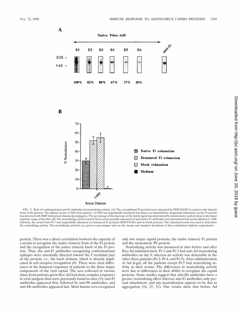
bodies to play a direct role in neutralizing activity. Humanserum diluted 1/100 was partially exhausted of anti-Fi antibod-ies by sequential incubation with Fi proteins. The depletionwas performed by NDS-PAGE to conserve the native trimericform of the Fi protein and by SDS-PAGE to produce the Fimonomeric form. Figure 9A shows the sequential exhaustion,on native trimeric Fi protein, of diluted serum after six inde-pendent contacts with the native protein. The loss of the signalwas measured by densitometry and indicated partial (but sig-nificant) exhaustion of anti-trimeric Fi antibodies. Figure 9Bshows the percent neutralization obtained with serum ex-hausted on the native trimeric form or the monomeric form ofFi protein and on mock protein. The partial depletion (72%)of antibodies recognizing the native trimeric form of Fi pro-teins significantly decreased the neutralizing activity.
Synergistic effect of anti-Fi and anti-Pb antibodies on viralneutralization. The direct effect of anti-Fi and anti-Pb anti-bodies on neutralizing activity was determined with the twokinds of antibodies purified independently from sera from Pt-1and Pt-3 (day 30). The anti-Fi antibodies recognizing confor-mational epitopes were obtained by passing the serum (diluted1/100) over a nitrocellulose membrane containing the nativetrimeric form of the Fi protein. The anti-Pb antibodies weresimilarly purified from the same diluted serum by using amembrane containing the recombinant Pb protein. The puri-fied antibodies were eluted from the membrane, and one-quarter of the purified antibody solution was used in an Adneutralization assay. We showed that to obtain viral neutral-ization, a mixture of anti-Fi and anti-Pb antibodies from Pt-1serum was necessary (Fig. 10). This result was confirmed withthe purified anti-Fi and anti-Pb antibodies from the serum ofPt-3 (data not shown). The results demonstrated a synergistic
effect between the conformational anti-Fi antibodies and theanti-Pb antibodies in the Ad-neutralizing activity.
DISCUSSION
In this study, we examined in detail the Ad-specific humoralimmune response induced in patients with lung cancer given asingle dose of Rec-Ad by fiberoptic bronchoscopy (18). Assaysof the anti-Ad response before the administration of Ad–b-gal,on Rec-Ad particle-coated plates, showed that the patients hadpreexisting immunity to adenovirus. After Rec-Ad administra-tion, three patients developed strong immunity to Ad, whichremained at a maximum after 3 months. Pt-1 had strong im-munity to Ad before the administration of Rec-Ad, and thiswas unaffected by the treatment. Although Rec-Ad did notreplicate, a single administration of Ad induced a very stronghumoral immune response. This result is consistent with ourprevious findings in animal models (3).
FIG. 7. Immunoblot pattern of serum specific for trimeric Fi knob domains. Suspensions (20 ml) of recombinant Ad5 Fi knob domains (Ad5 Fi-AT386) wereseparated by NDS-PAGE. The size of the protein was 60 kDa. The sera were tested as described in the legend to Fig. 4. D0 to D90, days 0 to 90, respectively.
TABLE 1. Anti-Ad neutralizing antibodies in seraof humans injected with Rec-Ad
Time ofcollectiona
% Neutralizationb in serum from:
Pt-1c Pt-2c Pt-3c Pt-4c Pt-5d
D0 100 0 42 0 0D30 100 50 100 40 0D60 100 52 100 20 0
a Sera of patients were collected before (D0) and on days 30 (D30) and 60(D60) after a single injection of Rec-Ad.
b Neutralization obtained with a 1/200 serum dilution.c Given 109 PFU of Ad–b-gal.d Given 108 PFU of Ad–b-gal.
TABLE 2. Relation between neutralization and therecognition of Pb and native Fi proteins
Patient Time ofcollectiona % Neutralizationb
Recognition of:
Pb Native Fi
Pt-1c D0 100 1111 1111D30 100 1111 1111D60 100 1111 1111
Pt-2c D0 0 1/2 1D30 50 111 111D60 52 111 111
Pt-3c D0 42 1111 111D30 100 1111 1111D60 100 1111 1111
Pt-4c D0 0 1/2 11D30 40 11 1111D60 20 11 1111
Pt-5d D0 0 2 1/2D30 0 2 1/2D60 0 1/2 1
a D0, before injection of Rec-Ad; D30 and D60, 30 and 60 days after injectionof Rec-Ad.
b Neutralization obtained at 1/200 serum dilution.c Given 109 PFU of Ad–b-gal.d Given 108 PFU of Ad–b-gal.
VOL. 72, 1998 IMMUNE RESPONSE TO ADENOVIRUS CAPSID PROTEINS 2393
on June 20, 2018 by guesthttp://jvi.asm
.org/D
ownloaded from

Recognition of the different capsid proteins was analyzed byWestern blotting to determine more precisely the molecularbasis of the humoral immune response. This analysis made itpossible to detect the recognition of each individual viral cap-sid protein, unlike the results obtained with Rec-Ad particle-coated plates, which showed an overall response to the vector.It is difficult to differentiate between Fi protein (62 kDa) andprotein IIIa (66 kDa) recognition by the sera. Before admin-istration of Ad–b-gal, sera from Pt-1 and Pt-3 recognized twocapsid proteins, Fi/P.IIIa and Pb, and sera from Pt-2 and Pt-4recognized only the Fi/P.IIIa protein. Sera from Pt-2, Pt-3, andPt-4 gave a complex response after injection. Specific anti-Pb
IgG appeared in Pt-2 and Pt-4 sera, and specific anti-Hx IgGantibodies appeared in Pt-3 and Pt-4 sera. The heterogeneityof the response to the capsid proteins was also assayed with thesera of six other patients (data not shown). Molecular analysiswas required to better understand the humoral immune re-sponse to capsid components, because of this complex re-sponse. Therefore, recombinant Pb and Fi proteins were used.The use of recombinant Fi protein made it possible to differ-entiate between Fi and P.IIIa recognition by the sera. Moreimportantly, the results of this study demonstrate that anti-Fiantibodies were composed essentially of antibodies recogniz-ing conformational epitopes of the trimeric form of the Fi
FIG. 8. Role of anti-Pb antibodies in neutralizing activity. (A) The recombinant Pb proteins were separated by SDS-PAGE. The immunoblot was sequentiallyincubated with serum (Pt-1, day 60) diluted 1/100. Sequential exhaustion on the Pb protein was detected with HRP-labeled anti-human Ig conjugate. The percentageof anti-Pb antibodies exhausted from the serum was determined after quantification by the Image Quant program. Densitometry analysis was done in the linear responserange of the film. After the final exhaustion (E6), we detected only 29% of the initial signal. (B) The partially exhausted serum (1/400) was tested for its capacity toneutralize the viral particle. The same serum was sequentially exhausted on mock proteins (six times) and was tested in the same way for the presence of anti-Adneutralizing antibodies. The neutralizing activities are given as percentages and are the means and standard deviations of three individual triplicate experiments.
2394 GAHERY-SEGARD ET AL. J. VIROL.
on June 20, 2018 by guesthttp://jvi.asm
.org/D
ownloaded from
protein. There was a direct correlation between the capacity ofa serum to recognize the native trimeric form of the Fi proteinand the recognition of the native trimeric knob of the Fi pro-tein. Thus, the anti-Fi antibodies recognizing conformationalepitopes were essentially directed toward the C-terminal partof the protein, i.e., the knob domain, which is directly impli-cated in cell receptor recognition (8). There were clear differ-ences in the humoral responses of patients to the three majorcomponents of the viral capsid. The sera collected at varioustimes from patients given Rec-Ad had more complex responsesto viral antigens than were previously found in mice (3); anti-Fiantibodies appeared first, followed by anti-Pb antibodies, andanti-Hx antibodies appeared last. Most human sera recognized
only two major capsid proteins, the native trimeric Fi proteinand the monomeric Pb protein.
Neutralizing activity was measured in vitro before and afterRec-Ad administration. Pt-1 and Pt-3 had anti-Ad neutralizingantibodies on day 0, whereas no activity was detectable in theother three patients (Pt-2, Pt-4, and Pt-5). After administrationof Ad–b-gal, all the patients except Pt-5 had neutralizing ac-tivity in their serum. The differences in neutralizing activitywere due to differences in their ability to recognize the capsidproteins. Some studies suggest that anti-Hx antibodies have agreater neutralizing effect whereas anti-Fi antibodies only pre-vent attachment, and any neutralization appears to be due toaggregation (16, 25, 31). Our results show that before Ad
FIG. 9. Role of conformational anti-Fi antibodies in neutralizing activity. (A) The recombinant Fi proteins were separated by NDS-PAGE to conserve the trimericform of the protein. The diluted serum (1/100) from patient 1 at D60 was sequentially incubated (six times) on immunoblots. Sequential exhaustion on the Fi proteinwas detected with HRP-labeled anti-human Ig conjugates. The percentage of the decrease in the initial signal was determined by densitometry analysis done in the linearresponse range of the film. (B) The neutralizing activity toward Ad in serum partially exhausted of anti-native Fi antibodies was determined with serum diluted at 1/400.Likewise, the serum from Pt-1 was sequentially exhausted on denatured Fi proteins (SDS-PAGE) and on mock proteins. The exhausted serum was used to determinethe neutralizing activity. The neutralizing activities are given as percentages and are the means and standard deviations of three individual triplicate experiments.
VOL. 72, 1998 IMMUNE RESPONSE TO ADENOVIRUS CAPSID PROTEINS 2395
on June 20, 2018 by guesthttp://jvi.asm
.org/D
ownloaded from

administration, the sera of two patients (Pt-2 and Pt-4) withonly anti-Fi antibodies had no neutralizing activity whereas thesera of two other patients (Pt-1 and Pt-3), with high levels ofanti-Fi and anti-Pb antibodies, had significant neutralizing ac-tivity. After administration, the Fi and Pb capsid proteins werealso recognized by sera from Pt-2 and Pt-4, and the sera ofthese patients contained neutralizing activity. Similar resultswere obtained with sera from nine other patients (data notshown).
To confirm the importance of anti-Pb and anti-Fi antibodiesin the neutralizing activity observed, we exhausted the serum ofone of these. The partial exhaustion of anti-Pb or anti-Fi an-tibodies resulted in a decrease in neutralization activity. Onlyanti-Fi antibodies that recognized conformational epitopes ofthe native trimeric Fi proteins were implicated. Our resultsalso show that anti-Fi and anti-Pb antibodies have a synergisticeffect in neutralization. These components are essential forvirus infection, because the Fi proteins are implicated in virusattachment and the Pb protein is involved in internalization ofthe virus by binding to the cell integrin receptor.
The binding of Pb to cell integrins can be inhibited by sol-uble synthetic Arg-Gly-Asp (RGD) peptides. The RGD se-quence is conserved within a highly variable region in the Pbprotein of four Ad serotypes. The RGD epitope has recentlybeen shown to escape antibody neutralization (21). Steric hin-drance from the Fi and a few bound IgG molecules probablyprevents IgG binding to all RGD sites on the Pb within theintact virus (21). Greber et al. showed that penetration of Adparticles into cells requires a stepwise disassembly program(7). The Fi proteins are released, the Pb proteins are dissoci-ated, and the other proteins are degraded or shed. Theseauthors also showed that, although dissociated from the rest ofthe virus, the Fi proteins remained associated with the cellmembrane. They proposed that the Fi proteins released fromthe Ad particle are essential for virus endocytosis, after the
Pb-integrin interaction (7). Boudin and Boulanger showed thatAd penton capsomers could be dissociated into its two constit-uents, Pb and Fi. This Ad penton capsomer dissociation wasobtained with the anti-Fi antibody but not with the anti-Pbantibody (2). In conclusion, these observations suggest that theAd particles may be uncoated by strong Fi-antibody or Fi-receptor interactions, leading to release of the Fi proteins andmaking the Pb proteins accessible to anti-Pb neutralizing an-tibodies.
Since the Rec-Ad was administered directly into the pa-tients’ lungs, it should be interesting to determine mucosalimmunity. It has previously been shown that lymphocytes ob-tained from bronchoalveolar lavage secrete both interleukin-2and interleukin-4, indicating that Th1 and Th2 cells are acti-vated in the airways (33). The profile of cytokines produced bybronchoalveolar lymphocytes before and after Rec-Ad admin-istration should help us to better understand the local tumor-specific immune response in these patients.
Mucosal antibodies to Ad and transgene product should alsobe induced in the lungs of these patients, because it had beenshown that recombinant Ad administered intranasally to miceinduces secretory IgA specific for the transgene product (5).Gallichan et al. also showed that mucosal immunization ofmice with a recombinant Ad induces both mucosal and sys-temic immune responses. Similarly, Van Ginkel et al. showedthat intratracheal administration of Ad–b-gal results in highlevels of systemic IgG and mucosal IgA antibodies to Ad andb-gal product. The sera collected from these mice had neutral-ization activity (28). In preliminary experiments, we detectedneutralizing antibodies and IgG antibodies recognizing Adcapsid components in the bronchoalveolar lavage fluid of thesepatients. Thus, the levels of neutralizing antibodies and thespecific antiviral antigen IgG in the serum seem to reflect thehumoral response in bronchoalveolar lavage fluid. The muco-sal IgA response is also a critical point and is still underinvestigation.
In conclusion, since the formation of neutralizing antibodiesmay prevent gene transfer when recombinant Ad is adminis-tered (4, 34, 35), immunomodulation of the immune systemcould be a strategy to modify the Ad immune response. Wehave shown that the transgene product expression can be pro-longed by treating the rodents with cyclosporin (6). Llan et al.showed that oral tolerization to Ad antigens permits long-termexpression of the transgene product when Rec-Ad vectors areused (13). The development of anti-Ad neutralizing antibodiesand cytotoxic lymphocytes was markedly inhibited in the tol-erant rats, but not in the control rats. A better understandingof the mechanisms by which antibodies neutralize Ad shouldpermit us to modulate the immune system and adapt it forgene therapy.
ACKNOWLEDGMENTS
We thank Transgene (Strasbourg, France) for providing Ad–b-gal,and we thank C. Trancrede, P. Saulnier, and E. Gautier for providingthe human sera.
This study was supported by grants from the Institut National de laSante et la Recherche Medical (INSERM), from the Association pourla Recherche contre le Cancer (ARC), La Ligue contre le cancer, theCRTG from Institut Cochin de Genetique Moleculaire (ICGM), andthe Association Francaise contre les myopathies (AFM). D. Godfrin isa recipient of a Ministere de la Recherche et la Technologie (MRT)doctoral fellowship, and H. Gahery-Segard is supported by a fellowshipfrom ECS (Ensemble Contre le SIDA, Sidaction).
REFERENCES
1. Bergelson, J. M., J. A. Cunningham, G. Droguett, E. A. Kurt-Jones, A.Krithivas, J. S. Hong, M. S. Horwitz, R. L. Crowell, and R. W. Finberg. 1997.
FIG. 10. Synergistic effect of anti-native Fi and Pb antibodies on neutralizingactivity. The anti-native Fi and anti-Pb antibodies were purified after incubationof serum from Pt-1 (diluted 1/100) on immunoblots containing trimeric native Fiproteins or Pb proteins. The purified antibodies were used to determine theneutralizing activity. Human anti-native Fi antibodies (anti-Fi), anti-Pb antibod-ies (anti-Pb), and the mixture of the two antibodies (anti-Fi 1 anti-Pb) were usedin the neutralization experiments. A negative control experiment was performedwith mock proteins. The same serum was incubated on immunoblots containingmock proteins. After being washed, the membrane was incubated with an elutionsolution. The solution (mock) was tested for neutralizing activity. The neutral-izing activities are given as percentages and are the means and standard devia-tions of two individual triplicate experiments.
2396 GAHERY-SEGARD ET AL. J. VIROL.
on June 20, 2018 by guesthttp://jvi.asm
.org/D
ownloaded from

Isolation of a common receptor for coxsackie B viruses and adenoviruses 2and 5. Science 275:1320–1323.
2. Boudin, M.-L., and P. Boulanger. 1981. Antibody-triggered dissociation ofadenovirus penton capsomer. Virology 113:781–786.
3. Gahery-Segard, H., V. Juillard, J. Gaston, R. Lengagne, A. Pavirani, P.Boulanger, and J.-G. Guillet. 1997. Humoral response to the capsid compo-nents of recombinant adenoviruses: routes of immunization modulate virus-induced Ig subclass shifts. Eur. J. Immunol. 27:653–659.
4. Gahery-Segard, H., V. Molinier-Frenkel, C. Le Boulaire, P. Saulnier, P.Opolon, R. Lengagne, E. Gautier, A. Le Cesne, L. Zitvogel, A. Venet, C.Schatz, M. Courtney, T. Le Chevallier, T. Tursz, J.-G. Guillet, and F. Farace.1997. Phase I trial of recombinant adenovirus gene transfer in lung cancer.Longitudinal study of the immune responses to transgene and viral products.J. Clin. Invest. 100:2218–2226.
5. Gallichan, W. S., D. C. Johnson, F. L. Graham, and K. L. Rosenthal. 1993.Mucosal immunity and protection after intranasal immunization with recom-binant adenovirus expressing herpes simplex virus glycoprotein B. J. Infect.Dis. 168:622–629.
6. Gilgenkrantz, H., D. Duboc, V. Juillard, D. Couton, A. Pavirani, J.-G. Guil-let, P. Briand, and A. Kahn. 1995. Transient expression of genes transferredin vivo into heart using first-generation adenoviral vectors: role of the im-mune response. Hum. Gene Ther. 6:1265–1274.
7. Greber, U. F., M. Willetts, P. Webster, and A. Helenius. 1993. Stepwisedismantling of adenovirus 2 during entry into cells. Cell 75:477–486.
8. Hong, S. S., L. Karayan, J. Tournier, D. T. Curiel, and P. A. Boulanger. 1997.Adenovirus type 5 knob binds to MHC class I a2 domain at the surface ofhuman epithelial and B lymphoblastoid cells. EMBO J. 16:2294–2306.
9. Horwitz, M. S. 1990. Adenoviridae and their replication, p. 1679–1721. InB. N. Fields (ed.), Virology. Raven Press, New York, N.Y.
10. Huang, S., R. I. Endo, and G. R. Nemerow. 1995. Upregulation of integrinsavb3 and avb5 on human monocytes and T lymphocytes facilitates adenovi-rus-mediated gene delivery. J. Virol. 69:2257–2263.
11. Juillard, V., P. Villefroy, D. Godfrin, A. Pavirani, A. Venet, and J.-G. Guillet.1995. Long-term humoral and cellular immunity induced by a single immu-nization with replication-defective adenovirus recombinant vector. Eur.J. Immunol. 25:3467–3473.
12. Karayan, L., B. Gay, J. Gerfaux, and P. A. Boulanger. 1994. Oligomerizationof recombinant penton base of adenovirus type 2 and its assembly with fiberin baculovirus-infected cells. Virology 202:782–795.
13. Llan, Y., R. Prakash, A. Davidson, V. Jona, G. Droguett, M. S. Hortwitz, N.Roy Chowdhury, and J. Roy Chowdhury. 1997. Oral tolerization to adeno-viral antigens permits long-term gene expression using recombinant adeno-viral vectors. J. Clin. Invest. 99:1098–1106.
14. Mathias, P., T. Wickham, M. Moore, and G. Nemerow. 1994. Multipleadenovirus serotypes use av integrins for infection. J. Virol. 68:6811–6814.
15. Novelli, A., and P. A. Boulanger. 1991. Deletion analysis of functional do-mains in baculovirus-expressed adenovirus type 2 fiber. Virology 185:365–376.
16. Philipson, L., K. Lonberg-Holm, and U. Pettersson. 1968. Virus-receptorinteraction in an adenovirus system. J. Virol. 2:1064–1075.
17. Quantin, B., L. D. Perricaudet, S. Tajbakhsh, and J.-L. Mandel. 1992.Adenovirus as an expression vector in muscle cells in vivo. Proc. Natl. Acad.Sci. USA 89:2581–2584.
18. Rosenecker, J., K.-H. Harms, R. M. Bertele, A. Pohl-Koppe, E. V. Mutius, D.Adam, and T. Nicolai. 1996. Adenovirus infection in cystic fibrosis patients:implications for the use of adenoviral vectors for gene transfer. Infection24:5–8.
19. Rosenfeld, M. A., K. Yoshimura, B. C. Trapnell, K. Yoneyama, E. R.Rosenthal, W. Dalemans, M. Fukayama, J. Bargon, L. E. Stier, L. Stratford-
Perricaudet, M. Perricaudet, W. B. Guggino, A. Pavirani, J.-P. Lecocq, andR. G. Crystal. 1992. In vivo transfer of the human cystic fibrosis transmem-brane conductance regulator gene to the airway epithelium. Cell 68:143–155.
20. Sparer, T. E., R. A. Tripp, D. L. Dillehay, T. W. Hermiston, W. S. M. Wold,and L. R. Gooding. 1996. The role of human adenovirus early region 3proteins (gp19K, 10.4K, 14.5K, and 14.7K) in a murine pneumonia model.J. Virol. 70:2431–2439.
21. Stewart, P. L., C. Y. Chiu, S. Huang, T. Muir, Y. Zhoa, B. Chait, P. Mathias,and G. R. Nemerow. 1997. Cryo-EM visualization of an exposed RGDepitope on adenovirus that escapes antibody neutralization. EMBO J. 16:1189–1198.
22. Stratford-Perricaudet, L. D., M. Levrero, J. F. Chasse, M. Perricaudet, andP. Briand. 1990. Evaluation of the transfer and expression in mice of anenzyme-encoding gene using a human adenovirus vector. Hum. Gene Ther.1:241–256.
23. Stratford-Perricaudet, L. D., I. Makeh, M. Perricaudet, and P. Briand. 1992.Widespread long-term gene transfer to mouse skeletal muscles and heart.J. Clin. Invest. 90:626–630.
24. Tomko, R. P., R. Xu, and L. Philipson. 1997. HCAR and MCAR: the humanand mouse cellular receptors for subgroup C adenoviruses and group Bcoxsackieviruses. Proc. Natl. Acad. Sci. USA 94:3352–3356.
25. Toogood, C. I. A., J. Crompton, and R. T. Hay. 1992. Antipeptide antiseradefine neutralizing epitopes on the adenovirus hexon. J. Gen. Virol. 73:1429–1435.
26. Tripathy, S. K., H. B. Black, E. Goldwasser, and J. M. Leiden. 1996. Immuneresponses to transgene-encoded proteins limit the stability of gene expres-sion after injection of replication-defective adenovirus vectors. Nat. Med.2:545–550.
27. Tursz, T., A. Le Cesne, P. Baldeyrou, E. Gautier, P. Opolon, C. Schatz, A.Pavirani, M. Courtney, D. Lamy, T. Ragot, P. Saulnier, A. Andremont, R.Monier, M. Perricaudet, and T. Le Chevalier. 1997. Phase I study of arecombinant adenoviral-mediated gene transfer in lung cancer patients.J. Natl. Cancer Inst. 88:1857–1863.
28. Van Ginkel, F. W., C. Liu, J. W. Simecka, J. Y. Dong, T. Greenway, R. A.Frizzell, H. Kiyono, J. R. McGhee, and D. W. Pascual. 1995. Intratrachealgene delivery with adenoviral vector induces elevated systemic IgG andmucosal IgA antibodies to adenovirus and b-galactosidase. Hum. GeneTher. 6:695–903.
29. Wickham, T. J., P. Mathias, D. A. Cheresh, and G. R. Nemerow. 1993.Integrins alpha v beta 3 and alpha v beta 5 promote adenovirus internaliza-tion but not virus attachment. Cell 73:309–319.
30. Wilson, J. M. 1993. Vehicles for gene therapy. Nature 365:691–692.31. Wohlfart, C. 1988. Neutralization of adenoviruses: kinetics, stoichiometry,
and mechanisms. J. Virol. 62:2321–2328.32. Yang, Y., C. J. E. Hildegund, and J. M. Wilson. 1994. MHC class I-restricted
cytotoxic T lymphocytes to viral antigens destroy hepatocytes in mice in-fected with E1-deleted recombinant adenoviruses. Immunity 1:433–442.
33. Yang, Y., Q. Li, H. C. J. Ertl, and J. M. Wilson. 1995. Cellular and humoralimmune responses to viral antigens create barriers to lung-directed genetherapy with recombinant adenoviruses. J. Virol. 69:2004–2015.
34. Yang, Y., G. Trinchieri, and J. M. Wilson. 1995. Recombinant IL-12 preventsformation of blocking IgA antibodies to recombinant adenovirus and allowsrepeated gene therapy to mouse lung. Nat. Med. 1:890–893.
35. Zabner, J., B. W. Ramsey, D. P. Meeker, M. L. Aitken, R. Balfour, R. L.Gilson, J. Launspach, R. A. Moscicki, S. M. Richards, T. A. Standaert, J.Williams-Warren, S. C. Wadsworth, A. Smith, and M. J. Welsh. 1996. Re-peat administration of an adenovirus vector encoding cystic fibrosis trans-membrane conductance regulator to the nasal epithelium of patients withcystic fibrosis. J. Clin. Invest. 97:1504–1511.
VOL. 72, 1998 IMMUNE RESPONSE TO ADENOVIRUS CAPSID PROTEINS 2397
on June 20, 2018 by guesthttp://jvi.asm
.org/D
ownloaded from