immune reconstitution in multiple myeloma
TRANSCRIPT
Immune reconstitutionin multiple myeloma
Inger Nijhof, MD PhDAmsterdam UMC – VU medical center, The Netherlands

Outline
Ø Multiple myeloma
Ø Immune reconstitution
Ø Secundary MGUS
Ø Clinical meaning?

Introduction
• Multiple myeloma (MM) is a malignancy of terminally differentiated plasma cells characterized by • a decrease in both the number and the functionality
of immune effector cells (ie, natural killer [NK] and T helper cells)
• an increase in immune suppressor cells (ie, regulatory T-cells and myeloid-derived suppressor cells)
• and osteoclast activation
• leading to tumor progression, infection, and osteolytic bony lesions
Wadhera et al. Blood. 2011;118(11):2985-2987

Voorbeeld voettekst | juli 2018

Introduction
• Malignant plasma cell clones almost always produce a single unique monoclonal heavy and/or light chain with a constant isotype
• This is represented on serum protein electrophoresis (SPEP) as an M-spike/ m-protein • This is used as a marker for diagnosis and monitoring of disease and response in MM
Voorbeeld voettekst | juli 2018

• In the course of MM, patients may develop monoclonal bands of different isotypes to the original myeloma M-protein
• Several terms have been used to describe this phenomenon, including:• abnormal protein band, • oligoclonal protein bands, • transient mono- or oligoclonal gammopathy, • apparent isotype switch, • oligoclonal humoral response, • atypical serum immunofixation pattern, • clonal isotype switch (CIS),• and in myeloma patients, secondary monoclonal gammopathy of
undetermined significance (sMGUS)Voorbeeld voettekst | juli 2018
Introduction
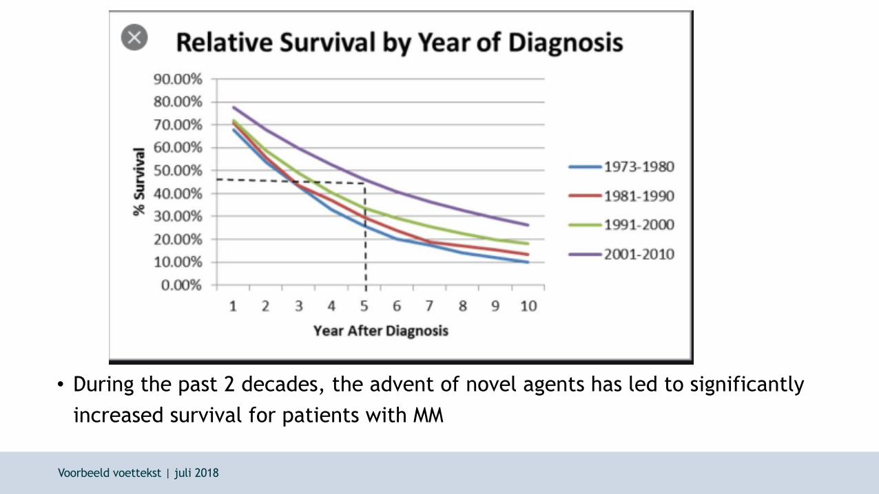
• During the past 2 decades, the advent of novel agents has led to significantly increased survival for patients with MM
Voorbeeld voettekst | juli 2018

• Howevers, unfortunately MM is still uncurable.
• MM knows a relapsing and remitting course
Keats. 2012

Introduction
• High-dose melphalan with autologous hematopoietic stem cell transplantation (ASCT) is the standard of care for fit patients with MM and is aimed at
achieving long-term remission
• Robust post-ASCT immune system reconstitution has been shown to correlate with deeper responses and improved clinical outcomes
• Although currently, no ‘specific validated immune signature’ has been
determined to predict superior survival among patients with MM, studies have reported on a clonal isotype switch (CIS) after ASCT
Voorbeeld voettekst | juli 2018

Clonal isotype switch/secundary MGUS
• Normally, MM relapse will present with the original monoclonal protein
documented at diagnosis and not with the CIS protein
• This would suggest that the transient CIS bands might represent part of the post-transplant immune reconstitution process and might even possess some
anti-MM activity?
Voorbeeld voettekst | juli 2018

Example of CIS/sMGUS
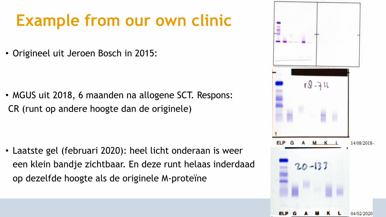
Example from our own clinic
• Origineel uit Jeroen Bosch in 2015:
• MGUS uit 2018, 6 maanden na allogene SCT. Respons:CR (runt op andere hoogte dan de originele)
• Laatste gel (februari 2020): heel licht onderaan is weer een klein bandje zichtbaar. En deze runt helaas inderdaad
op dezelfde hoogte als de originele M-proteïne
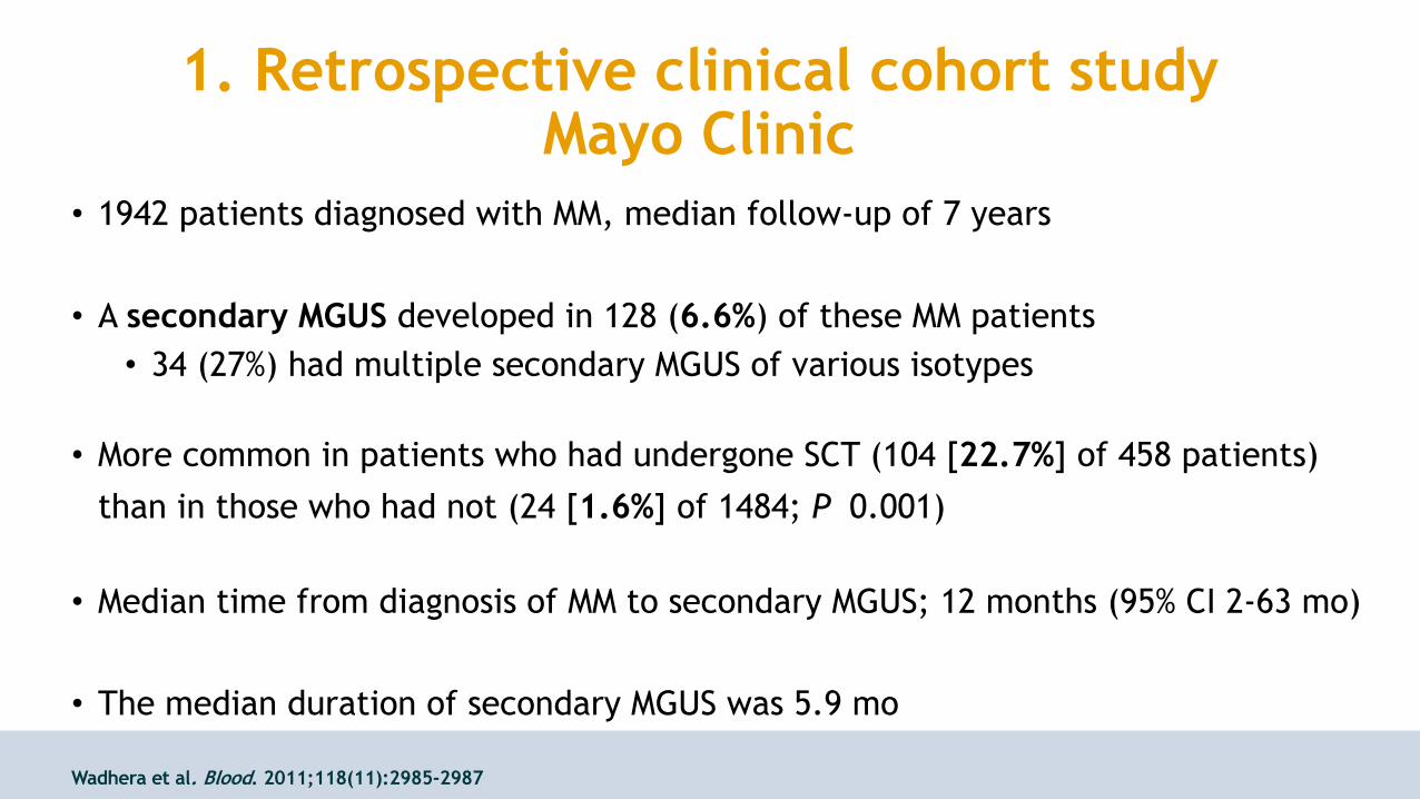
1. Retrospective clinical cohort studyMayo Clinic
• 1942 patients diagnosed with MM, median follow-up of 7 years
• A secondary MGUS developed in 128 (6.6%) of these MM patients• 34 (27%) had multiple secondary MGUS of various isotypes
• More common in patients who had undergone SCT (104 [22.7%] of 458 patients)
than in those who had not (24 [1.6%] of 1484; P 0.001)
• Median time from diagnosis of MM to secondary MGUS; 12 months (95% CI 2-63 mo)
• The median duration of secondary MGUS was 5.9 mo
Wadhera et al. Blood. 2011;118(11):2985-2987
• Most secondary MGUS M proteins were small;• detectable by immunofixation only in 84 patients (66%), • 0.2 to 0.9 g/dL in 29 patients (23%), • and ≥1 g/dL in 15 patients (12%)
• In most patients (87%), the underlying MM was responsive to therapy at the time of secondary MGUS diagnosis
• Median overall survival (OS) of the study cohort was 41 months
• OS was significantly superior among patients who developed secondary MGUS compared with the rest of the cohort (73 vs 38 months, respectively; P = 0.001)
Wadhera et al. Blood. 2011;118(11):2985-2987
1. Retrospective clinical cohort studyMayo Clinic
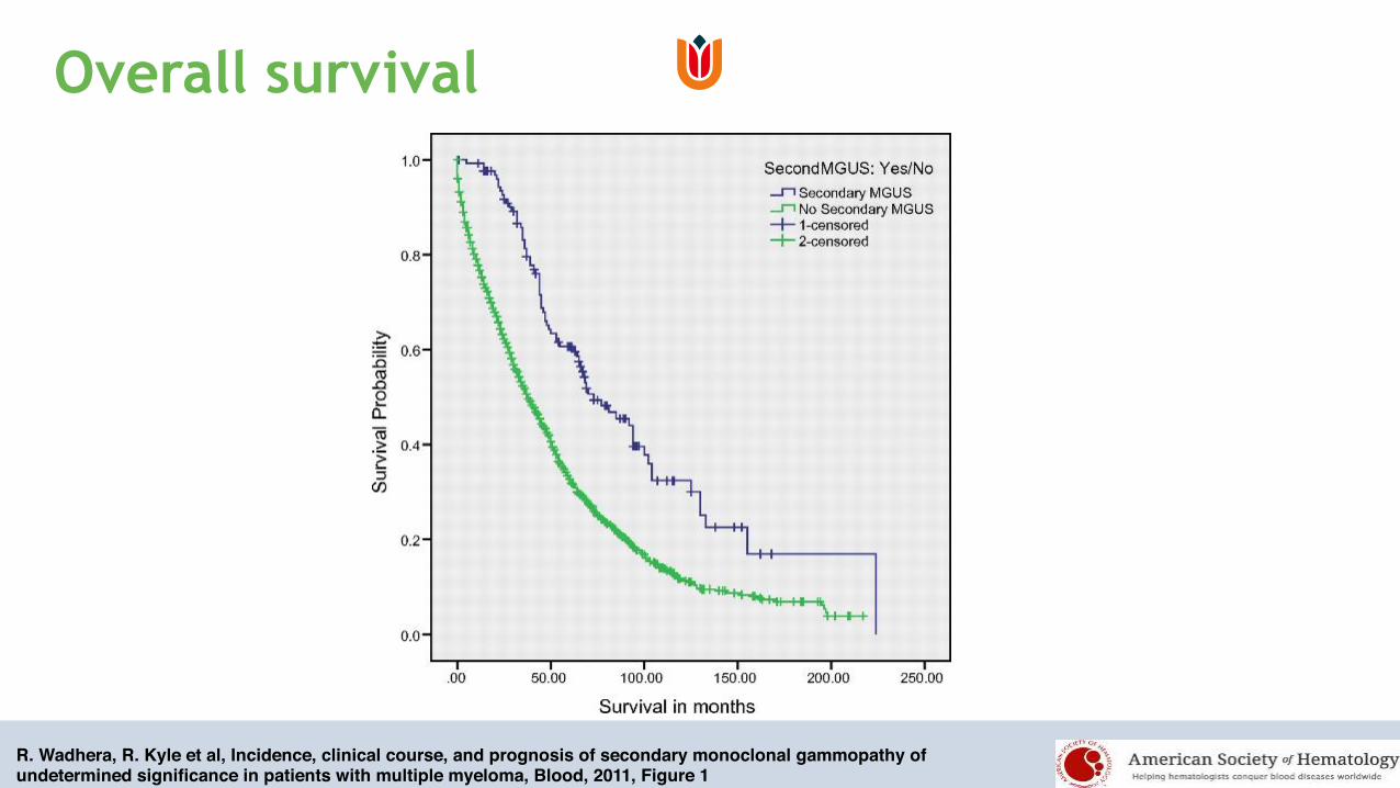
R. Wadhera, R. Kyle et al, Incidence, clinical course, and prognosis of secondary monoclonal gammopathy of undetermined significance in patients with multiple myeloma, Blood, 2011, Figure 1
Overall survival
Survival
• Survival of patients diagnosed since the year 2000 (n = 1088); Also here, OS was superior in patients with second MGUS (n = 86) versus those
without (n = 1002; median 77 vs 51 months, respectively; P = 0.001)
• On multivariate analysis, secondary MGUS and date of diagnosis of MM were independently predictive of OS (P = 0.001 for both factors)
Wadhera et al. Blood. 2011;118(11):2985-2987
2. Patients and Methods
• Patients with MM who had undergone ASCT from 2007 to 2016 were included in the present study
• The percentage of natural killer cells, B-cells, and T-cells was measured using
flow cytometry in pre- and post- ASCT bone marrow samples
• CIS was defined as the appearance of a new serum monoclonal spike on serum protein electrophoresis and immunofixation that differed from original heavy
or light chain detected at diagnosis
Ye et al. Clinical Lymphoma, Myeloma & Leukemia. 2019
2. Results• 177 patients with MM who had undergone ASCT during the study period
• A CIS was detected in 39 patients (22%)• Seventeen patients (46%) had only 1 new monoclonal protein • However, 10 (25%) had developed ≥ 4 monoclonal bands
• The newly detected monoclonal proteins were small, < 0.5 g/dL in 34 patients (87%)
• The median interval to the occurrence of a CIS was 7.1 mo (range, 1.9-32 mo)
• Patients with a relapse had an isotype that differed from a CIS, confirming the benign nature of this phenomenon
Ye et al. Clinical Lymphoma, Myeloma & Leukemia. 2019

2. Correlations
• A significantly greater incidence of CIS was found in patients who had received lenalidomide, an immunomodulatory drug, before ASCT (30.4% vs.
11.2%; P = 0.001), patients with IgA myeloma (21% vs. 7%; P = 0.01), and patients who had achieved minimally a very good partial response before
ASCT (52% vs. 23%; P = 0.023)
• Also, a CIS occurred more frequently in patients without suppressed uninvolved immunoglobulin (92% vs. 8%; P = 0.001)
Ye et al. Clinical Lymphoma, Myeloma & Leukemia. 2019

2. Immune Subset Recovery in Patients With CIS
• The number of peripheral lymphocytes and monocytes are prognostic factors in MM
• No differences between pre- or post-ASCT peripheral absolute lymphocyte or monocyte counts in patients who had experienced post-ASCT CIS and those who had not
• Similarly, no significant difference was found in the CD4, CD8, NK, or B-cell percentages or in the CD4/CD8 ratio in the pre-ASCT BM samples from patients with or without CIS
• The appearance of post-ASCT CIS was associated with lower CD8 T-cell percentages (43% in patients without CIS vs. 28% in patients with CIS; P = .015) and a higher CD4/CD8 ratio (0.2 vs. 2.8; P = .001; both with differences that were statistically significantly), suggestive of faster T-cell compartment reconstitution in this patient group
Ye et al. Clinical Lymphoma, Myeloma & Leukemia. 2019
Dosani T, et al. Blood Cancer J 2017; 7:e579.Porrata LFet al. Blood 2001; 98:579-85.

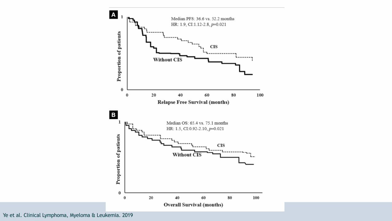
2. PFS and OS and correlations• The presence of post-ASCT CIS correlated significantly with improved PFS
(52.2 vs. 36.6 months; P = 0.21) and OS (75.1 vs. 65.4 months; P = 0.021)
• Age, cytogenetics, response category, presence of CIS, and low lactate
dehydrogenase were shown to influence PFS on univariate analysis
• Cytogenetics, lactate dehydrogenase, and CIS presence were also significantly associated with the MM response on multivariate analysis
• All patients who had experienced a relapse had an isotype different from that
of the CIS, highlighting the benign nature of this phenomenon
Ye et al. Clinical Lymphoma, Myeloma & Leukemia. 2019
Ye et al. Clinical Lymphoma, Myeloma & Leukemia. 2019

2. Conclusion
• A prospective analysis of 177 patients with multiple myeloma undergoing autologous stem cell transplant
• found that 22% developed new and small concentrations of monoclonal
protein after transplant that differed from that originally identified at diagnosis
• This phenomenon had a benign nature and correlated with improved survival
and more robust bone marrow immune reconstitution beyond the B-cell compartment
Ye et al. Clinical Lymphoma, Myeloma & Leukemia. 2019

Discussion
• Gene sequencing of heavy chain variable region in 7 patients with post-ASCT CIS did not show a clonal relationship to the original malignant clone isotype
highlighting nonmalignant B cells as the likely origin of CIS
Ye et al. Clinical Lymphoma, Myeloma & Leukemia. 2019Guikema JE, Vellenga E, et al. Br J Haematol 1999; 104:748-54.

3. Allogeneic SCT and sMGUS/CIS
• 138 patients who had undergone allogeneic stem cell transplantations
• 67 (48.2%) patients developed sMGUS, after a median latency of 6.9 months• 25 patients had only one new protein band (18.0%), • 9 (6.5%) had 2 bands, • 8 (5.8%) had 3 bands, • and 25 (18.0%) had 4 or more
• sMGUS occurred more often in patients with deeper responses, at least very
good partial response after allo SCT, compared to partial response or less (54.8% vs. 26.5%; P=0.005)
Schmitz et al. Haematologica. 2014

3. Secondary MGUS after allogeneic SCT in MM
• In most cases it was not possible to quantify the level of sMGUS, mostly levels too low• Abnormal protein bands could be quantified in only 4 patients, with a maximum
level of 11 g/L
• The median duration of all sMGUS cases was 4.47 months (range 0.0-74.5 months)
• There was no progression of sMGUS to MM or other lymphoproliferative diseases
• Clinicians should be aware of the benign nature of this phenomenon, and secondary monoclonal gammopathy of undetermined significance should not be confused with
relapse or progression of disease
Schmitz et al. Haematologica. 2014
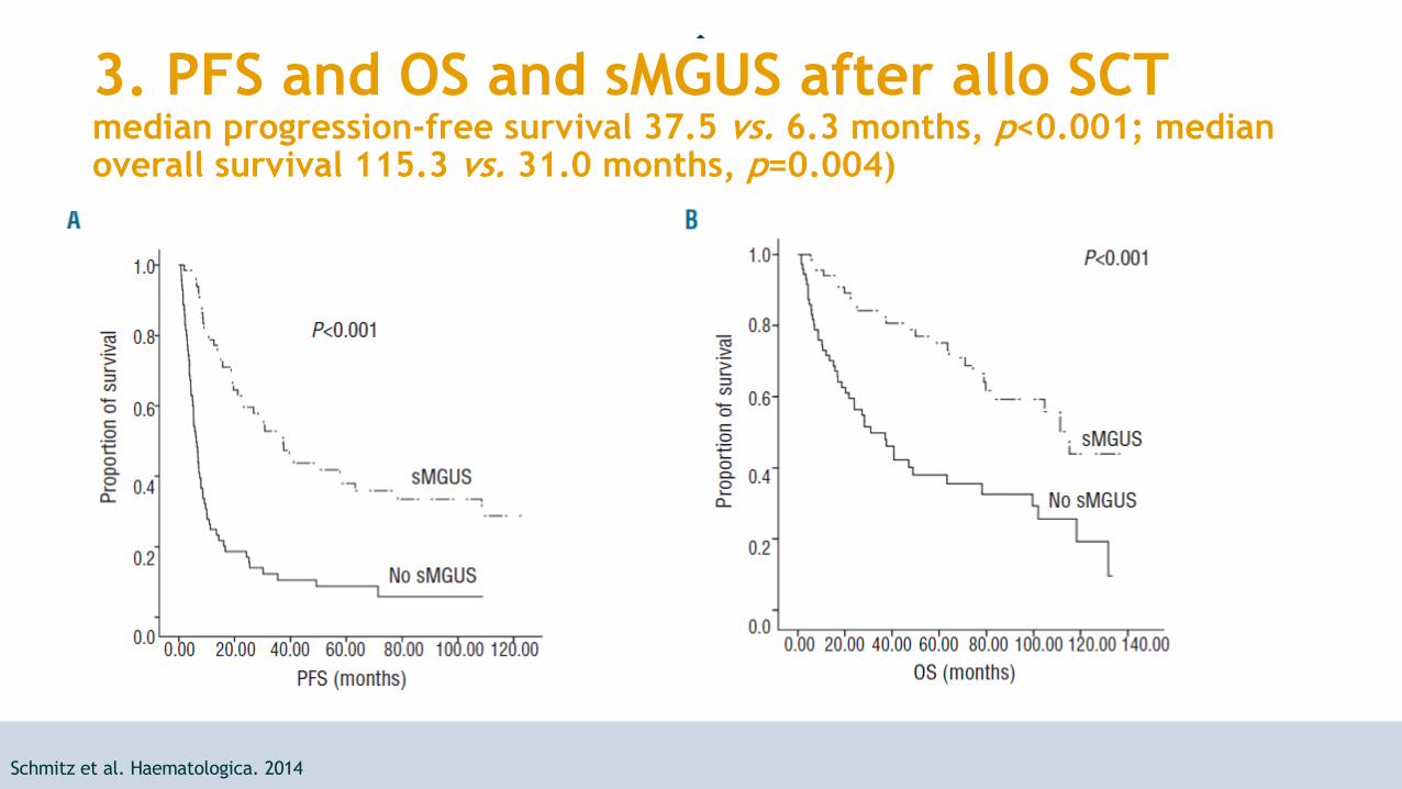
3. PFS and OS and sMGUS after allo SCTmedian progression-free survival 37.5 vs. 6.3 months, p<0.001; median overall survival 115.3 vs. 31.0 months, p=0.004)
Schmitz et al. Haematologica. 2014

Overall conclusions• The emergence of sMGUS reflects a strong humoral immune response and is a sign of
immune reconstitution after allo-SCT, autologous SCT, or novel agent-containing
regimens
• A higher frequency of sMGUS is observed in patients with high-quality responses, which suggests that major tumor reduction contributes to strong immune reconstitution and
development of oligoclonal bands
• There is no evidence that these abnormal protein bands are related to the myeloma clone • Prior studies suggest that new serum M-components after auto-SCT are not
produced by myeloma cells but rather by the regenerating B-cell compartment

• Furthermore, sMGUS not only occurs in patients but also after treatment for other hematologic malignancies, and even solid organ transplantations
• Development of sMGUS seems a favorable prognostic factor for PFS and OS,
independent of the response achieved
• The favorable prognosis conferred by sMGUS suggests that the oligoclonal bands may also be involved in an anti-myeloma immune response
• Importantly, in order to avoid unnecessary treatment clinicians should be aware that
sMGUS does not represent disease recurrence or development of a new malignancy
Overall conclusions